carbonic anhydrases in the reproductive system …jultika.oulu.fi/files/isbn9514266641.pdfcarbonic...
TRANSCRIPT
CARBONIC ANHYDRASES IN THE REPRODUCTIVE SYSTEMWith special emphasis on isoenzymes VI, IX, XII, and a novel nuclear nonclassical form
PEPEKARHUMAA
Department of Anatomy and Cell Biology,University of Oulu
OULU 2002

PEPE KARHUMAA
CARBONIC ANHYDRASES IN THE REPRODUCTIVE SYSTEMWith special emphasis on isoenzymes VI, IX, XII, and a novel nuclear nonclassical form
Academic Dissertation to be presented with the assent ofthe Faculty of Medicine, University of Oulu, for publicdiscussion in the Auditorium of the Department ofAnatomy and Cell Biology, on May 17th, 2002, at 12noon.
OULUN YLIOPISTO, OULU 2002

Copyright © 2002University of Oulu, 2002
Reviewed byProfessor Jorma ParankoDocent Jorma Toppari
ISBN 951-42-6664-1 (URL: http://herkules.oulu.fi/isbn9514266641/)
ALSO AVAILABLE IN PRINTED FORMATActa Univ. Oul. D 671, 2002ISBN 951-42-6663-3ISSN 0355-3221 (URL: http://herkules.oulu.fi/issn03553221/)
OULU UNIVERSITY PRESSOULU 2002

Karhumaa, Pepe, Carbonic anhydrases in the reproductive system With specialemphasis on isoenzymes VI, IX, XII, and a novel nuclear nonclassical formDepartment of Anatomy and Cell Biology, University of Oulu, P.O.Box 5000, FIN-90014University of Oulu, Finland Oulu, Finland2002
Abstract
Carbonic anhydrases (CAs) are a group of zinc-containing metalloenzymes that catalyze theinterconversion of carbon dioxide and bicarbonate (CO2 + H2O ⇔ HCO3
- + H+). They are present inalmost all organs and are implicated in various biological functions, the most important of which isparticipation in the regulation of ion, water, and acid-base balance. Recently, some members of theCA gene family have been suggested to promote cell proliferation and to act as trophic growth factors.
The present study was undertaken to examine the distribution of CA isoenzymes in thereproductive system, to attain a more detailed view on their linkage to the reproductive processes andto neonatal development.
The expression of membrane-bound CA IX and CA XII was studied in the female and malereproductive tracts by immunohistochemistry and western blotting. CA XII was found to beexpressed in the basolateral plasma membrane of luminal and glandular epithelia in human uterus. Inhuman efferent ducts, it was located in the basolateral plasma membrane of luminal epithelium,where it coexpressed with Aquaporin-1. In epididymal duct, CA XII was only expressed in occasionalepithelial cells. These cells coexpressed CA II, suggesting that they represent apical mitochondria-rich cells (AMRC). CA IX was also expressed in the basolateral plasma membrane of luminalepithelium in human efferent ducts, but its expression was not uniform among the tubules. Thesefindings suggest that basolateral plasma membrane-associated CA IX and CA XII contribute, alongwith CA II and CA IV, to the regulation of acid-base balance and water transport in the reproductivetract.
Western blotting of rat Leydig tumor cells and testis for CA II revealed an unidentified 66-kDapolypeptide band. The polypeptide was successfully purified from several rat tissues using CAinhibitor affinity chromatography. The amino acid sequence of the polypeptide showed it to beidentical to NonO/p54nrb, a non-POU domain-containing octamer-binding protein previouslyimplicated in transcriptional regulation. The recombinant NonO/p54nrb was shown to display CAactivity, and the antibody to it predominantly immunostained the nuclei in lymphocytes, where CAactivity was also detected histochemically. Accordingly, the nuclear Leydig cell CAimmunoreactivity represents NonO/p54nrb. It is classified as a novel, nonclassical CA, and it mayparticipate in pH-related events in the nucleus.
Human and rat milk was found to contain CA VI by immunohistochemistry and western blotting.The enzyme purified from human milk by CA inhibitor affinity chromatography was confirmed byPNGase F digestion and amino acid sequence as CA VI. The CA VI concentrations in humancolostral milk were approximately eight times higher than those in mature milk (34.7 mg/l vs. 4.5 mg/l). Secretion of CA VI into milk is suggested by its localization in the alveolar epithelium of the ratmammary gland. The structural and functional stability of CA VI in an acidic milieu, its suggestedgrowth-supporting function in taste bud stem cells, and its high concentration in colostrum suggestthat it is an essential factor for the growth and development of the newborn alimentary canal.
Keywords: efferent ducts, milk, testis, uterus, carbonic anhydrases

To my family


Acknowledgements
This work was carried out in the Department of Anatomy and Cell Biology, University ofOulu. I wish to express my sincere gratitude to my supervisor, Professor HannuRajaniemi, M.D., Ph.D, Head of the Department of Anatomy and Cell Biology, and to myother supervisors, Professor Seppo Parkkila, M.D., Ph.D., and Dr. Kari Kaunisto, M.D.,Ph.D., for their guidance during these years. I also wish to thank Professor Juha Peltonen,M.D., Ph.D., Professor Juha Tuukkanen, D.D.S., Ph.D, and Docent Kalervo Metsikkö,Ph.D., for their interest towards my work.
I am very grateful to Docents Jorma Paranko, M.D., Ph.D., and Jorma Toppari, M.D.,Ph.D., for their valuable comments and constructive criticism at the time of thecompletion of this thesis.
I would like to thank my co-authors: Dr. Anna-Kaisa Parkkila, M.D., Ph.D., ProfessorJuha Tapanainen, M.D., Ph.D., Research Associate Professor Abdul Waheed, Ph.D.,Professor William S. Sly, M.D., Dr. Özlem Türeci, M.D., Dr. Silvia Pastoreková, Ph.D.,Professor Jaromir Pastorek, Ph.D., Professor Philip W. Tucker, Ph.D., Dr. Ching-JungHuang, Ph.D., Dr. Jeffrey Grubb, B.A., Assistant Research Professor Gul Shah, Ph.D.,and Dr. Jukka Leinonen, D.D.S. I also wish to thank Jyrki Aatsinki, M.Sc., and MarittaPietilä, M.Sc., for teaching me cell biology techniques.
In addition, I would like to thank Ms. Lissu Hukkanen, Ms. Paula Soininen, Ms.Pirkko Peronius, Mr. Eero Oja, Mr. Mika Kihlström, and Ms. Seija Leskelä for theirskillful technical assistance.
I am grateful to Mr. Malcolm Hicks, M.A., for his careful revision of the language ofone original paper and Sirkka-Liisa Leinonen, Lic.Phil., for her careful revision of thelanguage of the manuscript of this thesis.
Finally, I owe my deepest gratitude to my dear wife Merja and my son Wille. I willnever forget their love and �patience� during these years.
This study was supported by grants from The Emil Aaltonen Foundation, The FinnishMedical Society Duodecim, and The Finnish Medical Society.
Oulu, March 2002 Pepe Karhumaa

Abbreviations
αGST α-glutathione S-transferaseAE Anion exchangerAMRC Apical mitochondria-rich cellAQP1 Aquaporin-1Asp Aspartic acidATP Adenosine triphosphateBSA Bovine serum albuminCA Carbonic anhydrasecAMP Cyclic adenosine 3´,5´ monophosphateCA-RP Carbonic anhydrase-related proteincDNA Complementary deoxyribonucleic acidCHIP28 Channel-forming integral protein of 28 kDaCys CysteineDAB 3,3'-diaminobenzidine tetrahydrochlorideCHO Chinese hamster ovaryDNA Deoxyribonucleic acidEGF Epidermal growth factorEu EuropiumFITC Fluorescein isothiocyanate HHMI Howard Hughes Medical InstituteHis HistidineIEF Isoelectric focusingIgA Immunoglobulin AIGF Insuline-like growth factorIgG Immunoglobulin GkDa KiloDaltonMALDI-MS Matrix-assisted laser desorption ionization mass spectrometry mRNA Messenger ribonucleic acidNBC Sodium bicarbonate cotransporterNGF Nerve growth factorNHE Sodium/proton exchanger
NonA No-on-transient ANonO Non-POU (Pit-Oct-Unc) domain-containing octamer-binding protein p54nrb Nuclear RNA-binding protein, 54 kDaPAGE Polyacrylamide gel electrophoresisPBS Phosphate-buffered salinepI Isoelectric pointPMSF Phenylmethylsulphonyl fluoridePNGase F Endoglycosidase FPSF Polypyrimidine tract-binding protein-associated splicing factorPVDF Polyvinylidene difluorideQ Glutamine RPTP Receptor-type protein tyrosine phosphataseRT-PCR Reverse transcriptase-polymerase chain reactionSD Standard deviationSDS Sodium dodecyl sulphateTBST Tris-buffered saline with Tween-20TGF Transforming growth factorTR-IFMA Time-resolved fluoroimmunoassay TRITC Tetramethylrhodamine isothiocyanate TSH Thyroid-stimulating hormoneVHL Von Hippel-Lindau

List of original publications
This thesis is based on the following articles, which are referred to in the text by theirRoman numerals:
I Karhumaa P, Parkkila S, Türeci Ö, Waheed A, Grubb JH, Shah G, Parkkila A-K,Kaunisto K, Tapanainen J, Sly WS & Rajaniemi H (2000) Identification of carbonicanhydrase XII as the membrane isozyme expressed in the normal human endome-trial epithelium. Mol Hum Reprod 6:68-74.
II Karhumaa P, Kaunisto K, Parkkila S, Waheed A, Pastoreková S, Pastorek J, Sly WS& Rajaniemi H (2001) Expression of the transmembrane carbonic anhydrases, CAIX and CA XII, in the human male excurrent ducts. Mol Hum Reprod 7:611-616.
III Karhumaa P, Parkkila S, Waheed A, Parkkila A-K, Kaunisto K, Tucker PW, HuangC-J, Sly WS & Rajaniemi H (2000) Nuclear NonO/p54nrb protein is a nonclassicalcarbonic anhydrase. J Biol Chem 275:16044-16049.
IV Karhumaa P, Leinonen J, Parkkila S, Kaunisto K, Tapanainen J & Rajaniemi H(2001) The identification of secreted carbonic anhydrase VI as a constitutive glyco-protein of human and rat milk. Proc Natl Acad Sci USA 98:11604-11608.


Contents
Abstract AcknowledgementsAbbreviations List of original publications 1 Introduction . . . . . . . . . . . . . . . . . . . . . . . . . . . . . . . . . . . . . . . . . . . . . . . . . . . . . . . . 152 Review of the literature . . . . . . . . . . . . . . . . . . . . . . . . . . . . . . . . . . . . . . . . . . . . . . . 17
2.1 Carbonic anhydrase gene families . . . . . . . . . . . . . . . . . . . . . . . . . . . . . . . . . . 172.1.1Mammalian CA isoenzymes . . . . . . . . . . . . . . . . . . . . . . . . . . . . . . . . . . . 17
2.1.1.1 CA VI . . . . . . . . . . . . . . . . . . . . . . . . . . . . . . . . . . . . . . . . . . . . . 202.1.1.2 CA IX . . . . . . . . . . . . . . . . . . . . . . . . . . . . . . . . . . . . . . . . . . . . . 212.1.1.3 CA XII . . . . . . . . . . . . . . . . . . . . . . . . . . . . . . . . . . . . . . . . . . . . 22
2.2 Sperm maturation and transport in the male reproductive tract . . . . . . . . . . . . 232.3 Sperm transport in the female reproductive tract . . . . . . . . . . . . . . . . . . . . . . . 242.4 pH regulation in the male reproductive tract . . . . . . . . . . . . . . . . . . . . . . . . . . . 262.5 pH regulation in the female reproductive tract . . . . . . . . . . . . . . . . . . . . . . . . . 272.6 Bicarbonate in reproductive physiology . . . . . . . . . . . . . . . . . . . . . . . . . . . . . . 282.7 Carbonic anhydrase in the male reproductive tract . . . . . . . . . . . . . . . . . . . . . . 292.8 Carbonic anhydrase in the female reproductive tract . . . . . . . . . . . . . . . . . . . . 302.9 Effects of CA inhibitors on reproduction . . . . . . . . . . . . . . . . . . . . . . . . . . . . . 322.10 Carbonic anhydrase in mammary gland and milk protein composition
and functions . . . . . . . . . . . . . . . . . . . . . . . . . . . . . . . . . . . . . . . . . . . . . . . . . . . 323 Aims of the present study . . . . . . . . . . . . . . . . . . . . . . . . . . . . . . . . . . . . . . . . . . . . . . 344 Materials and methods . . . . . . . . . . . . . . . . . . . . . . . . . . . . . . . . . . . . . . . . . . . . . . . . 35
4.1 Collection of human samples (I-IV) . . . . . . . . . . . . . . . . . . . . . . . . . . . . . . . . . 354.2 Collection of rat samples (III, IV) . . . . . . . . . . . . . . . . . . . . . . . . . . . . . . . . . . . 354.3 Cell culture (I, III) . . . . . . . . . . . . . . . . . . . . . . . . . . . . . . . . . . . . . . . . . . . . . . . 364.4 Chinese hamster ovary (CHO) cells expressing CA XII (I) . . . . . . . . . . . . . . . 364.5 Antibodies and immunoreagents (I-IV) . . . . . . . . . . . . . . . . . . . . . . . . . . . . . . 364.6 Immunohistochemistry (I-IV) . . . . . . . . . . . . . . . . . . . . . . . . . . . . . . . . . . . . . . 374.7 SDS-polyacrylamide gel electrophoresis (SDS-PAGE) (I-IV) . . . . . . . . . . . . . 374.8 Western blotting (I-IV) . . . . . . . . . . . . . . . . . . . . . . . . . . . . . . . . . . . . . . . . . . . 384.9 Isoelectric focusing (I) . . . . . . . . . . . . . . . . . . . . . . . . . . . . . . . . . . . . . . . . . . . 384.10 Northern blotting (I) . . . . . . . . . . . . . . . . . . . . . . . . . . . . . . . . . . . . . . . . . . . . . 38

4.11 Inhibitor affinity purification of CAs (I, III, IV) . . . . . . . . . . . . . . . . . . . . . . . . 394.12 Protein sequence analysis (III, IV) . . . . . . . . . . . . . . . . . . . . . . . . . . . . . . . . . . 394.13 Production and purification of recombinant nonO (III) . . . . . . . . . . . . . . . . . . 394.14 Carbonic anhydrase assay (III) . . . . . . . . . . . . . . . . . . . . . . . . . . . . . . . . . . . . . 404.15 Histological staining of CA activity and electron microscopy (III) . . . . . . . . . 404.16 Binding of nonO protein and CA II to the p-amino methyl benzene
sulfonamide-Affigel 10 column (III) . . . . . . . . . . . . . . . . . . . . . . . . . . . . . . . . 414.17 Deglycosylation studies (IV) . . . . . . . . . . . . . . . . . . . . . . . . . . . . . . . . . . . . . . . 414.18 Fluoroimmunoassay (IV) . . . . . . . . . . . . . . . . . . . . . . . . . . . . . . . . . . . . . . . . . 41
5 Results . . . . . . . . . . . . . . . . . . . . . . . . . . . . . . . . . . . . . . . . . . . . . . . . . . . . . . . . . . . . 425.1 Expression of transmembrane CAs in the human male and female
reproductive tracts . . . . . . . . . . . . . . . . . . . . . . . . . . . . . . . . . . . . . . . . . . . . . . . 425.1.1CA IX (II) . . . . . . . . . . . . . . . . . . . . . . . . . . . . . . . . . . . . . . . . . . . . . . . . . 425.1.2CA XII (I ,II) . . . . . . . . . . . . . . . . . . . . . . . . . . . . . . . . . . . . . . . . . . . . . . . 42
5.2 Identification of a novel nonclassical carbonic anhydrase and its expressionin human and rat Leydig cells (III) . . . . . . . . . . . . . . . . . . . . . . . . . . . . . . . . . . 43
5.3 CA VI in milk (IV) . . . . . . . . . . . . . . . . . . . . . . . . . . . . . . . . . . . . . . . . . . . . . . 446 Discussion . . . . . . . . . . . . . . . . . . . . . . . . . . . . . . . . . . . . . . . . . . . . . . . . . . . . . . . . . 45
6.1 CA IX and CA XII in the human reproductive tract . . . . . . . . . . . . . . . . . . . . . 456.2 Identification of a novel nonclassical CA, NonO/p54nrb-protein . . . . . . . . . . 486.3 Secretion of CA VI in milk . . . . . . . . . . . . . . . . . . . . . . . . . . . . . . . . . . . . . . . . 49
7 Conclusions . . . . . . . . . . . . . . . . . . . . . . . . . . . . . . . . . . . . . . . . . . . . . . . . . . . . . . . . 518 References . . . . . . . . . . . . . . . . . . . . . . . . . . . . . . . . . . . . . . . . . . . . . . . . . . . . . . . . . 52
1 Introduction
Carbonic anhydrases (CAs) are zinc-containing metalloenzymes that participate in theregulation of ion, water, and acid-base balance by catalyzing the reversible hydration ofcarbon dioxide in a reaction: CO2 + H2O ⇔ HCO3
- + H+ (Sly & Hu 1995). CAs includethree evolutionary unrelated gene families termed alpha-, beta-, and gamma-CAs. In theanimal kingdom, all of the heretofore identified CAs belong to the alpha-CA family(Hewett-Emmet & Tashian 2000). Eleven enzymatically active isoenzymes in the alphafamily have been identified so far, including four cytoplasmic, two mitochondrial, fourmembrane-associated, and one secreted form (Fujikawa-Adachi et al. 1999).
Before this study, two carbonic anhydrase isoenzymes, CA II and CA IV, had beenidentified and localized in human reproductive organs. The expression of CA IV wasfound in the apical plasma membrane of epithelial cells in epididymis and proximal vasdeferens, and CA II in the cytoplasm of epithelial cells in seminal vesicle, vas deferens,and sporadically in epididymis (Kaunisto et al. 1990, Parkkila S et al. 1993a). Varioushistochemical studies in human and animal reproductive organs suggest, however, thatother CA isoenzymes are possibly also expressed (Korhonen et al. 1966, Friedley &Rosen 1975, Ekstedt et al. 1991, Ekstedt & Ridderstråle 1992).
The exact physiological role of these isoenzymes in the reproductive tract is not fullyunderstood. CA II and CA IV probably participate in the acidification of epididymalfluid, whereas CA II in the seminal vesicle may promote alkalinization of the ejaculate(Kaunisto et al. 1990, Parkkila s et al. 1993a). CA I and CA II are expressed in humanplacenta and foetal membranes, suggesting that they are implicated in the regulation ofacid-base balance in amniotic fluid and the developing foetus (Mühlhauser et al. 1994).After birth, breast milk is the primary food for the newborn. In addition to nutrients, itcontains a number of bioactive factors, including growth factors and host defence agents,which contribute to the growth and development of the newborn (Kunz et al. 1999).Salivary CA VI has been implicated as a developmental factor in taste bud growth(Thatcher et al. 1998). During the early postnatal period, however, saliva secretion by thenewborn infant is minimal due to the immaturity of the salivary glands (Davidson 1982,Scott 1979), and milk as a good saliva substitute (Herod 1994) may compensate for thislow secretion during the neonatal period.

16
In the present study, the distribution of different CA isoenzymes was investigated inthe reproductive system, to achieve a more comprehensive view of their role inreproductive functions and neonatal development.
2 Review of the literature
2.1 Carbonic anhydrase gene families
To date, three independent CA gene families have been discovered: α-CA, β-CA and γ-CA (Hewett-Emmett & Tashian 1996, Hewett-Emmett 2000). The earlier postulation thatα-CAs would be restricted to the animal kingdom and plant green algae(Chlamydomonas), the β-CAs to plants and eubacteria, and the γ-CAs to archaebacteriaand eubacteria has been disproven, since the plant Arabidopsis has homologues of all thethree families (Hewett-Emmett & Tashian 1996). In addition, recent findings show thatboth α- and β-CA genes are present in many plants, lower eukaryotes and invertebrates,but α-CAs are clearly dominant in vertebrates (Hewett-Emmett 2000).
Based on X-ray crystallographic studies, the active center of α-CAs contains one zincion that is surrounded by three conserved histidine residues and one water molecule(Kannan et al. 1977, Eriksson & Liljas 1991). In addition, the active center of α-CAsconsists of 33 other residues, whose homology varies between the isoenzymes (Fujikawa-Adachi et al. 1999). The structural studies on β-CAs and γ-CAs are less numerous. Theactive center of γ-CAs also contains three histidines, as in α-CAs, but with differentspacing in the linear sequence (Hewett-Emmett & Tashian 1996). Mitsuhashi et al. (2000)suggested that zinc in the active center of β-CAs is coordinated by a Cys-Asp-His-Cystetrad, which is strictly conserved in the gene family. They found no water molecule in azinc-liganding radius in β-CAs, indicating the existence of distinct catalyzing sites for thesame CO2 hydration as in α-CAs and probably also in γ-CAs. Kimber & Pai (2000)suggested that zinc in the active center of β-CAs is surrounded by Cys-His-Cys, and thatα-CAs and β-CAs are likely to share a common CO2 hydration mechanism.
2.1.1 Mammalian CA isoenzymes
Carbonic anhydrase was first purified from red blood cells (Meldrum & Roughton 1932,1933). Thereafter, numerous biochemical studies have demonstrated the presence of CA

18
in different tissue or cell homogenates. The localization of CA in tissues became possiblein 1953, when Kurata (1953) reported a novel histochemical technique for demonstratingCA activity in tissues. However, this technique does not differentiate betweenisoenzymes, which has, in turn, been facilitated by isoenzyme-specific antibodies andimmunohistochemical techniques. To date, eleven active CA isoenzymes have beencharacterized in the animal kingdom, including four cytoplasmic (CA I, CA II, CA IIIand CA VII), two mitochondrial (CA VA and CAVB), one secreted (CA VI), and fourmembrane-associated (CA IV, CA IX, CA XII and CA XIV) forms (Fig. 1).
CA I is a moderate-activity isoenzyme present in erythrocytes and, at lower levels, inepithelial cells of the gastrointestinal tract, capillary and corneal endothelium, lens of theeye, islets of Langerhans, placenta and fetal membranes (Lönnerholm et al. 1985, Ventaet al. 1987, Mühlhauser et al. 1994, Parkkila et al. 1994). CA II has the highest activity ofall isoenzymes, and it is present in certain cell types of almost all tissues. It contributes,e.g. to H+ secretion by gastric parietal cells, renal tubular cells, and osteoclasts and toHCO3
- secretion by pancreatic duct cells, ciliary body epithelium, choroid plexus,salivary gland acinar cells, and distal colonic epithelium (Sly & Hu 1995). In largeintestine and gallbladder, CA II is involved in NaCl and water absorption (Swenson 1991,Parkkila & Parkkila 1996). CA II also promotes CO2 exchange in erythrocytes, lung, andkidney. It has also been suggested to take part in fatty acid and amino acid synthesis (Sly& Hu 1995). CA III has the lowest activity of the isoenzymes, and it is present abundantlyin slow-twitch (type I fiber) red skeletal muscle and, in lesser amounts, in certain othertissues (Carter et al. 1979, Jeffery et al. 1980, Shima et al. 1983, Väänänen et al. 1985,Väänänen & Autio-Harmainen 1987, Nishita & Matsushita 1989, Spicer et al. 1990). Ithas been suggested to protect cells from oxidative damage, and thus also to affect growth-signaling pathways (Räisänen et al. 1999). CA VII mRNA has been detected in baboonsalivary glands, rat lung, and mouse brain (Montgomery et al. 1991, Ling et al. 1994,Lakkis et al. 1997). The recombinant protein of CA VII has been shown to be a high-activity isoenzyme, but tissue expression of the protein has not been described yet(Earnhardt et al. 1998). Mitochondrial CA was first identified by Dodgson et al. (1980),and its function was linked to gluconeogenesis and ureagenesis and the regulation ofinsulin secretion (Dodgson et al. 1983, Metcalfe et al. 1985, Sly & Hu 1995, Parkkila etal. 1998). Recently, it has become clear that there are two mitochondrial CAs, termed CAVA and CA VB (Fujikawa-Adachi et al. 1999, Shah et al. 2000). The expression ofhuman CA VA has been demonstrated in liver (Fujikawa-Adachi et al. 1999). The mRNAof CA VB has been shown in normal human heart and skeletal muscle by northernblotting. In human pancreas, salivary glands, kidney, and spinal cord, the mRNA ofhuman CA VB could be detected using reverse transcription-PCR (Fujikawa-Adachi etal. 1999). CA VI is the secretory CA, which exclusive secretion has been found so faronly into saliva by salivary glands. CA IV is a membrane-anchored CA expressed in theapical surface of epithelial cells. It is expressed in, e.g. certain segments of nephron andgastrointestinal epithelial cells, the endothelial cells of certain capillary beds, and theepithelium of epididymis and vas deferens (Brown et al. 1990, Hageman et al. 1991,Ghandour et al. 1992, Fleming et al. 1993, Parkkila S et al. 1993a, Sender et al. 1994,Fleming et al. 1995, Kaunisto et al. 1995, Parkkila et al. 1996). CA IX and CA XII arerecently characterized transmembrane proteins, which have been linked to oncogenesis.However, their presence in non-malignant tissues has been demonstrated as well. The

19
third transmembrane isoform, CA XIV, has recently been identified and cloned frommouse and human tissues (Mori et al. 1999, Fujikawa-Adachi et al. 1999). Its mRNA hasbeen demonstrated in human heart, brain, liver, and skeletal muscle using the northernblotting technique and in colon, small intestine, urinary bladder, kidney, and spinal cordusing more sensitive techniques (Fujikawa-Adachi et al. 1999). In situ hybridization inmouse kidney showed that CA XIV mRNA is expressed in the proximal convolutedtubule and in the outer border of the inner stripe of the outer medulla (Mori et al. 1999).Kaunisto et al. (2002) have recently described immunohistochemical localization of CAXIV protein in mouse and rat kidney. It was found to be present in the proximal tubulesand thin limbs of Henle. A study by Parkkila et al. (2000a) demonstrated the presence ofCA XIV protein in neuronal membranes and axons in different parts of human and mousebrain. The mammalian CA gene family also contains three CA-related proteins (CA-RPVIII, X, and XI) and two subtypes of receptor-type protein tyrosine phosphatases (RPTP-β and γ). These proteins share the CA-like domain but lack CA activity (Tashian et al.2000). CA-RPs have been found from cDNA libraries and named in the order of theirdiscovery. mRNAs of CA-RP VIII, X and XI have been detected mainly in brain but alsoin a variety of other tissues (Kato 1990, Skaggs et al. 1993, Ling et al. 1994, Lakkis et al.1997a, Lakkis et al. 1997b, Fujikawa-Adachi et al. 1999, Tashian et al. 2000). Thelocalization and function of these proteins are so far unknown. The CA-like domain hasalso been shown in RPTPs (Krueger & Saito 1992, Levy et al. 1993, Barnea et al. 1993).The extracellular region of RPTPβ has been shown to be identical to a chondroitinsulphate proteoglycan, phosphacan (Maurel et al. 1994), which provides a binding site fora cell surface signal-transducing molecule, contactin (Peles et al. 1995). The exactfunctions of RPTPs are, however, unknown.
Fig. 1. Subcellular localization of the active CA isoenzymes. CA I, II, III, and VII arecytoplasmic, CA VA and VB are mitochondrial, CA VI is secreted, CA IV is associated with theapical plasma membrane, and CA IX and XII are associated with the basolateral plasmamembrane. CA XIV is located in both apical and basolateral plasma membranes.

20
2.1.1.1 CA VI
CA VI is so far the only known secretory isoenzyme of the CA gene family. It was firstfound in ovine parotid gland and saliva by Fernley et al. (1979). Feldstein and Silverman(1984) purified it from rat saliva and determined its amino acid composition. Murakamiand Sly (1987) and Kadoya et al. (1987) purified it independently from human saliva.The amino acid sequence of sheep CA VI was reported by Fernley et al. (1988), and thecDNA sequence of human CA VI was determined by Aldred et al. (1991). The molecularweight of CA VI polypeptide is 39-46 kDa (Feldstein & Silverman 1984, Kadoya et al.1987, Murakami & Sly 1987, Fernley 1991a,b, Parkkila et al. 1991a, Ogawa et al. 1992).The molecule contains two 3-kDa N-linked oligosaccharide chains, which can be cleavedoff by endo-β-N-acetylglucosaminidase F but not by endo-β-N-acetylglucosaminidase H,indicating that the oligosaccharides are of a complex type (Murakami & Sly 1987). CAVI has two cysteine residues that form an intramolecular disulfide bond, which probablyenables the enzyme to retain its enzymatic activity in an acidic milieu (Fernley et al.1988, Aldred et al. 1991, Parkkila et al. 1997). The CA domain of CA VI is highlyhomologous to four other CAs (CA IV, CA IX, CA XII and CA XIV), and they formtogether a cluster of �extracellular� CAs (Fujikawa-Adachi et al. 1999, Mori et al. 1999).The gene for bovine CA VI reported a few years ago showed some similarity to themembrane-bound CA (CA IV and CA IX) gene structures (Jiang & Gupta 1999). Thehuman CA6 gene is located in the distal short arm of chromosome 1 at a position wheresome human malignancies and genetic diseases frequently exhibit deletions,rearrangements, or single mutations (Sutherland et al. 1989, White et al. 1998).
CA VI is secreted into saliva by the serous acinar cells of the parotid andsubmandibular glands (Kadoya et al. 1987, Parkkila et al. 1991a, Ogawa et al. 1992). Itssecretion is controlled by the autonomic nervous system (Fernley 1991). It has beenestimated that human CA VI represent about 3 % of total protein in parotid saliva, themean +/- SD concentration being 47.0 +/- 39.2 mg/ml in radioimmunoassay (Fernley etal. 1995). Fluoroimmunoassays of total human saliva have demonstrated mean +/- SDconcentrations of 6.8 +/- 4.3 mg/ml (Parkkila S et al. 1993b). The CA VI concentrationalso shows circadian periodicity, being very low during sleep and higher during thedaytime and after meals (Parkkila et al. 1995).
In addition to its salivary expression, CA VI has been shown to be present in serum byfluoroimmunoassay and western blotting, in occasional acinar cells of lacrimal gland byimmunohistochemical methods, and in pancreas by the RT-PCR method (Kivelä et al.1997, Ogawa et al. 1995, Fujikawa-Adachi et al. 1999).
The exact physiological functions of CA VI have remained undefined. It has beenproposed to neutralize locally the protons produced by cariogenic bacteria, thusprotecting teeth from caries (Leinonen et al. 1999). Low CA VI concentrations have beenshown to be associated with increased caries prevalence, particularly in subjects withpoor oral hygiene (Kivelä et al. 1999). It has also been suggested to neutralize excess acidin the upper gastrointestinal tract and pancreas (Parkkila et al. 1997, Fujikawa-Adachi etal. 1999). In fact, patients with verified oesophagitis or oesophageal, gastric, or duodenalulcer have reduced salivary CA VI concentrations compared to patients with non-acidpeptic disease (Parkkila et al. 1997). A novel role for CA VI was discovered by Thatcheret al. (1998), who demonstrated that gustin, a salivary factor involved in taste function, is
21
CA VI. Low salivary CA VI concentrations have been shown to be associated with theloss and distortion of taste and smell after an influenza-like illness with apoptotic-likechanges in taste buds (Henkin et al. 1999a). Treatment with zinc normalized the CA VIconcentrations and the senses of taste and smell in some cases (Henkin et al. 1999b). Thetaste bud morphology was also normalized in these patients, suggesting that CA VI mightfunction as a trophic factor for taste bud stem cells (Henkin et al. 1999b). CA VI has alsobeen shown to have characteristics similar to the nerve growth factor (NGF) (Henkin etal. 1988). In addition, CA VI activates calmodulin-dependent bovine brain cAMPphosphodiesterase, which is a factor involved in taste function (Law et al. 1987).
Recently, a novel stress-inducible intracellular form of CA VI (CA VI type B) hasbeen identified. It has been suggested to participate in intracellular pH changes inducedby stress (Sok et al. 1999).
2.1.1.2 CA IX
Both cDNA (Pastorek et al. 1994) and the gene for CA IX (Opavský et al. 1996) havebeen cloned and characterized. The predicted protein has been shown to consist of asignal peptide, a proteoglycan-related sequence, a carbonic anhydrase domain, atransmembrane segment, and a short intracellular tail (Pastorek et al. 1994). CA IX(initially termed MN) was discovered in HeLa cells, and its participation in oncogenesiswas initially suggested by the facts that its expression correlated with the tumorigenity ofHeLa x fibroblast hybrids and its expression was also detected in immunoblots in variouscarcinomas but not in corresponding normal tissues (Závada et al. 1993). In 1996, a renalcell carcinoma-related antigen, G250, was sequenced and found to be homologous to CAIX (Oosterwijk et al. 1996).
CA IX is a glycoprotein of 54 and 58 kDa mass expressed at the basolateral plasmamembrane of epithelial cells and, in some cases, also in the nucleus (Pastoreková et al.1992). Its binding to DNA has also been demonstrated (Pastorek et al. 1994). The findingby Závada et al. (1993) that CA IX is expressed in various neoplasms but not incorresponding normal tissues has been confirmed by several groups (Liao et al. 1994,Uemura et al. 1997, Turner et al. 1997, Liao et al. 1997, Saarnio et al. 1998, Vermylen etal. 1999). However, CA IX has been found in certain non-malignant cells, including theepithelium of stomach, small intestine, colon, and gallbladder, the basal layer ofoesophageal epithelium, and occasional cells in uterine cervical epithelium (Pastorek etal. 1994, Liao et al. 1994, Pastoreková et al. 1997, Turner et al. 1997). CA IX has alsobeen found in the ductal cells of liver and pancreas (Pastoreková et al. 1997) and,recently, in occasional acinar cells in pancreas (Kivelä et al. 2000). In a recent extensivestudy, Ivanov et al. (2001) identified several other normal tissues expressing CA IX.Among the reproductive organs, they reported its expression in efferent ducts, rete testis,and ovary.
Although the exact function of CA IX is still unknown, its role in non-malignant cellproliferation and intercellular communication has been suggested based on twoindependent observations. First, the expression of CA IX in intestine is mainly restrictedto the rapidly proliferating area, i.e. crypts of Lieberkühn (Saarnio et al. 1998). Second,
22
its expression is regulated by cell density. CA IX expression is induced in dense culturesof HeLa cells but not in rapidly growing sparse cultures. (Pastoreková et al. 1992, Závadaet al. 1993, Pastoreková et al. 1997, Saarnio et al. 1998). It has also been shown tocontribute to cell adhesion properties via its proteoglycan-related domain (Závada et al.1997, 2000). Its role in malignant cell transformation has also been suggested by thefinding that transfection of NIH 3T3 fibroblasts with CA IX causes typical morphologicalfeatures of in vitro malignant transformation, including cell proliferation and anchorageindependence (Pastorek et al. 1994). The transcriptional regulators of CA IX identified sofar include von Hippel-Lindau and p53 tumor suppressor proteins (Ivanov et al. 1998,Kaluzová et al. 2000). In addition, a novel silencer element within the CA IX promoterhas been characterized (Kaluz et al. 1999). The use of CA IX as a biomarker of certaincarcinomas has been suggested, and it has been shown to be an excellent target forimmunotherapy in renal cell carcinoma (Steffens et al. 1997, Uemura et al. 1997, 1999).
2.1.1.3 CA XII
CA XII was cloned and characterized by two different groups independently (Türeci et al.1998, Ivanov et al. 1998). Its cDNA sequence predicted a 354-amino acid polypeptidewith a molecular mass of 39,448 Da, and it had potential sites for asparagineglycosylation (Türeci et al. 1998, Ivanov et al. 1998). The enzyme has features of thetype I membrane protein, and its sequence includes a 29-amino acid signal sequence, a261-amino acid CA domain, an additional short extracellular segment, a 26-amino acidhydrophobic transmembrane domain, and a 29-amino acid C-terminal cytoplasmic tailthat contains two potential phosphorylation sites. The CA domain has three zinc-bindinghistidine residues found in active CAs and shows 30-42 % homology with the other CAs.When expressed in COS cells, the cDNA produced a 43- to 44-kDa protein inmembranes, and PNGase F digestion reduced its molecular mass to 39 kDa, consistentwith the removal of two oligosaccharide chains (Türeci et al. 1998). The recombinant CAXII protein is an active isoenzyme, whose catalytic properties resemble those of a high-activity membrane-associated CA IV (Ulmasov et al. 2000). Its 4.3- to 4.5-kb mRNA hasbeen demonstrated in various tissues, including kidney, colon, prostate, pancreas, ovary,testis, lung, and brain using the northern blotting technique (Türeci et al. 1998, Ivanov etal. 1998). However, RT-PCR analysis showed a much wider tissue distribution pattern(Türeci et al. 1998). Similarly to CA IX, CA XII is over-expressed in certain cancers andtumor cell lines (Türeci et al. 1998, Ivanov et al. 1998, Kivelä et al. 2000, Parkkila et al.2000b,c, Ivanov et al. 2001), and it is involved in von Hippel-Lindau (VHL)-mediatedcarcinogenesis. Wild-type VHL protein clearly down-regulates the over-expression of CAXII in parental renal cell carcinoma cell lines (Ivanov et al. 1998). Recent studies havefurther demonstrated the presence of CA XII protein in many normal tissues, includingreproductive organs (efferent ducts, prostate gland, uterus, ovary, and breast) (Ivanov etal. 2001). In addition, the expression of both CA XII and CA IX is induced underhypoxic conditions in tumors and cultured tumour cells (Ivanov et al. 2001).
The function of CA XII in normal and malignant tissues is so far unknown. Recently,Ivanov et al. (1998) suggested that, in carcinogenesis, it may acidify the immediate

23
extracellular milieu surrounding the cancer cells and thus create a microenvironmentconducive to tumor growth and spread.
2.2 Sperm maturation and transport in the male reproductive tract
The spermatozoa are formed within the seminiferous tubules of the testes in a complexprocess called spermatogenesis. The continuous maintenance of this process is enabledby androgens secreted mainly by testicular Leydig cells located in the interstitial tissueoutside the seminiferous tubules. In addition, the epididymal maturation of spermatozoais also an androgen-dependent process (Robaire & Hermo 1988). Once formed within theseminiferous tubules, the immotile spermatozoa are released into luminal fluid andtransported to the epididymis, where they gain the ability to move and fertilize the ovum(Yanagimachi 1994).
The testicular spermatozoa are transported passively to the rete testis, which is abranched reservoir of the openings of the seminiferous tubules. From the rete testis, thetransport of spermatozoa to the epididymis takes place via the efferent ducts, whosenumber varies between studies and individuals (Stieve 1930, Holstein 1969, Jonté &Holstein 1987, Saitoh et al. 1990). The epithelium lining these ducts is columnar andconsists of two cell types called ciliated and nonciliated cells (Robaire & Hermo 1988).Both cell types are capable of performing endocytosis, and ciliated cells also maintain themovement of luminal fluid and sperms. Nonciliated cells are mainly responsible for theabsorption of water and ions. The efferent ducts absorb most of the fluid discharged fromthe testis with spermatozoa, thus increasing the epididymal sperm concentration (Clulowet al. 1994).
The epididymis can be divided into three parts, called caput, corpus, and cauda. In thehuman epididymis, the caput is mainly filled by efferent ducts, which open to theepididymal duct near the border between the caput and corpus (Saitoh et al. 1990, Yeunget al. 1991). The human ductus epididymis contains four types of epithelial cells calledprincipal cells, basal cells, apical mitochondria-rich cells (AMRC), and halo cells (Reid &Cleland 1957, Martan et al. 1964, Robaire & Hermo 1988, Palacios et al. 1991). Principaland basal cells are the main cell types, the former being involved in secretion andabsorption, while the latter probably participate in detoxification processes or act asscavenger cells (Robaire & Hermo 1988, Veri et al. 1993, Yeung et al. 1994). AMRC aremost abundant in the proximal epididymis, and their number declines towards the cauda(Palacios et al. 1991). These cells correspond to rat narrow and clear cells, which areinvolved in the acidification of epididymal fluid (Brown et al. 1992, Martìnez-Carcìa etal. 1995, Adamali & Hermo 1996). Halo cells are considered intraepithelial macrophagesor lymphocytes (Hoffer et al. 1973, Dym & Romrell 1975, Wang & Holstein 1983).
In mammals, the transit of spermatozoa through the epididymis usually takes 10-13days, whereas in humans the estimated transit time is 2-6 days (Amann & Howards 1980,Johnson & Varner 1988, Robaire & Hermo 1988). The epididymal segment where mostspermatozoa attain their full fertilizing capacity appears to be the proximal cauda. Thespermatozoa from that region are capable of moving progressively, which is characteristicof spermatozoa preceding fertilization, and bind to zona-free hamster ova in vitro at a

24
higher percentage than spermatozoa obtained from more proximal locations(Yanagimachi 1994, Turner 1995). To attain the capacity to fertilize, sperm undergoesmany maturational changes during its transit in the epididymal duct (Yanagimachi 1994).These include, for instance, changes in plasma membrane lipids, proteins andglycosylation, alterations in the outer acrosomal membrane, gross morphological changesin acrosome in some species, and cross-linking of nuclear protamines and proteins of theouter dense fiber and fibrous sheath. Numerous studies have questioned whether thehuman epididymal and efferent ducts are necessary for sperm maturation. In these studies,sperms that have bypassed the epididymis partly or completely or have also passed theefferent ducts and rete testis have still been able to fertilize eggs (Schyosman & Bedford1986, Silber et al. 1988, Silber 1988, 1989, Schyosman 1993). However, these studieshave been conducted on subjects with an abnormal reproductive tract, and spermatozoaaspirated from these subjects are not comparable to sperm aspirated from thecorresponding segment in normal subjects. In addition, after surgical bypassing of theproximal excurrent ducts, vas deferens may become able to substitute the bypassedsegments. Thus, these studies may, misleadingly, underevaluate the importance of thehuman epididymis and efferent ducts in sperm maturation (Cooper 1993, Bedford 1994,Turner 1995, Jones 1999). Alternatively, the requirement for post-testicular spermmaturation is not so essential in men as in other mammals (Yanagimachi 1994, Jones1999), or this maturation may be faster and require only a brief exposure to some part ofthe post-testicular tract (Bedford 1994, Turner 1995). The cauda epididymidis (andproximal ductus deferens) are the regions where spermatozoa are stored beforeejaculation (Turner 1995, Jones 1999). When ejaculation occurs, the stored spermatozoawith the surrounding fluid are mixed with the alkaline secretions of the male accessorysex glands and deposited to the vagina.
2.3 Sperm transport in the female reproductive tract
Spermatozoa are actively transported from the vagina via the cervical canal and theuterine cavity to the ampulla of the oviducts, where fertilization occurs. In the humanvagina, the ejaculated semen is deposited near the external cervical opening where theenvironment is very acidic due to lactic acid and thus hostile to spermatozoa (Harper1988). The alkaline pH of the ejaculate protects spermatozoa in this acidic environment(Speroff et al. 1994). This protection is, however, temporal, and most spermatozoa onlyremain motile in the vagina for a few hours (Fordney-Settlage 1981, Speroff et al. 1994).The spermatozoa are transported into the cervical canal by the pressure alterations in thevagina due to the female orgasm assisted by the normal motility of sperm (Fox et al.1970, Speroff et al. 1994). During their transit in the female reproductive tract, thespermatozoa are stored in cervical crypts (Harper 1988). The passage of spermatozoathrough the cervix is thought to maintain the muscle contractions of the reproductive tractwall and some properties of the spermatozoa. The interactions between sperm and mucusand the motility of spermatozoa during the transport are important, and one cause ofinfertility is presumably the impaired sperm movement through cervical mucus (Speroff

25
et al. 1994). The change in the composition of cervival mucus at mid-cycle also affectsthe passage (Fordney-Settlage 1981, Harper 1988, Barratt & Cooke 1991).
The release of spermatozoa from human cervical crypts may continue for several days(Yanagimachi 1994). Motile sperm has been recovered in the uterine cavity up to 24 hafter intercourse (Rubinstein et al. 1951, Moyer et al. 1970), occasionally even at 85 h(Harper 1988). The transport of spermatozoa from the cervix to the uterotubal junction ismainly attributable to uterine wall contractions (Yanagimachi 1994). The humanendometrium prepares for ovulation by secreting a unique kind of fluid to the uterinelumen. The fluid has a different protein pattern, ionic composition, and volume than at theother stages of the cycle (Casslén & Nilsson 1984, Casslén 1986, Harper 1988). This fluidserves to suspend spermatozoa and to keep them viable during the transport process, andit also contains macrophages that remove dead and nonviable spermatozoa.
The uterotubal junction and the lower isthmus in some animals act as a site of storagefor spermatozoa before the arrival of the ovum to the site of fertilization (Suarez 1998).During their storage, spermatozoa remain weakly motile and attached to the mucosalsurface of luminal epithelial cells, especially in specific epithelial crypts (Cooper et al.1979, Yanagimachi 1994, Suarez 1998). Whether the human oviduct has this function is,however, not fully determined (Harper 1988, Williams et al. 1993, Baillie et al. 1997).
The ampulla of the oviduct is the site of fertilization, and motile spermatozoa can befound there up to 85 h after intercourse. The transport of spermatozoa through theoviducts is a combination of sperm motility, fluid flow, and contractive movements of theoviduct walls (Harper 1988).
During its transport in the female reproductive tract, spermatozoa first undergo amaturational change called capacitation. Capacitation includes a variety of changes in thesperm plasma membrane, intracellular ions, metabolism, adenylate cyclase-cAMPsystem, nucleus, and acrosome (Yanagimachi 1994, Fraser 1995). Human spermatozoa donot need to experience the uterus or the isthmic region of the oviduct to becamecapacitated, and it has been proposed that capacitation is initiated and possibly alreadycompleted in the cervix (Speroff et al. 1994, Yanagimachi 1994).
Capacitation is followed by an acrosome reaction, which occurs when spermatozoacome into close contact with the ovum in the ampulla of the oviduct. The acrosomereaction enables spermatozoa to penetrate through the zona pellucida and fuse with theegg plasma membrane. In this reaction, the plasma membrane and the outer acrosomalmembrane fuse, enabling release of the acrosomal content. Acrosome contains certainenzymes, e.g. hyaluronidase and acrosin, important for the events preceding fertilization(Yanagimachi 1994).
Sperm hyperactivation occurs before the acrosome reaction (Yanagimachi 1994).Hyperactivation takes place in the oviduct and helps the spermatozoa to swim in theviscous oviduct fluid and to penetrate the zona pellucida (Stauss et al. 1995, Suarez1996).
If fertilization occurs, the fertilized ovum enters the uterine lumen at the morula stage4 days after ovulation. Approximately one day later, the morula develops into ablastocyst. Implantation begins about 7 days after ovulation. The implantation processinvolves apposition and adhesion of the blastocyst to the uterine surface, followed byinvasion of the trophoblast. During apposition, uterine fluid is pinocytosed bymicroprotrusions of the apical surface of the luminal endometrial epithelium, resulting in
26
a close association of the blastocyst with the uterine epithelium. Apposition is followedby adhesion of the outer layer of the trophoblast cells to the uterine epithelium, whichinitiates the invasion of trophoblasts. In humans, the invasion process is called interstitialinvasion, since the syncytiotrophoblasts invade first between uterine epithelial cells andthereafter through the basal lamina (Klentzeris 1997).
2.4 pH regulation in the male reproductive tract
The pH regulation of luminal fluid in the male reproductive tract has been studied only inanimals, since its examination in men is difficult. In rats, approximately 96 % of the fluiddischarged from testis with spermatozoa is reabsorbed in the efferent ducts (Clulow et al.1994). This is thought to be important for the completion of sperm maturation and storagein the epididymis (Ilio & Hess 1994, Eddy et al. 1996, Hess et al. 1997). The fluidabsorption is driven by sodium (Ilio & Hess 1994). Hansen et al. (1999) have shown thatamiloride, an inhibitor of Na+/H+ exchanger (NHE), reduces fluid reabsorption by 70 %.Bicarbonate is reabsorbed from the efferent duct lumen at the same percentage as thefluid (96 %). The concentration of this ion is approximately the same at the both ends ofthe efferent ducts, and pH remains almost unaltered along the ducts (Newcombe et al.2000). Acidification of epididymal fluid is a common feature among species, and itprobably also occurs in men (Wales et al. 1966, Levine & March 1971, Levine & Kelly1978, Au & Wong 1980, Rodriguez-Martinez et al. 1990, Caflisch & DuBose 1990).
Although pH regulation in the male reproductive tract is a complex process, recentimmunohistochemical studies have shed light on the role of different ion transportproteins in this process. The proteins in efferent duct epithelium that may participate inpH regulation include cytoplasmic and/or membrane-bound carbonic anhydrases innonciliated (and ciliated) cells (Cohen et al. 1976, Goyal et al. 1980), apical Na+/H+
exchanger isoform 3 (NHE-3) in nonciliated cells (Bagnis et al. 2001, Kaunisto &Rajaniemi 2002), apical and cytoplasmic H+ATPase in nonciliated cells (Herak-Kramberger et al. 2001), and basolateral Cl-/HCO3
- exchanger (AE2) in ciliated epithelialcells (Jensen et al. 1999). Epididymal duct epithelium also expresses various iontransporters capable of participating in pH regulation. Jensen et al. (1999a,b)demonstrated intense basolateral expression of both AE2 and Na+/HCO3
- cotransporter(NBC1) in the epithelium of rat proximal epididymal duct and faint expression in distalepididymal duct epithelium. In rats, CA IV is expressed in the apical plasma membrane ofthe principal cells mainly in corpus, distal caput, and proximal cauda epididymidis and ina few cells in the other parts of cauda (Kaunisto et al. 1995). NHE3 is abundantly presentin the apical plasma membrane of the principal cells in the epididymal epithelium(Pushkin et al. 2000, Kaunisto et al. 2001, Bagnis et al. 2001). Evidence of itsinvolvement in sodium-dependent acidification has also been obtained in in vitrofunctional studies using inhibitors to NHE (Bagnis et al. 2001). NHE1 is expressed inbasolateral plasma membrane throughout rat epididymal epithelium and NHE2 in theapical plasma membrane of the principal cells of caput, corpus, and cauda (Chew et al.2000). Narrow and clear cells of rat epididymal epithelium and the epithelium ofproximal vas deferens express both vacuolar H+ATPase and cytoplasmic CA II (Brown et

27
al. 1992, Breton et al. 1996, Kaunisto et al. 1995, Brown et al. 1999, Hermo et al. 2000).These cells also express apical NBC3 (Pushkin et al. 2000). In addition, narrow cells alsoexpress basolateral NBC1 and AE2 (Jensen et al. 1999a,b).
Of the ion transport proteins expressed in the human reproductive tract, only carbonicanhydrase has been demonstrated. CA II is expressed in occasional cells in epididymalduct epithelium (Kaunisto et al. 1990) and CA IV at the apical plasma membrane of theepithelial cells in epididymal duct and in proximal ductus deferens (Parkkila et al. 1993).
2.5 pH regulation in the female reproductive tract
The pH of the human vagina is acidic throughout the menstrual cycle. However, Fox etal. (1973) have shown that, during coitus, seminal plasma increases vaginal pH rapidlyfrom 4.3 to 7.2 due to its high bicarbonate concentration. In human cervical mucus, pHvaries during the menstrual cycle, the highest values being found at the time of ovulation(Lamar et al. 1940, Moghissi 1966, McDonald & Lumley 1970). Breckenridge et al.(1950) found, however, no cyclic variation in pH. The pH appears to vary from 6.5 to 7.5during the cycle (McDonald et al. 1970, Breckenridge et al. 1950), although highervalues have also been measured (Meaker & Glaser 1929, Miller & Kurzrok 1932,Moghissi 1966). The pH of human uterine fluid appears to vary around 7.0, depending onthe stage of the cycle (pH 6.6-7.6), and to drop transiently to slightly acidic at the time ofovulation (Fox et al. 1982, Maas et al. 1983). Intercourse increases the pH of uterine fluidby 0.2 to 0.95 pH units for about 30 minutes, and this increase is not attributable to thebicarbonate ions of seminal plasma (Fox et al. 1982). Thus, the female reproductive tractepithelium may provide an alkaline environment for maintaining the motility of spermafter coitus (Fox et al. 1982). The pH of normal human oviduct fluid is not known, butthe pH of hydrosalphinx fluid ranges within 7.2-7.7, and its bicarbonate concentration is20 +/-7.22 mM (David et al. 1973, Strandell et al. 1998). The pH of human follicularfluid has been estimated to vary between 7 and 8 (Shalgi et al. 1972, Imoedembe et al.1993, Dale et al. 1998).
The proteins so far identified that may participate directly in the pH regulation ofuterine fluid are carbonic anhydrase and vacuolar H+ATPase, which are found in bothglandular and luminal epithelia (Korhonen et al. 1966, Friedley & Rosen 1975, Ge &Spicer 1988, Skinner et al. 1999). The expression of the latter has not been examined inhuman uterus. Endometrial epithelial cells in animals have been shown to secretebicarbonate in vitro in response to various stimulants (Kyriakides & Levin 1973, Levin &Scargill 1987, Chan et al. 1997, Fong et al. 1998). Proteins that participate in pHregulation in cervix uteri and oviducts have not been extensively studied. In human, CAhas been shown to be expressed in cervical and oviductal epithelia (Korhonen et al. 1966,Friedley & Rosen 1975).
28
2.6 Bicarbonate in reproductive physiology
During the sperm transport, bicarbonate plays an important role, first in caudaepididymidis, where spermatozoa are stored in a quiescent state (Carr & Acott 1984, Carret al. 1985). Low intraluminal pH acidifies the cytoplasm of spermatozoa, suppressingtheir motility (Acott & Carr 1984, Carr et al. 1989, Carr & Acott 1989). It has also beendemonstrated that bicarbonate directly stimulates sperm motility through activation ofadenylate cyclase (Okamura et al. 1985, Tajima et al. 1987). Accordingly, the lowbicarbonate concentration in the epididymis is attributable to the suppressed motility ofspermatozoa during storage (Okamura et al. 1985, 1988).
The motility of spermatozoa increases when they come into contact with seminalplasma during ejaculation (Lindholmer 1974). The bicarbonate present in that fluid isknown to enhance sperm motility, which crucially facilitates their entry into the cervicalcanal (Okamura et al. 1985, Speroff et al. 1994). The alkaline seminal fluid also buffersthe low pH of the vaginal milieu, providing protection to the spermatozoa against acidity(Speroff et al. 1994). In addition, the lowered levels of bicarbonate in semen are at leastpartly responsible for the poor sperm motility in some infertile patients (Okamura et al.1986).
Low endocervical pH may impair the sperm-mucus interaction and lead to reducedfertility (Zavos et al. 1980, Jenkins et al. 1989). Douching of vagina with sodium-bicarbonate improves the penetration of sperm into the cervical canal (Ansari et al. 1980,Everhardt et al. 1990).
In addition to its role in the regulation of sperm motility, bicarbonate also participatesin other processes occurring in the female reproductive tract. Bicarbonate has been shownto be the key factor in capacitation and also to participate in the acrosome reaction(Harrison 1996, Sabeur & Meizel 1995). Moreover, bicarbonate has been shown to inducehyperactivated motility of sperm (Stauss et al. 1995). All these processes are known toinvolve cAMP, whose synthesis is stimulated by bicarbonate. cAMP increases spermprotein phosphorylation through cAMP-dependent protein kinase (Yanagimachi 1994,Harrison 1996, Chen et al. 2000). Bicarbonate also increases intracellular pH, which maybe essential in these processes (Yanagimachi 1994). One goal of bicarbonate action is tocause sperm plasma membrane alterations during and after capacitation, possibly due tothe increase of cAMP (Purohit et al. 1998, Harrison 1996, Harrison & Miller 2000,Gadella & Harrison 2000). Bicarbonate also acts as a dispersing factor for cumulus andcorona cells and thereby facilitates the penetration of sperm to zona pellucida (Stambaughet al. 1969, Boatman & Robbins 1991).
After fertilization, bicarbonate/CO2 is required for embryo cleavage and embryonicformation, particularly at the blastocyst stage (Boatman 1997). Strandell et al. (1998)demonstrated that proper pH is important and that low levels are detrimental forembryonic development in hydrosalphinx fluid. Bicarbonate also helps the humanembryo to recover from acidosis (Phillips et al. 2000). Böving (1959, 1965) hashypothesized that CO2 is an important signal for implantation. Accordingly, the CO2produced as a metabolic by-product of the developmentally active blastocyst that hasentered the uterus results in a local pH increase in the uterine epithelium after itshydration by carbonic anhydrase. CO2 has also been suggested to be involved in elicitingthe decidual reaction (Hetherington 1968a,b). Moreover, bicarbonate has been shown to

29
be an important ion in in vitro fertilization media (Bhattacharyya & Yanagimachi 1988,Suzuki et al. 1994).
2.7 Carbonic anhydrase in the male reproductive tract
The presence of carbonic anhydrase in the male reproductive organs was first demon-strated about 50 years ago. Mawson and Fisher (1952) showed biochemically that rat dor-solateral prostate homogenate contains a large amount of CA activity. Many investigatorshave later confirmed the finding (Fisher et al. 1955, Pincus & Bialy 1963, Leiter 1964,McIntosh 1969). Biochemical studies have also demonstrated that human and rat seminalvesicle and rat coagulating gland contain CA activity (Miyake & Pincus 1959, Pincus &Bialy 1963, Leiter 1964, Maren 1967). The CA activity in rat prostate and seminal vesiclewas found to be regulated by androgens (Miyake & Pincus 1959, Pincus & Bialy 1963).Interestingly, Leiter (1964) did not find CA activity in human prostate. Some years later,Hodgen et al. (1969, 1971) suggested the presence of a testis-specific isoenzyme. Theidentity of this isoenzyme has remained obscure, however.
Histochemical studies have demonstrated CA activity in testis, efferent ducts,epididymis, ductus deferens, seminal vesicle, and prostate in some species (Waldeyer &Häusler 1959, Cohen et al. 1976, Goyal et al. 1980, Ridderstråle et al. 1985, Rodriguez-Martinez et al. 1990, Ekstedt et al. 1991, Ekstedt & Ridderståle 1992). In testis, CAactivity has been located in Sertoli and Leydig cells and in interstitial tissue in rat andboar (Cohen 1976, Ridderstråle et al. 1985, Ekstedt et al. 1991). Endothelial cells ofcapillaries and some larger vessels in testis have been shown to contain CA activity inmany species, including human (Cohen 1976, Ridderstråle et al. 1985, Ekstedt et al.1991, Ekstedt et al. 1992). However, Goyal et al. (1980) failed to demonstrate CAactivity in bovine testicular seminiferous epithelium or interstitial tissue. Late spermatidsin some species showed CA activity (Ridderståle et al. 1985, Ekstedt et al. 1991). Inefferent ducts, mainly nonciliated, and in boar also ciliated cells, expressed CA activity inapical and basolateral plasma membranes, nucleus, and cytoplasm (Waldeyer & Häusler1959, Cohen et al. 1976, Goyal et al. 1980, Ekstedt et al. 1991, Ekstedt & Ridderståle1992).
Immunohistochemical studies have shown that spermatozoa contain CA II (Parkkila etal. 1991), but no isoenzymes have been reported in other cell types in human testis. It isnoteworthy that the mRNA of CA II in chicken, mouse, and human testis diverges fromthe somatic CA II mRNA in the 5´ and 3´ untranslated regions (Mezquita et al. 1994,Mezquita et al. 1999). Immonohistochemical studies have also shown CA II to beexpressed in rat dorsolateral prostate epithelium, and its expression is regionallycontrolled in a complex way by sex steroids. (Härkönen & Väänänen 1988, Härkönen etal. 1991). CA II is also expressed in rat seminal vesicle epithelium, where it is underandrogen control (Härkönen & Väänänen 1988). There are also studies demonstrating CAII expression in rat epididymal (principal cells mainly in corpus and narrow and clearcells elsewhere) and coagulating gland epithelium (Härkönen & Väänänen 1988,Kaunisto et al. 1995, Wilhelm et al. 1998, Brown et al. 1999). However, the expression of

30
CA II in clear cells is still controversial (Kaunisto et al. 1995, Brown et al. 1999, Hermoet al. 2000). CA IV is coexpressed with CA II in the principal cells of rat corpusepididymidis (Kaunisto et al. 1995), and this expression has been shown to be undertestosterone control (Kaunisto et al. 1999).
Both CA II and CA IV have also been detected in the human reproductive tract, andtheir expression pattern is different from that in rats. The seminal vesicle epithelium andoccasional epithelial cells in epididymis express CA II (Kaunisto et al. 1990). Theepithelium in the ampulla and the distal parts of vas deferens contain both CA II and CAIV (Kaunisto et al. 1990, Parkkila et al. 1993). All segments of the human epididymisexpress CA IV (Parkkila et al. 1993). CA XII mRNA has been demonstrated in humantestis and prostate using the northern blot and RT-PCR techniques (Ivanov et al. 1998,Türeci et al. 1998). In a more recent study, Ivanov et al. (2001) demonstrated theexpression of CA IX in the epithelia of efferent ducts and rete testis. They also found thatCA XII is expressed in efferent duct and prostate epithelia. Moreover, the expression ofCA III in testis has been demonstrated (Tashian 1989), and the sequence of CA-RP VIIIhas been identified from the cDNA library of human testis (Skaggs et al. 1993). Asummary of CAs in the human male reproductive tract is shown in Table 1.
2.8 Carbonic anhydrase in the female reproductive tract
Carbonic anhydrase activity was first demonstrated biochemically by Common (1941) inhen oviduct homogenate. Later, its presence in human and other mammalian oviducts(Lutwak-Mann 1955) and uterus has been detected (Lutwak-Mann 1955, Matsuda 1964,Nicholls & Board 1967). The CA activity has been shown to vary during the estrouscycle, pregnancy, and in response to hormonal treatment. It is notable that estrogenstimulates CA activity in some species, whereas progesterone is the stimulant in others,including human (Lutwak-Mann 1955, Pincus & Bialy 1963, Matsuda 1964, Nicholls1967, Maren 1967, Hodgen & Falk 1971, Falk & Hodgen 1972). Biochemical CAactivity has been also demonstrated in cervical mucus (Chantler et al. 1977) and in theovary of guinea pig and rabbit (Lutwak-Mann 1955, Friedley & Rosen 1975). Theproblem inherent in the biochemical measurements of CA activity in tissue homogenatesis blood contamination. Erythrocytes contain high concentrations of active isoenzymes,CA I and CA II, which may mask tissue-specific CA activity.
The existence of a uterus-specific isoenzyme has been proposed in human uterus,which could be mainly responsible for the rise in CA activity in response to hormonalstimulation (Hodgen & Falk 1971, Falk & Hodgen 1972, Ganguly et al. 1978). However,no such isoenzyme has been identified so far.
Histochemical studies have confirmed the presence of CA activity in reproductivetissues. These studies have shown marked differences in CA expression between species.In humans, CA staining was found in the surface epithelium of ovary and in the granulosacells of maturing follicles. Oviduct epithelium showed occasional basal staining, whilesmooth muscle was constantly stained for CA activity (Friedley & Rosen 1975). Thestaining distribution in uterine luminal and glandular epithelial cells was variable anddiffuse, except in the luteal phase, when it was confined more basally, as in oviducts(Korhonen et al. 1966, Friedley & Rosen 1975). No alteration was, however, seen in the

31
overall staining intensity during the cycle (Friedley & Rosen 1975). In the vaginal part ofportio uteri, CA activity was located basally in squamous epithelium (Korhonen et al1966). Pig oviduct epithelium has been shown to contain membrane-bound CA in thesperm-storing region and cytoplasmic CA in the ampulla (Rodriquez-Martinez et al.1991).
Three immunohistochemical studies identifying CA isoenzymes in the femalereproductive tract have been published (Ge & Spicer 1988, Liao et al. 1994, Ivanov et al.2001). The study by Ge & Spicer (1988) showed that almost all cells in mouse and ratovary express one or all of the isoenzymes I, II and III. In mouse oviduct, CA IIexpression was most intense in close proximity to the ovary, while CA II was absent in ratoviduct. CA III was expressed in the oviducts of both species. In murine uterus, CA IIwas expressed in the glandular and luminal epithelia extending into endocervicalepithelium, whereas rat uterine luminal epithelium was devoid of the enzyme. Theexpression of CA II appeared to vary during the estrous cycle only in mouse uterineglandular epithelium. CA III was expressed in luminal and glandular epithelia in bothspecies. In this animal study, however, Ge and Spicer (1988) used antibodies againsthuman CA isoenzymes. CA IX expression has been detected in occasional epithelial cellsin cervix uteri (Liao et al. 1994, Ivanov et al. 2001). In addition, Ivanov et al. (2001)demonstrated CA IX expression in the surface epithelium and rete ovarii of human ovary.The mRNA of CA XII has also been demonstrated in ovary by northern blotting and inovary, uterus, and breast using RT-PCR (Ivanov et al. 1998, Türeci et al. 1998). In anextensive study, Ivanov et al. (2001) demonstrated the expression of CA XII in some cellsof human cervix epithelium, in uterine glandular epithelia at the proliferative phase, andin the surface coelomic epithelium of ovary. A summary of CAs in the human femalereproductive tract is shown in Table 1.
Table 1. Histochemical and immunohistochemical localization of CAs in humanreproductive system.
Organ Localization of CAsCervix CA activity in epithelium a,b, CA IX and XII in reserve cells of the glands (rare)c,d, CA XII in
basal cells of squamous mucosa (focal)c
Endometrium CA activity in luminal and glandular epitheliuma,b, CA XII in proliferative glandular epithelium (focal)c
Oviduct CA activity in luminal epithelium and smooth muscleb
Ovary CA activity in surface epithelium and granulosa cellsb, CA IX (diffuse) and CA XII (focal) in sur-face coelomic epithelium and CA IX in rete ovarii (diffuse)c
Testis CA activity in endothelial cells of capillaries and some large vesselse, CA IX in rete testis (dif-fuse)c, CA II in spermatozoaf
Efferent ducts CA IX and CA XII in epithelium (diffuse)c
Epididymis CA II in occasional epithelial cellsg, CA IV in epithelium and subepithelial smooth muscle layerh
Vas deferens CA II in epitheliumg and CA IV in epithelium and subepithelial smooth muscle layerh
Seminal vesicle CA IIg and CA XII (focal)c in epitheliumBreast CA XII in lobular and ductal units (focal)c
Prostate CA XII in epithelium (focal)c

32
2.9 Effects of CA inhibitors on reproduction
Harris & Goto (1984) demonstrated that inhibition of CA activity in fowl testis andductus deferens with acetazolamide resulted in a reduced volume of spermatozoa andseminal plasma per collection. Similarly, Setchell & Waites (1967) demonstrated amarked reduction in the rate of testicular fluid secretion in ram after administration ofacetazolamide. The role of CA in the acidification of epididymal fluid is stillcontroversial. Au & Wong (1980) reported that systemic administration of acetazolamideinhibits acidification of epididymal fluid in rats, while Caflisch & DuBose (1990) foundno effect. In a more recent study, Breton et al. (1998) showed marked inhibition in invitro acidification of rat proximal vas deferens fluid with acetazolamide.
The effects of CA inhibition in the female reproductive tract have also been studied. Ithas been shown that carbonic anhydrase is involved in rat and mouse endometrialbicarbonate secretion. (Kyriakides & Levin 1973, Fong et al. 1998). Acetazolamide hasbeen shown to abolish the disaggregating effect of tubal fluid on the coronal cells of theovum (facilitates the penetration of spermatozoa into zona pellucida), which is known tobe a bicarbonate-dependent process (Stambaugh et al. 1969). Acetazolamide also reducesthe number of implantations, according to Böving (1963), and terminates the pregnancy ifadministered directly into uterine lumen (Pincus and Bialy 1963). Systemicadministration of acetazolamide does not, however, have this effect (Edgren et al. 1971).
2.10 Carbonic anhydrase in mammary gland and milk protein composition and functions
CA activity has been located histochemically in the alveolar capillaries of goat mammarygland, and this activity correlates with the milk yield (Cvek et al. 1998). Recently, focalexpression of CA XII was reported in the lobular and ductal units of human breast(Ivanov et al. 2001, Table 1).
The protein content of total milk is highest in colostrum (during the first few postpartaldays) and decreases thereafter until mature milk is secreted (over three weeks afterparturition). (Emmet & Rogers 1997, Kunz et al. 1999).
The human milk protein fraction contains nutrients, enzymes, growth factors,hormones, and host defence agents, most of which are glycosylated (Kunz et al. 1999).These components are present as soluble forms in the whey fraction, as casein micelles,or bound within the fat globule membrane. The major proteins in the human wheyfraction are lactoferrin, secretory IgA, serum albumin, and α-lactalbumin. Thepredominant caseins are β-casein and κ-casein. The concentration of whey proteins tends
a Korhonen et al. 1966, b Friedley & Rosen 1975, c Ivanov et al. 2001, d Liao et al. 1994, e Ridderstråle et al.1985,f Parkkila et al. 1991, g Kaunisto et al. 1990, h Parkkila et al. 1993Diffuse, ≥40% of cells within a field stain positively; Focal, <40% of cells within a field stain positively; Rare,<5% of cells within a field stain positively
33
to decrease and the concentration of caseins to increase during the first few days oflactation.
Both nutritive and non-nutritive properties of milk proteins are important for the infant(Kunz et al. 1999). As nutritive factors, milk proteins are sources of peptides, aminoacids, and nitrogen. The non-nutritive effects of milk proteins are also numerous, andtheir major role is to promote infantile growth and development. These proteins act as, forexample, anti-microbial factors (e.g. secretory IgA, lactoferrin, and lysozyme), anti-inflammatory agents (e.g. secretory IgA, lactoferrin, lysozyme, platelet-activating factoracetylhydrolase, and epidermal growth factor (EGF)), transporters (e.g. lactoferrin), anddigestive enzymes (e.g. bile-salt stimulated lipase) (Hamosh 1998, Xanthou 1998, Kunzet al. 1999). In addition, proteins are involved in the maturation and development of thegastrointestinal tract. These include, for instance, growth factors, such as EGF,transforming growth factor (TGF), nerve growth factor (NGF), and IGF I and II,hormones, such as insulin, thyroxin and cortisol, and other proteins, such as bombesin,lactoferrin, and neurotensin (Kunz et al. 1999, Xu 1996).
Many milk proteins are multifunctional. For example, lactoferrin, a 692-amino-acidglycoprotein, has bactericidal, antiviral, bacteriostatic, antiadhesive, immunomodulating,and anti-inflammatory activities. In addition, lactoferrin contributes to growth promotionin the intestine and liver and to transcriptional regulation (Hamosh 1998).

3 Aims of the present study
Expression of cytoplasmic CA I and II and membrane-bound CA IV has previously beendetected in the male and female reproductive organs. Their function is linked to spermmotility, fertilization, and embryonic development through acidification of epididymalfluid, alkalinization of seminal plasma and uterine fluid, and regulation of acid-basebalance in amniotic fluid and the developing foetus. The wide distribution of CA activityin histochemical studies of the reproductive organs suggests, however, that otherisoenzymes are also expressed in these organs and that CAs have a much broarder role inthe regulation of reproductive functions than has been thought earlier. The overall aim ofthis study was to provide a more comprehensive view of the expression of different CAisoenzymes in the male and female reproductive systems and of their linkages toreproductive functions.
The specific aims were:1. to determine the expression of recently identified membrane-bound CA IX and CA
XII in the male and female reproductive tracts, 2. to identify the CA isoenzyme(s) present in testicular Leydig cells, and3. to elucidate the presence of CA VI in mammary gland and milk.

4 Materials and methods
4.1 Collection of human samples (I-IV)
Samples of human uterus, testis, efferent ducts, and epididymis were obtained alongsideroutine histopathological specimens taken during surgical operations. The specimenswere fixed immediately in Carnoy�s fluid (absolute ethanol + chloroform + glacial aceticacid 6:3:1) for 6 h at 4°C, dehydrated, and embedded in paraffin wax in a vacuum oven at58°C. Sections of 5 µm were placed on microscope slides and used forimmunohistochemistry. Western blotting analyses of efferent ducts and epididymis wereperformed using the same paraffin-embedded specimens. They were first deparaffinizedin xylene for 5 minutes, washed in 100% ethanol for 5 minutes, and extracted with SDS-PAGE sample buffer according to Conti et al. (1988).
Colostral milk samples were obtained from 9 mothers on the 2-4th days postpartum,and saliva samples were obtained at the same time from two 3-day-old infants beforenursing. Mature milk samples were obtained from four mothers on the 90th daypostpartum. The milk samples were centrifuged (15000 x g) at 4°C for 10 minutes and thesupernatants were subjected to SDS-PAGE. Informed consent was obtained from eachmother, and the research was carried out according to the provisions of the Declaration ofHelsinki.
4.2 Collection of rat samples (III, IV)
Rat milk samples were collected from fentanyl-fluanisone anesthetized (3ml/kg, JanssenPharmaceutica, Beerse, Belgium) rats of the Sprague-Dawley strain on the second daypostpartum. Mammary gland specimens were taken from adult female rats and from ratstwo days before and after parturition. Other tissues were taken from adult rats of the samestrain. The tissue samples were fixed in Carnoy´s fluid and embedded in paraffin orhomogenized in ice-cold 0.1 M Tris-SO4 buffer, pH 8.7, containing 1 mMphenylmethylsulphonyl fluoride (PMSF), 1 mM benzamidine, and 1 mM o-

36
phenanthroline as protease inhibitors and used for western blotting or CA purification.For CA purification, the samples were sonicated after homogenization and centrifuged at100,000 x g for 30 min. Thereafter, the supernatants were collected and subjected to CApurification.
4.3 Cell culture (I, III)
Leydig tumor cells (LC-540; CCL-43; American Type Culture Collection, Rockville,USA) were grown for 3 days to confluency, trypsinized and centrifuged at 1000 RPM for10 minutes, after which the cell pellet was subjected to western blotting. Forimmunohistochemistry, CHO cells transfected with CA XII cDNA and LC-540 cellswere grown on plastic chamber slides and fixed in 4 % neutral-buffered formaldehydebefore immunostaining.
4.4 Chinese hamster ovary (CHO) cells expressing CA XII (I)
The cell lines expressing CA XII were produced in the laboratory of Dr. William S. Sly atSt Louis University School of Medicine, St Louis, MO. cDNAs of either wild-type ortruncated human CA XII were ligated into the mammalian expression vector pCXN(Türeci et al. 1998). To produce a secretory form of CA XII, a stop codon was introducedat Q260. These gene constructs were used to transfect CHO-K1 cells by electroporation.After selection in 400 µg/ml G418 for 10 days, colonies were isolated and cultured.Clones secreting high levels of human CA XII into the medium were identified by CAactivity assay (Sundaram et al. 1986).
4.5 Antibodies and immunoreagents (I-IV)
The polyclonal antiserum against the secretory form of human CA XII was raised in thelaboratory of Dr. William S. Sly. The CA XII secreted into the medium by transfectedCHO cells was affinity-purified using a sulfonamide-agarose resin and used to prepareantibody as previously described (Zhu & Sly 1990, Waheed et al. 1996). The polyclonalrabbit antisera to human (CA I, II, IV, V, VI, histidine-tagged CA XII fusion protein) andrat (CA II and VI) carbonic anhydrases and the monoclonal antibody to human CA IX(M75) have been produced and characterized previously (Parkkila et al. 1990, 1993,1996, Saarnio et al. 1999, Türeci et al. 1998, Kaunisto et al. 1995, Leinonen et al. 2001,Pastoreková et al. 1992). The generation and use of rabbit antiserum against αGST-nonOfusion protein was described by Yang et al. (1993). The antibodies against aquaporin-1(AQP1) (Delporte et al. 1996) and rat P450c17 were generous gifts from Dr. Bruce Baum(NIH, Bethesda, MD) and Dr. Michael R Waterman (Vanderbilt University, Nashville,
37
TN), respectively. The polyclonal antibody to human Ki-67 was commercialy available(Zymed Laboratories, San Francisco, CA).
The following secondary antibodies were used for immunohistochemistry: Fluoresceinisothiocyanate (FITC)-conjugated swine anti-rabbit IgG (Dakopatts, Copenhagen,Denmark), FITC-conjugated goat anti-rabbit IgG (Dakopatts), FITC-conjugated goat anti-guinea pig IgG (Sigma, St. Louis, MO), tetramethylrhodamine isothiocyanate (TRITC)-conjugated swine anti-rabbit IgG (Dakopatts), biotinylated swine anti-rabbit IgG(Dakopatts), and biotinylated goat anti-mouse IgG (Dakopatts).
The alkaline phosphatase-conjugated goat anti-rabbit IgG, alkaline phosphatase-conjugated goat anti-mouse IgG, and peroxidase-conjugated goat anti-rabbit IgG forwestern blottings were from Bio-Rad Laboratories (Richmond, CA).
4.6 Immunohistochemistry (I-IV)
The localization of carbonic anhydrase isoenzymes, nonO/p54nrb, P450c17, aquaporin-1,and Ki-67, was examined using the immunoperoxidase or immunofluorescencetechnique. The immunoperoxidase staining of Carnoy-fixed tissue sections wasperformed using the biotin-streptavidin complex method (Guesdon et al. 1979) with thefollowing steps: (1) Pre-treatment of sections with undiluted cow colostral whey (Hi-Col,Oulu, Finland) for 40 min and rinsing in PBS. (2) Incubation for 1 h or overnight withprimary antibody diluted in 1% BSA-PBS. (3) Treatment with cow colostral whey for 40min and rinsing in PBS. (4) Incubation for 1 h with secondary antibody diluted in 1%BSA-PBS. (5) Incubation for 30 min with peroxidase-conjugated streptavidin (Dakopatts)diluted in PBS. (6) Incubation for 2 min in DAB solution containing 9 mg3,3'-diaminobenzidine tetrahydrochloride (Fluka, Buchs, Switzerland) in 15 ml PBS + 5µl 30% H2O2. The sections were properly washed in PBS after the steps 2, 4, and 5. Step3 was only employed in (I).
Immunofluorescence staining was used for paraffin-embedded endometrium sectionsand neutral-buffered formaldehyde-fixed cells. For endometrium sections, the steps werebriefly: (1) Pre-treatment of sections with 1% BSA-PBS for 40 min. (2) Incubation for 1 hwith primary antibody diluted in 1% BSA-PBS. (3) Incubation for 1 h with FITC-conjugated secondary antibody diluted in 1% BSA-PBS. The sections were washedthoroughly after both incubations.
When cultured cells were stained, the steps were otherwise the same but 0.1% BSA-PBS was used instead of 1% BSA-PBS, and 0.05% saponin was present in all incubationsand washings to permeabilize the cells.
4.7 SDS-polyacrylamide gel electrophoresis (SDS-PAGE) (I-IV)
The electrophoreses were performed in a Mini-Protean electrophoresis unit (Bio-RadLaboratories, Richmond, CA) under reducing conditions according to Laemmli (1970),using a 10% acrylamide separating gel and a 4% acrylamide stacking gel. The protein
38
concentrates exposed to the gels were measured spectrophotometrically using a Bio-Radprotein assay kit (Bio-Rad Laboratories). The molecular mass standards were purchasedfrom Bio-Rad. The proteins were visualized from the gels with Colloidal Coomassiestaining (Novex, San Diego, CA).
4.8 Western blotting (I-IV)
The separated proteins were transferred electrophoretically from the gels on to a PVDFmembrane (Millipore Corporation, Bedford, MA) in a Novex Blot Module (Novex). Themembranes were first incubated with TBST buffer (10 mM Tris-HCl, pH 7.5, 150 mMNaCl, 0.05% Tween-20) containing 10% cow colostral whey (Hi-Col) for 30 min at roomtemperature followed by primary antibody diluted in TBST buffer for 1 h at roomtemperature or overnight at 4°C. The membrane was washed five times for 5 min withTBST buffer and incubated with secondary antibody diluted in TBST buffer for 30-60min at room temperature. After washing four times for 5 min in TBST buffer, thepolypeptides were visualized by chemiluminescence substrates (Bio-Rad Laboratories;Pierce, Rockford, IL).
4.9 Isoelectric focusing (I)
Isoelectric focusing (IEF) was carried out using Novex Pre-Cast vertical IEF gels (pH 3-10) (Novex) containing 5% polyacrylamide and 2% ampholytes, following the procedurerecommended by Novex. The electrophoreses were performed in a Xcell II Mini-Cell unit(Novex) at a constant power of 2 watts per gel for 2 h with a voltage limit of 500 V.
4.10 Northern blotting (I)
Northern blotting was performed in the laboratory of Dr. Özlem Türeci at the Universityof Saarland, Homburg, Germany. The procedure has been described in detail previously(Türeci et al. 1998). Briefly, RNA was extracted from renal tumor, normal kidney, anduterus using guanidium thiocyanate-phenol-chloroform (Chomczynski & Sacchi 1987).Gels containing 10 µg of RNA per lane were blotted onto nylon membranes (Hybond N,Amersham, Buckinghamshire, UK). After prehybridization, the membranes werehybridized with the specific 32P-labeled CA XII cDNA probe (Türeci et al. 1998). Themembranes were washed at progressively higher stringencies, followed byautoradiography.

39
4.11 Inhibitor affinity purification of CAs (I, III, IV)
Human endometrium specimens, rat tissue specimens, and cultured cells werehomogenized and/or sonicated in ice-cold 0.1 M Tris-SO4 buffer, pH 8.7, containing 1mM PMSF, 1 mM benzamidine, and 1 mM o-phenanthroline as protease inhibitors. Thehomogenates were centrifuged at 100000 x g for 30 min, and the supernatants weresubjected to affinity purification. The particle pellets were suspended in 0.1 M Tris-SO4buffer containing 1% TritonX-100 and incubated for 30 min on ice. The suspensionswere centrifuged (13000 x g) for 10 min, and the supernatants were subjected to affinitypurification. Human colostrum (15 ml) was centrifuged (35000 x g) for 30 min, and theclear supernatant (10 ml) was collected and mixed with 30 ml of ice-cold 0.1 M Tris-SO4buffer, pH 8.7, containing 1 mM benzamidine as a protease inhibitor, and subjected toaffinity purification. Inhibitor affinity chromatography was performed using the CM Bio-Gel A coupled to p-aminomethyl benzenesulfonamide as described (Parkkila et al. 1990).Briefly, samples were mixed with the buffer and the affinity gel in a rotatory shaker for1h to overnight at +4°C. The affinity gel was collected on to a cellulose filter in a funnelby gravitation and washed, after which the enzyme was eluted with elution buffer.
4.12 Protein sequence analysis (III, IV)
The trypsin-digested polypeptides were sequenced in the HHMI Biopolymer/W.M. KeckFoundation Biotechnology Resource Laboratory at Yale University. Their protocolincluded matrix-assisted laser desorption ionization mass spectrometry (MALDI-MS)followed by analysis with ProFound and PeptideSearch programs.
4.13 Production and purification of recombinant nonO (III)
Recombinant nonO was obtained from Dr. Philip W. Tucker at the University of Texas,Austin, TX. Production and purification included the following steps: The coding regionof nonO with a six-histidine tag at the N-terminus was amplified by PCR with Pfu DNApolymerase (Stratagene, La Jolla, CA) and cloned into the transfer vector of the BAC �BAC system (Gibco BRL, Life Technologies, Inc., Rockville, MD). Baculoviral DNA-containing nonO integrated into the polyhedron gene was isolated from Ecoli strainDH10BAC and transfected into sf9 insect cells (9 x 10⁵ cells). Large-scale proteinexpression was generated by infecting the nonO-containing virus into insert cellsincubated at 27 °C for 72 h. Infected cells were harvested, resuspended in HK buffer (20mM HEPES, pH 7.9, 100 mM KCl, 15 mM imidazole, and protease inhibitor; RocheMolecular Biochemicals, Indianapolis, IN), lysed by sonication, and centrifuged at 5500rpm for 30 min. The supernatant was incubated with Ni-NTA beads (Qiagen, Valencia,CA) for 1 h at 4ºC. The beads were washed 3 times with HK buffer and then eluted withelution buffer (20 mM HEPES, pH 7.9, 100 mM KCl, 20% glycerol, 250 mM imidazole,

40
protease inhibitor). The eluted protein was dialyzed against buffer D and stored at �70ºCin aliquots. The purity of the preparations was estimated by SDS-PAGE to exceed 90%.
4.14 Carbonic anhydrase assay (III)
The CA activity of recombinant nonO protein was determined in the laboratory of DrWilliam S. Sly as previously described (Maren 1960, Sundaram et al. 1986). This methodis based on the rapid pH change due to CA activity in the test solution. Briefly, the A testtube (containing phenol red (indicator color) in distilled water) is placed next to a pHstandard tube (phenol red + water + phosphate, pH 7.2). CO2 is continuously bubbled tosaturate the test solution. After 60 seconds of uninterrupted CO2 flow, diluted enzymeand thereafter barbital buffer, pH 7.9, are added, and timing by a stopwatch is begun. Thereaction reaches the end point when the indicator color matches that of the 7.2 pHstandard. Uncatalyzed time represents the time required for spontaneous return of the testsolution to acidity due to the uncatalyzed hydration of CO2 when only water instead ofenzyme is added prior to the association of the barbital buffer. The CA activity unit wascalculated as follows: (uncatalyzed time � catalyzed time)/catalyzed time. Specificactivity was represented as CA activity units/mg of purified protein.
4.15 Histological staining of CA activity and electron microscopy (III)
The rat lymph nodes were stained for CA activity by the method of Hansson (1967), inwhich cobalt and phosphorous precipitate at sites of carbonic anhydrase activity, andammonium sulphide converts this complex into a visible CoS precipitate. Briefly, thesections were floated on the surface of the incubation medium for 6 min. This mediumwas prepared by adding a freshly prepared solution of 0.75g NaHCO3 in 40 ml distilledwater to a solution containing 1 ml of 0.2 M CoSO4, 6 ml of 0.5 M H2SO4 and 10 ml of67 mM KH2PO4. Before mixing the NaHCO3 solution, CO2 was blown over the surfaceof the medium for 10 minutes. Thereafter, the sections were placed into a rinsing solution(9 ml of 67 mM KH2PO4 and 1 ml of 67 mM Na2HPO4 in a litre of physiological saline)followed by visualization in the blackening solution (0.5% (NH4)2S in distilled water),rinsing in distilled water, and embedding in Epon. Sections of 66 nm were taken andstudied using transmission electron microscopy. Control stainings were performed inincubation medium containing sodium acetazolamide (Diamox, Lederle Parenterals, Inc.,Carolina, Puerto Rico) at a final concentration of 10 mM.
41
4.16 Binding of nonO protein and CA II to the p-amino methyl benzene sulfonamide-Affigel 10 column (III)
The binding studies were carried out in the laboratory of Dr. William S. Sly. Briefly,Sulfonamide-Affigel 10 resin (200 µl) was washed and equilibrated with 10 mM Tris-SO4 buffer (pH 7.5). 7 µg of nonO protein alone or together with 7 µg of purified humanCA II was mixed with equilibrated affinity resin in the absence or presence of 2 mMacetazolamide at 4°C for 30 min. Unbound protein was recovered after centrifugation.After two washes with 0.5 ml of 10 mM Tris-SO4 buffer (pH 7.5), the bound protein waseluted with 1 ml of 0.5 M sodium perchlorate in 0.1 M sodium acetate (pH 5.6). Theeluted proteins were concentrated and the perchlorate was dialyzed out on Centricontubes using 10 mM Tris-SO4 buffer (pH 7.5). Unbound and bound proteins were analyzedby SDS-PAGE, followed by western blotting.
4.17 Deglycosylation studies (IV)
Purified milk and salivary CA VI (1 µg) were digested with PNGase F digestions asdescribed earlier (Petäjä-Repo et al. 1991). The deglycosylated and nondeglycosylatedproteins were subjected to SDS-PAGE followed by Colloidal Coomassie staining(Novex, San Diego, CA).
4.18 Fluoroimmunoassay (IV)
The CA VI concentrations in human milk samples (colostrum n=9, mature milk n=4)were quantified using competitive time-resolved fluoroimmunoassay (TR-IFMA) asdescribed in detail earlier for saliva samples (Parkkila et al. 1993). Briefly, purifiedhuman salivary CA VI was labelled with Eu labelling reagent according to themanufacturer´s instructions (Wallac, Turku, Finland). Sheep anti-rabbit microtitrationstrips (Wallac) were washed with 200 µl of Delfia wash solution (Wallac). Anti-humanCA VI diluted 1:25000 in Neo hTSH Assay Buffer (Wallac) was applied to themicrotitration wells (200 µl/well). After incubation for 4 h at room temperature withcontinuous shaking, the wells were washed six times with the wash solution. The labelledsalivary CA VI specimens (100000 counts/well), standards, or milk samples (50 µl of1:100 diluted milk) were added to the wells, and the incubation was brought to 200 µl/well (all dilutions in Neo hTSH Assay Buffer). The mixture was incubated in a shaker atroom temperature for 20 h, followed by washing of the wells six times with the washsolution and shaking for 5 min with enhancement solution (200 µl/well, Wallac).Thereafter, fluorescences were measured with a 1234 Delfia Research Fluorometer(Wallac).
5 Results
5.1 Expression of transmembrane CAs in the human male and female reproductive tracts
5.1.1 CA IX (II)
Human male efferent ducts were found to express CA IX (Table 2). This expression wasconfined to ductal epithelium but varied in the tubules. In some tubule sections, virtuallyall epithelial cells were stained for CA IX, including both ciliated and nonciliated cells.The presence of CA IX in efferent ducts was confirmed by western blotting. Since thefunction of CA IX has been linked to cell proliferation, its colocalization with aproliferation marker, Ki-67, in efferent ducts was also studied. The cellular or tubularexpression patterns of CA IX had no correlation with that of Ki-67, suggesting anotherrole for CA IX in efferent ducts. No staining of CA IX was found in epididymal duct ortestis (Karhumaa et al. unpublished observation).
5.1.2 CA XII (I ,II)
The two antibodies to CA XII employed here recognized a major 45-kDa and a minor 40-kDa band on the western blots of the CHO cells expressing wild-type CA XII, as reportedearlier (Türeci et al. 1998). Subjection of the transfected CHO cells toimmunocytochemistry in conjunction with these antibodies confirmed the distinct plasmamembrane expression.
Human uterus was found to express CA XII. The expression was confined to thebasolateral plasma membrane of luminal and glandular epithelia. Staining intensity wasslightly stronger in the deep part of uterine glandular epithelia. In cervix uteri, onlyoccasional epithelial cells were faintly positive. In one oviduct sample tested, epitheliumshowed a faint reaction for CA XII (Karhumaa et al. unpublished observation). The
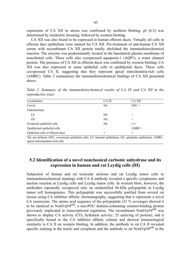
43
expression of CA XII in uterus was confirmed by northern blotting. pI (6.3) wasdetermined by isoelectric focusing, followed by western blotting.
CA XII was also found to be expressed in human efferent ducts. Virtually all cells inefferent duct epithelium were stained for CA XII. Pre-treatment of anti-human CA XIIserum with recombinant CA XII protein totally abolished the immunohistochemicalreaction. The enzyme was predominantly located in the basolateral plasma membrane ofnonciliated cells. These cells also coexpressed aquaporin-1 (AQP1), a water channelprotein. The presence of CA XII in efferent ducts was confirmed by western blotting. CAXII was also expressed in some epithelial cells in epididymal ducts. These cellscoexpressed CA II, suggesting that they represent apical mitochondria-rich cells(AMRC). Table 2 summarizes the immunohistochemical findings of CA XII presentedabove.
Table 2. Summary of the immunohistochemical results of CA IX and CA XII in thereproductive tract.
5.2 Identification of a novel nonclassical carbonic anhydrase and its expression in human and rat Leydig cells (III)
Subjection of human and rat testicular sections and rat Leydig tumor cells toimmunohistochemical stainings with CA II antibody revealed a specific cytoplasmic andnuclear reaction in Leydig cells and Leydig tumor cells. In western blots, however, theantibodies repeatedly recognized only an unidentified 66-kDa polypeptide in Leydigtumor cell homogenates. This polypeptide was successfully purified from several rattissues using CA inhibitor affinity chromatography, suggesting that it represents a novelCA isoenzyme. The amino acid sequence of the polypeptide (33 % coverage) showed itto be identical to NonO/p54nrb, a non-POU domain-containing octamer-binding proteinpreviously implicated in transcriptional regulation. The recombinant NonO/p54nrb wasshown to display CA activity (CO2 hydration activity; 25 units/mg of protein), and itspecifically bound to the CA inhibitor affinity column and showed immunologicalsimilarity to CA II on western blotting. In addition, the antibody to rat CA II revealedspecific staining in the nuclei and cytoplasm and the antibody to rat NonO/p54nrb in the
Localization CA IX CA XIICervix Nd OEC +Endometrium LE Nd + GE Nd +Oviductal epithelial cells Nd (+)Epididymal epithelial cells - AMRC +Epithelial cells of efferent duct + +Nd, not defined; OEC, occasional epithelial cells; LE, luminal epithelium; GE, glandular epithelium; AMRC,apical mitochondria-rich cells

44
nuclei of lymphocytes. Furthermore, CA activity was demonstrated histochemically inthe nuclei of lymphocytes.
5.3 CA VI in milk (IV)
The subjection of human colostrum to western blotting for CA VI revealed a major 42-kDa polypeptide band identical in size to purified human salivary CA VI. Similarly,identical-sized 42-kDa and 36-kDa polypeptide bands were also obtained from rat milkand rat purified salivary CA VI samples subjected to immunoblotting with specificantibody to rat salivary CA VI. The 42-kDa polypeptide was purified from humancolostrum on CA inhibitor affinity chromatography. PNGase F digestion of the purifiedhuman milk and salivary CA VI reduced their molecular size from 42 kDa to 36 kDa,indicating that both glycopolypeptides have a similar-sized polypeptide backbone. Toconfirm that the human milk CA is identical to the salivary isoenzyme VI, the purifiedcolostral isoenzyme was isolated from a SDS gel, trypsin-digested, and sequenced withMALDI-MS. The analysis of the sequence data with the ProFound and PeptideSearchdatabases revealed 100% identity with human salivary CA VI. The sequencedpolypeptides covered 40% of the full-length CA VI.
The mean concentrations of CA VI were 34.7 mg/l (range 10.0-78.4 mg/l, n=9) incolostrum and 4.5 mg/l (2.6-6.9 mg/l, n=4) in mature milk. The CA VI levels in the salivaof two infants were 1.9 and 3.6 mg/l, representing about half of the concentration inadults (Parkkila et al. 1993).
The resting mammary gland and the glands of adult female rat two days before andafter parturition showed a faint positive reaction for CA VI in alveolar epithelia. Alveolarmilk, however, showed a strong reaction. Rat mammary gland homogenates subjected towestern blotting showed the most intense band for CA VI in the lactating gland, amoderate band in glandular tissue two days before parturition, and only a faint band in theresting gland.
6 Discussion
6.1 CA IX and CA XII in the human reproductive tract
Although histochemical studies have demonstrated a wide distribution of CA activity inthe male and female reproductive tracts (Friedley & Rosen 1975, Rodriquez-Martinez etal. 1991, Ekstedt et al. 1991, Ekstedt & Ridderståle 1992), only three isoenzymes havebeen identified earlier in human. Cytoplasmic CA II has been demonstrated in seminalvesicle epithelium and in occasional cells in the ductal epithelium of epididymis(Kaunisto et al. 1990). CA IV is expressed in the apical plasma membrane of allepithelial cells throughout the epididymal duct and proximal vas deferens (Parkkila et al.1993). The expression of CA IX has been demonstrated in occasional epithelial cells ofthe uterine cervix, where its function is unclear (Liao et al. 1994). In the seminal vesicle,CA II has been suggested to participate in the alkalinization of the ejaculate, and therebyin the activation of sperm motility (Kaunisto et al. 1990). In the epididymis, CA II andCA IV are probably involved in the acidification of epididymal fluid, thereby helping tokeep the spermatozoa immotile during storage (Kaunisto et al. 1990, Parkkila S et al.1993a).
The results of the present study show that two plasma membrane-bound isoenzymes,CA IX and CA XII, are expressed in the basolateral plasma membrane of the epithelia inthe uterus and male excurrent ducts.
CA XII was present in the glandular and luminal epithelia of the endometrium duringthe proliferative and luteal phases and in occasional cervical epithelial cells. Recently,Ivanov et al. (2001) reported CA XII expression in glandular epithelium during theproliferative phase and in the basal cells of cervical epithelium.
In the human male reproductive tract, CA XII and CA IX were expressed in excurrentduct epithelium. CA IX expression was restricted exclusively to certain segments of theefferent ducts, while CA XII expression was detected in all segments, predominantly innonciliated cells. CA XII was also expressed in certain cells of the epididymal duct.These cells also expressed CA II, suggesting that they represent AMRCs. These cellscorrespond to rat narrow and clear cells, which are known to be involved in theacidification of epididymal fluid (Brown et al. 1992, Martìnez-Carcìa et al. 1995,

46
Adamali & Hermo 1996). Similar distribution of CA IX and CA XII in efferent ducts hasbeen reported later by Ivanov et al. (2001), although the cell types remained undefined.
The functional role of CA XII is possibly linked to the water and bicarbonateabsorption in efferent ducts and the acidification of luminal epididymal fluid in AMRC.There are several lines of evidence to support this role of CA XII. First, CA XII iscolocated with a water channel protein, AQP1, in nonciliated cells, which display amarked water-absorbing capacity (Brown et al. 1993). Water is absorbed from the lumendriven by sodium (and chloride) ions through AQP1 water channels (Brown et al. 1993,Ilio & Hess 1994, Clulow et al. 1994, Fisher et al. 1998). Second, CA XII is expressed inthe proximal convoluted tubule of the kidney, where AQP1 is also expressed (Nielsen etal. 1993, Parkkila et al. 2000b). Third, CA XII is colocated with AQP2 in the sameepithelial cells in the kidney collecting tubule (Parkkila et al. 2000b). Fourth, CA XII isalso expressed in other water-reabsorbing epithelia, such as colon and gallbladder (Kiveläet al. 2000, S. Parkkila, unpublished observation). Moreover, Tsuruoka et al. (2001) havereported that the inhibition of basolateral CA in the kidney proximal tubule decreasesbicarbonate (60%) and fluid (30%) absorption.
Ion and fluid transport in the male reproductive tract is a complex system involvingseveral ion transport proteins, many of which are still unidentified. The sodium-dependent water absorption in efferent ducts is probably driven by Na+K+-ATPase, whichis located at the basolateral plasma membrane of luminal epithelia (Ilio & Hess 1994).The apical entry of sodium into the epithelial cell is possibly mediated by the apicallylocated Na+/H+ exchanger, NHE-3 (Bagnis et al. 2001, Kaunisto & Rajaniemi 2002).Hansen et al. (1999) have shown that amiloride reduces fluid reabsorption by 70 %,probably by inhibiting the Na+/H+ exchanger. Water can be transported via the waterchannel protein isoform, AQP1, which is located in nonciliated epithelial cells (Brown etal. 1993). Recently, Herak-Kramberger et al. (2001) demonstrated the expression ofH+ATPase at the apical pole and the cytoplasm of nonciliated cells, providing anothermechanism for protons to exit the cell. In the renal proximal tubule, basolateral CA hasbeen suggested to eliminate CO3
2- gradients across the membrane (Muller-Berger et al.1997). In efferent ducts, basolateral CA XII could promote the elimination of excessHCO3
-/CO32- from the extracellular space transported by a so far unidentified bicarbonate
transporter, thus facilitating bicarbonate and water absorption (Fig. 2). CA XII would acteffectively enough, since it has high catalytic activity that is only slightly lower than inthe most efficient isoenzyme CA II (Ulmasov et al. 2000).
The narrow and clear cells of epididymal epithelium and proximal vas deferenscontain both vacuolar H+ATPase and cytoplasmic CA II. These cells also contain apicalNBC and basolaterally located NBC and AE2. The acidification of luminal fluid inproximal vas deferens has been studied using inhibitors of these proteins. The vacuolarH+ATPase inhibitor, bafilomycin, decreased acidification by 80 %, and acetazolamideinhibited proton secretion by 46 % (Breton et al. 1998). Acidification was also markedlydecreased when basolateral bicarbonate transport was inhibited. Thus, CA II may producethe protons secreted into the lumen by vacuolar H+ATPase, and CA XII may facilitate theelimination of bicarbonate from extracellular space, as in the nonciliated cells of efferentducts.
It is noteworthy that all above cell types where CA XII is expressed showsmorphological changes in estrogen receptor-α knockout mice (Hess et al. 2000). This

47
receptor is essential for fluid reabsorption in efferent ducts, and without its expression themale is infertile (Hess 2000). Therefore, it would be important to study in the futurewhether CA XII is under estrogen control and whether it thus participates in the eventsleading to infertility in animals missing estrogen receptor-α.
There is only limited information of the presence of ion transporters in endometrialepithelium, where the functional role of CA XII remains obscure. However, endometrialepithelium shows some similarities to efferent duct epithelium and AMRC. Like theepithelium of efferent ducts, endometrial epithelium absorbs sodium and transports water(uterine fluid volume is highest at ovulation and decreases before implantation),expressing Na+-K+-ATPase at its basolateral surface (Casslén & Nilsson 1984, Casslén1986, Ge & Spicer 1988, Deachapunya et al. 1999). Cytoplasmic CA and vacuolar H+-ATPase are expressed in endometrial epithelium (Korhonen et al. 1966, Friedley & Rosen1975, Skinner et al. 1999), and CHIP28 (AQP1) mRNA is expressed in uterus (Li et al.1994). In addition, the pH of uterine fluid changes during the menstrual cycle and coitus(pH is lowest at mid-cycle and rises thereafter and during coitus) (Maas et al. 1983, Foxet al. 1982). As a high-activity isoenzyme, CA XII may be involved in the regulation ofwater transport and the acid-bace balance of uterine fluid and, thus, participate in themaintenance of proper pH and fluid volume during fertilization and implantation. Thefinding that acetazolamide decreased the number of implantations in rabbits (Böving1963) is still somewhat controversial but accordant with this hypothesis.
The endometrium undergoes extensive growth and morphological organization duringthe menstrual cycle. This process is dependent on proteolytic enzymes and, thus, oncoordinated regulation of extracellular pH. The function of CA XII in endometrium ispossibly linked to this pH regulation, since CA XII has been suggested to be involved inthe acidification of the extracellular space, which facilitates tumor spread (Ivanov et al.1998), and CA XII is strongly expressed in the basal part of the endometrium, whereendometrial repair takes place. Knudsen et al. (1969) found no acetazolamide inhibitionof the proliferation of endometrium in rabbits. However, the influence of the drug on theproliferation of human endometrium has not been studied and, on the other hand, theexpression of CA XII has not been studied in rabbit endometrium.
The functional role of CA IX in the reproductive tract remains undetermined here. Itis expressed in epithelial cells of some normal tissues, shows high expression inneoplastic cells (Liao et al. 1994, Pastorek et al. 1997, Turner et al. 1997, McKiernan etal. 1997, Saarnio et al. 1998, Vermylen et al. 1999, Pastoreková et al. 1997, Kivelä et al.2000), and its function has been linked to cell proliferation (Pastoreková et al. 1992,Závada et al. 1993, Pastoreková et al. 1997, Saarnio et al. 1998). This role in theepithelium of efferent ducts is, however, unlikely, since its expression does not correlatewith that of Ki-67, a well-known marker of cell proliferation. CA IX has also beensuggested to participate in intercellular communication and cell adhesion (Pastoreková etal. 1997, Závada et al. 2000). Whether CA IX has these functions in efferent ductsremains to be established.

48
Fig. 2. Hypothetical model illustrating the suggested role of CA XII in the nonciliated cells ofefferent ducts. See text for details.
6.2 Identification of a novel nonclassical CA, NonO/p54nrb-protein
A novel CA of 66 kDa was purified in the present study from several rat tissues, usinginhibitor affinity chromatography developed for CA purification. Amino acid sequencingof the polypeptide revealed that it represents the previously cloned and characterizednuclear protein, nonO/p54nrb, a non-POU (Pit-Oct-Unc) domain-containing octamer-binding protein, which is homologous to the nuclear 54 kDa RNA-binding protein (Yanget al. 1993, Dong et al. 1993). Conventional octamer-binding proteins are a family oftranscription factors that contain a DNA-binding motif, the POU domain. NonO also hashomology with PSF, an essential mammalian splicing factor, and NonA, a Drosophilaoptomotor protein of unknown function (Yang et al. 1993). NonO lacks the POU domain,but it binds both RNA and DNA and is implicated in transcriptional regulation (Yang etal. 1993, Dong et al. 1993, Yang et al. 1997, Kamat et al. 1995, Lamb et al. 1992, Basuet al. 1997, Ladias et al. 1992, Hallier et al. 1996, Xin et al. 1992). NonO was also shownto possess CA activity. This is a novel and interesting finding, because no other class ofmammalian proteins except CAs has been shown to bind specifically to the CA inhibitoraffinity chromatography matrix and to contain CA catalytic activity. It is noteworthy thatthe catalytic activity of nonO is higher than that determined for CA III, for example(Jeffery & Carter 1980). Significant immunological cross-reactivity between CA II andnonO was also shown. The reason for this cross-reactivity is not known, but oneexplanation is the minor sequence homology found between CA II and nonO. Thepredicted amino acid sequence of nonO shares no structural elements required for

49
conventional CA activity. It lacks all conserved histidines, which are involved in zincbinding and have heretofore been considered essential for CA activity (Kannan et al.1977, Eriksson & Liljas 1991). Nor did it show structural similarity to β or γCAs (Dr. R.Tashian, personal communication). Because the CA activity and zinc binding capacity ofCA II are retained when His119 is substituted with glutamine (Lesburg et al. 1997), apolyglutamine stretch, Q29-Q38, in the nonO protein is a potential site for Zn bindingand could also be a potential site for the CA activity of nonO. However, the tertiarystructure of nonO could give more information of the CA catalytic site in nonO. CAcatalytic activity in nonO explains at least part of the nuclear CA activity seen inhistochemical CA stainings. It is also possible that CA activity is an important factor intranscriptional regulation, which would represent a novel function for CA. In addition,whether this CA activity in nonO affects steroid synthesis in Leydig cells will warrantattention in the near future.
6.3 Secretion of CA VI in milk
The present results show that CA VI is secreted into human and rat milk. The CA VIconcentration in human colostral milk is approximately eightfold compared to maturemilk. The concentration of CA VI in mature milk corresponds to the mean level reportedearlier for adult saliva (Parkkila S et al. 1993b). The CA VI concentrations in saliva werefound to be much lower in newborns than in adults (Parkkila S et al. 1993b). In addition,the saliva secretion rate of a newborn is low (Davidson 1982). Therefore, it isconceivable that the high concentrations of CA VI in colostrum possibly compensate forthe low salivary CA VI and thus play an important functional role in the newborngastrointestinal canal during the early postnatal period.
Recently, Thatcher et al. (1998) demonstrated that gustin, a salivary factor involved intaste function, is CA VI. Thereafter, CA VI has been suggested to function as a trophicfactor that promotes the growth and development of taste buds through its action on tastebud stem cells (Thatcher et al. 1998, Henkin et al. 1999a,b). This view has beensupported by the clinical disorder demonstrated by Henkin et al. (1999a), who describedthe association of a decreased concentration of salivary CA VI with pathological changesin taste buds anatomically consistent with apoptosis in a group of patients with loss anddistortion of taste and smell after an influenza-like illness. Zinc treatment caused therecovery of taste and smell in patients who showed an increase in synthesis/secretion ofCA VI (Henkin et al. 1999b). The zinc treatment also normalized the taste budmorphology. CA VI has also been shown to have characteristics similar to the nervegrowth factor (NGF) (Henkin et al. 1988). It also specifically displaces NGF from itsbinding sites on purified taste bud membranes (Lum & Henkin 1977). CA VI activatescalmodulin-dependent bovine brain cAMP phosphodiesterase, which is a factor involvedin taste function (Law et al. 1987). In addition, CA inhibitors used for different clinicalpurposes, cause taste distortions (Hansson 1961, Graber & Kelleher 1988, Miller &Miller 1990). These observations together with the present results suggest that the highconcentration of CA VI in colostrum may be linked to the development of the taste andsmell function in a newborn. Interestingly, CA VI retains its structural and functional

50
stability in the acidic milieu of gastric secretions (Parkkila et al. 1997), which enables itto act as a trophic factor even in the lower gastrointestinal tract.
CA VI may be a multifunctional protein, similarly to many other glycoproteins presentin milk (Kunz et al. 1999, Rodriquez-Palmero et al. 1999). As a glycoprotein and acid-base balance-modulating protein, it may have antimicrobial (Hooper et al. 1995), anti-inflammatory, immunomodulating, and mucosa-protecting effects (Parkkila et al. 1997).In fact, the results from our laboratory have shown that CA VI may bind to IgA and IgG(Parkkila et al. 1990, Kivelä et al. 1997), and it may thus be implicated in the knownantimicrobial and anti-inflammatory functions of these immunoglobulins (Xanthou1998). It will be challenging in the future to clarify the functions in which CA VIparticipates and to determine the possible defects resulting from its absence in milk. Thepresence or absence of CA VI in different milk formulas is another unsolved question,and this information would be important for the development of new formulas.

7 Conclusions
1. CA XII is expressed in the surface and glandular epithelial cells in the humanendometrium and in only occasional epithelial cells in the uterine cervix. It probablyregulates the extracellular pH on the basolateral surface of epithelial cells.
2. CA IX and CA XII are both expressed in the human excurrent ducts. CA XII isexpressed in the nonciliated cells of the efferent ducts and the apical mitochondria-rich cells (AMRC) in the epididymal duct. CA IX is expressed in almost all cells insome tubule sections of the efferent ducts and is almost absent in the others. In theefferent ducts where CA XII is expressed in the same cells as AQP1, CA XII may beinvolved in ion-coupled water absorption from testicular fluid. In AMRC, it mayparticipate in the acidification of epididymal fluid. The function of CA IX in theefferent ducts remains unclear.
3. NonO/p54nrb is a novel nuclear nonclassical CA with no amino acid sequencehomology to the other known CAs. It has CA activity and immunological similarityto CA II. In the reproductive tract, it is expressed at least in testicular Leydig cells andthe uterus.
4. CA VI is present in human and rat milk. Its concentration in human colostrum isseveral times higher than that in mature milk or saliva, suggesting that it is anessential regulating factor in the development of a newborn.

8 References
Acott TS & Carr DW (1984) Inhibition of bovine spermatozoa by caudal epididymal fluid: II. Inter-action of pH and a quiescence factor. Biol Reprod 30:926-935.
Adamali HI & Hermo L (1996) Apical and narrow cells are distinct cell types differing in their struc-ture, distribution, and functions in the adult rat epididymis. J Androl 17:208-222.
Aldred P, Fu P, Barrett G, Penschow JD, Wright RD, Coghlan JP & Fernley RT (1991) Human se-creted carbonic anhydrase: cDNA cloning, nucleotide sequence, and hybridization histochemis-try. Biochemistry 30:569-575.
Amann RP & Howards SS (1980) Daily spermatozoal production and epididymal spermatozoal re-serves of the human male. J Urol 124:211-215.
Ansari AH, Gould KG & Ansari VM (1980) Sodium bicarbonate douching for improvement of thepostcoital test. Fertil Steril 33:608-612.
Au CL & Wong PYD (1980) Luminal acidification by the perfused rat cauda epididymis. J Physiol309:419-427.
Bagnis C, Marsolais M, Biemesderfer D, Laprade R & Breton S (2001) Na+/H+-exchange activity andimmunolocalization of NHE3 in rat epididymis. Am J Physiol Renal Physiol 280:F426-F436.
Baillie HS, Pacey AA, Warren MA, Scudamore IW & Barratt CL (1997) Greater numbers of humanspermatozoa associate with endosalpingeal cells derived from the isthmus compared with thosefrom the ampulla. Hum Reprod 12:1985-1992.
Barnea G, Silvennoinen O, Shaanan B, Honegger AM, Canoll PD, D'Eustachio P, Morse B, Levy JB,Laforgia S, Huebner K, Musacchio JM, Sap J & Schlessinger J (1993) Identification of a carbonicanhydrase-like domain in the extracellular region of RPTPγ defines a new subfamily of receptortyrosine phosphatases. Mol Cell Biol 13:1497-1506.
Barratt CLR & Cooke ID (1991) Sperm transport in the human female reproductive tract � a dynamicinteraction. Int J Androl 14:394-411.
Basu A, Dong B, Krainer AR & Howe CC (1997) The intracisternal A-particle proximal enhancer-binding protein activates transcription and is identical to the RNA- and DNA-binding proteinp54nrb/NonO. Mol Cell Biol 17:677-686.
Bedford JM (1994) The status and state of the human epididymis. Hum Reprod 9:2187-2199.Bhattacharyya A & Yanagimachi R (1988) Synthetic organic pH buffers can support fertilization of
guinea pig eggs, but not as efficiently as bicarbonate buffer. Gamete Res 19:123-129.Boatman DE & Robbins RS (1991) Bicarbonate:carbon-dioxide regulation of sperm capacitation, hy-
peractivated motility, and acrosome reactions. Biol Reprod 44:806-813.Boatman DE (1997) Responses of gametes to the oviductal environment. Hum Reprod 12 Natl Suppl
JBFS 2:133-149.

53
Breckenridge MAB, Pederson AB & Pommerenke WT (1950) A pH study of human cervical secre-tion. Fertil Steril 1:427-434.
Breton S, Hammar K, Smith PJ, Brown D (1998) Proton secretion in the male reproductive tract: in-volvement of Cl--independent HCO3
- transport. Am J Physiol 275:C1134-C1142.Breton S, Smith PJS, Lui B & Brown D (1996) Acidification of the male reproductive tract by a pro-
ton pumping (H+)-ATPase. Nature Med 2:470-472.Brown D, Lui B, Gluck S & Sabolic I (1992) A plasma membrane proton ATPase in specialized cells
of rat epididymis. Am J Physiol 263:C913-C916.Brown D, Verbavatz J-M, Valenti G, Lui B & Sabolic I (1993) Localization of the CHIP28 water
channel in reabsorptive segments of the rat male reproductive tract. Eur J Cell Biol 61:264-273.Brown D, Zhu XL & Sly WS (1990) Localization of membrane-associated carbonic anhydrase type
IV in kidney epithelial cells. Proc Natl Acad Sci USA 87:7457-7461.Brown S, Tyszkowski R, Sabolic I & Brown D (1999) Postnatal development of H+ATPase (proton-
pump)-rich cells in rat epididymis. Histochem Cell Biol 111:97-105.Böving BG (1959) Implantation. Ann NY Acad Sci 75:700-725.Böving BG (1965) Implantation mechanisms. In: Hartman CG (eds) Mechanisms concerned with
conception, Pergamon Press, New York, p 321-396.Caflisch CR & DuBose TDJr (1990) Direct evaluation of acidification by rat testis and epididymis:
role of carbonic anhydrase. Am J Physiol 258:E143-E150.Carr DW & Acott TS (1984) Inhibition of bovine spermatozoa by caudal epididymal fluid: I. Studies
of a sperm motility quiescence factor. Biol Reprod 30:913-925.Carr DW & Acott TS (1989) Intracellular pH regulates bovine sperm motility and protein phosphor-
ylation. Biol Reprod 41:907-920.Carr DW, Usselman MC & Acott TS (1985) Effects of pH, lactate, and viscoelastic drag on sperm
motility: a species comparison. Biol Reprod 33:588-595.Carter N, Jeffery S, Shiels A, Edwards Y, Tipler T & Hopkinson DA (1979) Characterization of hu-
man carbonic anhydrase III from skeletal muscle. Biochem Genet 17:837-853.Casslén B & Nilsson B (1984) Human uterine fluid, examined in undiluted samples for osmolarity
and the concentrations of inorganic ions, albumin, glucose, and urea. Am J Obstet Gynecol150:877-881.
Casslén B (1986) Uterine fluid volume. Cyclic variations and possible extrauterine contributions. JReprod Med 31:506-510.
Chan HC, Fong SK, So SC, Chung YW & Wong PYD (1997) Stimulation of anion secretion by β�adrenoceptors in the mouse endometrial epithelium. J Physiol 501.3:517-525.
Chantler EN, Lo K & Elstein M (1977) Carbonic anhydrase activity in human cervical mucus and itsresponse to various contraceptives regimes. Br J Obstet Gynaecol 84:705-707.
Chen Y, Cann MJ, Litvin TN, Iourgenko V, Sinclair ML, Levin LR & Buck J (2000) Soluble adenylylcyclase as an evolutionarily conserved bicarbonate sensor. Science 289:625-628.
Chew SBC, Leung GPH, Leung PY, Tse CM & Wong PYD (2000) Polarized distribution of NHE1and NHE2 in the rat epididymis. Biol Reprod 62:755-758.
Chomzynski P & Sacchi N (1987) Single-step method of RNA isolation by acid guanidium thiocy-anate-phenol-chloroform extraction. Anal Biochem 162:156-159.
Clulow J, Jones RC & Hansen LA (1994) Micropuncture and cannulation studies of fluid compositionand transport in the ductuli efferentes testis of the rat: comparisons with the homologous metane-phric proximal tubule. Exp Physiol 79:915-928.
Cohen JP, Hoffer AP & Rosen S (1976) Carbonic anhydrase localization in the epididymis and testisof the rat: histochemical and biochemical analysis. Biol Reprod 14:339-346.
Common RH (1941) Carbonic anhydrase in various organs of the hen. J Agr Sci 31:412-414.

54
Conti CJ, Larcher F, Chesner J & Aldaz CM (1988) Polyacrylamide gel electrophoresis and immu-noblotting of proteins extracted from paraffin-embedded tissue sections. J Histochem Cytochem36:547-550.
Cooper GW, Overstreet JW & Katz DF (1979) The motility of rabbit spermatozoa recovered from thefemale reproductive tract. Gamete Res 2:35-42.
Cvek K, Dahlborn K & Ridderstråle Y (1998) Localization of carbonic anhydrase in the goat mam-mary gland during involution and lactogenesis. J Dairy Res 65:43-54.
Dale B, Menezo Y, Cohen J, DiMatteo L & Wilding M (1998) Intracellular pH regulation in the hu-man oocyte. Hum Reprod 13:964-970.
David A, Serr DM & Czernobilsky B (1973) Chemical composition of human oviduct fluid. FertilSteril 24:435-439.
Davidson M (1982) The gastrointestinal tract. In: Rudolph AM, Hoffman JIE & Axelrod S (eds) Pedi-atrics, Appleton-Century-Crofts, Norwalk, p 915-919.
Delporte C, Chen ZJ & Baum BJ (1996) Aquaporin 1 expression during the cell cycle in A5 cells.Biochem Biophys Res Commun 228:223-228.
Dodgson SJ, Forster RE, Schwed DA & Storey BT (1983) Contribution of matrix carbonic anhydraseto citrulline synthesis in isolated guinea pig liver mitochondria. J Biol Chem 258:7696-7701.
Dodgson SJ, Forster RE, Storey BT & Mela L (1980) Mitochondrial carbonic anhydrase. Proc NatlAcad Sci USA 77:5562-5566.
Dong B, Horowitz DS, Kobayashi R & Krainer AR (1993) Purification and cDNA cloning of HeLacell p54nrb, a nuclear protein with two RNA recognition motifs and extensive homology to humansplicing factor PSF and Drosophila NONA/BJ6. Nucleic Acids Res 21:4085-4092.
Dym M & Romrell LJ (1975) Intraepithelial lymphocytes in the male reproductive tract of rats andrhesus monkeys. J Reprod Fertil 42:1-7.
Earnhardt JN, Qian M, Tu C, Lakkis MM, Bergenhem NC, Laipis PJ, Tashian RE & Silverman DN(1998) The catalytic properties of murine carbonic anhydrase VII. Biochemistry 37:10837-10845.
Eddy EM, Washburn TF, Bunch DO, Goulding EH, Gladen BC, Lubahn DB & Korach KS (1996)Targeted disruption of the estrogen receptor gene in male mice causes alteration of spermatogen-esis and infertility. Endocrinology 137:4796-4805.
Edgren RA, Knudsen KA & Jones RC (1971) On the role of carbonic anhydrase in reproductive proc-esses. J Reprod Fertil 27:301-302.
Ekstedt E & Ridderstråle Y (1992) Histochemical localization of carbonic anhydrase in the testis andepididymis of the rabbit. Acta Anat 143:258-264.
Ekstedt E, Ridderstråle Y, Plöen L & Rodriguez-Martinez H (1991) Histochemical localization ofcarbonic anhydrase in the testis and epididymis of the boar. Acta Anat 141:257-261.
Emmet PM & Rogers IS (1997) Properties of human milk and their relationship with maternal nutri-tion. Early Hum Dev 49 Suppl: S7-S28.
Eriksson AE & Liljas A (1991) X-ray crystallographic studies of carbonic anhydrase isoenzymes I,II, and III. In: Dodgson SJ, Tashian RE & Carter ND (eds) The Carbonic Anhydrases. CellularPhysiology and Molecular Genetics, Plenium Press, New York, p 33-48.
Everhardt E, Dony JMJ, Jansen H, Lemmens WAJG, Doesburg WH (1990) Improvement of cervicalmucus viscoelasticity and sperm penetration with sodium bicarbonate douching. Hum Reprod5:133-137.
Falk RJ & Hodgen GD (1972) Carbonic anhydrase isoenzymes in normal human endometrium anderythrocytes. Am J Obstet Gynecol 112:1047-1051.
Feldstein JB & Silverman DN (1984) Purification and characterization of carbonic anhydrase fromthe saliva of the rat. J Biol Chem 259:5447-5453.
Fernley RT (1991a) Carbonic anhydrases secreted in the saliva. In: Dodgson SJ, Tashian RE, Gros G& Carter ND (eds) The Carbonic Anhydrases. Cellular Physiology and Molecular Genetics. Ple-num Press, New York, p 365-373.

55
Fernley RT (1991b) Secreted carbonic anhydrases. In: Botré F, Gros G & Storey BT (eds) Carbonicanhydrase. From Biochemistry and Genetics to Physiology and Clinical Medicine. VCH Verlags-gesellschaft, Weinheim, p 178-185.
Fernley RT, Farthing J & Cooper EJ (1995) Radioimmunoassay for salivary carbonic anhydrase inhuman parotid saliva. Arch Oral Biol 40:567-569.
Fernley RT, Wright RD & Coghlan JP (1979) A novel carbonic anhydrase from the ovine parotidgland. FEBS Lett 105:299-302.
Fernley RT, Wright RD & Coghlan, JP (1988) Complete amino acid sequence of ovine salivary car-bonic anhydrase. Biochemistry 27:2815-2820.
Fisher JS, Turner KJ, Fraser HM, Saunders PTK, Brown D & Sharpe RM (1998) Immunoexpressionof aquaporin-1 in the efferent ducts of the rat and marmoset monkey during development, its mod-ulation by estrogens, and its possible role in fluid resorption. Endocrinology 139:3935-3945.
Fisher MI, Tikkala AO & Mawson CA (1955) Zinc, carbonic anhydrase and phosphatase in the pro-static glands of the rat. Can J Biochem Physiol 33:181-190.
Fleming RE, Crouch EC, Ruzicka CA & Sly WS (1993) Pulmonary carbonic anhydrase IV: develop-mental regulation and cell-specific expression in the alveolar capillary endothelium. Am J Physiol265:L627-L635.
Fleming RE, Parkkila S, Parkkila A-K, Rajaniemi H, Waheed A & Sly WS (1995) Carbonic anhy-drase IV expression in rat and human gastrointestinal tract: Regional, cellular, and subcellular lo-calization. J Clin Invest 96:2907-2913.
Fong SK, Liu CQ & Chan HC (1998) Cellular mechanisms of adrenaline-stimulated anion secretionby the mouse endometrial epithelium. Biol Reprod 59:1342-1348.
Fordney-Settlage D (1981) A review of cervical mucus and sperm interactions in humans. Int J Fertil26:161-169.
Fox CA, Colson RH & Watson BW (1982) Continuous measurement of vaginal and intra-uterine pHby radio-telemetry during human coitus. In: Hoch Z & Lief HL (eds) Sexology, Excerpta Medica,Amsterdam, p 110-113.
Fox CA, Meldrum SJ & Watson BW (1973) Continuous measurement by radiotelemetry of vaginalpH during human coitus. J Reprod Fertil 33:69-75.
Fox CA, Wolff HS & Baker JA (1970) Measurement of intra-vaginal and intra-uterine pressures dur-ing human coitus by radio-telemetry. J Reprod Fertil 22:243-251.
Fraser LR (1995) Cellular biology of capacitation and the acrosome reaction. Hum Reprod 10 Suppl1:22-30.
Friedley NJ & Rosen S (1975) Carbonic anhydrase activity in the mammalian ovary, fallopian tube,and uterus: histochemical and biochemical studies. Biol Reprod 12:293-304.
Fujikawa-Adachi K, Nishimori I, Sakamoto S, Morita M, Onishi S, Yonezawa S & HollingsworthMA (1999a) Identification of carbonic anhydrase IV and VI mRNA expression in human pancreasand salivary glands. Pancreas 18:329-335.
Fujikawa-Adachi K, Nishimori I, Taguchi T & Onishi S (1999) Human mitochondrial carbonic an-hydrase VB. cDNA cloning, mRNA expression, subcellular localization, and mapping to chromo-some X. J Biol Chem 274:21228-21233.
Fujikawa-Adachi K, Nishimori I, Taguchi T & Onishi S (1999) Human carbonic anhydrase XIV(CA14) cDNA cloning, mRNA expression, and mapping to chromosome 1. Genomics 61:74-81.
Fujikawa-Adachi K, Nishimori I, Taguchi T, Yuri K & Onishi S (1999) cDNA sequence, mRNA ex-pression, and chromosomal localization of human carbonic anhydrase-related protein, CA-RP XI.Biochim Biophys Acta. 1431:518-524.
Gadella BM & Harrison RAP (2000) The capacitating agent bicarbonate induces protein kinase A-dependent changes in phospholipid transbilayer behavior in the sperm plasma membrane. Devel-opment 127:2407-2420.

56
Ganguly S, Sarkar D & Ghosh JJ (1978) Carbonic anhydrase of human endometrium: biochemical &clinical aspects. Indian J Biochem Biophys 15:222-226.
Ge ZH & Spicer SS (1988) Immunocytochemistry of ion transport mediators in the genital tract offemale rodents. Biol Reprod 38:439-452.
Goyal HO, Ferguson JG & Hrudka F (1980) Histochemical activity of carbonic anhydrase in testicu-lar and excurrent ducts of immature, mature intact and androgen-deprived bulls. Biol Reprod22:991-997.
Graber M & Kelleher S (1988) Side effects of acetazolamide, the champagne blues. Am J Med 84,979-980.
Guesdon JL, Ternynck T & Avrameas S (1979) The use of avidin-biotin interaction in immunoenzy-matic techniques. J Histochem Cytochem 27:1131-1139.
Hageman GS, Zhu XL, Waheed A & Sly WS (1991) Localization of carbonic anhydrase IV in spe-cific capillary bed of the human eye. Proc Natl Acad Sci USA 88:2716-2720.
Hallier M, Tavitian A & Moreau-Gachelin F (1996) The transcription factor spi-1/PU.1 binds RNAand interferes with the RNA-binding protein p54nrb. J Biol Chem 271:11177-11181.
Hamosh M (1998) Protective function of proteins and lipids in human milk. Biol Neonate 74:163-176.
Hansen LA, Clulow J & Jones RC (1999) The role of Na+-H+ exchange in fluid and solute transportin the rat efferent ducts. Exp Physiol 84:521-527.
Hansson HPJ (1961) On the effect of carbonic anhydrase inhibition on the sense of taste; and unusualside effect of a medication. Nord Med 65:566-567.
Hansson HPJ (1967) Histochemical demonstration of carbonic anhydrase activity. Histochemie11:112-128.
Harper MJK (1988) Gamete and zygote transport. In: Knobil E & Neill J (eds) The Physiology of Re-production, Raven Press, New York, p 103-134.
Harris GC Jr & Goto K (1984) Carbonic anhydrase activity of the reproductive tract tissues of agedmale fowls and its relationship to semen production. J Reprod Fertil 70:25-30.
Harrison RAP & Miller NGA (2000) cAMP-dependent protein kinase control of plasma membranelipid architecture in boar sperm. Mol Reprod Dev 55:220-228.
Harrison RAP (1996) Capacitation mechanisms, and the role of capacitation as seen in eutherianmammals. Reprod Fertil Dev 8:581-594.
Henkin RI, Law JS & Nelson NR (1988) The role of zinc on the trophic growth factors, nerve growthfactor and gustin. In: Trace elements in man and animals 6. Plenium press, New York and London,p 385-387.
Henkin RI, Martin BM & Agarwal RP (1999a) Decreased parotid saliva gustin/carbonic anhydraseVI secretion: An enzyme disorder manifested by gustatory and olfactory dysfunction. Am J MedSci 318:380-391.
Henkin RI, Martin BM & Agarwal RP (1999b) Efficacy of exogenous oral zinc in treatment with car-bonic anhydrase VI deficiency. Am J Med Sci 318:392-405.
Herak-Kramberger CM, Breton S, Brown D, Kraus O & Sabolic I (2001) Distribution of the vacuolarH+ATPase along the rat and human male reproductive tract. Biol Reprod 64:1699-1707.
Hermo L, Adamali HI & Andonian S (2000) Immunolocalization of CA II and H+V-ATPase in epi-thelial cells of the mouse and rat epididymis. J Androl 21:376-391.
Herod EL (1994) The use of milk as a saliva substitute. J Public Health Dent 3:184-189.Hess RA (2000) Estrogen and the male reproductive tract. In: Andrology 2000. Proc. 1st European
Congress of Andrology. Francevilla F, Francavilla S & Forti G (eds) Collana di Studi Abruzzesi,L�Aquila, Italia, p 279-298.
Hess RA, Bunick D, Lee K-H, Bahr J, Taylor JA, Korach KS & Lubahn DB (1997) A role for oes-trogens in the male reproductive system. Nature 390:509-512.

57
Hess RA, Bunick D, Lubahn DB, Zhou Q & Bouma J (2000) Morphologic changes in efferent duc-tules and epididymis in estrogen receptor-α knockout mice. J Androl 21:107-121.
Hetherington CM (1968) Induction of deciduomata in the mouse by carbon dioxide. Nature 219:863-864.
Hetherington CM (1968) The development of deciduomata induced by two nontraumatic methods inthe mouse. J Reprod Fertil 17:391-393.
Hewett-Emmet D (2000) Evolution and distribution of the carbonic anhydrase gene families. In: Che-gwidden WR, Carter ND & Edwards YH (eds) The Carbonic Anhydrases: New Horizons.Birkhäuser Verlag Basel, Switzerland, p 29-76.
Hodgen GD & Falk RJ (1971) Estrogen and progesterone regulation of carbonic anhydrase isoen-zymes in guinea pig and rabbit uterus. Endocrinology 89:859-864.
Hodgen GD, Gomes WR & Vandemark NL (1971) A testicular isoenzyme of carbonic anhydrase.Biol Reprod 4:224-228.
Hodgen GD, Gomes WR, & Vandemark NL (1969) Carbonic anhydrase isoenzymes in rat erythro-cytes, kidney and testis. Fed Proc 28:2878.
Hoffer AP, Hamilton DW & Fawcett DW (1973) The ultrastructure of principal cells and intraepithe-lial leucocytes in the initial segment of the rat epididymis. Anat Rec 175:169-202.
Holstein AF (1969) Morphologische studieren am nebenhoden des menschen. Zwanglose abhandlun-gen aus dem gebiet der normalen und patologischen. Anatomie 20:1-90.
Hooper LV, Beranek MC, Manzella SM & Baenziger JU (1995) Differential expression of GalNAc-4-sulfotransferase and GalNAc-transferase results in distinct glycoforms of carbonic anhydraseVI in parotid and submaxillary glands. J Biol Chem 270:5985-5993.
Härkönen PL & Väänänen HK (1988) Androgen regulation of carbonic anhydrase II, a major solubleprotein in rat lateral prostate tissue. Biol Reprod 38:377-384.
Härkönen PL, Mäkelä SI, Valve EM, Karhukorpi E-K & Väänänen HK (1991) Differential regulationof carbonic anhydrase II by androgen and estrogen in dorsal and lateral prostate of the rat. Endo-crinology 128:3219-3227.
Ilio KY & Hess RA (1994) Structure and function of the ductuli efferentes: a review. Microsc ResTech 29:432-467.
Imoedembe D, Chan R, Ramadan I & Sigue A (1993) Changes in follicular fluid gas and pH duringcarbon dioxide pneumoperitoneum for laparoscopic aspiration and their effect on human oocytefertilisability. Fertil Steril 59:177-182.
Ivanov S, Liao S-Y, Ivanova A, Danilkovitch-Miagkova A, Tarasova N, Weirich G, Merrill MJ, Proe-scholdt MA, Oldfield EH, Lee J, Zavada J, Waheed A, Sly W, Lerman MI & Stanbridge EJ (2001)Expression of hypoxia-inducible cell-surface transmembrane carbonic anhydrases in human can-cer. Am J Pathol 158:905-919.
Ivanov SV, Kuzmin I, Wei M-H, Pack S, Geil L, Johnson BE, Stanbridge EJ & Lerman MI (1998)Down-regulation of transmembrane carbonic anhydrases in renal cell carcinoma cell lines bywild-type von Hippel-Lindau transgenes. Proc Natl Acad Sci USA 95:12596-12601.
Jeffery S & Carter ND (1980) A comparison of carbonic anhydrase III isozymes from human, ba-boon, pig, and sheep muscle. Comp Biochem Physiol 66B:439-441.
Jeffery S, Edwards Y & Carter N (1980) Distribution of CA III in fetal and adult human tissues. Bi-ochem Genet 18:843-849.
Jenkins JM, Anthony FW, Purdie B, Gilbert D, Noon R & Masson GM (1989) Acidic endocervicalmucus: a potentially reversible cause of subfertility. Contemp Rev Obstet Gynaecol 1:273-278.
Jensen LJ, Schmitt BM, Berger UV, Nsumu NN, Boron WF, Hediger MA, Brown D & Breton S(1999) Localization of sodium bicarbonate cotransporter (NBC) protein and messenger ribonucle-ic acid in rat epididymis. Biol Reprod 60:573-579.
Jensen LJ, Stuart-Tilley AK, Peters LL, Lux SE, Alper SL & Breton S (1999) Immunolocalization ofAE2 anion exchanger in rat and mouse epididymis. Biol Reprod 61:973-980.
58
Jiang W & Gupta D (1999) Structure of the carbonic anhydrase VI (CA6) gene: evidence for two dis-tinct groups within the α-CA gene family. Biochem J 344:385-390.
Johnson L & Varner DD (1988) Effect of daily sperm production but not age on transit time of sper-matozoa through the human epididymis. Biol Reprod 39:812-817.
Jones RC (1999) To store or mature spermatozoa? The primary role of the epididymis. Int J Androl22:57-67.
Jonte G & Holstein AF (1987) On the morphology of the transitional zones from the rete testis intothe ductuli efferentes and from the ductuli efferentes into the ductus epididymidis. Investigationson the human testis and epididymis. Andrologia 19:398-412.
Kadoya Y, Kuwahara H, Shimazaki M, Ogawa Y & Yagi T (1987) Isolation of a novel carbonic an-hydrase from human saliva and immunohistochemical demonstration of its related isoenzymes insalivary gland. Osaka City Med J 33:99-109.
Kaluz S, Kaluzová M, Opavský R, Pastoreková S, Gibadulinova A, Dequiedt F, Kettmann R & Pas-torek J (1999) Transcriptional regulation of the MN/CA 9 gene coding for the tumor-associatedcarbonic anhydrase IX. identification and characterization of a proximal silencer element. J BiolChem 274:32588-32595.
Kaluzová M, Pastoreková S, Pastorek J & Kaluz S (2000) P53 tumour suppressor modulates tran-scription of the TATA-less gene coding for the tumour-associated carbonic anhydrase MN/CA IXin MaTu cells. Biochim Biophys Acta 1491:20-26.
Kamat JP, Basu A, Satyamoorthy K, Xu MQ, Showe L & Howe CC (1995) IPEB transcription factorregulating the intracisternal A particle gene during F9 cell differentiation is expressed at sites oflymphoid development. Mol Reprod Dev 41:8-15.
Kannan KK, Petef M, Fridborg K, Cid-Dresidner H & Lövgren S (1977) Structure and reactions ofcarbonic anhydrases: imidatsole binding to human carbonic anhydrase B and the mechanism ofaction of carbonic anhydrases. FEBS Lett 73:115-119.
Kato K (1990) Sequence of a novel carbonic anhydrase-related polypeptide and its exclusive presencein Purkinje cells. FEBS Lett 271:137-140.
Kaunisto K & Rajaniemi H (2002) Expression and localization of the Na+/H+ exchanger isoformNHE-3 in the rat efferent ducts. J Androl 23:237-241.
Kaunisto K, Fleming RE, Kneer J, Sly WS & Rajaniemi H (1999) Regional expression and androgenregulation of carbonic anhydrase IV and II in the adult rat epididymis. Biol Reprod 61:1521-1526.
Kaunisto K, Moe OW, Pelto-Huikko M, Traebert M & Rajaniemi H (2001) An apical membrane Na+/H+ exchanger isoform, NHE-3, is present in the rat epididymal epithelium. Pflugers Arch-Eur JPhysiol 442:230-236.
Kaunisto K, Parkkila S, Parkkila AK, Waheed A, Sly WS & Rajaniemi H (1995) Expression of car-bonic anhydrase isoenzymes IV and II in rat epididymal duct. Biol Reprod 52:1350-1357.
Kaunisto K, Parkkila S, Rajaniemi H, Waheed A, Grubb J & Sly WS (2002) Carbonic anhydrase XIV:Luminal expression suggests key role in renal acidification. Kidney Int, in press.
Kaunisto K, Parkkila S, Tammela T, Rönnberg L & Rajaniemi H (1990) Immunohistochemical lo-calization of carbonic anhydrase isoenzymes in the human male reproductive tract. Histochemis-try 94:381-386.
Kimber MS & Pai EF (2000) The active site architecture of Pisum sativum beta-carbonic anhydraseis a mirror image of that of alpha-carbonic anhydrases. EMBO J 19:1407-1418.
Kivelä A, Parkkila S, Saarnio J, Karttunen TJ, Kivelä J, Parkkila A-K, Waheed A, Sly WS, GrubbJH, Shah G, Türeci Ö & Rajaniemi H (2000) Expression of a novel transmembrane carbonic an-hydrase isozyme XII in normal human gut and colorectal tumors. Am J Pathol 156: 577-584.
Kivelä AJ, Parkkila S, Saarnio J, Karttunen TJ, Kivelä J, Parkkila A-K, Pastoreková S, Pastorek J,Waheed A, Sly WS & Rajaniemi H (2000) Expression of transmembrane carbonic anhydraseisoenzymes IX and XII in normal human pancreas and pancreatic tumours. Histochem Cell Biol114:197-204.

59
Kivelä J, Parkkila S, Parkkila A-K & Rajaniemi H (1999) A low concentration of carbonic anhydraseisoenzyme VI in whole saliva is associated with caries prevalence. Caries Res 33:178-184.
Kivelä J, Parkkila S, Waheed A, Parkkila A-K, Sly WS & Rajaniemi H (1997) Secretory carbonicanhydrase isoenzyme (CA VI) in human serum. Clin Chem 43:2318-2322.
Klentzeris LD (1997) The role of endometrium in implantation. Hum Reprod 12 Natl Suppl JBFS2:170-175.
Knudsen KA, Jones RC & Edgren RA (1969) Effect of a carbonic anhydrase inhibitor (Diamox) onthe progesterone-stimulated rabbit uterus. Endocrinology 85:1204-1205.
Korhonen LK, Korhonen E, Hyyppä M & Punnonen R (1966) Histochemical demonstration of car-bonic anhydrase activity in the uterus. Histochemie 6:164-167.
Krueger NX & Saito H (1992) A human transmembrane protein-tyrosine-phosphatase, PTPζ, is ex-pressed in brain and has an N-terminal receptor domain homologous to carbonic anhydrases. ProcNatl Acad Sci USA 89:7417-7421.
Kunz C, Rodriguez-Palmero M, Koletzko B & Jensen R (1999) Nutritional and biochemical proper-ties of human milk, part I: general aspects, proteins, and carbohydrates. Clin Perinatol 26:307-333.
Kurata Y (1953) Histochemical demonstration of carbonic anhydrase activity. Stain Technol 28: 231-233.
Kyriakides C & Levin RJ (1973) Endometrial mechanisms involved in the electrical activity associ-ated with the secretion of uterine fluid. J Physiol 229:17-18.
Ladias JA, Hadzopoulou-Cladaras M, Kardassis D, Cardot P, Cheng J, Zannis V & Cladaras C (1992)Transcriptional regulation of human apolipoprotein genes ApoB, ApoCIII, and ApoAII by mem-bers of the steroid hormone receptor superfamily HNF-4, ARP-1, EAR-2, and EAR-3. J BiolChem 267:15849-15860.
Laemmli UK (1970) Cleavage of structural proteins during the assembly of the head of bacteriophageT4. Nature 227:680-685.
Lakkis MM, Bergenhem NCH, O�Shea KS & Tashian RE (1997) Expression of the acatalytic carbon-ic anhydrase VIII gene, Car-8, during mouse embryonic development. Histochem J 29:135-141.
Lakkis MM, O�Shea KS & Tashian RE (1997) Differential expression of the carbonic anhydrasegenes for CA VII (Car7) and CA-RP VIII (Car8) in mouse brain. J Histochem Cytochem 45:657-662.
Lakkis MM, O�Shea KS & Tashian RE (1997) Differential expression of the carbonic anhydrasegenes for CA VII (Car7) and CA-RP VIII (Car8) in mouse brain. J Histochem Cytochem 45:657-662.
Lamar JK, Shettles LB & Delfs E (1940) Cyclic penetration of human cervical mucus to spermatozoain vitro. Am J Physiol 129:234-241.
Lamb BT, Satyamoorthy K, Solter D, Basu A, Xu MQ, Weinmann R & Howe CC (1992) A DNAelement that regulates expression of an endogenous retrovirus during F9 cell differentiation isE1A dependent. Mol Cell Biol 12:4824-4833.
Law JS, Nelson N, Watanabe K & Henkin RI (1987) Human salivary gustin is a potent activator ofcalmodulin-dependent brain phosphodiesterase. Proc Natl Acad Sci USA 84:1674-1678.
Leinonen J, Kivelä J, Parkkila S, Parkkila A-K & Rajaniemi H (1999) Salivary carbonic anhydraseisoenzyme VI is located in the human enamel pellicle. Caries Res 33:185-190.
Leinonen J, Parkkila S, Kaunisto K, Koivunen P & Rajaniemi H (2001) Secretion of carbonic anhy-drase isoenzyme VI (CA VI) from human and rat lingual serous von Ebner`s glands. J HistochemCytochem 49:657-662.
Leiter E (1964) Studies on carbonic anhydrase activity in the male accessory sex organs. Invest Urol2:58-70.
Lesburg CA, Huang C-C, Christianson DW & Fierke CA (1997) Histidine → carboxamide ligandsubstitutions in the zinc binding site of carbonic anhydrase II alter metal coordination geometrybut retain catalytic activity. Biochemistry 36:15780-15791.
60
Levin RJ & Scargill M (1987) Role of bicarbonate and chloride ions in rat endometrial electrogenicsecretory activity. J Physiol 391:21.
Levine N & Kelly H (1978) Measurement of pH in rat epididymis in vivo. J Reprod Fertil 52:333-335.Levine N & Marsh DJ (1971) Micropuncture studies of the electrochemical aspects of fluid and elec-
trolyte transport in individual seminiferous tubules, the epididymis and the vas deferens in rats. JPhysiol 213:557-570.
Levy JB, Canoll PD, Silvennoinen O, Barnea G, Morse B, Honegger AM, Huang J-T, CannizzaroLA, Park S-H, Druck T, Huebner K, Sap J, Ehrlich M, Musacchio JM & Schlessinger J (1993)The cloning of a receptor-type protein tyrosine phosphatase expressed in the central nervous sys-tem. J Biol Chem 268:10573-10581.
Li X, Yu H & Koide SS (1994) The water channel gene in human uterus. Biochem Mol Biol Int32:371-377.
Liao SY, Aurelio ON, Jan K, Závada J & Stanbridge EJ (1997) Identification of the MN/CA9 proteinas a reliable diagnostic biomarker of clear cell carcinoma of the kidney. Cancer Res 57:2827-2831.
Liao SY, Brewer C, Závada J, Pastorek J, Pastorekovà S, Manetta A, Berman ML, DiSaia PJ & Stan-bridge EJ (1994) Identification of the MN antigen as a diagnostic biomarker of cervical intraepi-thelial squamous and glandular neoplasia and cervical carcinomas. Am J Pathol 145:598-609.
Lindholmer CH (1974) The importance of seminal plasma for human sperm motility. Biol Reprod10:533-542.
Ling B, Bergenhem NCH, Dogson SJ, Forster RE & Tashian RE (1994) Determination of mRNA lev-els for six carbonic anhydrase isozymes in rat lung. Isozyme Bull 27:47.
Lum CYK & Henkin RI (1977) Binding of mouse 2.5 S nerve growth factor (NGF) to purified bovinetaste bud membrane (TBM): a new radioreceptor assay for NGF. Fed Proc 36:968.
Lutwak-Mann C (1955) Carbonic anhydrase in the female reproductive tract. Occurrence, distribu-tion, and hormonal dependence. J Endocrin 13:26-38.
Lönnerholm G, Selking Ö & Wistrand PJ (1985) Amount and distribution of carbonic anhydrases CAI and CA II in the gastrointestinal tract. Gastroenterology 88: 1151-1161.
Maas DHA, Reiss G & Braun D (1983) pH, pCO2, and lactate concentration in human uterine fluid.Arch Androl 11:188.
Maren TH (1960) A simplified micromethod for the determination of carbonic anhydrase and its in-hibitors. J Pharmacol Exp Ther 130:26-29.
Maren TH (1967) Carbonic anhydrase: chemistry, physiology, and inhibition. Physiol Rev 47:595-781.
Martínez-Carcía F, Regadera J, Cobo P, Palacios J, Paniagua R & Nistal M (1995) The apical mito-chondria-rich cells of the mammalian epididymis. Andrologia 27:195-206.
Martan J, Risley P & Hruban Z (1964) Holocrine cells of the human epididymis. Fertil Steril 15:180-187.
Matsuda R (1964) A progestin assay by the measurement of carbonic anhydrase activity. Adv ObstetGynecol 16:1-13.
Maurel P, Rauch U, Flad M, Margolis RK & Margolis RU (1994) Phosphacan, a chondroitin sulfateproteoglycan of brain that interacts with neurons and neural cell-adhesion molecules, is an extra-cellular variant of a receptor-type protein tyrosine phosphatase. Proc Natl Acad Sci USA 91:2512-2516.
Mawson CA & Fischer MI (1952) Carbonic anhydrase and zinc in prostate gland of the rat and rabbit.Arch Biochem Biophys 36:485-486.
McDonald RR & Lumley IB (1970) Endocervical pH measured in vivo through the normal menstrualcycle. Obstet Gynecol 35:202-206.
McIntosh J (1969) Carbonic anhydrase isoenzymes in the eryhrocytes and dorsolateral prostate of therat. Biochem J 14:463-467.

61
McKiernan JM, Buttyan R, Bander NH, Stifelman MD, Katz AE, Chen M-W, Olsson CA & SawczukIS (1997) Expression of the tumor-associated gene MN: a potential biomarker for human renal cellcarcinoma. Cancer Res 57:2362-2365.
Meaker SR & Glaser W (1929) The hydrogen ion concentration of the endocervical secretions. SurgGynecol Obstet 48:73-78.
Meldrum NU & Roughton FJW (1932) Some properties of carbonic anhydrase, the carbon dioxideenzyme present in blood. J Physiol (London) 75:15-16.
Meldrum, NU & Roughton FJW (1933) Carbonic anhydrase. Its preparation and properties. J. Phys-iol. (London) 80:113-142.
Metcalfe HK, Monson JP, Drew PJ, Iles RA, Carter ND & Cohen RD (1985) Inhibition of glucone-ogenesis and urea synthesis in isolated rat hepatocytes by acetazolamide. Biochem Soc Trans13:255.
Mezquita J, Pau M & Mezquita C (1994) A novel carbonic anhydrase II mRNA isolated from maturechicken testis displays a TATA box and other promoter sequences in a leader 5' untranslated re-gion not present in somatic tissues. Gene 147:231-235.
Mezquita P, Mezquita C & Mezquita J (1999) Novel transcripts of carbonic anhydrase II in mouseand human testis. Mol Hum Reprod 5:199-205.
Miller EG & Kurzrok R (1932) Biochemical studies of human semen. III Factors affecting migrationof sperm through the cervix. Am J Obstet Gynecol 24:19-26.
Miller LG & Miller SM (1990) Altered taste secondary to acetazolamide therapy. J Fam Pract 31:199-200.
Mitsuhashi S, Mizushima T, Yamashita E, Yamamoto M, Kumasaka T, Moriyama H, Ueki T,Miyachi S & Tsukihara T (2000) X-ray structure of β-carbonic anhydrase from the red alga,Porphyridium purpureum, reveals a novel catalytic site for CO2 hydration. J Biol Chem 275:5521-5526.
Miyake T & Pincus G (1959) Hormonal influences on the carbonic anhydrase concentration in theaccessory reproductive tracts of the rat. Endocrinology 65:64-72.
Moghissi KS (1966) Cyclic changes of cervical mucus in normal and progestin-treated women. FertilSteril 17:663-675.
Montgomery JC, Venta PJ, Eddy RL, Fukushima Y-S, Shows TB & Tashian RE (1991) Characteri-zation of the human gene for a newly discovered carbonic anhydrase, CA VII, and its localizationto chromosome 16. Genomics 11: 835-848.
Mori K, Ogawa Y, Ebihara K, Tamura N, Tashiro K, Kuwahara T, Mukoyama M, Sugawara A, OzakiS, Tanaka I & Nakao K (1999) Isolation and characterization of CA XIV, a novel membrane-bound carbonic anhydrase from mouse kidney. J Biol Chem 274:15701-15705.
Moyer DL, Rimdusit S & Mishell DRJr (1970) Sperm distribution and degradation in the human fe-male reproductive tract. Obstet Gynecol 35:831-840.
Mühlhauser J, Crescimanno,C, Rajaniemi H, Parkkila S, Milovanov AP, Castellucci M & KaufmannP (1994) Immunohistochemistry of carbonic anhydrase in human placenta and fetal membranes.Histochemistry 101: 91-98.
Müller-Berger S, Nesterov VV & Frömter E (1997) Partial recovery of in vivo function by improvedincubation conditions of isolated renal proximal tubule. II. Change of Na-HCO3 cotransport stoi-chiometry and of response to acetazolamide. Pflügers Arch-Eur J Physiol 434:383-391.
Murakami H & Sly WS (1987) Purification and characterization of human salivary carbonic anhy-drase. J Biol Chem 262:1382-1388.
Newcombe N, Clulow J, Man SY & Jones RC (2000) pH and bicarbonate in the ductuli efferentestestis of the rat. Int J Androl 23:46-50.
Nicholls RA & Board JA (1967) Carbonic anhydrase concentration in endometrium after oral pro-gestins. Am J Obstet Gynecol 99:829-832.
62
Nielsen S, Smith BL, Christensen EI, Knepper MA & Arge P (1993) CHIP28 water channels are lo-calized in constitutively water-permeable segments of nephron. J Cell Biol 120:371-383.
Nishita T & Matsushita H (1989) Immunocytochemical localization of carbonic anhydrase isozymeIII in equine thymus. Histochemistry 91:39-42.
Ogawa Y, Chang CK, Kuwahara H, Hong SS, Toyosawa S & Yagi T (1992) Immunoelectron micro-scopy of carbonic anhydrase isozyme VI in rat submandibular gland: comparison with isozymesI and II. J Histochem Cytochem 40:807-817.
Ogawa Y, Toyosawa S, Inagaki T, Hong S-S & Ijuhin N (1995) Carbonic anhydrase isozyme VI inrat lacrimal gland. Histochemistry 103:387-394.
Okamura N, Tajima Y & Sugita Y (1988) Decrease in bicarbonate transport activities during epidi-dymal maturation of porcine sperm. Biochem Biophys Res Commun 157:1280-1287.
Okamura N, Tajima Y, Ishikawa H, Yoshii S, Koiso K & Sugita Y (1986) Lowered levels of bicar-bonate in seminal plasma cause the poor sperm motility in human infertile patients. Fertil Steril45:265-272.
Okamura N, Tajima Y, Soejima A, Masuda H & Sugita Y (1985) Sodium bicarbonate in seminal plas-ma stimulates the motility of mammalian spermatozoa through direct activation of adenylate cy-clase. J Biol Chem 260:9699-9705.
Oosterwijk E, De Weijert M, Van Bokhoven A, Brakenhoff RH, Peelen WP & Debruyne FMJ (1996)Molecular characterization of the renal cell carcinoma-associated antigen G250. Proc Amer AssocCancer Res 37:461.
Opavský R, Pastorekovà S, Zelník V, Gibadulinovà A, Stanbridge EJ, Zàvada J, Kettman R & Pas-torek J (1996) Human MN/CA9 gene, a novel member of the carbonic anhydrase family: structureand exon to protein domain relationships. Genomics 33:480-487.
Palacios J, Regadera J, Nistal M & Paniagua R (1991) Apical mitochondria-rich cells in the humanepididymis: an ultrastructural, enzymohistochemical, and immunohistochemical study. Anat Rec231:82-88.
Parkkila A-K, Parkkila S, Juvonen T & Rajaniemi H (1993) Carbonic anhydrase isoenzymes II and Iare present in the zona glomerulosa cells of the human adrenal gland. Histochemistry 99:37-41.
Parkkila A-K, Scarim AL, Parkkila S, Waheed A, Corbett JA & Sly WS (1998) Expression of car-bonic anhydrase V in pancreatic B-cells suggests role for mitochondrial carbonic anhydrase in in-sulin secretion. J Biol Chem 273: 24620-24623.
Parkkila S & Parkkila A-K (1996) Carbonic anhydrase in the alimentary tract. Roles of the differentisozymes and salivary factors in the maintenance of optimal conditions in the gastrointestinal ca-nal. Scand J Gastroenterol 31:305-317.
Parkkila S, Kaunisto K & Rajaniemi H (1991a) Location of the carbonic anhydrase isoenzymes VIand II in human salivary glands by immunohistochemistry. In: Botré F, Gros G & Storey BT (eds)Carbonic Anhydrase. From Biochemistry and Genetics to Physiology and Clinical Medicine.VCH Verlagsgesellschaft, Weinheim, p 254-257.
Parkkila S, Kaunisto K, Kellokumpu S & Rajaniemi H (1991b) A high activity carbonic anhydraseisoenzyme (CA II) is present in mammalian spermatozoa. Histochemistry 95:477-482.
Parkkila S, Kaunisto K, Rajaniemi L, Kumpulainen T, Jokinen K & Rajaniemi H (1990) Immunohis-tochemical localization of carbonic anhydrase isoenzymes VI, II and I in human parotid and sub-mandibular glands. J Histochem Cytochem 38:941-947.
Parkkila S, Parkkila A-K & Rajaniemi H (1995) Circadian periodicity in salivary carbonic anhydraseVI concentration. Acta Physiol Scand 154:205-211.
Parkkila S, Parkkila A-K, Juvonen T & Rajaniemi H (1994) Distribution of carbonic anhydrase isoen-zymes I, II and VI in the human alimentary tract. Gut 35: 646-650.
Parkkila S, Parkkila A-K, Juvonen T, Waheed A, Sly WS, Saarnio J, Kaunisto K, Kellokumpu S &Rajaniemi H (1996) Membrane-bound carbonic anhydrase IV is expressed in the luminal plasmamembrane of the human gallbladder epithelium. Hepatology 24:1104-1108.

63
Parkkila S, Parkkila A-K, Kaunisto K, Waheed A, Sly WS & Rajaniemi H (1993a) Location of amembrane-bound carbonic anhydrase isoenzyme (CA IV) in the human male reproductive tract.J Histochem Cytochem 41:751-757.
Parkkila S, Parkkila A-K, Lehtola J, Reinilä A, Södervik H-J, Rannisto M & Rajaniemi H (1997) Sal-ivary carbonic anhydrase protects gastroesophageal mucosa from acid injury. Dig Dis Sci42:1013-1019.
Parkkila S, Parkkila A-K, Rajaniemi H, Shah GN, Grubb JH, Waheed A & Sly WS (2000a) Expres-sion of membrane-associated carbonic anhydrase XIV on neurons and axons in mouse and humanbrain. Proc Natl Acad Sci USA 98:1918-1923.
Parkkila S, Parkkila A-K, Saarnio J, Kivelä J, Karttunen TJ, Kaunisto K, Waheed A, Sly WS, TüreciÖ, Virtanen I & Rajaniemi H (2000b) Expression of the membrane-associated carbonic anhydraseisozyme XII in the human kidney and renal tumors. J Histochem Cytochem 48:1601-1608.
Parkkila S, Parkkila A-K, Vierjoki T, Ståhlberg T & Rajaniemi H (1993b) Competitive time-resolvedimmunofluorometric assay for quantifying carbonic anhydrase VI in saliva. Clin Chem 39:2154-2157.
Parkkila S, Rajaniemi H, Parkkila A-K, Kivelä J, Waheed A, Pastoreková S, Pastorek J & Sly WS(2000c) Carbonic anhydrase inhibitor suppresses invasion of renal cancer cells in vitro. Proc NatlAcad Sci USA 97:2220-2224.
Pastorek J, Pastoreková S, Callebaut I, Mornon JP, Zelník V, Opavský R, Zat´ovicová M, Liao S, Por-tetelle D, Stanbridge EJ, Závada J, Burny A & Kettmann R (1994) Cloning and characterizationof MN, a human tumor-associated protein with a domain homologous to carbonic anhydrase anda putative helix-loop-helix DNA binding segment. Oncogene 9:2877-2888.
Pastorekovà S, Závadová Z, Ko�tál M, Babu�íková O & Závada J (1992) A novel quasi-viral agent,MaTu, is a two-component system. Virology 187:620-626.
Pastoreková S, Parkkila S, Parkkila A-K, Opavský R, Zelník V, Saarnio J & Pastorek J (1997) Car-bonic anhydrase IX, MN/CA IX: Analysis of stomach complementary DNA sequence and expres-sion in human and rat alimentary tracts. Gastroenterology 112:398-408.
Peles E, Nativ M, Campbell PL, Sakurai T, Martinez R, Lev S, Clary DO, Schilling J, Barnea G,Plowman GD, Grumet M & Schlessinger J (1995) The carbonic anhydrase domain of receptor ty-rosine phosphatase ß is a functional ligand for the axonal cell recognition molecule contactin. Cell82: 251-260.
Petäjä-Repo UE, Merz WE & Rajaniemi HJ (1991) Significance of the glycan moiety of the rat ovar-ian luteinizing hormone/chorionic gonadotropin (CG) receptor and human CG for receptor-hor-mone interaction. Endocrinology 128:1209-1217.
Phillips KP, Léveillé M-C, Claman P & Baltz JM (2000) Intracellular pH regulation in human preim-plantation embryos. Hum Reprod 15:896-904.
Pincus G & Bialy G (1963) Carbonic anhydrase in steroid-responsive tissues. Recent Prog Horm Res19:201-250.
Purohit SB, Laloraya M & Kumar PG (1998) Bicarbonate-dependent lipid ordering and protein ag-gregation are part of the nongenomic action of progesterone on capacitated spermatozoa. J Androl19:608-618.
Pushkin A, Clark I, Kwon T-H, Nielsen S & Kurtz I (2000) Immunolocalization of NBC3 and NHE3in the rat epididymis: colocalization of NBC3 and the vacuolar H+-ATPase. J Androl 21:708-720.
Reid BL & Cleland KW (1957) The structure and function of the epididymis. I. The histology of therat epididymis. Aust J Zool 5:223-246.
Ridderstråle Y, Ekstedt E & Plöen L (1985) Localization of carbonic anhydrase in testis. Acta PhysiolScand 124 Suppl 542:408.
Robaire B & Hermo L (1988) Efferent ducts, epididymis, and vas deferens: Structure, functions, andtheir regulation. In: Knobil E & Neill J (eds) The Physiology of Reproduction, Raven Press, NewYork, p 999-1080.
64
Rodriguez-Martinez H, Ekstedt E & Einarsson S (1990) Acidification of epididymal fluid in the boar.Int J Androl 13:238-243.
Rodriguez-Martinez H, Ekstedt E & Ridderstrale Y (1991) Histochemical localization of carbonic an-hydrase in the female genitalia of pigs during the oestrous cycle. Acta Anat 140:41-47.
Rodriguez-Martinez H, Hurst M & Ekstedt E (1990) Localization of carbonic anhydrase activity inthe developing boar testis. Acta Anat 139:173-177.
Rodriguez-Palmero M, Koletzko B, Kunz C & Jensen R (1999) Nutritional and biochemical proper-ties of human milk: II: lipids, micronutrients, and bioactive factors. Clin Perinatol 26:335-359.
Rubinstein BB, Strauss H, Lazarus ML & Hankin H (1951) Sperm survival in women: motile spermin the fundus and tubes of surgical cases. Fertil Steril 2:15-19.
Räisänen SR, Lehenkari P, Tasanen M, Rahkila P, Härkonen PL & Väänänen HK (1999) Carbonicanhydrase III protects cells from hydrogen peroxide-induced apoptosis. FASEB J 13:513-522.
Saarnio J, Parkkila S, Parkkila A-K, Haukipuro K, Pastoreková S, Pastorek J, Kairaluoma MI & Kart-tunen TJ (1998) Immunohistochemical study of colorectal tumors for expression of a novel trans-membrane carbonic anhydrase, MN/CA IX, with potential value as a marker of cell proliferation.Am J Pathol 153:279-285.
Saarnio J, Parkkila S, Parkkila A-K, Waheed A, Casey MC, Zhou XY, Pastoreková S, Pastorek J,Karttunen T, Haukipuro K, Kairaluoma MI & Sly WS (1998) Immunohistochemistry of carbonicanhydrase isozyme IX (MN/CA IX) in human gut reveals polarized expression in the epithelialcells with the highest proliferative capacity. J Histochem Cytochem 46:497-504.
Saarnio J, Parkkila S, Parkkila A-K, Waheed A, Karttunen TJ & Sly WS (1999) Cell-specific expres-sion of mitochondrial carbonic anhydrase in the human and rat gastrointestinal tract. J HistochemCytochem 47:517-524.
Sabeur K & Meizel S (1995) Importance of bicarbonate to the progesterone-initiated human spermacrosome reaction. J Androl 16:266-271.
Saitoh K, Terada T & Hatakeyama S (1990) A morphological study of the efferent ducts of the humanepididymis. Int J Androl 13:369-376.
Schoysman R & Bedford JM (1986) The role of the human epididymis in sperm maturation and spermstorage as reflected in the consequences of epididymovasostomy. Fertil Steril 46:293-299.
Schoysman R (1993) Clinical situations challenging the established concept of epididymal physiolo-gy in the human. Acta Eur Fertil 24:55-60.
Scott J (1979) Qualitative and quantitative changes in the histology of the human submandibular sal-ivary gland during post natal growth. J Biol Buccale 7:341-352.
Sender S, Gros G, Waheed A, Hageman GS & Sly WS (1994) Immunohistochemical localization ofcarbonic anhydrase IV in capillaries of rat and human skeletal muscle. J Histochem Cytochem42:1229-1236.
Setchell BP (1967) Fluid secretion by the testis. J Reprod Fertil 14:347-348.Shah GN, Hewett-Emmett D, Grubb JH, Migas MC, Fleming RE, Waheed A & Sly WS (2000) Mi-
tochondrial carbonic anhydrase CA VB: differences in tissue distribution and pattern of evolutionfrom those of CA VA suggest distinct physiological roles. Proc Natl Acad Sci USA 97:1677-1682.
Shalgi R, Kraicer PF & Soferman N (1972) Gases and electrolytes of human follicular fluid. J ReprodFertil 28:335-340.
Shima K, Tashiro K, Hibi N, Tsukosia Y & Hirai H (1983) Carbonic anhydrase III. Immunohisto-chemical localization in human skeletal muscle. Acta Neuropathol (Berlin) 59:237-239.
Silber SJ (1988) Pregnancy caused by sperm from vasa efferentia. Fertil Steril 49:373-375.Silber SJ (1989) Apparent fertility of human spermatozoa from the caput epididymis. J Androl
10:263-269.Silber SJ, Balmaceda J, Borrero C, Ord T & Asch R (1988) Pregnancy with sperm aspiration from
the proximal head of the epididymis: a new treatment for congenital absence of the vas deferens.Fertil Steril 50:525-528.

65
Skaggs LA, Bergenhem NC, Venta PJ & Tashian RE (1993) The deduced amino acid sequence ofhuman carbonic anhydrase-related protein (CARP) is 98% identical to the mouse homologue.Gene 126:291-292.
Skaggs LA, Bergenhem NCH, Venta PJ & Tashian RE (1993). The deduced amino acid sequence ofhuman carbonic anhydrase-related protein (CARP) is 98% identical to the mouse homologue.Gene 126:291-292.
Skinner MA, MacLaren LA & Wildeman AG (1999) Stage-dependent redistribution of the V-ATPaseduring bovine implantation. J Histochem Cytochem 47:1247-1254.
Sly WS & Hu PY (1995) Human carbonic anhydrases and carbonic anhydrase deficiences. Annu RevBiochem 64: 375-401.
Sok J, Wang X-Z, Batchvarova N, Kuroda M, Harding H & Ron D (1999) CHOP-dependent stress-inducible expression of a novel form of carbonic anhydrase VI. Mol Cell Biol 19:495-504.
Speroff L, Glass RH & Kase NG (1994) Clinical and gynecologic endocrinology and infertility. Wil-liams & Wilkins, Baltimore, p 231-250.
Spicer SS, Ge Z-H, Tashian RE, Hazen-Martin DJ & Schulte BA (1990) Comparative distribution ofcarbonic anhydrase isoenzymes III and II in rodent tissues. Am J Anat 187:55-64.
Stambaugh R, Noriega C & Mastroianni LJr (1969) Bicarbonate ion; the corona cell dispersing factorof rabbit tubal fluid. J Reprod Fertil 18:51-58.
Stauss CR, Votta TJ & Suarez SS (1995) Sperm motility hyperactivation facilitates penetration of thehamster zona pellucida. Biol Reprod 53:1280-1285.
Stauss CR, Votta TJ & Suarez SS (1995) Sperm motility hyperactivation facilitates penetration of thehamster zona pellucida. Biol Reprod 53:1280-1285.
Steffens MG, Boerman OC, Oosterwijk-Wakka JC, Oosterhof GO, Witjes JA, Koenders EB, OyenWJ, Buijd WC, Debruyne FMJ, Corstens FH & Oosterwijk E (1997) Targeting of renal cell car-cinoma with iodine-131-labelled chimeric monoclonal antibody G250. J Clin Oncol 15:1529-1537.
Strandell A, Sjögren A, Bentin-Ley U, Thorburn J, Hamberger L & Brännström M (1998) Hydrosal-phinx fluid does not adversely affect the normal development of human embryos and implantationin vitro. Hum Reprod 13:2921-2925.
Suarez SS (1996) Hyperactivated motility of sperm. J Androl 17:331-335.Suarez SS (1998) The oviductal sperm reservoir in mammals: mechanism of formation. Biol Reprod
58:1105-1107.Sundaram V, Rumbolo P, Grubb J, Strisciuglio P & Sly WS (1986) Carbonic anhydrase II deficiency:
diagnosis and carrier detection using differential enzyme inhibition and inactivation. Am J HumGenet 38:125-136.
Sutherland GR, Baker E, Fernandez KE, Callen DF, Aldred P, Coghlan JP, Wright RD & Fernley RT(1989) The gene for human carbonic anhydrase VI (CA6) is on the tip of the short arm of chro-mosome 1. Cytogenet Cell Genet 50:149-150.
Suzuki K, Ebihara M, Nagai T, Clarke NGE & Harrison RAP (1994) Importance of bicarbonate/CO2for fertilization of pig oocytes in vitro, and synergism with caffeine. Reprod Fertil Dev 6:221-227.
Swenson ER (1991) Distribution and functions of carbonic anhydrase in the gastrointestinal tract. In:Dodgson SJ, Tashian RE, Gros G & Carter ND (eds). The Carbonic Anhydrases. Cellular Physi-ology and Molecular Genetics. Plenum Press, New York, p 265-287.
Tajima Y, Okamura N & Sugita Y (1987) The activating effects of bicarbonate on sperm motility andrespiration at ejaculation. Biochem Biophys Acta 924:519-529.
Tashian RE (1989) The carbonic anhydrases: widening perspectives on their evolution, expressionand function. BioEssays 10:186-192.
66
Tashian RE, Hewett-Emmett D, Carter ND & Bergenhem NCH (2000) Carbonic anhydrase (CA)-re-lated proteins (CA-RPs), and transmembrane proteins with CA or CA-RP domains. In: Chegwid-den WR, Carter ND & Edwards YH (eds) The Carbonic Anhydrases: New Horizons. BirkhäuserVerlag Basel, Switzerland, p 105-120.
Thatcher BJ, Doherty AE, Orvisky E, Martin BM & Henkin RI (1998) Gustin from human parotidsaliva is carbonic anhydrase VI. Biochem Biophys Res Commun 250:635-641.
Tsuruoka S, Swenson ER, Petrovic S, Fujimura A & Schwartz GJ (2001) Role of basolateral carbonicanhydrase in proximal tubular fluid and bicarbonate absorption. Am J Physiol Renal Physiol280:F146-F154.
Türeci Ö, Sahin U, Vollmar E, Siemer S, Göttert E, Seitz G, Parkkila A-K, Shah GN, Grubb JH,Pfreundschuh M & Sly WS (1998) Human carbonic anhydrase XII: cDNA cloning, expression,and chromosomal localization of a carbonic anhydrase gene that is overexpressed in some renalcell cancers. Proc Natl Acad Sci USA 95:7608-7613.
Turner JR, Odze RD, Crum CP & Resnick MB (1997) MN antigen expression in normal, preneoplas-tic and neoplastic esophagus: a clinicopathological study of a new cancer-associated biomarker.Hum Pathol 28:740-744.
Turner TT (1995) On the epididymis and its role in the development of the fertile ejaculate. J Androl16:292-298.
Uemura H, Kitagawa H, Hirao Y, Okajima E, Debruyne FMJ & Oosterwijk E (1997) Expression oftumor-associated antigen MN/G250 in urologic carcinoma: potential therapeutic target. J UrolSuppl 157:377.
Uemura H, Nakagawa Y, Yoshida K, Saga S, Yoshikawa K, Hirao Y & Oosterwijk E (1999) MN/CAIX/G250 as a potential target for immunotherapy of renal cell carcinomas. Br J Cancer 81:741-746.
Ulmasov B, Waheed A, Shah GN, Grubb JH, Sly WS, Tu C & Silverman DN (2000) Purification andkinetic analysis of recombinant CA XII, a membrane carbonic anhydrase overexpressed in certaincancers. Proc Natl Acad Sci USA 97:14212-14217.
Venta PJ, Montgomery JC & Tashian RE (1987) Molecular genetics of carbonic anhydrase isozymes.Isozymes: Curr Top Biol Med Res 14: 59-72.
Veri JP, Hermo L & Robaire B (1993) Immunocytochemical localization of the Yf subunit of glutath-ione S-transferase shows regional variation in the staining of epithelial cells of the testis, efferentducts, and epididymis of the male rat. J Androl 14:23-44.
Vermylen P, Roufosse C, Burny A, Verhest A, Bosschaerts T, Pastoreková S, Ninane V & Sculier J-P (1999) Carbonic anhydrase IX antigen differentiates between preneoplastic malignant lesions innon-small cell lung carcinoma. Eur Respir J 14:806-811.
Väänänen HK & Autio-Harmainen H (1987) Carbonic anhydrase III: a new histochemical marker formyoepithelial cells. J Histochem Cytochem 35:683-686.
Väänänen HK, Paloniemi M & Vuori J (1985) Purification and localization of human carbonic anhy-drase III. Typing of skeletal muscle fibers in paraffin embedded sections. Histochemistry 83:231-235.
Waheed A, Okuyama T, Heyduk T & Sly WS (1996) Carbonic anhydrase IV: purification of a secre-tory form of the recombinant human enzyme and identification of the positions and importance ofits disulfide bonds. Arch Biochem Biophys 333:432-438.
Waldeyer A & Häusler G (1959) Histochemical studies of the carbonic anhydrase activity of vas def-erens and its accessory glands in the Mus Rattus. Acta Biol Germ Med 2:568-589.
Wales RG, Wallace JC & White IG (1966) Composition of bull epididymal and testicular fluid. J Re-prod Fertil 12:139.
Wang YF & Holstein AF (1983) Intraepithelial lymphocytes and macrophages in the human epidi-dymis. Cell Tissue Res. 233:517-521.
67
White PS, Jensen SJ, Rajalingam V, Stairs D, Sulman EP, Maris JM, Biegel JA, Wooster R & Bro-deur GM (1998) Physical mapping of the CA VI, ENO1, and SLC2A5 (GLUT5) genes and reas-signment of SLC2A5 to 1p36.2. Cytogenet Cell Genet 81:60-64.
Wilhelm B, Keppler C, Hoffbauer G, Lottspeich F, Linder D, Meinhardt A, Aumüller G & Seitz J(1998) Cytoplasmic carbonic anhydrase II of rat coagulating gland is secreted via the apocrine ex-port mode. J Histochem Cytochem 46:505-511.
Williams M, Hill CJ, Scudamore I, Dunphy B, Cooke ID & Barratt CL (1993) Sperm numbers anddistributions within the human fallopian tube around ovulation. Hum Reprod 8:2019-2026.
Xanthou M (1998) Immune protection of human milk. Biol Neonate 74:121-133.Xin J-H, Cowie A, Lachance P & Hassel JA (1992) Molecular cloning and characterization of PEA3,
a new member of Ets oncogene family that is differentially expressed in mouse embryonic cells.Genes Dev 6:481-496.
Xu R-J (1996) Developmant of the newborn GI tract and its relation to colostrum/milk intake: a re-view. Reprod Fertil Dev 8:35-48.
Yanagimachi R (1994) Mammalian Fertilization. In: Knobil E & Neill J (eds) The Physiology of Re-production, Raven Press, New York, p 189-317.
Yang Y-S, Hanke JH, Carayannopoulos L, Craft CM, Capra JD & Tucker PW (1993) NonO, a non-POU-domain-containing, octamer-binding protein, is the mammalian homolog of DrosophilanonAdiss. Mol Cell Biol 13:5593-5603.
Yang Y-S, Yang M-CW, Tucker PW & Capra JD (1997) NonO enhances the association of manyDNA-binding proteins to their targets. Nucleic Acids Res 25:2284-2292.
Yeung CH, Cooper TG, Bergmann M & Schulze H (1991) Organization of tubules in the human caputepididymidis and the ultrastructure of their epithelia. Am J Anat 191:261-279.
Yeung CH, Nashan D, Cooper TG, Sorg C, Oberpenning F, Schulze H & Nieschlag E (1994) Basalcells of the human epididymis-antigenic and ultrastructural similarities to tissue-fixed macrophag-es. Biol Reprod 50:917-926.
Závada J, Závadová Z, Machon O, Kutinova L, Nemeckova S, Opavský R & Pastorek J (1997) Tran-sient transformation of mammalian cells by MN protein, a tumor-associated cell adhesion mole-cule with carbonic anhydrase activity. Int J Oncol 10:857-863.
Závada J, Závadová Z, Pastorek J, Biesová Z, Jezek J & Velek J (2000) Human tumor-associated celladhesion protein MN/CA IX: identification of M75 epitope and of the region mediating cell ad-hesion. Br J Cancer 82:1808-1813.
Závada J, Závadová Z, Pastoreková S, Ciampor F, Pastorek J & Zelník V (1993) Expression of MaTu-MN protein in human tumor cultures and in clinical specimens. Int J Cancer 54:268-274.
Zavos PM & Cohen MR (1980) The pH of cervical mucus and the postcoital test. Fertil Steril 34:234-238.
Zhu XL & Sly WS (1990) Carbonic anhydrase IV from human lung. purification, characterization,and comparison with membrane carbonic anhydrase from human kidney. J Biol Chem 265:8795-8801.