carcinoma cell lines
TRANSCRIPT
British Joumal of Cancer (1997) 76(2), 189-197© 1997 Cancer Research Campaign
Retinoid metabolism and all-trans retinoic acid-inducedgrowth inhibition in head and neck squamous cellcarcinoma cell lines
BJM Braakhuis1, I Klaassen1, BM van der Leede2, J Cloos', RH Brakenhoff1, MP Copper1, T Teerlink3,HFJ Hendriks4, PT van der Saag2 and GB Snow1
'Department of Otolaryngology / Head and Neck Surgery, University Hospital Vrije Universiteit, Amsterdam; 2Hubrecht Laboratory, Netherlands Institute forDevelopmental Biology, Utrecht; 2Department of Clinical Chemistry, University Hospital Vrije Universieit, Amsterdam and 4Department of Physiology andKinetics, TNO Nutrition and Food Research Institute, Zeist, The Netherlands
Summary Retinoids can reverse potentially premalignant lesions and prevent second primary tumours in patients with head and necksquamous cell carcinoma (HNSCC). Furthermore, it has been reported that acquired resistance to all-trans retinoic acid (RA) in leukaemia isassociated with decreased plasma peak levels, probably the result of enhanced retinoid metabolism. The aim of this study was to investigatethe metabolism of retinoids and relate this to growth inhibition in HNSCC. Three HNSCC cell lines were selected on the basis of a largevariation in the all-trans RA-induced growth inhibition. Cells were exposed to 9.5 nm (radioactive) for 4 and 24 h, and to 1 and 10 gM (non-radioactive) all-trans RA for 4, 24, 48 and 72 h, and medium and cells were analysed for retinoid metabolites. At all concentrations studied,the amount of growth inhibition was proportional to the extent at which all-trans-, 13- and 9-cis RA disappeared from the medium as well asfrom the cells. This turnover process coincided with the formation of a group of as yet unidentified polar retinoid metabolites. The level ofmRNA of cellular RA-binding protein 11 (CRABP-11), involved in retinoid homeostasis, was inversely proportional to growth inhibition. Thesefindings indicate that for HNSCC retinoid metabolism may be associated with growth inhibition.
Keywords: CRABP-11, head and neck cancer, metabolism, retinoid, squamous
Retinoids are a class of compounds that consists of the naturalvitamin A derivatives, such as retinol, retinal, retinoic acid (RA)and their various metabolic products, and the synthetic derivativesthat are structurally related to these natural compounds (Dawsonand Hobbs, 1994). Natural retinoids are important for normalepithelial cell differentiation. A low vitamin A (retinol) plasmalevel and a low dietary intake of retinoids have been proven to berisk factors in various carcinomas (Hong and Itri, 1994). Manystudies report inhibiting effects of exogenous retinoids on theinduction and progression of cancer in various tissues (Lotan,1993). As for solid tumours, retinoids are particularly importantfor head and neck squamous cell carcinoma (HNSCC) (Benner etal, 1993). Three retinoids, 13-cis retinoic acid (13-cis RA) (Honget al, 1986), retinyl palmitate (Stich et al, 1988) and all-transretinoic acid (all-trans RA) (Koch, 1978), cause responses in40-70% of patients with leucoplakia, the most common premalig-nant lesion of the mucosa of the oral cavity (Van der Waal, 1992).It has also been demonstrated that administration of 13-cis RAcould successfully prevent and/or delay the occurrence of secondprimary tumours in the upper aerodigestive tract (Hong et al,1990). In a chemotherapeutic approach, single-agent 13-cis RAhas limited activity in advanced squamous cell carcinoma of the
Received 1 May 1996Revised 9 January 1997Accepted 14 January 1997
Correspondence to: BJM Braakhuis, Department of Otolaryngology/Headand Neck Surgery, University Hospital Vrije Universiteit, De Boelelaan 1117,PO Box 7057,1007 MB Amsterdam, The Nethertands
head and neck (Linnman et al, 1988). Thus far, retinoids appear tobe active in early-stage HNSCC, but their utility is limited by theinterpatient variability with respect to not only response, but alsoto side-effects. Another characteristic of treatment ofHNSCC withretinoids is that discontinuation of treatment leads invariably torecurrence of the lesion (Hong and Itri, 1994).
It is not known how retinoids are actually able to regulategrowth control. The association between vitamin A deficiencyand the development of cancer suggests that the intracellularretinoid-dependent pathways play a role in cancer development.Most of the actions of retinoids are thought to result from changesin gene expression mediated by nuclear retinoic acid receptors(RARs) and retinoid X receptors (RXRs) (Mangelsdorf et al,1994). Retinoids bind to these receptors, which act as transcriptionfactors upon dimerization. The expression of one such RAR-3, asdetermined by in situ hybridization was found to be selectivelyabsent in 31 of 52 leucoplakia cases and could be restored by treat-ment with 13-cis RA (Lotan et al, 1995). Restoration of expressionis associated with a clinical response to 13-cis RA. However,pretreatment levels of RAR-P do not predict the clinical response(Lotan et al, 1995). Sixty-five per cent of clinical HNSCC samplesshow a lack of RAR-, expression, as judged by in situ hybridiza-tion (Xu et al, 1994). No association, however, was found inHNSCC cell lines between all-trans RA sensitivity and the expres-sion of RARs, RXRs and the cellular retinoic acid-bindingproteins (CRABP) (Zou et al, 1994).
All-trans RA induces complete remission in most patients withacute promyelocytic leukaemia. However, relapses are frequentand resistance to the drug is developing. This resistance is associ-ated with unexpectedly low plasma levels of retinoids despite
189

190 BJM Braakhuis et al
continued treatment (Muindi et al, 1994). The interindividual vari-ation in retinoid pharmacokinetics is already known from otherstudies (Eckhoff et al, 1991; Adamson et al, 1993; Lee et al, 1993),and it is hypothesized that the variability in the pharmacokineticsof all-trans RA may result from differences in catabolic ratesdetermined or influenced by genetic or environmental factors.Thus, a poor metabolism may be associated with a response,whereas enhanced retinoid metabolism is associated with acquiredtherapy resistance (Muindi et al, 1994). CRABP-II levels may bethe cause of this resistance, as increased levels of this enzyme havebeen found in tumour cells from relapsed patients treated with all-trans RA (Delva et al, 1993). Also, the oxidative breakdown viathe cytochrome P450 enzyme system is a possible explanation(Rigas et al, 1993). Two lines of evidence for the latter possibilityhave been provided: a tenfold increase in the 4-oxo-all-trans RAglucuronide has been found in the urine of relapsed patients(Muindi et al, 1994) and ketoconazole and liarozole, inhibitors ofthe cytochrome P450 system, are able to attenuate this catabolism(Wouters et al, 1992; Rigas et al, 1993).
In vitro, variation in growth inhibition after exposure ofHNSCC to all-trans RA has been reported (Jetten et al, 1990;Sacks et al, 1995), and thus far this variation cannot be explained(Zou et al, 1994). The aim of this study was to investigate the pres-ence or absence of a variation in retinoid metabolism betweenHNSCC cell lines and whether this is related to the degree ofgrowth inhibition by all-trans RA.
MATERIALS AND METHODS
Cell lines
HNSCC cell lines were obtained from Dr TE Carey, University ofMichigan, Ann Arbor, MI, USA, and are described elsewhere(Carey, 1985). UM-SCC- 14C originated from a local recurrence ofcancer of the floor of the mouth, UM-SCC-22A and -35 fromhypopharyngeal tumours. Cells were cultured routinely in DMEM(Dulbecco's modified Eagle medium, ICN Biomedicals, Irvine,UK) with 5% fetal calf serum (FCS, Flow Laboratories) in 75-cm2flasks (Nunc, Roskilde, Denmark). Cellular doubling times were26 h for UM-SCC-14C, 52 h for UM-SCC-35 and 34 h for theUM-SCC-22A cell line.
Chemicals
All-trans RA was obtained from Acros Chimica (Geel, Belgium),4-oxo-trans- and cis-RA were kind gifts of Hoffmann-la Roche,Basle, Switzerland; retinol and 13-cis-RA were obtained fromSigma (St Louis, MO, USA). All compounds were dissolved as a10-2 M stock in dimethylsulphoxide (DMSO, JT Baker, Deventer,The Netherlands) and stored at -80°C. For each experimentfreshly prepared solutions were made, the first (10-3 M) beingmade with DMSO. Subsequent dilutions were prepared in cellculture medium. All handling with retinoids was performed insubdued light, tubes were wrapped in aluminium foil and oxida-tion was prevented by replacing the air by nitrogen.
Cell growth inhibition studies
Effects on the growth of HNSCC cells were determined using the'SRB assay'. Details of the assay, which measures the cellularprotein content, reflecting the actual cell number, have been
described previously (Braakhuis et al, 1993). In short, cells wereplated at a concentration of 1500 (UM-SCC-14C), 2000 (UM-SCC-22A) and 3000 (UM-SCC-35) cells per well in 150 gl ofDMEM and 5% FCS, and were allowed to attach and grow for72 h (the 'lag phase'). After this phase, it was found that control(incubated only with culture medium) cell growth was logarithmicfor a period up to 96 h. Consequently, all-trans RA was added in50 ,gl of medium, resulting in a final concentration that variedbetween 10-5 and 10-9 M. Growth was assessed after 72 h (the 'logphase'), by staining the cellular protein with sulphorhodamine B(SRB, Sigma) and spectrophotometric measurement of the absorp-tion at 540 nm with a microplate reader. IC50 values were esti-mated based on the absorption values and defined as theconcentration that corresponded to a reduction in growth of 50%compared with values for untreated control cells. When using thehighest concentration of all-trans RA, a 1% DMSO solution waspresent in the cell culture medium. Control experiments showedthat exposing the cells to this level ofDMSO without all-trans RAleads to a growth inhibition of between 10% and 25%.
In a separate set of experiments the effect of conditionedmedium was tested on the growth rate of UM-SCC-14C and -35cells. For this purpose flasks containing near-confluent UM-SCC-35- and -14C-cells were exposed to 10- and 10-8 M all-trans RAfor 24 h. These conditioned media were added to cells growing in96-well plates that were about to start their log phase. The cellswere exposed to this conditioned medium for another 72 h and thelevel of growth inhibition was determined by the 'SRB assay', asdescribed.
Exposure to radioactive all-trans RA
Near-confluent cultures, growing under normal conditions, weretreated for 4 and 24 h with 9.5 nM [ 1, 12-3H] all-trans RA (DupontNEN Research Products, Dordrecht, The Netherlands, sp. act.52.1 Ci mmol-1). After incubation the medium was removed andsaved at -80°C. Cells were rinsed with phosphate-bufferedsaline (pH 7.4, PBS), scraped in 1 ml of PBS and collected bycentrifugation. Cell pellets were stored at -80°C until extraction.Retinoids were extracted and analysed by reversed-phase high-performance liquid chromatography (HPLC) as described previ-ously (Pijnappel et al, 1993). The following standards wereincluded: 13- and 9-cis- and all-trans RA. The experiment wasperformed in duplicate.
Exposure to non-radioactive all-trans RA
Cells were cultured to near confluence in 75-cm2 flasks with 5 mlof medium (DMEM plus FCS). The cells were exposed to 10 and1 gM all-trans RA. At each time point (0, 4, 24, 48 and 72 h) 500 tlof the supematant was taken from a separate flask and stored at-80°C in the dark until analysis. For the analysis of the intracellularconcentration of retinoids, the flasks were washed twice with freshPBS (pH 7.4) and the cells were trypsinized. The number of livingcells (determined with trypan blue) was calculated and cell pelletswere washed twice with PBS and stored at -80°C in the dark.
Non-radioactive retinoids were determined by reversed-phaseHPLC after extraction with acetonitrile (Teerlink et al, 1997). AWaters (Milford, MA, USA) HPLC system was used, consisting ofa model 717 plus automatic sample injector, a model 616 gradientpump, a model 486 UV detector, and a temperature control moduleand column heater. Mobile phase was degassed online using a
British Journal of Cancer (1997) 76(2), 189-197 0 Cancer Research Campaign 1997

Retinoids in head and neck cancer 191
model DG2410 degasser from Uniflows (Tokyo, Japan).Millennium 2010 software from Waters was used for instrumentcontrol and data acquisition. Separation was performed on aSpherisorb ODS2 3-im column (100 x 4.6 mm) from PhaseSeparations (Deeside, UK) maintained at 30°C. Composition ofthe mobile phases and the binary gradient used were as describedby Eckhoff and Nau (1990). UV detection was performed at340 nm and retinoids were identified using external standardiza-tion. We included the following standards: 4-oxo-trans RA, 4-oxo-cis RA, 13-cis RA, all-trans RA and retinol. As we also intendedto measure the levels of unknown retinoid metabolites, the resultswere expressed as a percentage of the total area under the curve ofthe relevant part of the chromatogram (retention time between 6and 30 min).
Measurement of CRABP mRNA levels
Total RNA was isolated from cultured cells according to Gough(1988). Total RNA (20 jg) was loaded on a 1% agaroseformaldehyde gel and electrophoresed in 3-(N-morpholine)-propane sulphonic acid (MOPS) buffer essentially as described bySambrook et al (1989). The RNA was Northern blotted by capil-lary transfer in 10 x saline sodium citrate (SSC) (Sambrook et al,1989) onto genescreen plus filters (Dupont NEN). The filter wasbaked for 2 h at 80°C, prehybridized in 7% sodium dodecylsulphate (SDS), 0.5 M sodium phosphate buffer, 2 mm EDTA,pH 7.0, for 2 h at 65°C, and after addition of the denatured probe,hybridized at 65°C for 16 h. The probes were made by labellingthe isolated 0.6-kb XbaV/BamHI fragment containing humanCRABP-I cDNA (Astrom et al, 1991), the 1-kb EcoRI fragmentcontaining human CRABP-II cDNA (Astrom et al, 1991) and the0.2-kb fragment containing part of 18S rRNA cDNA with[a-32P]dCTP to a specific activity of approximately 109 dpm jig-by multiprimed elongation (Feinberg and Vogelstein, 1983). Afterhybridization the filters were washed twice with 2 x SSC, 0.2%SDS and twice with 0.2 x SSC, 0.2% SDS, at 65°C for 15 min, andthe bands visualized by autoradiography with Kodak X-AR 5 filmusing intensifying screens. 18S rRNA was used as an internalstandard to correct for the amount of RNA loaded on the gel.
RESULTS
Inhibition of cell proliferation
The three HNSCC cell lines were selected for their considerabledifference in their response to all-trans RA (Figure IA). UM-SCC-35was the most sensitive line with an IC50 value of 6.8 nm. UM-SCC-14C showed hardly any response, even after exposure to the rela-tively high concentration of 10-5 M. The third line, UM-SCC-22A,showed an intermediate type of response, with a moderate growthinhibition at the broad concentration range from 10-5 to 10-9 M.
Metabolism of 9.5 nM[3H]all-trans RAThe fate of [3H]all-trans RA was studied in the media and the cellpellets of all three cell lines. The cells were exposed to 9.5 nm all-trans RA, a concentration that is in the range found in humanplasma (Eckhoff et al, 1991). The concentration of [3H]all-transRA decreased in the medium and the time dependency of thiseffect differed between the cell lines (Figure 2). In UM-SCC-35this decrease started at 4 h and led to a total loss at 24 h exposure
A
125
100
8 75
CD 50
25
0
0 -9 -8 -7 -6 -5[all-trans-RA] log M
B
125
100
- 75
2 50
25
0
0 -9 -8 -7[4-oxo -trans-RA] log M
-6 -5
Figure 1 The SRB test was used to assess the antiproliferative effect of all-trans RA in three HNSCC cell lines (A). Li, UM-SCC-14C; 0, UM-SCC-22A;A, UM-SCC-35. The growth inhibiting effect of 4-trans-oxo RA on twoHNSCC cell lines is shown in B. Results of three separate experiments areindicated (means ± s.d.)
(Figure 2G and H). A similar decrease was seen for 9- and 13-cisRA. For this cell line the contribution of all-trans RA to the totalamount of retinoids was relatively low, being 22% at 4 h and 1.4%at 24 h. For the insensitive line, UM-SCC-14C, the concentrationof all-trans RA in the medium was the highest (19.5%) of all threecell lines at 24 h (Figure 2C and D) and for the UM-SCC-22A asomewhat lower value (9.4%) was observed (Figure 2E and F).The HPLC method of analysis enabled us to measure retinoidmetabolites. In the media of the cell cultures a number of peakscould be detected after 4 and 24 h exposure to [3H]all-trans RA,corresponding to retention times between 2 and 20 min (Figure 2).For UM-SCC-35 these peaks formed at 24 h the majority (86 %) ofthe total of labelled retinoids. These polar metabolites were lessprevalent in the two other cell lines, being 49% and 68% for UM-SCC-14 and 22A respectively.As the intracellular recovery was rather low, varying between
0.2% and 2.5% of the total amount of radioactivity added, onlyestimations of retinoid levels could be made. Intracellular retinoidlevels decreased in the course of time. UM-SCC-35 had the lowest
British Journal of Cancer (1997) 76(2), 189-1970 Cancer Research Campaign 1997

192 BJM Braakhuis et al
BA
C D
E
G
, I, _eI
0 5 10 15 20 25 30 35 0 5 10 15 20 25 30 35
Time (min)
Figure 2 High-performance liquid chromatogram after 4 (left) and 24 h (right) exposure to 9.5 nM [3H]all-trans RA. Results are expressed in c.p.m. on the y-axisafter multiplication by 10-3. Note the difference in disappearance of retinoids and formation of metabolites between the cell lines. Data are shown for the culturemedia. We included the following standards with the corresponding retention times in minutes: 13-cis-RA (24.3), 9-cis-RA (25.5), all-trans-RA (26.2). (A and B)Medium without cells; (C and D) UM-SCC-14C; (E and F) UM-SCC-22A; and (G and H) UM-SCC-35. This experiment was performed in duplicate. Arepresentative experiment is shown
levels of intracellular retinoids, most of them being polar metabo- Metabolism of 1 and 10 ,UM unlabelled all-trans RAlites. The differences between the lines, however, were not as largeas seen in the media.The observed decrease in retinoid levels in the cell culture
media is mainly due to retinoid turnover by the cells. Without cellsthe decrease in retinoid levels in the medium was minimal (Figure2 A and B).
When exposed to 1 gM all-trans RA, retinoid metabolites were
measured after various time points (Figure 3 and Table 1). Ingeneral, the pattern is similar to that seen with the exposure to thelower concentration, but the effect was less dramatic. Becausenow the exposure time is longer, the kinetics of disappearance of
British Journal of Cancer (1997) 76(2), 189-197
2
5
4
3
2
x
ci6
5
4
3
2
5
4
3
2
F
H
7
k.i
0 Cancer Research Campaign 1997

Retinoids in head and neck cancer 193
Medium without cells UM-SCC-14C UM-SCC-22A UM-SCC-351 ZO -- D--__ _--- 10 1 0
E~~~ ~~~..14- ~ .2 - . ,Tim(h rlm (h im() Ae h
0~~~~~~~~~~~0 ----I-----0ow
. 4 0 ~ ~ ~ ~ ~~~~~~~~~~~~~~~~~~~4 ------0-
0 - 0~~~~~~~~~~4
0 4 24 48 720 4 24 48 72 0 4 24 48 72 ~~ ~~ ~~~~~~~~~~04 24 48 72
Medium without cells UM-SCC-14C UM-SCC-22A UMsea r gw100 100 ..10-0
0 0~~~~~~~~~~~~~~~~~~~~~~~MZ
.) 40 40-04
0> I
0 4 24 40 72 0 4 24 40 72 0 4 24 la 0 4 40 72
Time (h) Time (h) Time (h) Time (h)
Figure 3 Measurement of retinoids in cell culture media after various periods of exposure to 1 0--6m all-trans RA. (A) values are expressed as a percentage
of the total area under the curve of the relevant part of the chromatogram (retention time between 6 and 30 min, see Figure 4); T= 0 has been set at 1O00%.B, values are expressed as a percentage of the total area under the curve of the total chromatogram at that given time point
0.010
0.008
D 0.006
0.003
0.001 . _
-0.001
5 10 15 20Retention time (min)
Figure 4 HPLC chromatogram of the cell culture medium24 h after exposure to 10-6 M all-trans RA. Peaks: 1, all-trnRA; 3, 13-cis RA; 4, retinol; 5, 4-oxo-1 3-cis RA. Note the L
between 6 and 21 min. AU, arbitrary units
the retinoids can be studied in more detail. Thethe major retinoids from the medium was highSCC-35 cell line. After 24 h this is already verreaches an apparent plateau at 48 h with 10% of tThe disappearance of retinoids from the othermore gradual, UM-SCC-14C being the slowestregard to the composition of the retinoids aobserved: the contribution of the unidentifiable pvisible as a number of peaks with retention times 1min, actually increased. These metabolites coulcall three cell lines, but was most remarkable ffollowed in order by UM-SCC-22A and -14C.high-performance liquid chromatogram of UM-Sexposure is given in Figure 4.
After exposure to I ltM all-trans RA, UM-.lowest and UM-SCC-22A the highest levelsretinoids. The intracellular concentrations wereconsidered accurate. Generally speaking, retinoidin the course of time, but no polar metabolites cotWhen the cells of the three cell lines were expo
trans RA, the pattern seen was similar to that witsure experiments (data not shown).
Growth inhibition by retinoid metabolites
We wished to investigate whether growth inhibition in the UM-SCC-35 cell line could be caused by the excessive formation of one
4 or more toxic retinoid metabolites. To test this hypothesis weperformed two types of experiments. Two cell lines were exposed toa well-known polar metabolite of all-trans RA, 4-oxo-trans RA.This compound was found to induce growth inhibition in the UM-
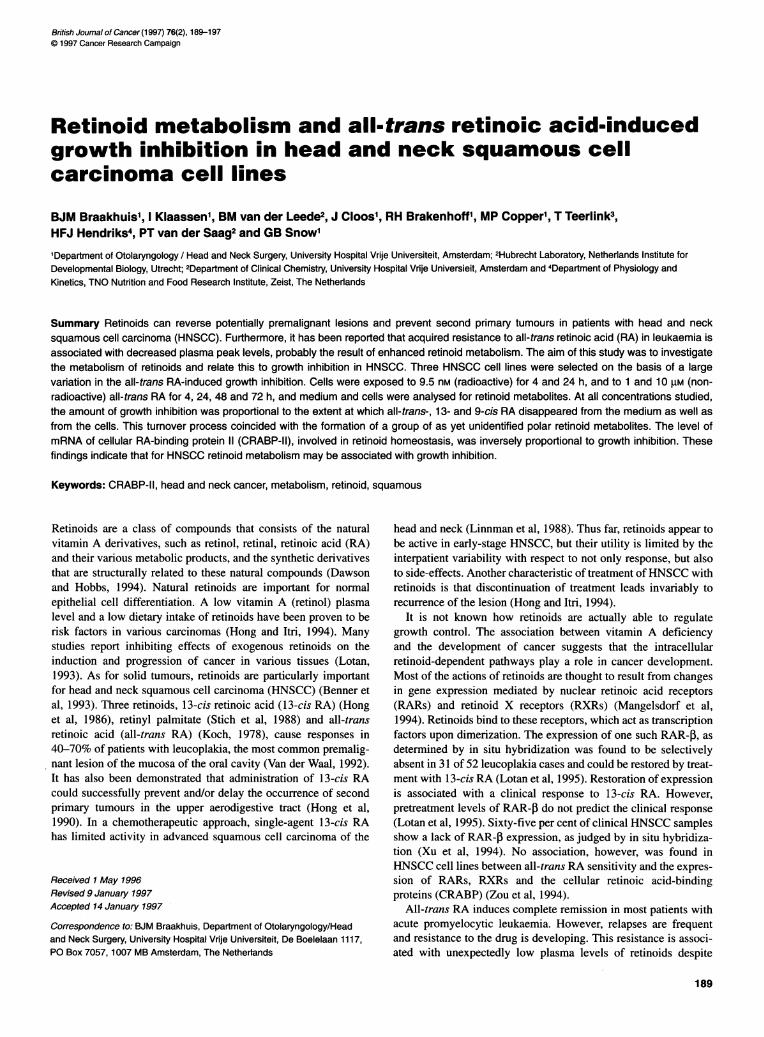
25 30 SCC-35 cell line (Figure 1B), although to a lesser extent thanall-trans RA, IC50 values being 39.0 and 6.8 nm respectively (Figure
of UM-SCC-35, IA). This metabolite was not active in the UM-SCC-14C cell line.ns RA; 2, 9-cis In a second set of experiments we argued that UM-SCC-35 cellsunidentified peaks might produce toxic metabolites and that these were released into
the medium. Thus, UM-SCC-35 cells were exposed for 24 h to10-6 and 10-1 M all-trans RA and it was found that this medium,
disappearance of when added to virgin UM-SCC-14C cells, minimally affected
lest for the UM- growth of these cells (Figure 4). Incubation of UM-SCC-35 cellsy significant and with conditioned medium of UM-SCC-14C cells (after treatment-he original level, with 10-8 M), however, caused a stronger antiproliferative effect
two cell lines is than incubation with the medium derived from UM-SCC-35 cell(Figure 3). With cultures. It appears that newly formed retinoids are less potent withshift could be respect to growth inhibition than the parent compounds they were
olar metabolites, derived from, but it must be added that, on an individual basis, the
between 6 and 21 concentration of these metabolites is significantly lower than theI be detected for concentration of parent retinoid (Table 1).
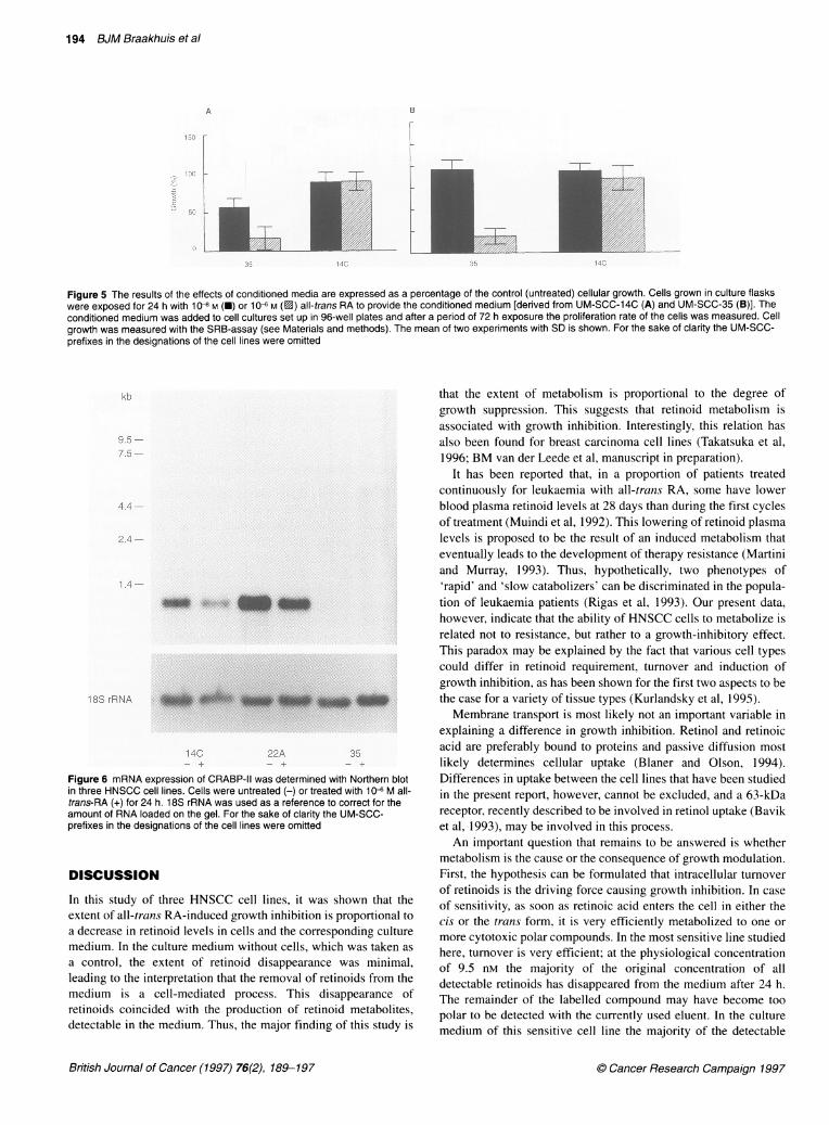
Anr exaMpleCof Measurement of CRABP expression,An example ofCC-35 after 24 h CRABP-I and -II are proteins involved in retinoid homeostasis and
they may be important with respect to metabolism and growthSCC-35 had the inhibition. We therefore analysed CRABP-I and -II expression byof intracellular Northern blotting and hybridization. CRABP-I had undetectabletoo low to be transcript levels in all cell lines, confirming previous results (Zou
levels decreased et al, 1994). The analysis of CRABP-II showed that UM-SCC-35uld be detected. had considerably lower transcript levels than the other two cell)sed to 10 JM all- lines (Figure 5). Exposure to 10-6 M all-trans RA for 24 h had anth the 1 ,UM expo- apparent down-regulating effect on CRABP-II mRNA levels in
UM-SCC-14C and -22A.
British Journal of Cancer (1997) 76(2), 189-197. Cancer Research Campaign 1997
n 7_
14C
B
IJ1
35 14C
Figure 5 The results of the effects of conditioned media are expressed as a percentage of the control (untreated) cellular growth. Cells grown in culture flaskswere exposed for 24 h with 10-8 M (-) or 106 M (0) all-trans RA to provide the conditioned medium [derived from UM-SCC-14C (A) and UM-SCC-35 (B)]. Theconditioned medium was added to cell cultures set up in 96-well plates and after a period of 72 h exposure the proliferation rate of the cells was measured. Cellgrowth was measured with the SRB-assay (see Materials and methods). The mean of two experiments with SD is shown. For the sake of clarity the UM-SCC-prefixes in the designations of the cell lines were omitted
kb
9.5
7.5-
2.4 .-~4
1.4 -~
18S rRNA
14C 22A 35
Figure 6 mRNA expression of CRABP-11 was determined with Northern blotin three HNSCC cell lines. Cells were untreated (-) or treated with 10- M all-trans-RA (+) for 24 h. 18S rRNA was used as a reference to correct for theamount of RNA loaded on the gel. For the sake of clarity the UM-SCC-prefixes in the designations of the cell lines were omitted
DISCUSSION
In this study of three HNSCC cell lines, it was shown that theextent of all-trans RA-induced growth inhibition is proportional toa decrease in retinoid levels in cells and the corresponding culturemedium. In the culture medium without cells, which was taken as
a control, the extent of retinoid disappearance was minimal,leading to the interpretation that the removal of retinoids from themedium is a cell-mediated process. This disappearance ofretinoids coincided with the production of retinoid metabolites,detectable in the medium. Thus, the major finding of this study is
that the extent of metabolism is proportional to the degree ofgrowth suppression. This suggests that retinoid metabolism isassociated with growth inhibition. Interestingly, this relation hasalso been found for breast carcinoma cell lines (Takatsuka et al,1996; BM van der Leede et al, manuscript in preparation).
It has been reported that, in a proportion of patients treatedcontinuously for leukaemia with all-trans RA, some have lowerblood plasma retinoid levels at 28 days than during the first cyclesof treatment (Muindi et al, 1992). This lowering of retinoid plasmalevels is proposed to be the result of an induced metabolism thateventually leads to the development of therapy resistance (Martiniand Murray, 1993). Thus, hypothetically, two phenotypes of'rapid' and 'slow catabolizers' can be discriminated in the popula-tion of leukaemia patients (Rigas et al, 1993). Our present data,however, indicate that the ability of HNSCC cells to metabolize isrelated not to resistance, but rather to a growth-inhibitory effect.This paradox may be explained by the fact that various cell typescould differ in retinoid requirement, turnover and induction ofgrowth inhibition, as has been shown for the first two aspects to bethe case for a variety of tissue types (Kurlandsky et al, 1995).Membrane transport is most likely not an important variable in
explaining a difference in growth inhibition. Retinol and retinoicacid are preferably bound to proteins and passive diffusion mostlikely determines cellular uptake (Blaner and Olson, 1994).Differences in uptake between the cell lines that have been studiedin the present report, however, cannot be excluded, and a 63-kDareceptor, recently described to be involved in retinol uptake (Baviket al, 1993), may be involved in this process.
An important question that remains to be answered is whethermetabolism is the cause or the consequence of growth modulation.First, the hypothesis can be formulated that intracellular turnoverof retinoids is the driving force causing growth inhibition. In case
of sensitivity, as soon as retinoic acid enters the cell in either thecis or the trans form, it is very efficiently metabolized to one or
more cytotoxic polar compounds. In the most sensitive line studiedhere, turnover is very efficient; at the physiological concentrationof 9.5 nm the majority of the original concentration of alldetectable retinoids has disappeared from the medium after 24 h.The remainder of the labelled compound may have become toopolar to be detected with the currently used eluent. In the culturemedium of this sensitive cell line the majority of the detectable
British Joumal of Cancer (1997) 76(2), 189-197
194 BJM Braakhuis et al
A
150 r
100 -e
CO
50 I
0
35
. Cancer Research Campaign 1997

Retinoids in head and neck cancer 195
Table 1 HPLC-analysis of cell culture medium after exposure to 106M all-trans retinoic acid
Cell line Exposure Recoverya All-trans RAa 13-cis RAa 9-cis RAa,b Retinola 4-oxo-cis-RAa Polar Remainderadtime (h) metabolitesac
None 0 100 89.4 + 3.1 5.1 + 1.8 1.8 + 0.8 2.6 + 1.2 u.d.l. 0.1 + 0.2 u.d.1.4 105.7 + 8.1 93.6 + 8.8 6.2 + 2.5 2.1 + 0.8 2.3 + 1.1 u.d.l. 0.2 + 0.5 1.0 + 1.224 106.7 + 4.7 91.7 + 7.2 8.4 + 2.4 2.8 + 0.9 2.3 + 1.1 u.d.l. 0.1 + 0.3 1.3 + 1.348 108.3 + 5.6 90.7 + 6.7 10.7 + 1.5 3.5 + 0.6 2.0 + 0.9 u.d.l. 0.1 + 0.3 1.4 + 1.472 110.9 + 11.4 88.1 + 11.5 14.4 + 1.8 4.8 + 0.7 1.7 + 0.5 u.d.l. 0.1 + 0.2 1.2 + 1.3
UM-SCC-14C 0 100 90.8 + 0.6 4.7 + 0.3 1.5 + 0.6 2.2 + 0.4 u.d.l. 0.3 + 0.4 0.5 + 0.74 119.6 + 42.2 106.3 + 39.0 6.9 ± 0.3 1.9 + 0.1 2.7 + 1.1 u.d.l. 0.8 + 1.2 1.0 + 0.8
24 109.5 + 52.7 93.5 + 49.1 9.3 + 1.4 2.6 + 0.3 0.7 + 1.0 0.6 + 0.4 1.9 + 1.2 1.6 + 0.248 95.6+0.1 76.3+3.7 11.7+1.7 3.5+1.1 0.5+0.3 0.4+0.1 1.9+0.8 1.0+1.572 81.8 + 7.5 55.1 + 1.5 13.2 + 5.5 4.5 + 2.4 0.6 + 0.1 1.1 + 0.9 5.8 + 2.2 2.6 + 0.3
UM-SCC-22A 0 100 88.2 + 4.2 6.1 + 2.2 1.9 + 1.2 1.7 + 0.8 u.d.l. u.d.l. 2.1 + 1.04 94.5 + 12.9 81.5 + 10.3 7.0 + 1.9 1.9 + 0.6 1.4 + 0.3 u.d.l. u.d.l. 2.7 + 0.424 67.6 ± 18.3 54.3 + 15.1 6.7 + 2.4 1.8 + 0.7 0.8 + 0.2 0.2 + 0.2 2.4 + 0.4 1.7 + 0.548 54.5 + 49.5 37.0 + 35.7 7.4 + 8.4 2.3 + 2.4 0.5 + 0.3 0.9 + 0.9 5.8 + 2.5 1.5 + 1.872 29.4 + 26.7 16.9 + 19.3 4.5 + 4.2 1.6 ± 1.5 0.3 + 0.2 0.8 + 0.7 5.1 + 2.0 1.0 + 1.0
UM-SCC-35 0 100 90.2 + 1.5 4.6 + 1.4 1.6 + 0.5 2.8 + 2.0 u.d.l. u.d.l. 0.7 + 0.84 93.7 + 21.0 82.2 + 17.6 4.7 + 3.1 1.6 + 0.6 2.4 + 1.5 0.4 + 0.5 1.8 + 1.1 1.0 + 0.724 29.8 + 18.9 10.0 + 8.0 2.0 + 1.9 0.5 + 0.5 2.3 + 0.8 0.6 + 0.8 12.8 + 7.5 2.1 + 2.948 10.6+6.2 0.9+0.9 0.4+0.4 0.1 ±0.1 2.0+0.9 0.6+0.7 5.7+3.6 1.5+1.9.72 9.0+9.1 0.2+0.4 u.d.l. u.d.l. 2.0+0.6 0.3+0.2 4.2+5.1 2.5+3.0
Cell culture media were analysed with HPLC after exposure for various times (mean of three separate experiments + s.d. is shown). aValues are expressed as apercentage of the total area under the curve of the relevant part of the chromatogram (retention time between 6 and 30 min, see Figure 4). T= 0 has been setat 100%. bA peak was observed between all-trans RA and 13-cis RA and based on literature data this peak was identified to represent 9-cis RA. cThis refers tocompounds identified by peaks with a retention time between 6 and 21 min (see Figure 4). aThis refers to compounds identified by peaks with a retention timebetween 21 and 30 min, with the exception of the known retinoids, 13-, 9-cis, all-trans RA and retinol. u.d.l., under detection limit.
retinoids are present in the form of 'polar metabolites'. It isconceivable that a sensitive cell line has a relatively high expres-sion of the enzymes involved in metabolism, for example oxida-tive enzymes such as the cytochrome P450s (Martini and Murray,1993; Rigas et al, 1993). The question arises whether unidentifiedmetabolites also have growth-inhibitory activity. Although it hasbeen reported that the 4-oxo-retinoic acid derivatives are consid-ered breakdown products in humans (Eckhoff et al, 1991), thesemolecules have also a transcription-activating capacity (Pijnappelet al, 1993). In addition, 4-oxo derivation of retinal to 4-oxoreti-naldehyde and the subsequent conversion to 4-oxoretinoic acidand 4-oxoretinol is suggested to be an important step duringXenopus embryogenesis (Blumberg et al, 1996). That study alsoshowed that all these 4-oxo products were able to bind to andtransactivate RARs. Our results also show that oxo derivativeshave growth-inhibiting capacity (Figure IB). The experimentswith conditioned medium provided no evidence that the sensitiveUM-SCC-35 cells produced a 'suicide' retinoid. The lack of effectof the conditioned medium to produce growth inhibition, however,could be attributed to the fact that the levels of the specificmetabolite were too low, perhaps because of further degradation.The peaks corresponding to the levels of the known 4-oxo-oxida-tion products were rather low.A second hypothesis can be formulated on the relationship
between growth inhibition and metabolism. Retinoid metabolism isa secondary event and is an attempt by the cell to neutralize thegrowth-inhibiting effect. In this scenario, all-trans RA and/or 1 3-cisRA are the key retinoids that cause growth inhibition and the othermetabolites must be considered as breakdown products. This
hypothesis is further supported by the notion that conditionedmedium of the insensitive cell line containing high levels of all-trans- and 13-cis RA appeared to be still very growth inhibitory forthe UM-SCC-35 cells. In addition, the expression of CRABP-IImRNA is in further favour of this hypothesis. It has been suggestedthat CRABP forms an intracellular buffer if the RA concentrationexceeds a certain level (Astrom et al, 1991; Adamson et al, 1993;Bavik et al, 1993; Delva et al, 1993; Griffiths et al, 1993; Blaner andOlson, 1994; Napoli et al, 1995). The present study supports thistheory, as the most sensitive cell line was found to have the lowestlevels of CRABP-II, and the addition of more RA failed to increaseits synthesis. In contrast, Zou et al (1994) reported that CRABP-IIexpression was not related to retinoid sensitivity in four HNSCCcell lines. An argument against the relation between high CRABP-IIlevels and insensitivity is the fact that all-tranis treatment does notup- but rather down-regulates CRABP-II mRNA expression. Thisphenomenon has already been observed for other epithelial celllines (Sanquer et al, 1993; Zou et al, 1994). The data, taken together,suggest that CRABP expression can be important but that nogeneral rule can be formulated. Further studies should elucidate theimportance of these molecules, and the possibility that the amountof protein is more important than the level of transcription cannot beexcluded. The positive correlation between a CRABP expressionand metabolism as has been found for CRABP-I in F9 teratocarci-noma stem cells (Boylan and Gudas, 1992) is in contrast with thecurrently reported results. The differences in the types of cells andthe function of these proteins may explain this discrepancy.
The levels of retinol in the medium of UM-SCC-35 cells remainhigh during the course of the exposure and deserve special attention.
British Journal of Cancer (1997) 76(2), 189-1970 Cancer Research Campaign 1997

196 BJM Braakhuis et al
The relatively low consumption of retinol from the medium of UM-SCC-35 suggests that the cells are not able to use retinol as a retinoidsource to produce retinoic acid. A low activity of one or moreenzymes of the group of alcohol dehydrogenases, involved in theconversion of retinol to retinoic acid, may be responsible for such aneffect (Harding and Duester, 1992; Napoli et al, 1995). As a conse-quence the cells may have adapted themselves to low intracellularlevels of all-tranis RA. Theoretically, any excess of all-trans RA isnot adequately buffered and may lead to cell death. This concept is inagreement with the hypothesis that metabolism is a secondary event.One can only speculate about the mechanism responsible for
retinoid induced growth inhibition. Studies on leucoplakia byLotan et al (1995) suggest that the induction of expression ofRAR-3 is important for growth suppression. The same group,however, could not find such a correlation when studying malig-nant HNSCC cell lines (Xu et al, 1994). We have also found noindication that the expression per se or induction by retinoic acidof RAR-o and -y, and RXR-ix is related to sensitivity of the celllines used (Copper et al, 1997). RAR-f mRNA levels were too lowto be measured. A recent in vitro study on breast cancer cellsshowed that RAR-a antagonists were as efficient in inhibitinggrowth as agonists (Dawson et al, 1995). This indicates thatbinding of a retinoid to a RAR may be important but that tran-scriptional activation on a retinoic acid-responsive element(RARE) is not a prerequisite. It is not clear whether this findingcan be extrapolated to the in vivo situation and whether it can beextended to other tumour types. No indications are available thatRAR-u. is important in growth inhibition of HNSCC.
ACKNOWLEDGEMENTSWe thank Hoffmann-la Roche for providing retinoids, Dr AAstrom, University of Michigan, Ann Arbor, MI, USA, forsupplying the cDNA clones of the CRABPs, and Ivar Steen and TSchoemaker for their technical assistance. This study wassupported by the Dutch Cancer Society, grants 95-926 (BJMB andGBS) and 93-558 (PvdS).
REFERENCES
Adamson PC, Boylan JF, Balis FM, Murphy RF, Godwin KA, Gudas Li andPoplack DG (1993a1) Time course of induction of metabolism of all-trans-retinoic acid and the up-regulation of cellular retinoic acid-binding protein.Caoncer Res 53: 472-476
Adamson PC, Pitot HC, Balis FM, Rubin J. Murphy RF and Poplack DG (19936)Vafiability in the oral bioavailability of all-trans-retinoic acid. J Natl Coincerhist 85: 993-996
Astrom A, Tavakkol A, Pettersson U, Cromie M, Elder JT and Voorhees JJ ( 1991)Molecular cloning of two human cellular retinoic acid-binding proteins(CRABP). Retinoic acid-induced expression of CRABP-11 but not CRABP-I inadult human skin in vivo and in skin fibroblasts in vitro. J Biol Clie,n 266:17662- 17666
Bavik CO. Levy F. Hellman U. Wernstedt C and Eriksson U (1993) The retinalpigment epithelial membrane receptor for plasma retinol-binding protein-isolation and cDNA cloning of the 63-kda protein. J Biol Clieot 268:20)540-20546
Benner SE. Lippman SM and Hong WK (1993) Retinoids in head and neck cancer.In Retinoids in Ontcology. Hong WK and Lotan R (eds). pp. 20)3-223. MarcelDekker: New York
Blaner WS and Olson JA (1994) Retinol and retinoic acid metabolism. In TheRetidnoids. Biology; Chemistry aiid Medicine. 2nd edn. Sporn MB. Roberts ARand Goodman DS (eds). pp. 229-256. Raven Press: New York
Blumberg B. Bolado J, Derguine F, Craig AJ, Moreno TA. Chakravarti D, Heyman.Buck J, Evans RM (1996) Novel retinoic acid receptor ligands in Xenopusembryos. Proc Noti A(cdSci USA 93: 4873-4878
Braakhuis BJM, Jansen G. Noordhuis P. Kegel A and Peters GJ (1993) Importanceof pharmacodynamics in the in vitro antiproliferative activity of the antifolatesmethotrexate and 10-ethyl-10-deazaaminopterin against human head and necksquamous cell carcinoma. Biocheom Phar-mocol 40: 2155-2161
Boylan JF and Gudas LJ (1992) The level of CRABP-I expression influences theamounts and types of all-trans-retinoic acid metabolites in F9 teratocarcinomastem cells. JBiol Clhetin 267: 21486-21491
Carey TE (I1985) Establishment of epidermoid carcinoma cell lines. In Hea(id al1dNeck Cancer, Wittes RE (ed.). pp. 287-314. John Wiley: New York
Copper MP. Klaassen 1. Brekenhoff RH, Cloos J. Snow GB and Braakhuis BJM(1997) All-trron,s Retinoic acid induced gene-expression and growth inhibitionin head and neck cancer cell lines. Or(al Oncol (in press)
Dawson MI and Hobbs PD (1994) The synthetic chemistry of retinoids. In TheReti/?oids. Biology. Chemistry-N aniid Medicine. 2nd edn. Sporn MB, Roberts ARand Goodman DS (eds.), pp. 5-178. Raven Press: New York
Dawson MI, Chao WR, Pine P. Jong L. Hobbs PD, Rudd CK, Quick TC. Niles RM.Zhang XK, Lombardo A. Ely KR. Shroot B and Fontana JA (1995) Correlatiolnof retinoid binding affinity to retinoic acid receptor alpha with retinoidinhibition of growth of estrogen receptor-positive MCF-7 mammary carcinomacells. Cancer Res 55: 4446-4451
Delva L, Cornic M, Balitrand N. Guidez F, Miclea JM. Delmer A. Teillet F. FenauxP, Castaigne S, Degos L and Chomienne C (1993) Resistance to all-trans-retinoic acid (ATRA) therapy in relapsing acute promyelocytic leukemia -study of in vitro ATRA sensitivity and cellular retinoic acid binding proteinlevels in leukemic cells. Bloo(d 82: 2175-21 81
Eckhoff C and Nau H ( 1990) Identification and quantitation of all-trans- and 1 3-cis-retinoic acid in human plasma. J Lip Re.s 31: 1445-1454
Eckhoff C. Collins MD and Nau H (1991) Human plasma all-trans-. 3-cis- and13-cis-4-oxoretinoic acid profiles during subchronic vitamin Asupplementation: Comparison to retinol and retinyl ester plasma levels. J Nmmtr121: 1016-1025
Feinberg AP and Vogelstein B (1983) A technique for radiolabeling DNA restrictionendonuclease fragments to high specific activity. Anatil Biochemn 132: 6-13
Gough NM (1988) Rapid and quantitative preparation of cytoplasmic RNA fromsmall numbers of cells. Anial Biochein 173: 93-95
Griffiths CEM. Elder JT. Bernard BA. Rossio P. Cromie MA. Finkel LJ. Shroot Band Voorhees JJ ( 1993) Comparison of CD271 (adapalene) and all-trans-retinoic acid in human skin - dissociation of epidermal effects and CRABP-IImessenger RNA expression. J Inrest Dermn(atol 101: 325-328
Harding P and Duester G (1992) Retinoic acid activation and thyroid hormonerepression of the human alcohol dehydrogenase gene ADH3. J Biol Clhemil 267:14145-14150
Hong WK, Endicott J and Itri L (1986) 13-cis retinoic acid in the treatmiient of oralleukoplakia. N Etigl J Med 315: 1501-15()5
Hong WK, Lippman SM, Itri LM. Karp DD. Lee JS, Byers RM, Schantz SP KramnerAM, Lotan R, Peters LJ, Dimery IW, Brown BW and Goepfert H (1990)Prevention of second primary tumors with isotretinion in squamous cellcarcinoma of the head and neck. N En,gl J Med 323: 795-81)1
Hong WK and Itri LM (I1994) Retinoids and human cancer. In The Rertinoidls.Biology! Chemistis amid Medicine, 2nd edn, Sporn MB, Roberts AR andGoodman DS (eds.). pp. 597-631. Raven Press: New York
Jetten AM, Kim JS, Sacks PG, Rearick JT. Lotan D, Hong HK and Lotan R ( 1990)Inhibition of growth and squamous-cell differentiation markers in culturedhuman head and neck squamous carcinoma cells by beta-all-trans retinoic acid.Intt J Canzcer- 45: 195-202
Koch HF ( 1978) Biochemical treatment of precancerous oral lesions: theeffectiveness of various analogues of retinoic acid. J Maxillofac Sur,i 6:59-63
Kurlandsky SB, Gamble MV, Ramakrishnan R and Blaner WS (1995) Plasmadelivery of retinoic acid to tissues in the rat. J Biol Clmemti 270: 17850)-17857
Lee JS. Newman RA, Lippman SM. Huber MH, Minor T, Raber MN, Krakoff IHand Hong WK ( 1993) Phase-I evaluation of all-trans-retinoic acid in adultswith solid tumors. J Cliii Oncol 11: 959-966
Lippman SM, Kessler JF, Al-Sarraf M, Alberts DS, Itri LM and Mattox D (I1988)Treatment of advanced squamous cell carcinom-a of the head and neck withisotretinoin: a phase It randomized trial. Invest Neit DI-lJgs 6: 51-56
Lotan R (1993) Retinoids and squamous cell differentiation. In Retinaoids illOmncology. Hong WK and Lotan R (eds), pp. 43-73. Marcel Dekker: New York
Lotan R, Xu XC, Lippman SM. Ro JY. Lee JS, Lee JJ and Hong WK (1995)Suppression of retinoic acid receptor-beta in premalignant oral lesions and itsup-regulation by isotretinoin. N Entgl J Med 332: 1405-1410
Mangelsdorf DJ, Umesono K and Evans RM (1994) The retinoid receptors. In TleRetimioids. Biology: ChemistrY (iand Medicine, 2nd edn. Sproni MB. Roberts ARand Goodman DS (eds). pp. 319-351. Raven Press: New York
British Journal of Cancer (1997) 76(2), 189-197 @ Cancer Research Campaign 1997

Retinoids in head and neck cancer 197
Martini R and Murray M (1993) Participation of p450-3a enzymes in rat hepaticmicrosomal retinoic acid 4-hydroxylation. Ar/ch Biocheoi Biophvs 303: 57-66
Muindi JF, Frankel SE, Miller WH, Young WC, Dmitrovsky E and Warell RP (1992)Continuous treatment with all-trans-retinoic acid causes a progressive decreasein plasma concentrations: implications of relapse and resistance in acutepromyelocytic leukemia. Blood 79: 299-303
Muindi JF, Scher HI, Rigas JR, Warrell RP and Young CW (1994) Elevatedplasma lipid peroxide content correlates with rapid plasma clearance ofall-trans-retinoic acid in patients with advanced cancer. Canlcer- Rex 54:2125-2128
Napoli JL, Boerman MHEM, Chai X, Zhai Y and Fiorella PD (1995) Enzymes andbinding proteins affecting retinoic acid concentrations. J Steroid Biochein MolBiol 53: 497-502
Pijnappel WWM, Hendriks HFJ, Folkers GE, Van Den Brink CE, Dekker EJ,Edelenbosch C, Van Der Saag PT and Durston AJ (1993) The retinoid ligand 4-oxo-retinoic acid is a highly active modulator of positional specification.Natir-e 366: 340-344
Rigas JR, Francis PA, Muindi JRF, Kris MG, Huselton C, Degrazia F, Orazem JP,Young CW and Warrell RP (1993) Constitutive variability in thepharmacokinetics of the natural retinoid, all-trans-retinoic acid, and itsmodulation by ketoconazole. J Natl Cancer Itnst 85: 1921-1926
Sacks PG, Harris D and Chou TC (1995) Modulation of growth and proliferation insquamous cell carcinoma by retinoic acid: A rationale for combination therapywith chemotherapeutic agents. Itit J Cancer 61: 409-415
Sanquer S, Eller MS and Gilchrest BA (1993) Retinoids and state of differentiationmodulate CRABP-II gene expression in a skin equivalent. J Invest Derinatol100: 148-153
Sambrook J, Fritsch EF and Maniatis T ( 1989) Molecul/or Cloniing: o LaboratoryMonual. Cold Spring Harbor Laboratory Press: New York
Stich HF. Homby AD and Mathew B (1988) Response of oral leukoplakias to theadministration of Vitamin A. Cancer Lett 40: 93-101
Takatsuka J, Takahashi N and De Luca L ( 1996) Retinoic acid metabolism andinhibition of cell proliferation: an unexpected liaison. Can1cer Res 56: 675-678
Teerlink T, Copper MP, Klassen I and Braakhuis BJM (1997) Simultaneous analysisof retinol, retinoic acid isomers and polar metabolites using on-columnconcentration after single-phase fluid extraction. J Clhrotatogr B (in press)
Van Der Waal I ( 1992) Oral precancerous lesions - present knowledge. DentZahnartc/ Z 47: 860-864
Wouters W, Van Dun J, Dillen A, Coene MC, Cools W and De Coster R (1992)Effects of liarozole, a new antitumoral compound, on retinoic acid-inducedinhibition of cell growth and on retinoic acid metabolism in MCF-7 humanbreast cancer cells. Ctancer Res 52: 2841-2846
Xu XC, Ro JY, Lee JS, Shin DM, Hong WK and Lotan R (1994) Differentialexpression of nuclear retinoid receptors in normal, premalignant, and malignanthead and neck tissues. Ccanicer Res 54: 3580-3587
Zou CP, Clifford JL, Xu XC, Sacks PG, Chambon P, Hong WK and Lotan R (1994)Modulation by retinoic acid (RA) of squamous cell differentiation, cellularRA-binding proteins, and nuclear RA receptors in human head and necksquamous cell carcinoma cell lines. Cancer Res 54: 5479-5487
C Cancer Research Campaign 1997 British Journal of Cancer (1997) 76(2), 189-197