cardiac imaging in atrial fibrillation
TRANSCRIPT

Noninvasive evaluation of the patient with angina
Michael Hall Division of Cardiology
University of Mississippi Medical Center
No disclosures

Objectives
1. Give an overview of the different diagnostic testing options in patients with angina
2. Discuss practical and technical considerations of noninvasive cardiac tests
2. Discuss sensitivities/specificities and cost effectiveness of noninvasive cardiac tests

Case
• 32 yo AAF with no PMH presents to ED with left-sided CP lasting 5 minutes at a time. Occurs with rest. No SOB/DOE. She has associated nausea and diaphoresis.
• Nonsmoker, no drugs
• Dad had MI at age 66
• BP 132/88mmHg, HR 76, RR 14, O2 sats 99% RA

Normal EKG Troponins x 2 WNL
CXR WNL CT PE Protocol was normal
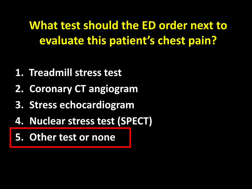
What test should the ED order next to evaluate this patient’s chest pain?
1. Treadmill stress test
2. Coronary CT angiogram
3. Stress echocardiogram
4. Nuclear stress test (SPECT)
5. Other test or none

Pretest probability of CAD
ACC/AHA Guidelines for Exercise Testing

Important factors for choosing a cardiac test
• Need/indication
• Test availability and local expertise
• Diagnostic accuracy
• Body habitus
• Side effects of medications
• Cost

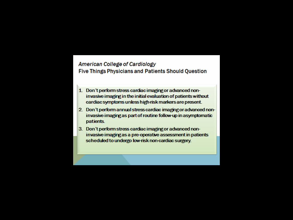
• “The tests usually aren’t necessary before low-risk surgery.”
• “Heart imaging tests can pose risks.”
• “Imaging tests can cost a lot.”

Mai Tone Lønnebakken and Eva Gerdts (2011). Contrast Echocardiography in Coronary Artery Disease,
Coronary Angiography - Advances in Noninvasive Imaging Approach for Evaluation of Coronary Artery
Disease.
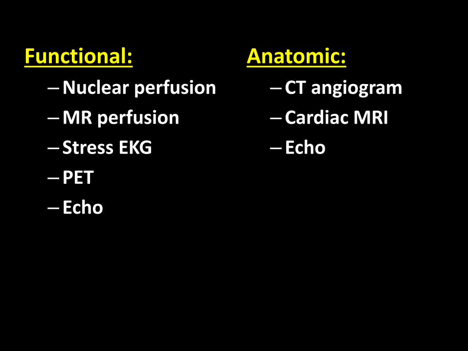
Functional:
–Nuclear perfusion
–MR perfusion
– Stress EKG
–PET
– Echo
Anatomic:
–CT angiogram
–Cardiac MRI
– Echo

Stress Testing
• In general, if patients can exercise, exercise is preferred over pharmacologic testing
• Exercise can be performed with either treadmill or bicycle
• Bruce protocol is most commonly used: monitor EKG, symptoms, BP and HR during graded exercise

Exercise Stress- Bruce Protocol • Reports exercise capacity in metabolic equivalents
(METS)
• METs reflect resting volume oxygen consumption per minute (VO2) for a 70kg, 40 yo man
• 1 MET = 3.5mL/min/kg of body weight
• Stage 1 is 1.7 mph at 10% grade= 5 METS
• Stage 2 is 2.5 mph at 12% grade= 7 METS
• Stage 3 is 3.4 mph at 14% grade= 9 METS
• Each stage goes for 3 minute intervals

Contraindications to exercise stress
Absolute – Recent MI ( within 2 days)
– recent significant change in resting ECG
– High risk unstable angina
– Uncontrolled arrhythmias causing symptoms or hemodynamic compromise
– Symptomatic severe AS
– Acute aortic dissection
– Acute PE
– Acute myo/pericarditis
– Uncontrolled symptomatic heart failure

Practical considerations for exercise stress
1. Can the patient adequately exercise?
2. Does the patient have an interpretable EKG?
– LBBB
– Pacing
– Pre-excitation (WPW)
– Digoxin
– LVH

Exercise stress
• ECG’s at rest, each exercise stage, peak exercise, and every 1-2 minutes in recovery for at least 5 minutes
• Also record HR, BP, patient’s perceived exertion, symptoms
• Completed when patient attains 85% of age-predicted maximum HR: 220- age or because of symptoms

Duke Treadmill Score
DTS= Exercise Time – (5 x Max ST) – (4 X Angina Index)
Angina Index: 0= no angina during exercise 1= non-limiting angina 2=Exercise limited angina
Risk: ≥ +5= low risk +4 to -10= Moderate Risk ≤ -11= High Risk
Mark DB et al. NEJM 1991 Shaw LJ et al. Circulation 1998
***Patients with a “low risk” exercise EKG incur ≤ 1% per year cardiac mortality.

Exercise capacity is strongly predictive of mortality
Myers J et al. NEJM, 2002
Each 1-MET increase in exercise capacity conferred a 12% improvement in survival
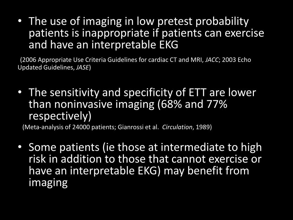
• The use of imaging in low pretest probability patients is inappropriate if patients can exercise and have an interpretable EKG
(2006 Appropriate Use Criteria Guidelines for cardiac CT and MRI, JACC; 2003 Echo Updated Guidelines, JASE)
• The sensitivity and specificity of ETT are lower than noninvasive imaging (68% and 77% respectively)
(Meta-analysis of 24000 patients; Gianrossi et al. Circulation, 1989)
• Some patients (ie those at intermediate to high risk in addition to those that cannot exercise or have an interpretable EKG) may benefit from imaging
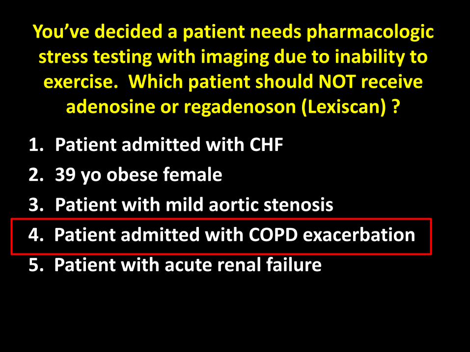
You’ve decided a patient needs pharmacologic stress testing with imaging due to inability to exercise. Which patient should NOT receive
adenosine or regadenoson (Lexiscan) ?
1. Patient admitted with CHF
2. 39 yo obese female
3. Patient with mild aortic stenosis
4. Patient admitted with COPD exacerbation
5. Patient with acute renal failure

Vasodilators (Adenosine, regadenoson)-preferred for assessment of myocardial perfusion
- Contraindications: asthma, severe hypotension, bradyarrhythmias - Withhold caffeine for 12 hours
LAD Lcx Lcx LAD
Pre-vasodilator Post-vasodilator
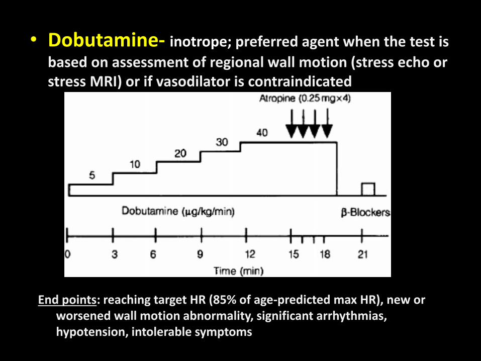
• Dobutamine- inotrope; preferred agent when the test is
based on assessment of regional wall motion (stress echo or stress MRI) or if vasodilator is contraindicated
End points: reaching target HR (85% of age-predicted max HR), new or worsened wall motion abnormality, significant arrhythmias, hypotension, intolerable symptoms
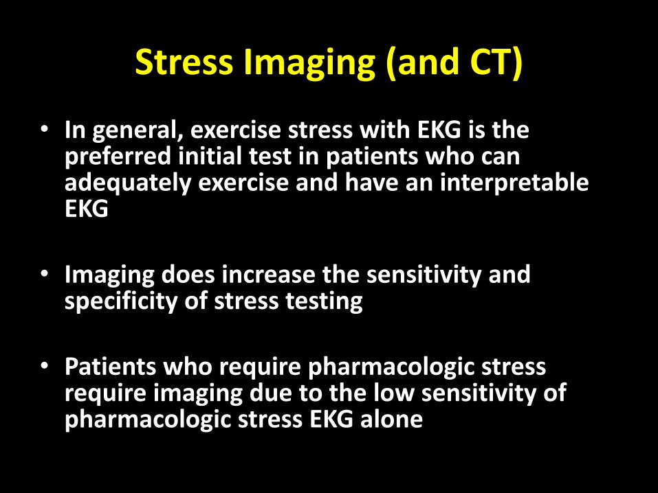
Stress Imaging (and CT)
• In general, exercise stress with EKG is the preferred initial test in patients who can adequately exercise and have an interpretable EKG
• Imaging does increase the sensitivity and specificity of stress testing
• Patients who require pharmacologic stress require imaging due to the low sensitivity of pharmacologic stress EKG alone

Stress ECHO (exercise or dobutamine)
• Grade myocardial wall segments using 17 segment model at rest and at stress
Each segment is graded as : 1. Normal or hyperdynamic 2. Hypokinetic 3. Akinetic 4. Dyskinetic 5. Aneurysmal
Both wall motion and perfusion are highly accurate (more accurate than EKG changes) for detection of CAD. However, wall motion is more specific and requires ischemia. Perfusion images are typically more sensitive

Stress echo
• Normal results: normal LV wall motion/thickening at rest and stress
• Abnormal: – Fixed wall motion abnormality- abnormal at rest and
stress- suggests previous MI – New or worsened wall motion abnormalities suggest
ischemia ***Variables associated with adverse outcomes are extensive ischemia (WMAs), poor EF% response, low ischemic threshold (early abnormalities), location of WMA in LAD territory
ASE Guidelines for Stress Echo (2007).

Short axis

Apical 4-chamber

Inducible inferior wall motion abnormality
Baseline Target HR

Other indications for stress echo
• Myocardial viability
• Evaluation of mitral valve disease
– Evaluate exercise PA systolic pressures (>60mmHg is indication for MV surgery)
• Evaluation of aortic valve disease
– Low gradient aortic stenosis

Stress echo
• Sensitivity 79%, Specificity 87% (wall motion abnormalities require ischemia)
• Sensitivity is reduced in people with poor acoustic windows (obesity, COPD, large breasts)
(Heijenbrok-Kal et al. Am Heart J, 2007)
• A normal stress echo is associated with excellent prognosis: 1-year cardiac event-free survival of 99% and 3-year survival of 97%
(McCully RB et al. JACC, 1998)

Nuclear (SPECT) Stress Test • Radioactive tracer
(Technetium-99) is administered
• Tracer decays spontaneously and emits energy (photons)
• Photons leave the body and are captures with a gamma camera
• During stress, metabolic changes drive agent into the cell
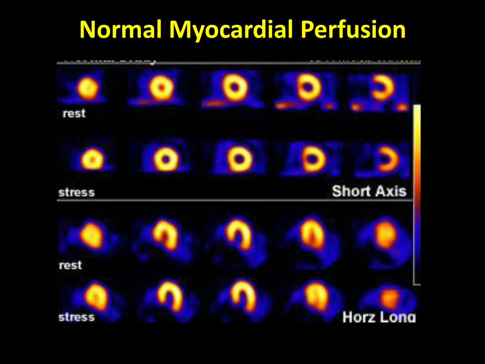
Normal Myocardial Perfusion
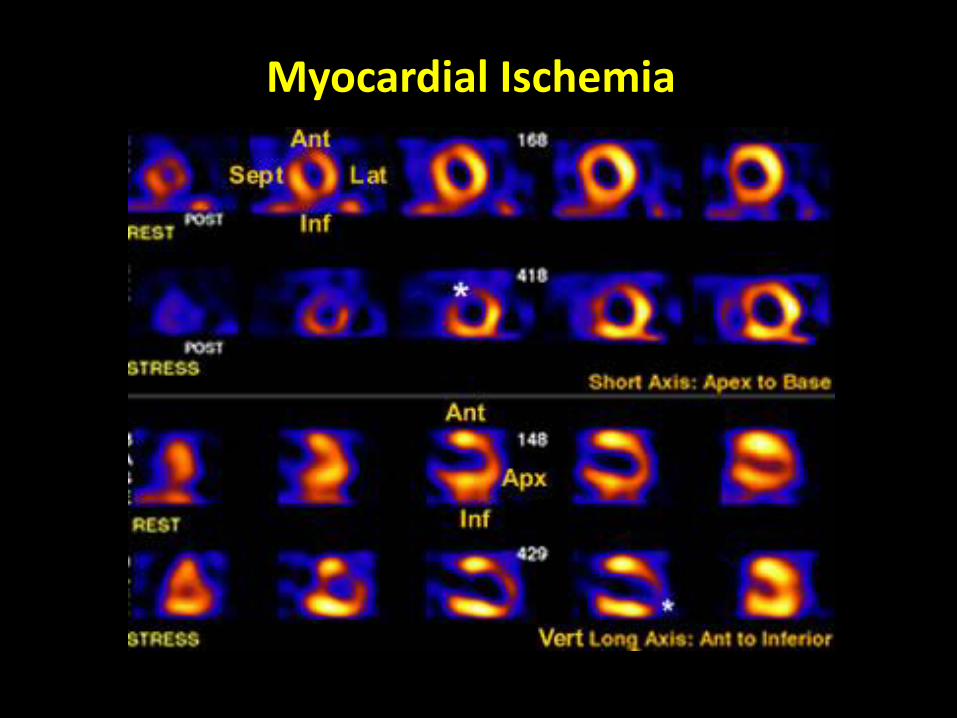
Myocardial Ischemia

SPECT Cons:
• Image quality in obesity, women (attenuation artifacts)
• Limited anatomic info
• Radiation dose (equivalent to >500 CXRs)
• Availability of technetium
Pros:
• Sensitive for ischemia (70-90%)
• No need for contrast dye
• Safe in patients with ICDs/PCMs
• Widely available
• Well validated
***In 21,000 patients with a normal SPECT and 2-year follow-up, annual hard cardiac event rate was 0.7%
Klocke FJ et al. ACC/AHA/ASNC Guidelines for radionuclide imaging, Circulation, 2003

No Field Bo Field Applied
Bo
M0
B1 out of the slide
q B0 B0
Cardiac MRI
Craig Hamilton, PhD, Biomedical Engineering, Wake Forest

Which of the following patients is safe for a stress cardiac MRI?
1. Patient with pacemaker
2. Patient with coronary stents
3. Patient on hemodialysis
4. Patient with gastric pacemaker

MRI Safety
MR safe
MR conditional=most metallic heart valves, Stents, prosthetic joints, dentures
MR unsafe= most pacemakers, insulin pumps, Most ICD’s, metallic foreign bodies

Nephrogenic Systemic Fibrosis (NSF)
• Rare, but serious complication thought to be related to toxic effect of gadolinium ions in patients with advanced renal failure/ESRD
• Causes fibrosis of skin, joints, eyes, internal organs
• Gadolinium contraindicated with GFR<30
• **No cases of NSF reported in patients with normal renal function**

Requirements
• Hemodynamically stable
• Can breath-hold
• Can lie flat
• Can’t be claustrophobic

Special considerations for CMR
• Inability to obtain adequate echo windows (CMR can slice through any tissue plane)
• Poor nuclear candidates (ie large breasts, obese, small size women)
• When you need accurate quantification of LV or RV function (ie those receiving chemotherapy)
• When you want a comprehensive test rather than multiple tests (provides anatomic and physiologic data)
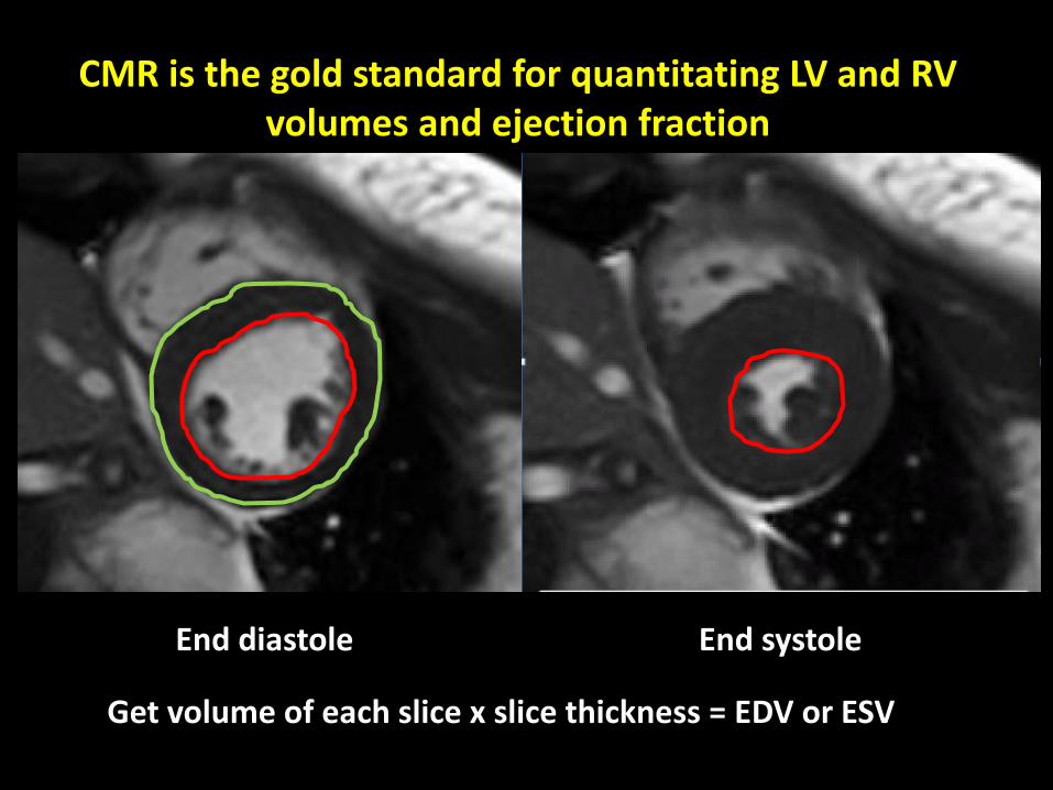
End diastole End systole
Get volume of each slice x slice thickness = EDV or ESV
CMR is the gold standard for quantitating LV and RV volumes and ejection fraction

CMR Adenosine Stress Test Sc
ou
t im
ages
Cin
e Im
ages
Stre
ss P
erfu
sio
n
Ad
dit
ion
al Im
ages
Res
t Pe
rfu
sio
n
Del
ayed
En
han
cem
en
t
Adenosine 140mcg/kg/min (3-6 minutes)
Gad Gad
Time (mins)
0 45


Rest Perfusion Stress Perfusion
Adenosine stress CMR


Peak dose dobutamine Stress perfusion
Dobutamine stress CMR

Time
Sign
al in
ten
sity
scar
Scar
Normal myocardium
Principles of late (delayed) gadolinium enhancement
T1 relaxation curve
Gadolinium is an inert biological tracer that distributes in extracellular water and cannot cross an intact cell membrane. Therefore, expanded extracelluar space (ie fibrosis or scar) causes a relative accumulation of gadolinium relative to normal myocardium
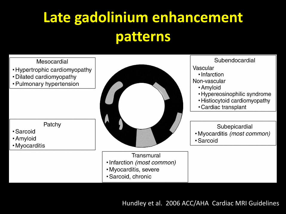
Late gadolinium enhancement patterns
Hundley et al. 2006 ACC/AHA Cardiac MRI Guidelines

Myocardial infarction

CMR detects infarcts missed by SPECT
Wagner et al. Lancet. 2003

Stress CMR is more sensitive than SPECT for detection of significant CAD
Greenwood et al. Lancet, 2012.
Greenwood et al. Circulation, 2014.
NPV 91% for CMR vs 79% for SPECT
CMR is more sensitive than SPECT in both sexes

Stress CMR strategy in the ED chest pain unit reduces health care expenditures in intermediate-
risk patients • Randomized 109 intermediate
to high risk participants to CPU observation vs inpatient care
• No difference in rate of cardiac events at 1 year
• Costs (including index visit) were lower in the CMR group compared to inpatient care
($3101 vs $4,742, p=0.004)
Mean cumulative cost after hospital discharge
Miller CD et al. JACC Imaging, 2011

Myocardial Viability
> 50% delayed enhancement in a myocardial segment is associated with lower likelihood of viability

Aneurysm with layering thrombus

Microvascular ischemia

Myocarditis
T2 (water) image showing myocardial edema

Takotsubo CMP

Coronary CT angiography
• Requires radiation and iodinated contrast (~120ml’s)
• Optimal with low and stable (<65 bpm) heart rate- use ECG gating; often need beta blockers
• Image quality/diagnostic accuracy reduced with obesity
• Requires breath holding
• Can be obscured in patients with highly calcified vessels

Coronary calcium scoring
• Usually acquired before contrasted angiogram
• Excessive calcium (CAC score >400 can cause overestimation of stenoses
• Good prognostic marker of future cardiac events
• Not as useful in acute chest pain settings- does not rule out ACS

Coronary CT angiography
• Highly sensitive for detection of CAD (near 100%)
• Negative predictive value ~99% makes this an ideal tool for acute chest pain setting
• Rapid test (<10 minutes)
• Can visualize coronary plaque characteristics
Sechtem U et al. European Heart Journal, 2012

High-risk plaques
-Low attenuation plaques -Predominantly non-calcified -positive remodeling
Motoyama S et al. JACC, 2009

Prospective ECG gating reduces radiation exposure
Sun. Cardiovasc Diagn Ther, 2012
CTA can be routinely performed with < 4mSv
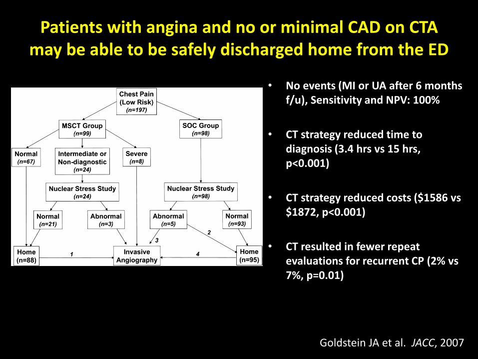
Patients with angina and no or minimal CAD on CTA may be able to be safely discharged home from the ED
• No events (MI or UA after 6 months f/u), Sensitivity and NPV: 100%
• CT strategy reduced time to diagnosis (3.4 hrs vs 15 hrs, p<0.001)
• CT strategy reduced costs ($1586 vs $1872, p<0.001)
• CT resulted in fewer repeat evaluations for recurrent CP (2% vs 7%, p=0.01)
Goldstein JA et al. JACC, 2007
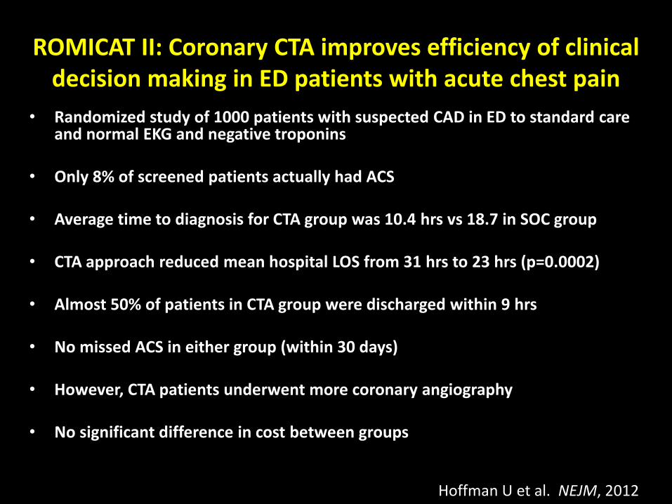
ROMICAT II: Coronary CTA improves efficiency of clinical decision making in ED patients with acute chest pain
• Randomized study of 1000 patients with suspected CAD in ED to standard care and normal EKG and negative troponins
• Only 8% of screened patients actually had ACS
• Average time to diagnosis for CTA group was 10.4 hrs vs 18.7 in SOC group
• CTA approach reduced mean hospital LOS from 31 hrs to 23 hrs (p=0.0002)
• Almost 50% of patients in CTA group were discharged within 9 hrs
• No missed ACS in either group (within 30 days)
• However, CTA patients underwent more coronary angiography
• No significant difference in cost between groups
Hoffman U et al. NEJM, 2012
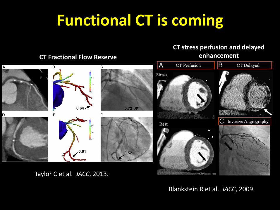
Functional CT is coming
Blankstein R et al. JACC, 2009.
CT Fractional Flow Reserve
CT stress perfusion and delayed enhancement
Taylor C et al. JACC, 2013.

Conclusions • Noninvasive cardiac testing can help answer the questions
“Is significant CAD present?”, and if so, “what is the patient’s prognosis?”
• In general, if a patient can exercise and has an interpretable EKG, consider exercise stress testing
• The choice of imaging modality to identify functional significance of CAD should be based on test availability, patient body habitus, local expertise, cost-effectiveness
• Perfusion imaging can also guide which patients may benefit from revascularization strategies (those with >10% of ischemic myocardium)
Dowsley T et al. Canadian Journal of Cardiology, 2013

Dowsley T et al. Canadian Journal of Cardiology, 2013

Thanks