cardiac involvement in mis-c patients simil-appendicitis
TRANSCRIPT
Page 1/9
Simil-appendicitis presentation may precedecardiac involvement in MIS-C patientsMatteo Trevisan ( [email protected] )
Universita degli Studi di Trieste Dipartimento di Scienze Mediche e Chirurgiche e della Salutehttps://orcid.org/0000-0003-1897-7701
Alessandro Amaddeo Institute for Maternal and Child Health: IRCCS materno infantile Burlo Garofolo
Andrea Taddio Institute for Maternal and Child Health: IRCCS materno infantile Burlo Garofolo
Alessandro Boscarelli Institute for Maternal and Child Health: IRCCS materno infantile Burlo Garofolo
Egidio Barbi Institute for Maternal and Child Health: IRCCS materno infantile Burlo Garofolo
Giorgio Cozzi Institute for Maternal and Child Health: IRCCS materno infantile Burlo Garofolo
Case report
Keywords: MIS-C, COVID-19, appendicitis, QT prolongation, myocarditis
Posted Date: May 21st, 2021
DOI: https://doi.org/10.21203/rs.3.rs-540399/v1
License: This work is licensed under a Creative Commons Attribution 4.0 International License. Read Full License
Page 2/9
AbstractBackground
Multisystem in�ammatory syndrome in children (MIS-C) is a new clinical entity characterized by asystemic hyperin�ammation triggered by SARS-CoV-2 infection in children and adolescents. Thiscondition could potentially involve all organs with main complications concerning cardiovascular system.Despite up to 90% of patients complain gastrointestinal symptoms (nausea, vomit and diarrhea), apresentation mimicking acute appendicitis has rarely been reported, and can be the presenting feature ofthe disease, potentially leading to misdiagnosis and delayed treatment.
Case presentation
A 15-year-old boy presented to the Emergency Department for a two-day history of fever, vomiting andmild abdominal pain. One month before, the patient complained ageusia and anosmia while his mothertested positive for Sars-CoV2 nasopharyngeal swab. At admission, laboratory tests showed leukocytosiswith lymphopenia and elevation of in�ammatory markers, while cardiac enzymes, electrocardiogram andechocardiography were unremarkable. An abdominal ultrasound displayed a thickening of terminal ileusand cecum with ascites. Because of the worsening abdominal pain and a physical examinationsuggestive of acute appendicitis, a laparoscopy was performed but no surgical condition was found.After surgery, fever and generalized malaise persisted, so a cardiac evaluation was repeated, showing arelevant increase in in�ammatory markers and cardiac enzymes. Electrocardiogram demonstrated a QTcprolongation with mild decrease in left ventricular ejection fraction at echocardiogram. A MIS-C wasdiagnosed and intravenous immunoglobulin along with a steroid treatment started. After 36 hours, thepatient presented a complete clinical recovery with fever cessation. Cardiac anomalies normalized inthree weeks.
Conclusion
MIS-C has been de�ned as a systemic in�ammation, involving at least two organs, after a previous SARS-CoV2 infection in children and adolescents. Physicians should be aware that while gastrointestinalmanifestations are common, a pseudo appendicitis presentation may also occur, leading to misdiagnosisand delayed treatment. This report suggests that in patients with symptoms suggestive of an acuteappendicitis, the presence of lymphopenia, hypoalbuminemia and ultrasound images of terminal ileusin�ammation, should raise the suspect for MIS-C even without initial overt signs of cardiac involvement.
BackgroundMultisystem in�ammatory syndrome in children (MIS-C), �rstly described by Riphagen and colleagues, ischaracterized by a systemic hyperin�ammation triggered by severe acute respiratory syndromecoronavirus 2 (SARS-CoV-2) infection in children and adolescents (1, 2). According to the Centre ofDisease Control and Prevention (CDC), case de�nition of MIS-C includes age of less than 21 years, fever
Page 3/9
for at least 24 hours, elevation of in�ammatory markers, serious illness leading to hospitalization or atleast two organs involvement (cardiac, renal, respiratory, hematological, gastrointestinal, dermatological,or neurological) with a history of possible SARS-CoV2 infection (positive real time-polymerase chainreaction, positive serology or contact with COVID-19 in the past 4 weeks). Usually developed after 4–6weeks from primary infection, MIS-C is the most dangerous complication of SARS-CoV2 infection inchildren (2, 3).
While adult patients with COVID-19 present gastrointestinal symptoms in only 15% of cases, up to 90% ofMIS-C patients complain abdominal pain, diarrhea and vomiting. Gastrointestinal symptoms may be the�rst symptoms in MIS-C patients mimicking other conditions such as gastrointestinal infections orin�ammatory bowel diseases (2, 4–6). For this reason, laboratory exams and abdominal ultrasound canbe helpful in differential diagnosis, though at the onset they can be indeterminate or unremarkable (2, 4,6, 7).
Cardiovascular involvement is present in up to 80% of MIS-C patients, usually arising after 6–8 days offever, with cardiogenic shock as its most life-threatening manifestation (2, 8). Due to a high prevalence ofintensive care needs, directly associated to the elevation of myocardial and in�ammatory markers, aprompt recognition and treatment of MIS-C patient is mandatory (9). Up to now, immunomodulanttreatment seems effective to recover from cardiac damage, but no studies evaluated long-termcardiovascular sequalae (2, 10–12).
Here, we report a case of MIS-C in an adolescent boy with pseudo-appendicitis symptoms followed bymyocarditis and heart conduction abnormalities.
Case PresentationWe report the case of a 15-year-old adolescent who presented to the pediatric emergency department witha two-day history of fever, vomiting and diarrhea and mild abdominal pain. His history was remarkablefor a period of anosmia and ageusia experienced one month before presentation. In that occasion, twonasopharyngeal swabs for SARS-CoV-2 tested negative, while his mother’s one tested positive.
At admission, he was febrile and reported a severe asthenia. Vital signs were normal, except for mildtachycardia (hearth rate 140 beats/min) and fever of 39°C. Re�ll time was lower than 2 seconds. Thecardio-thoracic examination was unremarkable, while a mild diffuse tenderness on abdominal palpationwas elicited. Laboratory tests showed mild leukocytosis (white blood cells 10480 mm3), withlymphopenia (550 mm3), elevation of C-reactive protein (CRP 137 mg/L, normal value < 5 mg/L), mildelevation of D-dimer (1249 ng/mL; n.v. < 500 ng/mL) and �brinogen within the normal values (430mg/dL, n.v. 174–434 mg/dL). Considering the history of recent ageusia and anosmia, the presence offever, asthenia and gastrointestinal symptoms within elevation of in�ammatory markers andlymphopenia, a diagnosis of MIS-C was suspected. Nevertheless, no signs of cardiac involvement werenoted: myocardial markers were in normal range (cardiac troponin 2 ng/L [n.v. <19 ng/L] and brain

Page 4/9
natriuretic peptide BNP 200 pg/mL [n.v. <300 pg/mL]) with normal electrocardiogram andechocardiography.
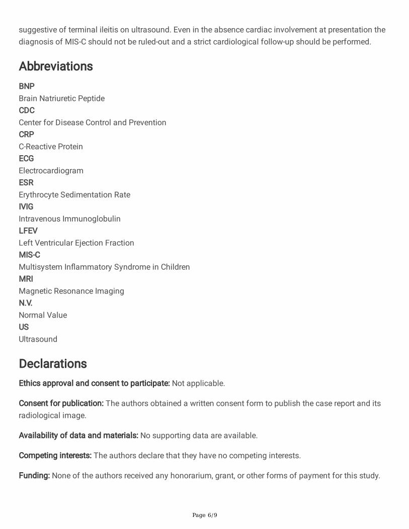
Twenty-four hours after admission the patient developed a progressive worsening abdominal pain in theright lower quadrant, with local guarding and rebound tenderness. Abdominal ultrasound showed athickening of the terminal ileus with ascites and mesenteric lymphadenopathy, while the appendix wasnot detected (Fig. 1).
A laparoscopic exploration was performed to rule out acute appendicitis. The ileus and cecum appearedthickened and in�amed, while the appendix was normal. Broad spectrum antibiotic treatment wasstarted.
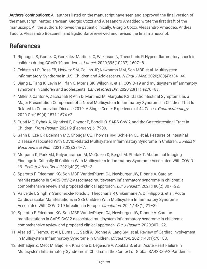
Four days after admission and two days after surgery, despite antibiotic therapy the patient was stillfebrile and markedly asthenic. Thus, a second cardiological evaluation was performed, showingincreased in�ammatory and myocardial markers (CRP 250 mg/L, cardiac troponin 65 ng/L, BNP 9195pg/mL), negative T waves along with prolonged QT interval (490msec) at ECG (Fig. 2) and a reduced leftventricular ejection fraction (LVEF 55%) with a tricuspid regurgitation at echocardiography. According tothe simultaneous presence of cardiac and abdominal involvement, a diagnosis of MIS-C was made andtreatment with intravenous immunoglobulins (2 gr/kg) and steroids (methylprednisolone 2 mg/kg) wasstarted. Due to the concomitant myocarditis, he received a prophylactic anticoagulation and antiplatelettherapy. After 24 hours the patient had a prompt recovery with cessation of fever, abdominal pain andmalaise. In few days in�ammatory and cardiac markers progressively decreased to normal values, whileECGs and echocardiogram normalized in three weeks. Exercise restriction was recommended for 6months, when the patient will undergo cardiac magnetic resonance imaging (MRI).
Discussion And ConclusionsHere, we reported the case of an adolescent with MIS-C in which gastrointestinal symptoms resembled anacute appendicitis. This simil-appendicitis presentation led to an unnecessary explorative laparoscopyand treatment delay. The subsequent cardiovascular involvement and the increasing in�ammatorymarkers allowed the right diagnosis and treatment with prompt and complete recovery.
Despite gastrointestinal symptoms (abdominal pain, emesis and diarrhea) are common features in MIS-Cpatients, only in a few cases these manifestations resemble an acute appendicitis (5, 6, 13–15).Moreover, when these manifestations precede other organ involvement a differential diagnosis betweenin�ammatory bowel disease, abdominal surgical conditions or severe infections could be di�cult (13–16). Typical laboratory �ndings in MIS-C patients are lymphopenia with neutrophilia, increased PT and D-dimer, hypoalbuminemia, hypertransaminasemia, elevation of in�ammatory (CRP, ESR, ferritin and�brinogen) and cardiac (troponin and BNP) markers (2). As in our case, among all these values, increasedin�ammatory markers, lymphopenia and hypoalbuminemia are common �ndings in patients withgastrointestinal symptoms (4, 6). In addition, reviewing abdominal imaging in MIS-C patients, commonUS �ndings are ascites, acalculous cholecystitis, bowel wall thickening and mesenteric adenitis (4, 6, 7).
Page 5/9
Nevertheless, as in our case, Tullie et al. described eight patients affected by MIS-C with acute abdomenpresentation, in some cases simultaneous with cardiac involvement, for whom symptoms and ultrasoundimaging were misleading. Notably, pseudo-appendicitis presentation may be associated to the �nding ofterminal ileitis or typhlitis on abdominal imaging or laparoscopy (6, 13). Therefore, in the �rst week ofdisease, gastrointestinal symptoms, laboratory exams and abdominal US could be misleading, especiallyif no other organ involvement is already present. In our case, retrospectively, the persistent asthenia couldbe seen as an early marker of cardiac involvement. Indeed, asthenia is almost ever present in MIS-Cpatient with cardiac manifestation (12). Nevertheless, no elevation of cardiac enzymes or decreasedventricular function were noted in the patient until the second week of disease.
In regard to the risk of intensive care admission and inotropic support in MIS-C patient, a prompt and rightdiagnosis is mandatory. Up to 80% of patients develop cardiovascular involvement ranging from onlymild elevation of cardiac markers (troponin and pro-BNP) to cardiogenic shock. If MIS-C shares someKawasaki’s disease features, the former usually results in more severe ventricular dysfunction andmyocarditis. Cardiogenic shock has been proposed to be the result of either myocardial viral damage or“cytokine storm” vasodilatation. Other cardiovascular complications encompass coronary arteryaneurism/dilatation and conduction abnormalities. According to a review of cardiac involvement in MIS-C, coronary artery dilatation is reported in up to 25% of patients, while heart conduction abnormalitiesshowed a 7–60% prevalence. Typical arrhythmias are �rst-degree atrioventricular block, QTc prolongationand ST segment changes. The American Heart Association suggests repeated ECG during the acutephase, but telemetry is needed if any arrythmias occur. From several large studies, earlyimmunomodulatory treatment seems to resolve cardiac damage in most of all cases, but little is knownabout cardiac sequelae in MIS-C patients. Once myocardial damage occurs, an expert consensusrecommends exercise restriction for 6 months with a cardiac MRI at 3 or 6 months to evaluate heartfunction.
Up to now, no randomized trials have been developed for treatment and management of MIS-C, but due tothe similarities between Kawasaki disease and MIS-C, an immunomodulatory approach with IVIG andsteroids is recommended. Moreover, antiplatelet treatment and prophylactic anticoagulation aresuggested once myocardial or coronary involvement are present. As in our case, several retrospectivestudies reported, in the majority of patients, normalization of myocardial markers, ECGs abnormalitiesand ventricular dysfunction after immunomodulatory treatment (10, 12). Nevertheless, longer studies andtrials are needed to evaluate treatments and chronic sequalae.
In conclusion, our case highlights how gastrointestinal involvement in MIS-C could mimic acuteappendicitis, and this presentation may precede cardiac involvement, leading to possible misdiagnosisand delayed treatment.
In patients with a recent exposure to SARS-CoV-2 a clinical presentation with fever, asthenia andgastrointestinal symptoms should be seen as highly suggestive of MIS-C. This suspect may be supportedby the presence of an increased in�ammatory markers, lymphopenia and hypoalbuminemia and images
Page 6/9
suggestive of terminal ileitis on ultrasound. Even in the absence cardiac involvement at presentation thediagnosis of MIS-C should not be ruled-out and a strict cardiological follow-up should be performed.
AbbreviationsBNPBrain Natriuretic PeptideCDCCenter for Disease Control and PreventionCRPC-Reactive ProteinECGElectrocardiogramESRErythrocyte Sedimentation RateIVIGIntravenous ImmunoglobulinLFEVLeft Ventricular Ejection FractionMIS-CMultisystem In�ammatory Syndrome in ChildrenMRIMagnetic Resonance ImagingN.V.Normal ValueUSUltrasound
DeclarationsEthics approval and consent to participate: Not applicable.
Consent for publication: The authors obtained a written consent form to publish the case report and itsradiological image.
Availability of data and materials: No supporting data are available.
Competing interests: The authors declare that they have no competing interests.
Funding: None of the authors received any honorarium, grant, or other forms of payment for this study.
Page 7/9
Authors’ contributions: All authors listed on the manuscript have seen and approved the �nal version ofthe manuscript. Matteo Trevisan, Giorgio Cozzi and Alessandro Amaddeo wrote the �rst draft of themanuscript. All the authors followed the patient clinically. Giorgio Cozzi, Alessandro Amaddeo, AndreaTaddio, Alessandro Boscarelli and Egidio Barbi reviewed and revised the �nal manuscript.
References1. Riphagen S, Gomez X, Gonzalez-Martinez C, Wilkinson N, Theocharis P. Hyperin�ammatory shock in
children during COVID-19 pandemic. Lancet. 2020;395(10237):1607–8.
2. Feldstein LR, Rose EB, Horwitz SM, Collins JP, Newhams MM, Son MBF, et al. MultisystemIn�ammatory Syndrome in U.S. Children and Adolescents. N Engl J Med. 2020;383(4):334–46.
3. Jiang L, Tang K, Levin M, Irfan O, Morris SK, Wilson K, et al. COVID-19 and multisystem in�ammatorysyndrome in children and adolescents. Lancet Infect Dis. 2020;20(11):e276–88.
4. Miller J, Cantor A, Zachariah P, Ahn D, Martinez M, Margolis KG. Gastrointestinal Symptoms as aMajor Presentation Component of a Novel Multisystem In�ammatory Syndrome in Children That IsRelated to Coronavirus Disease 2019: A Single Center Experience of 44 Cases. Gastroenterology.2020 Oct;159(4):1571-1574.e2.
5. Puoti MG, Rybak A, Kiparissi F, Gaynor E, Borrelli O. SARS-CoV-2 and the Gastrointestinal Tract inChildren. Front Pediatr. 2021;9 (February):617980.
�. Sahn B, Eze OP, Edelman MC, Chougar CE, Thomas RM, Schleien CL, et al. Features of IntestinalDisease Associated With COVID-Related Multisystem In�ammatory Syndrome in Children. J PediatrGastroenterol Nutr. 2021;72(3):384–7.
7. Morparia K, Park MJ, Kalyanaraman M, McQueen D, Bergel M, Phatak T. Abdominal ImagingFindings in Critically Ill Children With Multisystem In�ammatory Syndrome Associated With COVID-19. Pediatr Infect Dis J. 2021;40(2):e82–3.
�. Sperotto F, Friedman KG, Son MBF, VanderPluym CJ, Newburger JW, Dionne A. Cardiacmanifestations in SARS-CoV-2-associated multisystem in�ammatory syndrome in children: acomprehensive review and proposed clinical approach. Eur J Pediatr. 2021;180(2):307–22.
9. Valverde I, Singh Y, Sanchez-de-Toledo J, Theocharis P, Chikermane A, Di Filippo S, et al. AcuteCardiovascular Manifestations in 286 Children With Multisystem In�ammatory SyndromeAssociated With COVID-19 Infection in Europe. Circulation. 2021;143(1):21–32.
10. Sperotto F, Friedman KG, Son MBF, VanderPluym CJ, Newburger JW, Dionne A. Cardiacmanifestations in SARS-CoV-2-associated multisystem in�ammatory syndrome in children: acomprehensive review and proposed clinical approach. Eur J Pediatr. 2020;307–22.
11. Alsaied T, Tremoulet AH, Burns JC, Saidi A, Dionne A, Lang SM, et al. Review of Cardiac Involvementin Multisystem In�ammatory Syndrome in Children. Circulation. 2021;143(1):78–88.
12. Belhadjer Z, Méot M, Bajolle F, Khraiche D, Legendre A, Abakka S, et al. Acute Heart Failure inMultisystem In�ammatory Syndrome in Children in the Context of Global SARS-CoV-2 Pandemic.

Page 8/9
Circulation. 2020;142(5):429–36.
13. Tullie L, Ford K, Bisharat M, Watson T, Thakkar H, Mullassery D, et al. Gastrointestinal features inchildren with COVID-19: an observation of varied presentation in eight children. Lancet Child AdolescHealth. 2020;4(7):e19–20.
14. Jackson RJ, Chavarria HD, Hacking SM. A Case of Multisystem In�ammatory Syndrome in ChildrenMimicking Acute Appendicitis in a COVID-19 Pandemic Area. Cureus. 2020;12(mm):1–5.
15. Cabrero-Hernández M, García-Salido A, Leoz-Gordillo I, Alonso-Cadenas JA, Gochi-Valdovinos A,González Brabin A, et al. Severe SARS-CoV-2 infection in children with suspected acute abdomen: Acase series from a tertiary hospital in Spain. Pediatr Infect Dis J. 2020;39(8):E195–8.
1�. Mahajan N, Chang HT, Leeman R, Manalo R, Glaberson WR. Case of multisystem in�ammatorysyndrome in children presenting as fever and abdominal pain. BMJ Case Rep. 2020 Sep 8;13(9):1–5.
Figures
Figure 1
Abdominal ultrasound showing transmural thickening of the terminal ileus.

Page 9/9
Figure 2
Electrocardiogram showing sinus bradycardia (HR 50 beats/min) with QT tract prolongation of 494 msec,diffuse T-waves alteration and minimal right branch block.