cardiac muscle physiology - judoctors · pdf file03.10.2013 · cardiac muscle...
TRANSCRIPT

Cardiac muscle physiology
Calcium channels open when the membrane potential gets less negative.
And because they are slow they don't open as fast as the Na+ channels so the difference is in the timing.
As we know Calcium enters during phase two, and in Cardiac muscles what enters triggers the release of
calcium from the Sarcoplasmic Reticulum (Calcium induced calcium release), calcium from SR binds to
troponin C on actin filaments as a result troponin C will uncover binding sites on actin; so myosin binds to
these sites and the power stroke continues and contraction takes place ; However, in skeletal muscles the
release of calcium from SR is due to electrostatic transfer from the sarcolemma to the SR through T tubules
then Ryanodine-receptor channels are opened.
In Excitation contraction coupling the coupler is Calcium.
In order to Relax the cardiac muscle should decreasing intracellular calcium to its normal value (10-7 molar)
by:
1) Calcium up take back to the Sarcoplasmic Reticulum, through active transfer (calcium ATPase pump on
the SR) as the concentration of calcium in SR is relatively high (transfer against gradient) and this
happens in skeletal muscles as well.
But the difference between the cardiac and skeletal muscle is in the Calcium- Sodium exchanger on the
sarcolemma.
2) In cardiac muscle Ca+2 is exchanged with Na+ through an electrogenic pump (one Ca+2 out , three Na+
in), this pump works bilaterally which means if sodium concentration inside is high (in phase Zero) the
pump will work in the other direction (Na+ to outside, Ca+2 to inside) and in this case the calcium
getting inside will add to the calcium that will be entering through phase two and to calcium that is
released from the SR.
Note: Na+ - K+ pump maintains Na+ gradient necessary for the exchange of calcium with sodium.
3) Calcium ATPase pump on the sarcolemma of cardiac muscle.

The Calcium pump in the SR is a protein, this membrane protein is stimulated by another protein also found
on the SR called Phospholamban, phospholamban is always present but when it is phosphorelated it activates
the Calcium ATPase of the SR, then it uptakes calcium back to the SR against concentration, so
phosphorelation of phospholambin is one of the ways by which Calcium pump is activated, epinephrine
norepinephrine binds to Beta one receptors on the heart, it activates Adenylate cyclase which converts ATP to
cAMP, cAMP activates cAMP dependent protein kinase which phosphorelates phospholamban.
So epinephrine shortens time needed for relaxation which increases the heart rate.
The Sodium Calcium exchanger has a low affinity for calcium (it needs high concentrations to be activated)
but high capacity.
The Calcium pump (calcium ATPase) has very High affinity but low capacity.
Note: cardiac glycosides (digoxin) increases the contractility of cardiac muscle by binding to Na+ - K+ pump and
inhibiting it; so Na+ intracellular concentration increases, as a result the Calcium-Sodium exchanger will work
the other way which leads to increase in the intracellular calcium and finally increasing the contractility
(positive inotropic effect).
Now during abnormal states (very high intracellular calcium concentration) these pumps are not enough;
there is another sodium calcium exchanger in the wall of mitochondria, so during pathological states the
mitochondria will take excess calcium from cytoplasm.
Calcium concentration during diastole (relaxation) 10-7 , during systole (contraction)10-5 .
Effect of Ca++ channel blockers (Diltiazem), at 10 uMol/L the action potential is becoming reduced and the
force of contraction is decreased; we need this to decrease the oxygen consumption in Myocardial infarction.
At 30 uMol/L more decrease.
Cardiac Muscle action potential Vs. Skeletal Muscle :

Phase 0 -Depolarization phase (Na+ influx)
Phase 1 partial repolarization (Not in skeletal)
Phase 2 Plateau (depolarization not in skeletal) slow calcium channels
Phase 3 fast repolarization phase (K+ efflux
Phase 4 resting membrane potential
Let us see the effect of action potential on the mechanical response:
In skeletal muscles:
The action potential happens in the latent period then it is followed by mechanical response (contraction and
relaxation).
You can give another stimulus after the refractory period and you'll get another contraction (summation of
contraction), if the rate of stimulation is very high, you get sustained contraction (Tetanus).
In Cardiac muscle:
Very long refractory period because of the long action potential; so if you give another stimulus the cardiac
muscle will be already relaxed.

This prevents tetanus in cardiac muscle.
This is a skeletal muscle : two many stimulus
which leads to incomplete tetanus (saw shaped: contraction with some relaxation) or complete tetanus
(sustained contraction).
In skeletal muscle there is neuromuscular junction which is not present in cardiac muscle.
The Dr read the steps in his figure:
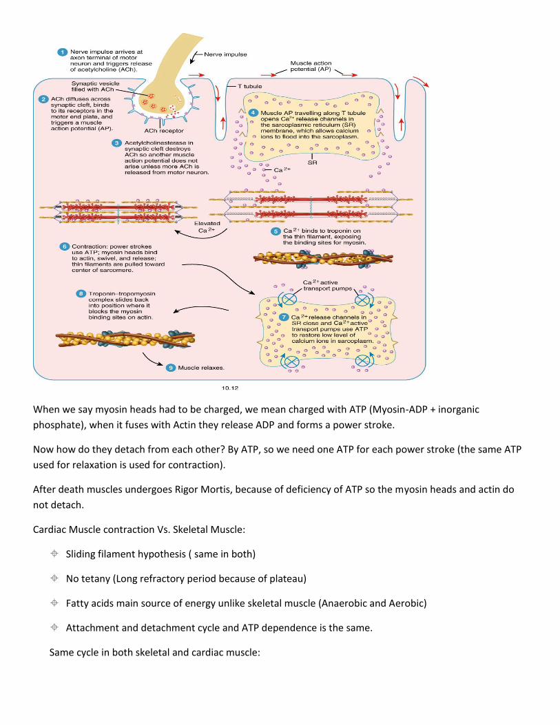
When we say myosin heads had to be charged, we mean charged with ATP (Myosin-ADP + inorganic
phosphate), when it fuses with Actin they release ADP and forms a power stroke.
Now how do they detach from each other? By ATP, so we need one ATP for each power stroke (the same ATP
used for relaxation is used for contraction).
After death muscles undergoes Rigor Mortis, because of deficiency of ATP so the myosin heads and actin do
not detach.
Cardiac Muscle contraction Vs. Skeletal Muscle:
Sliding filament hypothesis ( same in both)
No tetany (Long refractory period because of plateau)
Fatty acids main source of energy unlike skeletal muscle (Anaerobic and Aerobic)
Attachment and detachment cycle and ATP dependence is the same.
Same cycle in both skeletal and cardiac muscle:

Source of energy:
-in cardiac muscle the source of energy normally is ATP coming from Creatine phosphate.
-Creatine phosphate is storage to ATP.
-in myocardial infarction we measure CPK (Creatine PhosphoKinase), because this cycle is going to be
activated so there is release of this enzyme.
-creatine phosphate is the immediate source of energy and it is enough for about 10 seconds (in 100 meter
running race).
-other two sources of energy are the aerobic and anaerobic respiration.

Now we come to the last part of our lecture, Length-Tension Relationship in skeletal and cardiac muscle
(frank-starling law of the heart).
Within physiological limits, an increase in the length of the muscle increases the force of contraction.
Skeletal muscle normally works at its optimal length.
Cardiac muscle doesn't work at its optimal length (it works on less length); so you can increase the force of
contraction by increasing the length within physiological limits (the optimal length), but if it exceeds the
optimal length, the force will decrease.
Look at this figure:
You can see the contractile component, Series elastic component (tendon) and parallel elastic component
(proteins and elastic elements).
Let us suppose that the muscle is like a rubber band, when we pull the rubber band the amount of tension we
put in it before contraction we call it (Resting tension or passive tension). in order to shorten, the band must
overcome the force that pulls it (passive tension) and contracts, so there are two forces: 1) force to overcome
the passive tension 2)force to make contraction (the active tension).
If the rubber band (the muscle) was overstretched, the contraction would decrease.why? Because the passive
tension was so high to the extent that the passive tension equals the total tension ( no active tension).
Active tension cannot be measured directly
What can be measured?
(1) passive tension - tension required to extend a resting muscle
(2) total tension - active tension and passive combined
Active is calculated from 1 & 2
(AT = TT - PT)
Note that active tension falls away linearly with increasing length

The optimal length (2.2 micrometer), the maximum force
of contraction is when the length is optimal.
Note: in the figure above, in skeletal muscles the drop you see in the Total tension is caused by the Parallel
elastic elements, this does Not happen in the cardiac muscle since there are no parallel elastic elements.
What happens during heart failur (stagflation of blood), so dilation of ventricles to the extent that the cardiac
out put decreases.
The figure below shows why when overstretching the muscle the force of contraction decreases.
as you see this is because of decreasing of
overlap between Actin and Myosin filaments.
Cardiac Muscle length-tension relationship Vs Skeletal muscle:
Cardiac muscle works at much less than its maximum length in contrast to skeletal
Total, Active and Passive length-tension relationship differ (no drop in the total tension in cardiac
muscle).
Frank-Starling law of the heart is same in both.
