cardioprotective properties of opioid receptor agonists in ... · the selective µ or agonist...
TRANSCRIPT
1
Cardioprotective Properties of Opioid Receptor Agonists in Rats with Stress-Induced
Cardiac Injury
Ekaterina S. Prokudinaa B, Leonid N. Maslova* A, D, E, F, Natlia V. Naryzhnayaa B,G,
Sergey Yu. Tsibulnikova B,C,E, Yury B. Lishmanova,b D, John E. Madias PhD c D,E, Peter R.
Oeltgen PhD d D,E
aLaboratory of Experimental Cardiology, Cardiology Research Institute, Tomsk National
Research Medical Center, Russian Academy of Sciences 634012 Tomsk, Russia.
bLaboratory of Nuclear Medicine, National Research Tomsk Polytechnic University,
Tomsk, Russia.
cIcahn School of Medicine at Mount Sinai, and the Division of Cardiology, Elmhurst
Hospital Center, New York, New York, USA.
dDepartment of Pathology, University of Kentucky College of Medicine, Lexington, KY,
USA
* Correspondence:
Leonid N. Maslov, MD, PhD, DSci, Professor of Pathological Physiology
Laboratory of Experimental Cardiology,
Federal State Budgetary Scientific Institution «Research Institute for Cardiology»,
Kyevskaya 111A, 634012 Tomsk, Russia
Tel. +7 3822 262174
E-mail address: [email protected]
Short title: Stress cardiomyopathy and opioid receptors
Summary
Purpose The objectives of this study were to investigate the role of endogenous opioids in
the mediation of stress-induced cardiomyopathy (SIC), and to evaluate which opioid receptors
regulate heart resistance to immobilization stress. Methods Wistar rats were subjected to 24 h

2
immobilization stress. Stress-induced heart injury was assessed by 99mTc-pyrophosphate
accumulation in the heart. The opioid receptor (OR) antagonists (naltrexone, NxMB - naltrexone
methyl bromide, MR 2266, ICI 174.864) and agonists (DALDA, DAMGO, DSLET, U-50,488)
were administered intraperitoneally prior to immobilization and 12 h after the start of stress. In
addition, the selective µ OR agonists PL017 and DAMGO were administered
intracerebroventricularly prior to stress. Finally pretreatment with guanethidine was used. Results
Naltrexone did not alter the cardiac 99mTc-PP accumulation in stressed rats. NxMB aggravated
stress-induced cardiomyopathy (P = 0.005) (SIC). The selective µ OR agonist DALDA, which does
not cross the blood-brain barrier, completely prevented (P = 0.006) SIC. The µ OR agonist
DAMGO exhibited weaker effect than DALDA. The selective δ ligand (DSLET) and κ OR ligand
(U-50,488) did not alter stress-induced 99mTc-pyrophosphate accumulation in the heart.
Intracerebroventricular administration of the µ OR agonists aggravated SIC. Pretreatment with
guanethidine abolished this effect (P = 0.01). Guanethidine alone exhibited cardioprotective
properties. Conclusions A stimulation of central µ OR promotes an appearance of SIC. In contrast,
stimulation of peripheral µ OR contributes to an increase in cardiac tolerance to stress.
Key words: 99mTc-pyrophosphate, opioid receptors, takotsubo syndrome, stress,
cardiomyopathy
1. Introduction
In the mid-seventies of the last century it was found that restraint stress could cause
cardiomyopathy in pigs (Jönsson et al. 1975, Johansson et al. 1974), and food-shock stress could
induce cardiomyopathy in rats (Miller, Mallov, 1977). In 1990, stress-induced cardiomyopathy was
described in humans by Sato et. al. (Sato H et al. 1990). These authors called the condition tako-
tsubo syndrome (TS) because the left ventricle of patients afflicted with this pathology during
systole resembles the Japanese octopus fishing implement called “tako-tsubo” (Sato et al. 1990).
Later, the existence of the disease was confirmed by other cardiologists (Pavin et al. 1997;
3
Tsuchihashi et al. 2001; Kurisu et al. 2003; Akashi et al. 2003]. The incidence of TS was found to
be increased as a result of improved diagnostic methods. According to Khera et al. (Khera et al.
2016) from 2007 to 2012, the incidence of TS increased over 3-fold in the United States, as
reflected in the relevant literature. Thus, in 2007, there were 52 cases per 1 million, and in 2012
already 178 cases per million hospitalized patients were reported (Khera et al. 2016). The rate of
28-day mortality in patients with TS is similar to mortality in patients with ST-segment elevation
myocardial infarction (STEMI) (5.5% vs. 5.7%) (Stiermaier et al., 2016), while the rate of 1-year
mortality in patients with TS and in patients with STEMI is 12.5% vs. 9%, respectively. The long-
term mortality (during 3.8 ± 2.5 years) in patients with TS was significantly higher compared to the
one of STEMI patients (24.7% vs. 15.1%) (Stiermaier et al. 2016). In-hospital mortality in the TS
patients with cardiopulmonary failure is 18% (El-Battrawy et al. 2017). This high mortality may be
partially due to limitations of our knowledge about the pathogenesis of the disease, and as a result
to the lack of a specific effective therapy of TS. In our opinion, animal studies could contribute
important insights in the pathogenesis and management of TS.
Immobilization stress in rats has been found to promote the development of a state similar
to TS (Ueyama et al. 2002; 2004). Although researchers have been studying TS already for the past
26 years, the pathogenesis of this disease remains a mystery in many ways. Currently, the focus is
on activation of the adrenergic system in the pathogenesis of stress-induced cardiomyopathy (Chen
et al. 2017; Dilsizian et al. 2017; Pelliccia et al. 2017; Sestini et al. 2017; Casey et al. 2017; Kido
et al. 2017; Ceccacci et al. 2016; Chen et al. 2016; Christensen et al. 2016). Exogenous
catecholamines can cause the TS (Casey et al. 2017; Kido et al. 2017; Elikowski et al. 2017; Nazir
et al. 2017; Belliveau et al. 2016). Stress-induced cardiomyopathy (SIC) is often noted in patients
with pheochromocytoma (Elikowski et al. 2017; Agrawal et al. 2017; Zhang et al. 2017; Y-Hassan
et al. 2016). It is believed that SIC is an excessive activation of β-adrenergic receptors (Chen et al.
2017; Oras et al. 2017ab; Brunetti et al. 2016), so some authors mimic SIC by administration of
4
toxic doses of isoproterenol (Oras et al. 2017ab; Sachdeva et al. 2014). This approach seems to us
not quite correct, because in the case of using isoproterenol, other humoral factors that may be
involved in the mechanism of SIC origin are not considered. Many people find themselves in
severe stressful situations but TS does not occur. We hypothesize that the endogenous peripheral
opioid system plays a significant role in providing a deterrent mechanism to block TS from
developing. We have previously shown that the 24-hour immobilization stress can cause
accumulation of 99mTc-pyrophosphate in rat myocardium (Lishmanov et al. 1997) that according to
Miller, Mallov (Miller, Mallov, 1977) is an indicator of stress-induced myocardial injury.
According to our data the ligands of opioid receptors can modulate stress heart damage (Lishmanov
et al. 1997). The objectives of this study were to investigate the role of endogenous opioids in the
mediation of stress-induced cardiomyopathy, and to evaluate which opioid receptors regulate heart
resistance to immobilization stress.
2. Materials and Methods
2.1. Animals and Research Protocol
Male Wistar rats weighing 230 – 250 g were housed in groups of four rats per cage and
allowed free access to tap water and a standard laboratory rat chow. Animals were kept in an air-
conditioned room, where the temperature was maintained at 23±1°C, and the relative humidity was
kept at 60–70%. Animals were exposed to a 12 h day–night cycle.
Stress was induced by a 24-hour immobilization of animals in the supine position. The rats
were fixed with adhesive tape for each limb thus reducing trauma related to the procedure. Naïve
rats were used as control animals. They were not supplied with water and chow during
immobilization stress. Animals which were immobilized and injected intraperitoneally two times
with a solution of 0.9% NaCl (1 ml/kg) and were used as stress control. The first injection was
administered at 9.00 a.m. and the second injection at 21.00 p.m. The study was approved by the

5
Ethical Committee of the Cardiology Research Institute (approval number: 79, from 14.10.2016),
and it conformed to the European Union Directive 2010/63/EU.
In most experiments, ligands of opioid receptors (OR) were administered intraperitoneally
twice: 30 min before immobilization and 12 h after the start of exposure to the stress (Lishmanov et
al. 1997). In addition μ-OR agonists were administered intracerebroventricularly via a cannula
implanted in advance. The preferential μ and κ OR antagonist naltrexone was administered at a
dose of 0.5 mg/kg (n = 12) (Thomas et al. 1998). Naltrexone methyl bromide (NxMB), an OR
antagonist that does not cross the blood-brain barrier (BBB), was used at a dose of 5 mg/kg (n = 12)
(Browen et al. 1983). The half-life of methylnaltrexone in the blood plasma is 7.6 h (Misra et al.
1987) and the half-life of naltrexone in various mammalian species is from 4 to 10 h (Crabtree
1984). Therefore, these drugs are capable of providing long-term blockade of opioid receptors. A
preferential κ OR antagonist MR2266 ((-)2-(3-furyl methyl)-5,9-diethyl-2-hydroxy-6,7-
benzomorphan) (Lahti et al. 1985) was administered at a dose of 5 mg/kg (n = 12). The selective δ
OR antagonist ICI 174.864 (N,N-dially-Tyr-Aib-Aib-Phe-Leu-OH, where the Aib is α-
aminoisobutyric acid) was administered at a dose of 2.5 mg/kg (n = 12) (Dauge et al. 1988;
Rebrova et al. 2001). The selective μ OR agonist that does not cross the BBB DALDA (H-Tyr-D-
Arg-Phe-Lys-NH2) (Roques et al. 1990; Samii et al. 1994) was given at a dose of 0.1 mg/kg (n =
12) (Rebrova et al. 2001; Maslov et al. 2002). The selective μ OR agonist DAMGO (H-Tyr-D-Ala-
Gly-Nα-Me-Phe-Gly-ol) 20 was injected at a dose of 0.1 mg/kg (n = 12) (Rebrova et al. 2001). The
selective δ OR agonist DSLET (H-Tyr-D-Ser-Gly-Phe-Leu-Thr-OH) 20 was administered at a dose
of 0.1 mg/kg (n = 12) (Rebrova et al. 2001). The selective κ1 OR agonist (±)-U-50,488 (trans-(±)-
3,4-Dichloro-N-methyl-N-[2-(1-pyrrolidinyl)cyclohexyl] benzeneacetamide hydrochloride) (Lahti
et al. 1985; Von Voigtlander et al. 1982] was used at a dose of 8 mg/kg (n = 12) [Von Voigtlander
et al. 1982).
6
In addition, DAMGO was administered intracerebroventricularly and the selective μ OR
agonist PL017 (Tyr-Pro-Nα-Me-Phe-D-Pro-NH2) (Chang et al. 1983) was infused
intracerebroventricularly both at a dose of 20 µg two times: 30 min before immobilization and 12 h
after immobilization (Chang et al. 1983). Guanethidine monosulfate, a compound which depletes
peripheral storage of endogenous catecholamines (Maitre et al. 1971), was used at a dose of 50
mg/kg subcutaneously every day during 3 days (Maslov et al. 2009). The last injection of
guanethidine was performed 24 h before immobilization. Each group included 12 animals. A total
of 432 rats were included in the study. Control (naive) group included 96 animals.
2.2. Measurement of 99mTc-pyrophosphate accumulation
The evaluation of the extent of the stress-induced cardiac damage was studied by the
assessment of the level of myocardial accumulation of radioactive 99mTc-pyrophosphate (99mTc-PP),
which was administered intravenously in a dose of 150 MBq/kg 30 min after cessation of
immobilization (Miller, Mallov, 1977]. The animals were decapitated under ethyl ether anesthesia
100 minutes after the injection. Hearts were removed from thorax and perfused through aorta with
cold physiological saline (10 ml, 10○C). Registration of radioactivity was measured by the γ
counter RIS-A1-E “Doscalibrator” Amplitude Company, (Third Zapadnyi 15, Zelenograd,
Moscow, Russia. The accumulation of 99mTc-PP in the myocardial tissue was expressed as a
percent of administered dose per 1 g of heart tissue as % of total dose/g weight of heart x 100.
2.3. Surgical procedure
The cannula implantation was performed as described previously (Lishmanov et al. 2009].
Five to 7 days before the induction of stress, a cannula, consisting of a 30 gauge stainless steel
needle (SFM Hospital Products, Berlin, Germany), was inserted into the lateral cerebral ventricle of
rats and was fixed in the skull by dental cement. This procedure was undertaken in surgery and in
experimental procedures under pentobarbital anesthesia (50 mg/kg), and was facilitated by the use
of a stereotaxic apparatus (SEZh-5; Constructor Company, Acadimika Bogomoltsa 4, Kiev,
7
Ukraine). The cannula was inserted at the following coordinates from the bregma: AP –1.5 mm,
+2.0 mm; V –3.5 mm. The place of injection in the lateral cerebral ventricle was confirmed by
injecting methylene blue dye through the cannula at the end of the experiment. At the end of the
experiment, the brain was removed, placed in formalin and later sectioned. Correct placement of
the cannula was confirmed by the presence of dye in the cerebroventricular system in all rats.
2.4. Pharmacological agents
Naltrexone, U-50,488 were purchased from Sigma-Aldrich (USA). Naltrexone methyl
bromide and MR2266 were synthesized by Boehringer Inglheim KG (Inglheim am Rhein,
Germany). Peptides ICI 174.864, DAMGO, DSLET, PL017 were synthesized by Chiron
Mimotopes Peptide Systems (San Diego, USA). Peptide DALDA was synthesized by Laboratory of
Chemical Biology and Peptide Research, Clinical Research Institute of Montreal, Montreal,
Quebec, Canada. Guanethidine was synthesized by the International Laboratory (San Bruno, CA,
USA) and purchased from Advanced Technology and Industrial Co. (Hong Kong, China).
2.5. Statistical analysis
Results are expressed as mean ± SEM from indicated number of experiments. Statistical
comparison of means between groups was made by one-way ANOVA. The assumption of
normality distribution data has been tested by Shapiro-Wilk (SW) normality test. Values exceeding
the 95% probability limits (P < 0.05) were considered significant.
3. Results
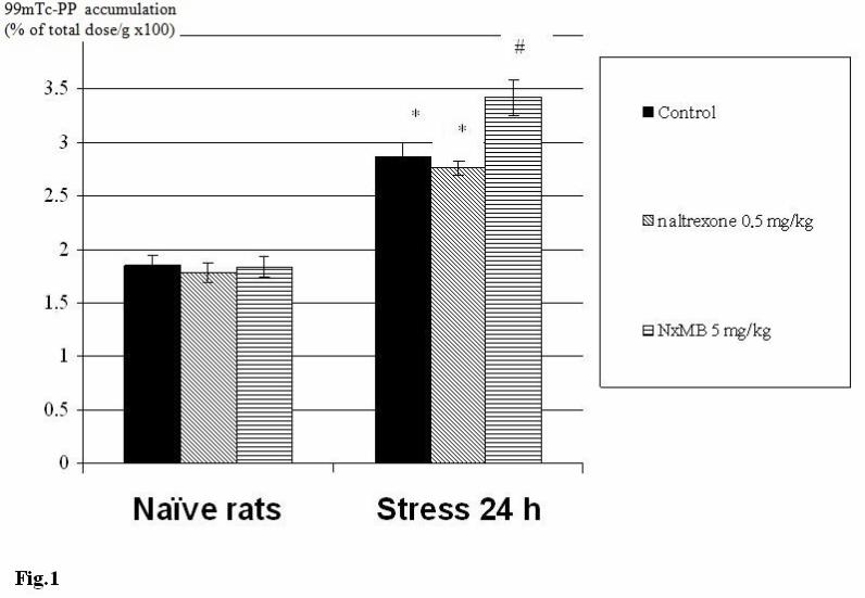
As shown in Figure 1, immobilization stress caused an increase in the 99mTc-PP
accumulation in the myocardium by 1.5 fold (P = 0.001) (~50%). Pretreatment with naltrexone did
not alter the cardiac 99mTc-PP accumulation in stressed rats (Fig. 1). On the contrary, injection of
NxMB contributed to an increase in stress-evoked 99mTc-PP uptake by the heart (P = 0.005) (Fig.
1). It should be noted that the administration of naltrexone or NxMB had no effect on the
accumulation of 99mTc-PP in the myocardium of naïve rats (Fig. 1). Pretreatment with DALDA

8
caused a decrease in the stress-induced accumulation of 99mTc-PP in the myocardium by 1.8 fold (P
= 0.006) (Fig. 2a). The selective μ OR agonist DAMGO resulted in a weaker similar effect,
reducing the stress induced 99mTc-PP heart uptake only by 1.36 fold (P = 0.01) (Fig. 2a). The
selective μ OR agonists had no effect on cardiac 99mTc-PP accumulation in naïve rats. In contrast,
intracerebroventricular administration of DAMGO, DALDA and PL017 enhanced the 99mTc-PP
accumulation in the heart after immobilization (P = 0.01) (Fig. 2b). It should be noted that the
intracerebroventricular administration of selective μ OR agonists had no effect on the cardiac
99mTc-PP accumulation in naïve rats. As shown in Figure 3, the selective δ OR antagonist ICI
174.864 and the preferential κ OR antagonist MR2266 did not alter the cardiac 99mTc-PP
accumulation in naïve and stressed rats. Also the selective δ OR agonist DSLET and the selective κ
OR agonist U-50,488 had no effect on the 99mTc-PP uptake by the heart in both groups of animals
(Fig. 4). Pretreatment with guanethidine contributed to a decrease in the cardiac 99mTc-PP
accumulation in stressed rats, compared with stress control group, and attenuated
intracerebroventricular DAMGO-induced 99mTc-PP accumulation in stressed rats (P = 0.01) (Fig.
5). Finally, guanethidine did not change the cardiac 99mTc-PP accumulation in naïve rats.
4. Discussion
Our results indicate that the immobilization stress causes damage to the heart. Our data are
consistent with the results of other investigators [Miller, Mallov, 1977] who also observed an
enhancement of 99mTc-PP accumulation in the myocardium of food-shock stressed rats.
Pretreatment with guanethidine prevented the stress-induced 99mTc-PP uptake by the heart of the
stressed rats that indicated the involvement of endogenous catecholamines in the pathogenesis of
SIC. In addition, these data indicate that our rat model of SIC is similar to TS because endogenous
catecholamines are involved in the appearance of both immobilization induced cardiomyopathy and
also TS [Riester et al. 2015; Smeijers et al. 2015; Sharkey et al. 2015; Nunez-Gil et al. 2015].
9
We found that administration of NxMB, which does not penetrate into the brain from the
bloodstream, aggravated SIC. On the other hand, naltrexone, which crosses the BBB, prevented the
emergence of SIC. These data indicated that endogenous agonists of central ORs are involved in
the pathogenesis of SIC. The endogenous agonists of peripheral ORs prevented the occurrence of
SIC. Endogenous agonists κ and δ OR perhaps are not involved in the development of this
cardiomyopathy, since antagonists and agonists of these ORs had no effect on the stress induced
99mTc-PP accumulation in the myocardium. It is well known that naltrexone and NxMB exhibit a
high affinity for μ OR. Therefore, it can be hypothesized that central and peripheral μ ORs are
involved in the regulation of cardiac tolerance to stress but their role in regulating the cardiac
tolerance to stress is different. Activation of the central μ OR aggravates the pathologic effect of
stress and a stimulation of the peripheral μ OR has a cardioprotective effect. Therefore, NxMB
exacerbates the pathogenic effect of stress. Naltrexone blocks both central and peripheral μ ORs
thereby excluding their involvement in the regulation of cardiac tolerance to impact of stress.
We decided to test this hypothesis using the selective μ OR agonist DALDA, which cannot
cross the BBB (Samii et al. 1994), and DAMGO, which exhibits antinociceptive effect (hot-plate
test in mice) at a dose of 2.5 mg/kg probably due to the stimulation of central μ OR. The
antinociceptive effect in hot-plate test indirectly indicates stimulation of the central opioid receptors
(Delay-Goyet et al. 1991). This opioid did not exhibit antinociceptive effect at a dose of 1.25
mg/kg in this test (Delay-Goyet et al. 1991). We used DAMGO at a dose of 0.1 mg/kg to avoid
activation of central OR. Both opioid peptides exhibit similar affinity to μ OR (Schiller et al.,
1989). The single most important difference between these opioids is the permeability to the BBB.
We found that DALDA showed a strong cardioprotective effect in the stressed rats, while the
cardioprotective effect of DAMGO was weaker, probably because small quantities of DAMGO can
cross the BBB, and partially occupy central μ OR. These results are in agreement with our
previously published data (Lishmanov et al. 2017).

10
In further experiments, we explored whether the activation of the central μ OR would affect
the heart resistance to stress. It turned out that intracerebroventricular administration of DAMGO,
or the selective μ OR agonist PL017 aggravated the stress-induced injury to the heart. Pretreatment
with guanethidine completely eliminated the negative effect of intracerebroventricular DAMGO
administration. This result suggested an involvement of the sympathetic nervous system (SNS) in
the mediation of stress-induced myocardial injury, and our hypothesis is in agreement with data of
other investigators (Hassen, Feuerstein. 1987; Kiritsy-Roy et al. 1989; Yamauchi et al. 1997) and
our previously published data (Lishmanov et al. 2017).
In 1987, Hassen and Feuerstein (Hassen, Feuerstein 1987) found that the stimulation of μ
OR in n. tractus solitarius leads to activation of the SNS. In 1989, Kiritsy-Roy et al. (Kiritsy-Roy et
al. 1989) experiments with awake rats showed that intracerebroventricular administration of
selective μ OR agonist DAMGO or δ OR agonist DPDPE leads to a 2 to 3 fold increase in the
plasma level of norepinephrine, and increase in plasma epinephrine concentration by several ten
folds. Maximum stimulation of SNS was achieved with the administration of 5 nM of DAMGO and
125 nM of DPDPE, with the latter compound having no effect on catecholamine levels when
administered at a dose of 5 nM (Kiritsy-Roy et al. 1989). In 1997, Yamauchi et al. (Yamauchi et al.
1997) found that the intracerebroventricular administration of β-endorphin (preferential μ and δ OR
agonist R) causes an increase in blood plasma epinephrine and norepinephrine levels in rats.
Naloxone (2 mg/kg intravenously) completely eliminated this effect of β-endorphin. These data
demonstrate that occupancy of central μ OR by agonists can promote an activation of SNS. In
contrast there are data that demonstrate following occupancy of peripheral μ OR by agonists results
in a limitation of norepinephrine release from sympathetic nerve terminals in the heart (Ledda et al.
1982; Ensinger et al. 1984; Von Kugelgen et al. 1985; Fuder et al. 1986; Szabo et al. 1986), and
epinephrine release from the adrenal glands is observed (Chen et al. 1989). However, most
publications indicate that the limitation of norepinephrine release from peripheral sympathetic

11
terminals is a result of presynaptic δ and κ OR activation (Von Kugelgen et al. 1985; Fuder et al.
1986; Szabo et al. 1986), and thus it is unclear why these receptor agonists (DSLET and U-50,488)
had no effect on SIC. Furthermore, it was found that κ OR agonists can inhibit synthesis of
catecholamines in chromaffin cells (Takekoshi et al. 2000). Opioid receptors have been found in
chromaffin cells (Saiani et al. 1982; Abood et al. 1995; Kampa et al. 1999) primarily consisting of
δ (Abood et al. 1995) or κ OR (Kampa et al. 1999). Kampa et al (Kampa et al. 1999) demonstrated
that human pheochromocytoma cells contain κ1 OR, fewer κ2 OR and minimal binding capacity for
δ and µ OR agonists sites. It remains unclear why exactly the activation of peripheral µ opioid
receptors increased cardiac tolerance to stress. In conclusion, it should be noted that in the
regulation of heart tolerance to the impact of ischemia and reperfusion an important role is played
by µ, δ, κ ORs (Abood et al. 1995), and in regulation of cardiac tolerance to the stress only µ OR
are involved according to our data. The reason for such differences in the functional role of ORs
remains unclear.
5. Conclusions
Naltrexone enhanced the cardiac tolerance to the immobilization stress. Naltrexone methyl
bromide aggravated stress-induced cardiomyopathy. The selective µ OR agonist DALDA, which
does not cross the BBB, completely prevented stress-induced heart injury. The selective µ OR
agonist DAMGO, which is capable of crossing the BBB, exhibited weaker effect than DALDA.
The selective δ and κ OR ligands did not alter the stress induced 99mTc-pyrophosphate
accumulation in the heart. Intracerebroventricular administration of the selective µ OR agonists
aggravated the stress-induced heart injury. Pretreatment with guanethidine abolished the noxious
effects of the immobilization stress. Guanethidine administered alone exhibited cardioprotective
properties. Thus, stimulation of central µ OR mediates the emergence of stress cardiomyopathy. In
contrast, stimulation of peripheral µ OR contributes to an increase in cardiac tolerance to stress.
12
Overall, these results support our hypothesis that opioid-mediated cardioprotection is mediated via
peripheral µ OR activation.
Conflict of interest: The authors declare that they have no conflict of interest.
Acknowledgements
The authors are grateful to Dr. Kevin Gormley (Division of Neuroscience & Behavioral
Research, NIDA NIH, Bethesda, USA) for providing the peptides (DAMGO, DSLET, ICI 174.864,
PL017). The authors are grateful to Professor P.W. Schiller (Clinical Research Institute of
Montreal, Montreal, Quebec, Canada) for kindly granting DALDA. The authors are grateful to Drs.
P Veerhoff and Dr Duttmann (Boehringer Inglheim KG, Inglheim am Rhein, Germany) who kindly
donated the naltrexone methyl bromide and MR2266.
This study was funded by Russian Science Foundation (grant 18-75-00001). The section
dedicated to naltrexone is framed within the framework of state. assignments AAAA-A15-
115120910024-0
References
ABOOD ME, TAO Q: Characterization of a delta opioid receptor in rat
pheochromocytoma cells. J Pharmacol Exp Ther 274:1566-1573, 1995.
AGRAWAL S, SHIRANI J, GARG L, SINGH A, LONGO S, LONGO A, FEGLEY M,
STONE L, RAZAVI M, RADOIANU N, NANDA S: Pheochromocytoma and stress
cardiomyopathy: Insight into pathogenesis. World J Cardiol 9: 255-260, 2017.
AKASHI YJ, NAKAZAWA K, SAKAKIBARA M, MIYAKE F, KOIKE H, SASAKA
K: The clinical features of takotsubo cardiomyopathy. QJM 96:563-573, 2003.

13
BELLIVEAU D, DE S: Reverse takotsubo cardiomyopathy following exogenous
epinephrine administration in the early postpartum period. Echocardiography 33: 1089-1091,
2016.
BROWEN DR, ROBERTSON MJ, GOLDBERG LI: Reversal of morphine-induced
catalepsy in the rat by narcotic antagonists and their quaternary derivatives.
Neuropharmacology 22: 317-321, 1983.
BRUNETTI ND, SANTORO F, DE GENNARO L, CORREALE M, GAGLIONE A,
DI BIASE M: Drug treatment rates with beta-blockers and ACE-inhibitors/angiotensin receptor
blockers and recurrences in takotsubo cardiomyopathy: A meta-regression analysis. Int J
Cardiol 214: 340-342, 2016.
CASEY RT, CHALLIS BG, PITFIELD D, MAHROOF RM, JAMIESON N, BHAGRA
CJ, VUYLSTEKE A, PETTIT SJ, CHATTERJEE KC: Management of an acute
catecholamine-induced cardiomyopathy and circulatory collapse: a multidisciplinary approach.
Endocrinol Diabetes Metab Case Rep 2017. pii: 17-0122. 2017
CECCACCI A, MANCONE M, CALCAGNO S, DE VINCENTIS G, SARDELLA G,
FEDELE F: Role of MIBG scintigraphy in reverse Tako-tsubo cardiomyopathy: Confirming a
pathophysiologic hypothesis. Int J Cardiol 223: 54-55, 2016.
CHANG KJ, WEI E, KILLIAN A CHANG JK: Potent morphiceptin analogs: structure
activity relationships and morphine-like activities. J Pharmacol Exp Ther 227:403-408, 1983.
CHEN W, DILSIZIAN V: Cardiac sympathetic disturbance in takotsubo
cardiomyopathy: primary etiology or a compensatory response to heart failure? JACC
Cardiovasc Imaging 9: 991-993, 2016.
CHEN W, DILSIZIAN V: Exploring the pathophysiology of takotsubo cardiomyopathy.
Curr Cardiol Rep 19: 53, 2017.
14
CHEN YM, DIXON WR, WAKADE AR: The effect of etorphine on the secretion of
endogenous catecholamines and total tritium evoked by nerve- and acetylcholine-stimulation in
perfused rat adrenal glands. Life Sci 44:167-74, 1989.
CHRISTENSEN TE, BANG LE, HOLMVANG L, SKOVGAARD DC, OTURAI DB,
SØHOLM H, THOMSEN JH, ANDERSSON HB, GHOTBI AA, IHLEMANN N, KJAER A,
HASBAK P: 123I-MIBG scintigraphy in the subacute state of takotsubo cardiomyopathy. JACC
Cardiovasc Imaging 9: 982-990, 2016.
CRABTREE BL: Review of naltrexone, a long-acting opiate antagonist. Clin Pharm 3:
273-280, 1984.
DAUGE V, ROSSIGNOL P, ROQUES BP: Comparison of the behavioural effects
induced by administration in rat nucleus accumbens or nucleus caudatus of selective μ and δ
opioid peptides or kelatorphan an inhibitor of enkephalin-degrading-enzymes.
Psychopharmacology (Berl) 96: 343-352, 1988.
DELAY-GOYET P, RUIZ-GAYO M, BAAMONDE A, GACEL G, MORGAT JL,
ROQUES BP: Brain passage of BUBU, a highly selective and potent agonist for delta opioid
receptors: in vivo binding and mu versus delta receptors occupancy. Pharmacol Biochem Behav
38:155-162, 1991.
EL-BATTRAWY I, LANG S, ANSARI U, SATTLER K, BEHNES M, SCHRAMM K,
FASTNER C, TÜLÜMEN E, ZHOU X, HOFFMANN U, BORGGREFE M, AKIN I:
Incidence and prognostic relevance of cardiopulmonary failure in takotsubo cardiomyopathy.
Sci Rep 7: 14673, 2017.
ELIKOWSKI W, MAŁEK-ELIKOWSKA M, KAROŃ J, MROZIŃSKA M, BASZKO
A, HORBACKA K: Takotsubo cardiomyopathy after intravenous epinephrine administration
following cardiac arrest provoked by pneumoperitoneum - a case report. Pol Merkur Lekarski
42: 165-169, 2017.

15
ENSINGER H, HEDLER L, SCHURR C, STARKE K: Ethylketocyclazocine decreases
noradrenaline release and blood pressure in the rabbit at a peripheral opioid receptor. Naunyn
Schmiedebergs Arch Pharmacol 328: 20-23, 1984.
FUDER H, BUDER M, RIERS HD, ROTHACHER G: On the opioid receptor subtype
inhibiting the evoked release of 3H-noradrenaline from guinea-pig atria in vitro. Naunyn
Schmiedebergs Arch Pharmacol 332: 148-155, 1986.
HASSEN AN, FEUERSTEIN G: μ-Opioid receptors in NTS elicit pressor responses
via sympathetic pathways. Am J Physiol 252: H156-H162, 1987.
JOHANSSON G, JONSSON L, LANNEK N, BLOMGREN L, LINDBERG P, POUPA
O: Severe stress-cardiopathy in pigs. Am Heart J 87: 451-457, 1974.
JÖNSSON L, JOHANSSON G, LANNEK N, LINDBERG P, POUPA O:
Histochemical and electron microscopic studies of acute cardiomyopathy induced by restraint
stress in pigs. Recent Adv Stud Cardiac Struct Metab 6: 461-470, 1975.
KAMPA M, MARGIORIS AN, HATZOGLOU A, DERMITZAKI I, DENIZOT
A, HENRY JF: κ1-Opioid binding sites are the dominant opioid binding sites in surgical
specimens of human pheochromocytomas and in a human pheochromocytoma (KAT45) cell
line. Eur J Pharmacol 364: 255-262, 1999.
KHERA R, LIGHT-MCGROARY K, ZAHR F, HORWITZ PA, GIROTRA S: Trends
in hospitalization for takotsubo cardiomyopathy in the United States. Am Heart J 172: 53-63,
2016.
KIDO K, GUGLIN M: Drug-induced takotsubo cardiomyopathy. J Cardiovasc
Pharmacol Ther 22: 552-563, 2017.
KIRITSY-ROY JA, MARSON L, VAN LOON GR: Sympathoadrenal, cardiovascular
and blood gas responses to highly selective mu and delta opioid peptides. J Pharmacol Exp
Ther 251:1096-1103, 1989.

16
KURISU S, SATO H, KAWAGOE T, ISHIHARA M, SHIMATANI Y, NISHIOKA K:
Tako-tsubo-like left ventricular dysfunction with ST-segment elevation: a novel cardiac
syndrome mimicking acute myocardial infarction. Am Heart J 143: 448-455, 2002.
LAHTI RA, MICKELSON MM, MCCALL JM, VON VOIGTLANDER PF: [3H]U-
69593 a highly selective ligand for the opioid κ receptor. Eur J Pharmacol 109: 281-284, 1985.
LEDDA F, MANTELLI L: Possible presynaptic inhibitory effect of etorphine on
synaptic nerve terminals of guinea-pig heart. Eur J Endocrinol 85: 247-250, 1982.
LISHMANOV YB, MASLOV LN, NARYZHNAYA NV: Cardioprotective effects of
stimulation of peripheral μ-opiate receptors and the role of opiatergic mechanisms in the
pathogenesis of stress-induced heart damage. Biull Eksp Biol Med 123; 239-241, 1997.
LISHMANOV YB, MASLOV LN, UGDYZHEKOVA DS: Participation of central and
peripheral 1 and 2 opioid receptors in arrhythmogenesis. Clin Exp Pharmacol Physiol 26:
716-723, 1999.
LISHMANOV YB, TSIBUL'NIKOV SY, NARYZHNAYA NV, KOROBOV MV,
MASLOV LN: The role of endogenous opioid system in the regulation of heart tolerance to
stress-induced damage. Bull Exp Biol Med 163: 25-27, 2017.
MAITRE L, STAEHELIN M: Guanethidine uptake and noradrenaline depletion in
noradrenaline storage particles of the rat heart. Biochem Pharmacol 20: 1233-1242, 1971.
MASLOV LN, LISHMANOV YB, OELTGEN PR, BARZAKH EI, KRYLATOV
AV, GOVINDASWAMI M: Activation of peripheral δ2 opioid receptors increases cardiac
tolerance to ischemia/reperfusion injury: Involvement of protein kinase C, NO-synthase, KATP
channels and the autonomic nervous system. Life Sci 84: 657-663, 2009.
http://dx.doi.org/10.1016/j.lfs.2009.02.016.
MASLOV LN, KRYLATOV AV, NARYZHAIA NV, SOLENKOVA NV,
LISHMANOV AIU, BOGOMAZ SA, GROSS GJ, STEFANO JB, LOKTIUSHINA BA:
17
Interactions of peripheral mu-opioid receptors and KATP-channels in regulation of cardiac
electrical stability in ischemia, reperfusion, and postinfarction cardiosclerosis. Ross Fiziol Zh
Im I M Sechenova 88: 842-850, 2002.
MILLER DG, MALLOV S: Quantitative determination of stress-induced myocardial
damage in rats. Pharmacol Biochem Behav 7: 139-145, 1977.
MISRA AL, PONTANI RB, VADLAMANI NL: Intravenous kinetics and metabolism
of [15,16-3H]naltrexonium methiodide in the rat. J Pharm Pharmacol. 39: 225-227, 1987.
NAZIR S, LOHANI S, TACHAMO N, GHIMIRE S, POUDEL DR, DONATO A:
Takotsubo cardiomyopath associated with epinephrine use: A systematic review and meta-
analysis. Int J Cardiol 229: 67-70, 2017.
NUNEZ-GIL IJ, BERNARDO E, FELTES G, ESCANED J, MEJÍA-RENTERÍA
HD, DE AGUSTÍN JA: Platelet function in Takotsubo cardiomyopathy. J Thromb
Thrombolysis 39: 452-458, 2015.
ORAS J, REDFORS B, ALI A, ALKHOURY J, SEEMAN-LODDING H, OMEROVIC
E, RICKSTEN SE: Early treatment with isoflurane attenuates left ventricular dysfunction and
improves survival in experimental Takotsubo. Acta Anaesthesiol Scand 61: 399-407, 2017a.
ORAS J, REDFORS B, ALI A, LUNDGREN J, SIHLBOM C, THORSELL A,
SEEMAN-LODDING H., OMEROVIC E, RICKSTEN SE: Anaesthetic-induced
cardioprotection in an experimental model of the Takotsubo syndrome - isoflurane vs. propofol.
Acta Anaesthesiol Scand 61: 309-321, 2017b.
PAVIN D, LE BRETON H, DAUBERT C: Human stress cardiomyopathy mimicking
acute myocardial syndrome. Heart 78: 509-511, 1997.
PELLICCIA F, KASKI JC, CREA F, CAMICI PG: Pathophysiology of takotsubo
syndrome. Circulation 135: 2426-2441, 2017.

18
REBROVA TY, MASLOV LN, LISHMANOV AY, TAM SV: Stimulation of mu and
delta-opiate receptors and tolerance of isolated heart to oxidative stress: the role of NO-
synthase. Biochemistry (Mosc). 66: 422-428, 2001.
RIESTER A, WEISMANN D, QUINKLER M, LICHTENAUER UD, SOMMEREY S,
HALBRITTER R: Life-threatening events in patients with pheochromocytoma. Eur J
Endocrinol 173: 757-764, 2015.
ROQUES BP, GACEL G, DAUGE V, BAAMONDE A, CALENCO G, TURCAUD S:
Novel approaches in the development of new analgesics. Neurophysiol Clin 20: 369-387, 1990.
SACHDEVA J, DAI W, KLONER RA: Functional and histological assessment of an
experimental model of Takotsubo's cardiomyopathy. J Am Heart Assoc 3: e000921, 2014.
SAIANI L, GUIDOTTI A. Opiate receptor-mediated inhibition of catecholamine release
in primary cultures of bovine adrenal chromaffin cells. J Neurochem 39: 1669-1676, 1982.
SAMII A, BICKEL U, STROTH U, PARDRIDGE WM: Blood-brain barrier transport
of neuropeptides: analysis with a metabolically stable dermorphin analogue. Am J Physiol 267:
E124-E131, 1994.
SATO H, TATEISHI H, UCHIDA T, ISHIHARA M, SHIMATANI Y, NISHIOKA K:
Tako-tsubo-like left ventricular dysfunction due to multivessel coronary spasm. In: Kodama K,
Haze K, Hon M. editors, Clinical aspect of myocardial injury: from ischemia to heart failure.
Tokyo: Kagakuyourosha. 56-64, 1990.
SCHILLER PW, NGUYEN TM-D, LEMIEUX C: Two new families of opioid peptide
analogs displaying extraordinary μ-receptor selectivity and preference for either peripheral or
central sites. In: Advances in the Biosciences. London: Pergamon Press, 75:85-88. 1989;
SESTINI S, PESTELLI F, LEONCINI M, BELLANDI F, MAZZEO C, MANSI L,
CARRIO I, CASTAGNOLI A: The natural history of takotsubo syndrome: a two-year follow-
19
up study with myocardial sympathetic and perfusion G-SPECT imaging. Eur J Nucl Med Mol
Imaging 44: 267-283, 2017.
SHARKEY SW, MCALLISTER N, DASSENKO D, LIN D, HAN K, MARON BJ:
Evidence that high catecholamine levels produced by pheochromocytoma may be responsible
for tako-tsubo cardiomyopathy. Am J Cardiol 115: 1615-1618, 2015.
SMEIJERS L, SZABÓ BM, VAN DAMMEN L, WONNINK W, JAKOBS BS,
BOSCH JA: Emotional, neurohormonal, and hemodynamic responses to mental stress in Tako-
Tsubo cardiomyopathy. Am J Cardiol 115: 1580-1586, 2015.
STIERMAIER T, MOELLER C, OEHLER K, DESCH S, GRAF T, EITEL C: Long-
term excess mortality in takotsubo cardiomyopathy: predictors, causes and clinical
consequences. Eur J Heart Fail 18: 650-656, 2016.
SZABO B, HEDLER L, ENSINGER H STARKE K: Opioid peptides decrease
noradrenaline release and blood pressure in the rabbit at peripheral receptors. Naunyn
Schmiedebergs Arch Pharmacol. 332: 50-56, 1986.
TAKEKOSHI K, ISHII K, KAWAKAMI Y, ISOBE K, NAKAI T: κ-Opioid inhibits
catecholamine biosynthesis in PC12 rat pheochromocytoma cell. FEBS Lett 477: 273-277,
2000.
THOMAS JB, ZHENG X, MASCARELLA SW, ROTHMAN RB, DERSCH CM,
PARTILLA JS: N-Substituted 9β-methyl-5-(3-hydroxyphenyl)morphans are opioid receptor
pure antagonists. J Med Chem 41: 4143-4149, 1998.
TSUCHIHASHI K, UESHIMA K, UCHIDA T, OH-MURA N, KIMURA K, OWA M:
Transient left ventricular apical ballooning without coronary artery stenosis: a novel heart
syndrome mimicking acute myocardial infarction: Angina Pectoris–Myocardial Infarction
Investigations in Japan. J Am Coll Cardiol 38: 11-18, 2001.
20
UEYAMA T, KASAMATSU K, HANO T, YAMAMOTO K, TSURUO Y, NISHIO I:
Emotional stress induces transient left ventricular hypocontraction in the rat via activation of
cardiac adrenoceptors: a possible animal model of ‘tako-tsubo’ cardiomyopathy. Circ J 66:
712-713, 2002.
UEYAMA T: Emotional stress-induced Tako-tsubo cardiomyopathy: animal model and
molecular mechanism. Ann N Y Acad Sci 1018: 437-444, 2004.
VON KUGELGEN I, ILLESS P, WOLF D, STARKE K: Presynaptic inhibitory opioid -
and -receptors in a branch of the rabbit ileocolic artery. Eur J Pharmacol 118: 97-103, 1985.
VON VOIGTLANDER PF, LEWIS RA: U-50,488, a selective kappa opioid agonist:
comparison to other reputed kappa agonists. Prog Neuropsychopharmacol Biol Psychiatry 6:
467-470, 1982.
YAMAUCHI N, SHIBASAKI T, WAKABAYASHI I, DEMURA H: Brain β-endorphin
and other opioids are involved in restraint stress-induced stimulation of the hypothalamic-
pituitary-adrenal axis, the sympathetic nervous system, and the adrenal medulla in the rat. Brain
Res 777: 140-146, 1997.
Y-HASSAN S: Clinical features and outcome of pheochromocytoma-induced takotsubo
syndrome: analysis of 80 published cases. Am J Cardiol 117: 1836-1844, 2016.
ZHANG R, GUPTA D, ALBERT SG: Pheochromocytoma as a reversible cause of
cardiomyopathy: Analysis and review of the literature. Int J Cardiol 249: 319-323, 2017.
Figure 1. Effect of the administration of 2 non-selective opioid receptor antagonists on the
immobilization stress induced myocardial injury measured by the level of accumulation of 99mТс
pyrophosphate
* Significant difference in comparison with naïve animals
# Significant differences compared to control stress
Figure 2a. Effect of the intraperitoneal administration of 2 peptide μ opioid receptor agonists on
the immobilization stress induced myocardial injury measured by the level of accumulation of 99mТс pyrophosphate. * Significant difference in comparison with naïve animals
# Significant differences compared to control stress
Figure 2b. Effect of the intracerebroventricular administration of 2 peptide μ opioid receptor
agonists on the immobilization stress induced myocardial injury measured by the level of
accumulation of 99mТс pyrophosphate
* Significant difference in comparison with naïve animals
# Significant differences compared to control stress
Figure 3. Effect of administration of δ and κ opioid receptor antagonists on the immobilization
stress induced myocardial injury measured by the level of accumulation of 99mТс pyrophosphate
* Significant difference in comparison with naïve animals
Figure 4. Effect of administration of selective δ and κ opioid receptor agonists on the
immobilization stress induced of myocardial injury measured by the level of accumulation of 99mТс pyrophosphate.
* Significant difference in comparison with naïve animals
Figure 5. Effect of subcutaneous administration of guanethidine and intracerebroventricular
administration of DAMGO on the immobilization stress induced myocardial injury measured by
the level of accumulation of 99mТс pyrophosphate
* Significant difference in comparison with naïve animals
# Significant differences compared to control stress