cardiovascular complications in eating disorderscdn.intechweb.org/pdfs/29055.pdf · cardiovascular...
TRANSCRIPT
9
Cardiovascular Complications in Eating Disorders
Beatriz Jáuregui Garrido1 and Ignacio Jáuregui Lobera2 1Department of Cardiology, Universitary Hospital “Virgen del Rocio”, Seville,
2Department of Nutrition and Bromatology. Pablo de Olavide University, Seville, Spain
1. Introduction
Eating disorders (mainly anorexia and bulimia) affect about 5 million persons each year in countries as the United States, and around 3% of young women in western countries have an eating disorder (Becker et al., 1991; Sullivan, 1995). Despite these figures, eating disorders are still not considered a serious type of mental disorder in some countries, resulting in a health care crisis for those currently suffering from these disorders, as well as for their families (Klump et al., 2009). Anorexia nervosa is a severe and potentially fatal disease with high rate of morbidity and mortality. One review has estimated the aggregate mortality rate at 0.56 % per year, or approximately 5.6 % per decade (Sullivan, 1995). At least one third of all deaths in patients with anorexia nervosa are estimated to be due to cardiac causes, mainly sudden death (Isner et al., 1985; Neumärker, 1997; Sharp & Freeman, 1993). Furthermore, the fact that many of these patients are not under medical control leads to the risk of potentially fatal complications, sometimes being underestimated . With respect to anorexia nervosa, it is an eating disorder characterized by a voluntary restriction of the intake, which causes significant weight loss and may reach severe malnutrition and death. Most often affects women (estimated ratio of 10:1) aged between 12 and 25 years, the adolescence being the time of highest risk. It has not been certain whether mortality rates are high for other eating disorders, such as bulimia nervosa and eating disorder not otherwise specified, the latter being the most common eating disorder diagnosis (Kaye, 2009).
Eating disorders are known to result in a variety of potentially serious medical complications. These are usually more severe in patients with anorexia nervosa because of the complications attendant to starvation but many can also be seen in patients with bulimia nervosa, mainly attributed to the purging behaviours in which these patients engage, including self-induced vomiting and laxative abuse (Birmingham & Beumont, 2004).
Cardiovascular complications are common, and they have been reported in up to 80% of the cases, mainly in the form of bradycardia, hypotension, arrhythmias, repolarization abnormalities and sudden death by up to 10% of the cases (Cooke et al., 1994; De Simone et al., 1994; Harris et al., 1993; Isner et al., 1985; Neumärker, 1997; St John Sutton et al., 1985). In recent years special attention has been directed to the QT interval electrocardiographic abnormalities (Higham & Campbell, 1994; Swenne & Larsson, 1999) and changes in
www.intechopen.com
Relevant Topics in Eating Disorders
188
myocardial mass and cardiac function detected in the echocardiographic study (Conri et al., 1989; Galetta et al., 2002; Lombardi, 1998; Monmeneu et al., 1999).
Several studies have demonstrated that approximately 30% of deaths in patients with anorexia nervosa are due to cardiac complications (Mont et al., 2003). Data indicates that 80% of patients with an eating disorder have a cardiac complication (Casiero & Frishman, 2006). In fact, food restriction can lead to increased vagal tone, bradycardia, orthostatic hypotension, syncope, arrhythmias, congestive heart failure, and sudden death (Casiero & Frishman, 2006). In addition, mitral valve prolapse has been shown in more than 30% of patients with eating disorders compared to 4% of controls (Johson et al., 1986). Among eating disorder patients, cardiac complications are most common in those with laxative and/or diuretic abuse, and data indicate that, at least at the early stages, cardiac abnormalities are reversible with weight restoration (Mont et al., 2003).
Cardiovascular complications for patients with bulimia nervosa can include arrhythmias, palpitations, orthostatic hypotension, and electrocardiographic disturbances, which may be due to electrolyte imbalances (Katzman et al., 2011; Rome & Ammerman, 2003; Walsh et al., 2000). Patients who engage in self-induced vomiting or diuretic or laxative abuse may also become dehydrated, which can result in dizziness and fainting (Pomeroy & Mitchell, 2002). The abuse of emetics as Ipecac can result in irreversible and potentially fatal cardiomyopathies (Halmi, 2002).
2. QT abnormalities and bradycardia
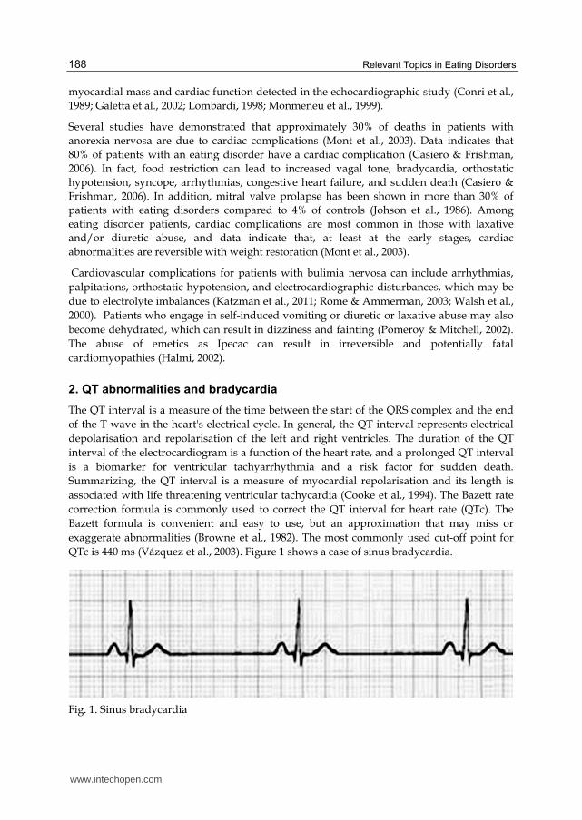
The QT interval is a measure of the time between the start of the QRS complex and the end of the T wave in the heart's electrical cycle. In general, the QT interval represents electrical depolarisation and repolarisation of the left and right ventricles. The duration of the QT interval of the electrocardiogram is a function of the heart rate, and a prolonged QT interval is a biomarker for ventricular tachyarrhythmia and a risk factor for sudden death. Summarizing, the QT interval is a measure of myocardial repolarisation and its length is associated with life threatening ventricular tachycardia (Cooke et al., 1994). The Bazett rate correction formula is commonly used to correct the QT interval for heart rate (QTc). The Bazett formula is convenient and easy to use, but an approximation that may miss or exaggerate abnormalities (Browne et al., 1982). The most commonly used cut-off point for QTc is 440 ms (Vázquez et al., 2003). Figure 1 shows a case of sinus bradycardia.
Fig. 1. Sinus bradycardia
www.intechopen.com
Cardiovascular Complications in Eating Disorders
189
With regards to eating disorder patients, QT interval abnormalities have been studied as a marker of sudden death and to assess the effect of refeeding. In fact, it has been proposed that these deaths are a result of cardiac arrhythmias for which a long QT interval on the electrocardiogram is a marker (Isner et al., 1985). Nevertheless, the measurement of the QT interval has a poor predictive value for the recognition of patients who are at particular risk of sudden death. A measured QT interval >600 ms is associated with a significant risk of sudden death (Jackmann et al., 1988), but few patients usually have such long QT intervals. Another QT parameter is the QT dispersion (QTd), which is the difference between the maximum QT and the minimum QT in all derivations. Finally, the dispersion of the corrected QT interval (QTcd) is the difference between the maximum and the minimum QTc interval in the 12 electrocardiographic derivations. The most used cut-off point for the dispersion is 60 ms (Bayés de Luna & Viñolas, 1996; Girola et al., 2001; Hill & Friedman, 1997; Lund et al., 2002; Toivonen, 2002).
Considering the main studies on the QT interval among patients with anorexia nervosa, there are some controversial results. Thus, some studies have not found long QT intervals (Powers et al., 1991; Gottdiener et al., 1978) while others have found long QT intervals (Cooke et al., 1994; Isner et al., 1985; Thurston & Marks, 1974). Apart from methodological problems, these conflicting results could be explained by difficulties in comparison due to the huge variability of the QT interval with heart rate (Cooke et al., 1994). In a recent study, QT and QTc intervals as well as the dispersion intervals QTd and QTcd were longer among anorectic women than in a control group (Vázquez et al., 2003).
In relation with the undernutrition state of anorexia nervosa, arrhythmias, especially bradycardia, are the best-known cardiac disturbances probably as a response to maintain body energy by means of a decrease of cardiac work. There have been different proposed pathogenic mechanisms to explain those disturbances: electrolyte loss, drugs adverse cardiovascular effects, decreased glycogen content in the cardiac cell, myofibril atrophy, interstitial oedema, mitochondrial swelling, and activation of calcium-dependent proteinases. With respect to the pathological basis of a long QT interval in anorexia nervosa there are some proposals too: cardiac muscle is lost in proportion to loss of body mass, but there is no convincing evidence of impairment of ventricular function in these patients, histological studies have not shown myocarditis, abnormalities of the hypothalamus are well described and, finally, a increased autonomic tone has been suggested as a possible mechanism for lengthening the QT interval (Clark & Wildenthal, 1986; Thurston & Marks, 1974; Palossy & Oó, 1977; Powers et al., 1991; Tolnay & von Althen, 1987).
In general, the most frequent findings among anorectic patients have been arrhythmias, bradycardia, atrial extrasystoles, isolated premature ventricular contractions and a significant increase QT and QTc, compared with healthy women of similar age (Panagiotopoulos et al., 2000; Swenne & Larsson, 1999; Vázquez et al., 2003). As a result of these findings, some authors have suggested that weight loss in patients with anorexia nervosa and eating disorders in general is a risk factor for the prolongation and dispersion of the QT interval (Galetta et al., 2002; Swenne & Larsson, 1999). In fact, a significant and negative correlation between QTc and body mass index has been reported (Vázquez et al., 2003). An increase of the QT interval dispersion represents regional differences in
www.intechopen.com
Relevant Topics in Eating Disorders
190
myocardial excitability recovery and may lead to and increased arrhythmogenic substrate, with a higher risk for ventricular arrhythmias clinically significant and a higher risk of sudden death. In fact, the increased QT interval and the increased QT dispersion have been associated with higher risk of ventricular arrhythmias in both patients with heart disease and healthy individuals. Thus, the predictive value of the increased QT interval dispersion as a marker of sudden acute ventricular arrhythmia or death has been demonstrated (Harris et al., 1993; Isner et al., 1985). Recently, parameters of QT variability have been proposed as surrogate markers for arrhythmia risk stratification in anorexia nervosa (Koschke et al., 2010). A meta-analysis of heart rate and QT interval alteration in anorexia nervosa showed that bradycardia and relationship between heart rate and body mass index decreases as the disease continues. Moreover QTc interval in anorectic patients was within normal range although significantly longer than in controls (Lesinskiene, et al., 2008).
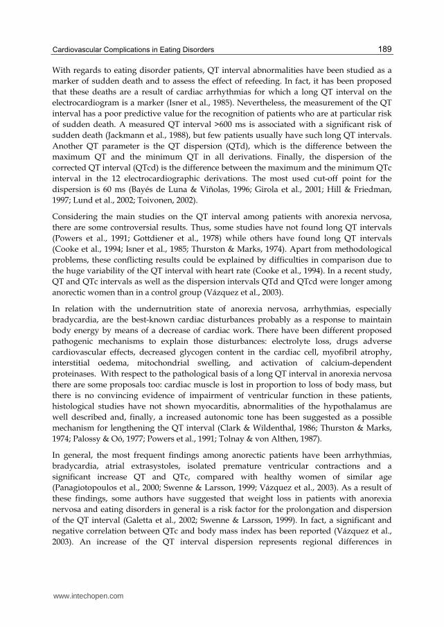
Considering the subtypes of anorexia nervosa, a significant increase has been found in weight/height ratio and body mass index from malnourished stage to weight restoration, paralleled by a significant decrease in QTd among anorectic patients (restricting type) (Nahshoni, et al., 2007). With respect to bulimia nervosa, compared with the controls, bulimic patients with a history of anorexia nervosa seem to have significantly more signal-averaged electrocardiography abnormalities (Takimoto et al., 2006). Besides the relationship between QT changes and potential fatal arrhythmias, those changes have shown to be related with low resting metabolic rate among chronic anorectic patients (Krantz et al., 2005). Despite the fact that the majority of studies have highlighted the QT disturbances in anorexia nervosa, a prolonged QT interval and QT dispersion have been reported in both anorexia nervosa and bulimia nervosa (Takimoto et al., 2004). In fact, QT and QTd have been shown to be significantly longer in all eating disorder subtypes than in controls. Moreover, QT interval and QTd were significantly correlated with the rate of body weight loss in bulimia nervosa (Takimoto et al., 2004). Figure 2 shows a long QT related to weight loss.
Fig. 2. Long QT interval
www.intechopen.com
Cardiovascular Complications in Eating Disorders
191
2.1 QT abnormalities and refeeding
The effect of the reached target weights on QT interval has been demonstrated, with a significant shortening of that interval by means of an adequate refeeding (Cooke et al., 1994). After admission, the QT prolongation and dispersion are usually normalized within the 3 days of refeeding (Swenne, 2000). After refeeding, a significant decrease in QT interval and QT dispersion has been observed as well as a normalization of the heart rate and heart rate variability (Mont et al., 2003). On the other hand, the QT/RR slope has been found significantly enhanced in anorectic patients compared with healthy controls, and this slope returns to normal range values after refeeding. This QT/RR slope seems to be significantly correlated with the body mass index in the patient group (Roche et al., 2005). During the follow-up of these cardiac abnormalities in female adolescents with anorexia nervosa, one year after refeeding a significant decrease in QT, QTc, QTd and QTcd has been reported (Ulger et al., 2006).
3. Structural changes
With respect to the cardiac structural changes in eating disorders those referred to the interventricular septum and the left ventricular mass have been the most studied ones. With regards to anorexia nervosa, echocardiography has shown a reduction in the dimensions of the interventricular septum (52% of patients), left ventricular free wall (61%), left atrium (31%) and left ventricular mass (61%) (Silvetti et al., 1998). More recently, by means of tissue Doppler imaging, when compared with control groups, anorectic patients have shown lower left ventricular mass, lower myocardial systolic wave peak of both lateral wall and septum, and comparable early (Em) and atrial (Am) diastolic waves, and Em/Am ratio. On the other hand, the ratio between transmitral peak E-wave and Em has shown significantly greater in anorectic patients than in controls, while no differences have been observed between thin and normal-weight females. In anorectic patients, Sm peak has been significantly related to left ventricular mass indexed, at both septum and lateral wall levels (Galetta et al., 2005). Anorexia nervosa has been associated with a significant reduction in the cyclic variation in the integrated backscatter (IBS) signal of the myocardium, which is also related to left ventricular hypotrophy (Franzoni et al., 2003).
With respect to the ventricular mass, left ventricular chamber dimension and mass seem to be significantly less in women with anorexia nervosa than in either the women of normal weight or the thin women, even after standardisation for body size or after controlling for blood pressure. Nevertheless it seems that there are no substantial changes in the left ventricular shape (De Simone et al., 1994). Some authors have reported a high frequency of mitral valve prolapse due to the disproportion between ventricle and valve among anorectic patients (Conri et al., 1989).
3.1 Structural changes and refeeding
Other studies have highlighted that the left ventricular mass index is significantly lower than that in normal subjects as well as the left ventricular end-diastolic and end-systolic volume indexes. Nevertheless, in spite of the reductions in left ventricular mass and volume indexes, left ventricular chamber architecture described as h/R ratio, mass to volume ratio,
www.intechopen.com
Relevant Topics in Eating Disorders
192
and short/long left ventricular axis ratio usually is normal. The same applies to the left ventricular afterload assessed as end-systolic meridional and circumferential wall stress. Moreover, among anorectic patients, ejection fraction, percent fractional shortening, and the relationship between end-systolic wall stress and ejection fraction have been all within normal limits. In some patients restudied after a 15% to 20% weight gain, left ventricular mass and volume indexes increased significantly but end-systolic wall stress and ejection fraction did not change (St John Sutton et al., 1985).
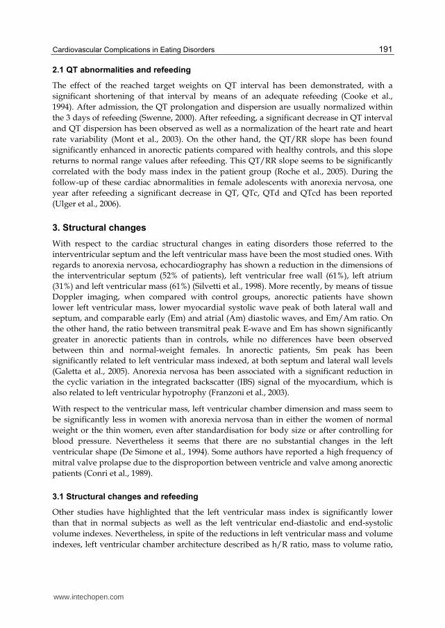
Fig. 3. Mitral valve prolapse
Control echocardiograms of anorectic patients after refeeding have shown an increase in left ventricular diameters and cardiac mass (Ulger et al., 2006). Apart from that, echocardiograms have shown an increase in cardiac output, an improvement in the exercise capacity, and a normalization of the heart rate and heart rate variability after refeeding (Mont et al., 2003). Other authors have reported that cardiac complications of anorexia nervosa are common, usually benign and always reversible after refeeding in hospital (Lupoglazoff et al., 2001). Figure 3 shows a mitral valve prolapse.
4. Left ventricular function
In a pioneer study (Gottdiener et al., 1978), the authors found that in spite of the structural changes, left ventricular systolic function, was unimpaired as indicated by normal echocardiographic fractional shortening, and by normal exercise augmentation of ejection fraction determined by radionuclide cineangiography. Another study showed that there was no evidence of arrhythmias at rest, during exercise, or with continuous electrocardiographic recording, concluding that young patients with anorexia nervosa appeared to have essentially normal cardiac function with a reduction in left ventricular muscle mass (Moodie & Salcedo, 1983). It seems that the down-regulation of left ventricular mass per se, like up-regulation of left ventricular mass, is not associated with abnormal left ventricular function (St John Sutton et al., 1985). Other authors have reported similar results (Dec et al., 1987) and a cardiovascular vagal hyperactivity in anorectic patients, which
www.intechopen.com
Cardiovascular Complications in Eating Disorders
193
appears to influence the ventricular diastolic dynamics. Heart rate variability and diastolic function analysis have been proposed as useful tools in monitoring anorexia-induced cardiac modifications (Galetta et al., 2003).
Apart from bradycardia, hypotension, tachycardia, a decrease in cardiac chamber size, impaired left ventricular contractility, electrocardiographic changes, pericardial effusion, and mitral valve prolapse, in patients with anorexia nervosa, some previous articles, as the above-mentioned ones have focused on the effects of anorexia nervosa on left ventricular function. However, atrial function in patients with anorexia nervosa has not been studied. The percentage of total transmitral flow contributed by atrial contraction is about 30% (Jovic & Troskot, 1977), which indicates the importance of atrial function in left ventricular filling, and a case of anorexia nervosa as cause of atrial failure has been described (Mizuno et al., 1998).
Cardiac disturbances in anorectic patients are essentially derived from malnutrition and starvation. On a cellular level this leads to diminish the protein synthesis, activation of calcium-dependent proteinases, mitochondrial swelling, decreased glycogen content, interstitial oedema, and myofibrillar atrophy. The physiologic consequences of the malnutrition include decreased contractile force of the ventricle, decreased cardiac output, and decreased diastolic compliance. Clinically, bradycardia, relative hypotension, mitral valve prolapse, and diminished exercise capacity may all be seen in these patients as well as the above mentioned atrial dysfunction (Abel et al., 1979; Fohlin, 1977; Goldberg et al., 1988; Gottdiener et al., 1978; Johnson et al., 1986; Meyers et al., 1986; Monmeneu et al., 1999; Moodie, 1987; Nutter et al., 1979; Tolnai & von Althen, 1987; Warren & Vande Wiele, 1973).
5. Electrolyte changes
Eating disorder patients may show varying degrees of dehydration, sodium and chloride depletion, mainly in patients who vomit, potassium deficiency in patients with laxatives abuse, and weaknesses of chlorine, sodium and potassium in patients using diuretics, with different depletions depending on the substance used. With respect to electrolyte disturbances, typical laboratory changes, although not exclusively seen in anorexia nervosa, include hyponatremia, hypokalaemia, and hypochloremia (Baumann et al., 2010).
The presence of the symptoms listed below usually indicates a water loss of about 25% (or more) and indicates a severe situation: oliguria, sunken eyes, dry mucous membranes (dry mouth), shrivelled skin, fever and muscle cramps. Along with this, blood parameters will show an increase of haematocrit value, and increase of the total proteins, increased urine density, and increased urea concentration in urine.
Considering hypochloremia, it would be moderate with a level within a range between 90-95 mEq/L, more severe between 80-90 mEq/L and severe (which would require urgent treatment) an amount of chlorine in blood below 80 mEq/L. The hypochloremia key signs are hypotension and paresis or intestinal ileus.
Hyponatremia is considered moderate when blood sodium levels between 130 and 138 mEq/L, severe with levels between 125 and 130 mEq/L, and very severe when the sodium level is below 125 mEq/L. The characteristic signs are: muscle contractures, muscle aches, muscle fibrillations and sometimes seizures. Hypotension is less common than in hypochloremia, but usual. The accepted normal range is 135-145 mEq/L.
www.intechopen.com
Relevant Topics in Eating Disorders
194
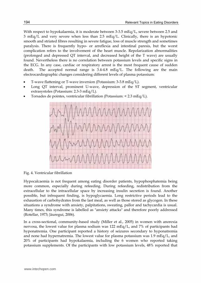
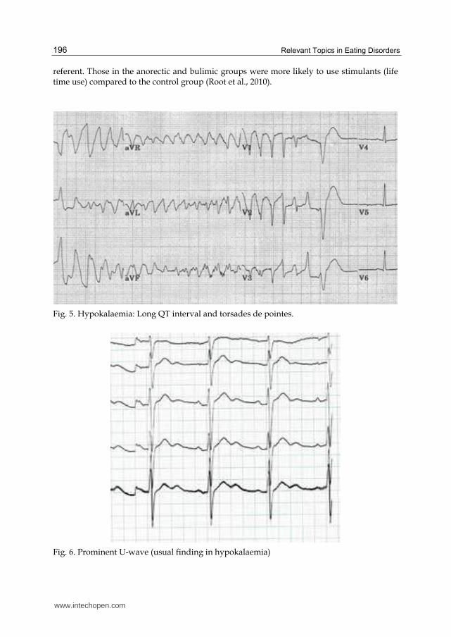
With respect to hypokalaemia, it is moderate between 3-3.5 mEq/L, severe between 2.5 and 3 mEq/L and very severe when less than 2.5 mEq/L. Clinically, there is an hypotonic smooth and striated fibres resulting in severe fatigue, loss of muscle strength and sometimes paralysis. There is frequently hypo- or arreflexia and intestinal paresis, but the worst complication refers to the involvement of the heart muscle. Repolarization abnormalities (prolonged and depressed QT interval, and decreased height of the T wave) are usually found. Nevertheless there is no correlation between potassium levels and specific signs in the ECG. In any case, cardiac or respiratory arrest is the most frequent cause of sudden death. The accepted normal range is 3.4-4.8 mEq/L. The following are the main electrocardiographic changes considering different levels of plasma potassium:
T-wave flattening or T-wave inversion (Potassium: 3-3.8 mEq/L). Long QT interval, prominent U-wave, depression of the ST segment, ventricular
extrasystoles (Potassium: 2.3-3 mEq/L). Torsades de pointes, ventricular fibrillation (Potassium: < 2.3 mEq/L).
Fig. 4. Ventricular fibrillation
Hypocalcaemia is not frequent among eating disorder patients, hypophosphatemia being more common, especially during refeeding. During refeeding, redistribution from the extracellular to the intracellular space by increasing insulin secretion is found. Another possible, but infrequent finding, is hypoglycaemia. Long restrictive periods lead to the exhaustion of carbohydrates from the last meal, as well as those stored as glycogen. In these situations a syndrome with anxiety, palpitations, sweating, pallor and tachycardia is usual. Many times, this syndrome is labelled as "anxiety attacks" and therefore poorly addressed (Rotellar, 1973; Jáuregui, 2006).
In a cross-sectional, community-based study (Miller et al., 2005) in women with anorexia nervosa, the lowest value for plasma sodium was 122 mEq/L, and 7% of participants had hyponatremia. One participant reported a history of seizures secondary to hyponatremia and none had hypernatremia. The lowest value for plasma potassium was 1.9 mEq/L, and 20% of participants had hypokalaemia, including the 6 women who reported taking potassium supplements. Of the participants with low potassium levels, 48% reported that
www.intechopen.com
Cardiovascular Complications in Eating Disorders
195
they regularly purged, whereas the remainder (52%) denied purging. In a previous large study in an outpatient treatment program has reported the absence of hypokalaemia in non-purgers anorectic patients (Rieger et al., 1978). Mean plasma potassium level was lower for the women who reported purging than for those who did not (3.5 vs. 3.7 mEq/L). A history of dysrhythmia was reported by 2% of the women and, in most cases, hypokalaemia was noted as the probable. Serum calcium levels were low in 6% of participants.
The reported serious cardiac abnormalities in patients with anorexia nervosa include dysrhythmias, some of which may be secondary to hypokalaemia (Sharp & Freeman, 1993; Bonne et al., 1993). Screening and monitoring patients with anorexia nervosa for cardiac risk factors and dysfunction is important, and this should include asking patients whether they purge or have histories of hypokalaemia and/or purging behaviours. Monitoring vital signs and performing electrocardiograms and serial measurements of plasma potassium are also important. This is important, as purging may increase the risk of hypokalaemia and subsequent cardiac dysrhythmias, and self-induced vomiting increases the risk of additional complications (Miller et al., 2005). In fact, QT prolongation and ventricular arrhythmia may develop in the setting of severe hypokalaemia, exposing patients to high risk of sudden cardiac event (Facchini et al., 2006).
6. Purging behaviours and effects of drugs (laxatives, diuretics, stimulants)
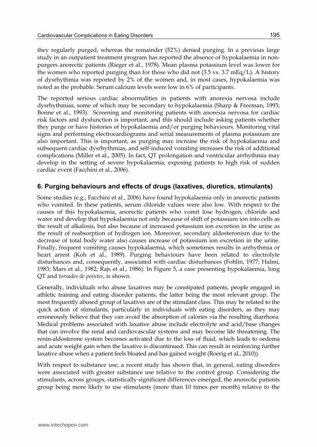
Some studies (e.g., Facchini et al., 2006) have found hypokalaemia only in anorectic patients who vomited. In these patients, serum chloride values were also low. With respect to the causes of this hypokalaemia, anorectic patients who vomit lose hydrogen, chloride and water and develop that hypokalaemia not only because of shift of potassium ion into cells as the result of alkalosis, but also because of increased potassium ion excretion in the urine as the result of reabsorption of hydrogen ion. Moreover, secondary aldosteronism due to the decrease of total body water also causes increase of potassium ion excretion in the urine. Finally, frequent vomiting causes hypokalaemia, which sometimes results in arrhythmia or heart arrest (Koh et al., 1989). Purging behaviours have been related to electrolyte disturbances and, consequently, associated with cardiac disturbances (Fohlin, 1977; Halmi, 1983; Mars et al., 1982; Rajs et al., 1986). In Figure 5, a case presenting hypokalaemia, long QT and torsades de pointes, is shown.
Generally, individuals who abuse laxatives may be constipated patients, people engaged in athletic training and eating disorder patients, the latter being the most relevant group. The most frequently abused group of laxatives are of the stimulant class. This may be related to the quick action of stimulants, particularly in individuals with eating disorders, as they may erroneously believe that they can avoid the absorption of calories via the resulting diarrhoea. Medical problems associated with laxative abuse include electrolyte and acid/base changes that can involve the renal and cardiovascular systems and may become life threatening. The renin-aldosterone system becomes activated due to the loss of fluid, which leads to oedema and acute weight gain when the laxative is discontinued. This can result in reinforcing further laxative abuse when a patient feels bloated and has gained weight (Roerig et al., 2010)).
With respect to substance use, a recent study has shown that, in general, eating disorders were associated with greater substance use relative to the control group. Considering the stimulants, across groups, statistically significant differences emerged, the anorectic patients group being more likely to use stimulants (more than 10 times per month) relative to the
www.intechopen.com
Relevant Topics in Eating Disorders
196
referent. Those in the anorectic and bulimic groups were more likely to use stimulants (life time use) compared to the control group (Root et al., 2010).
Fig. 5. Hypokalaemia: Long QT interval and torsades de pointes.
Fig. 6. Prominent U-wave (usual finding in hypokalaemia)
www.intechopen.com
Cardiovascular Complications in Eating Disorders
197
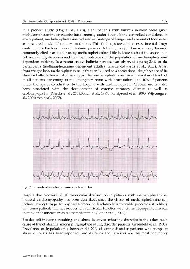
In a pioneer study (Ong et al., 1983), eight patients with bulimia nervosa were given methylamphetamine or placebo intravenously under double blind controlled conditions. In every patient, methylamphetamine reduced self-ratings of hunger and amount of food eaten as measured under laboratory conditions. This finding showed that experimental drugs could modify the food intake of bulimic patients. Although weight loss is among the most commonly cited reasons for using methamphetamine, little is known about the association between eating disorders and treatment outcomes in the population of methamphetamine dependent patients. In a recent study, bulimia nervosa was observed among 2.4% of the participants (methamphetamine dependent adults) (Glasner-Edwards et al., 2011). Apart from weight loss, methamphetamine is frequently used as a recreational drug because of its stimulant effects. Recent studies suggest that methamphetamine use is present in at least 5% of all patients presenting to the emergency room with heart failure and 40% of patients under the age of 45 admitted to the hospital with cardiomyopathy. Chronic use has also been associated with the development of chronic coronary disease as well as cardiomyopathy (Diercks et al., 2008;Karch et al., 1999; Turnipseed et al., 2003; Wijetunga et al., 2004; Yeo et al., 2007).
Fig. 7. Stimulants-induced sinus tachycardia
Despite that recovery of left ventricular dysfunction in patients with methamphetamine-induced cardiomyopathy has been described, since the effects of methamphetamine can include myocyte hypertrophy and fibrosis, both relatively irreversible processes, it is likely that some patients will not recover left ventricular function with either appropriate medical therapy or abstinence from methamphetamine (Lopez et al., 2009).
Besides self-inducing vomiting and abuse laxatives, misusing diuretics is the other main cause of hypokalaemia among purging-type eating disorder patients (Greenfeld et al., 1995). Prevalence of hypokalaemia between 4.6-20% of eating disorder patients who purge or abuse diuretics has been reported, and diuretics and laxatives are the most commonly
www.intechopen.com
Relevant Topics in Eating Disorders
198
abused purging medications by patients with purging-type eating disorders. In a former study (Mitchell et al., 1985) among bulimic patients, 33.9 % of diuretic abuse and 60.6% of laxative abuse was found, and may be that these figures underestimate the prevalence of diuretic abuse due to the fact that such medication is not only obtained by prescription but is also available in many over-the-counter formulations (92). Again the risk for cardiovascular disturbances is present by means of the relationship between diuretic abuse and hypokalaemia (Mascolo et al., 2011).
7. The heart response facing a higher metabolic demand (refeeding syndrome)
Refeeding syndrome has been considered one of the most serious complications related to anorexia nervosa, one which can occur, and possibly prove fatal, with oral as well as enteral and parenteral feeding. The syndrome involves a set of alterations caused by too rapid or unbalanced refeeding in a severely malnourished patient. The main signs and symptoms include sodium and water retention (leading to oedema and possible cardiac failure), hypophosphatemia due to intracellular disturbances (causing generalised systemic problems and a difficulty with energy storage), and the depletion of electrolytes, such as potassium and magnesium, as well as of vitamins (with the associated clinical consequences) and thiamine cofactor, which can lead to Wernicke’s syndrome and/or cardiomyopathy. The calorie intake with which the syndrome may occur varies widely among the reported cases (between 10 and 60 kcal/kg/day), while the presence of glucose in the alimentary canal of extremely malnourished patients is a determining factor that can trigger the process (Katzman, 2005; Marinella, 2005; Miller, 2008; O’Connor & Goldin, 2011; Solomon, 1990; Tey et al., 2005).
Refeeding syndrome must be prevented and, therefore, many studies have sought to establish guidelines for medical and nursing care. The recommendations are to achieve a weight gain of approximately 500 g/week and constantly monitor fluid intake. Administration of phosphorus and potassium may be beneficial during the initial weeks of refeeding and the potential value of high-calorie supplements should also be assessed. At all events, the calorie intake should be carefully matched to the initial baseline metabolic rate 7, as should the carbohydrate intake (De Cock et al., 2006; Hearing, 2004; Portsmouth Hospitals NHS Trust, 2007).
Refeeding syndrome consists of cardiovascular, neurologic, and hematologic complications, and can be associated with significant morbidity and mortality. Adverse effects of hypophosphatemia include cardiac failure, muscle weakness, immune dysfunction, and death. Cardiac sequelae are secondary to and occur early in the cascade of events that arise during refeeding. Congestive heart failure results from the decreased ventricular mass and myofibrillar atrophy, causing decreased stroke volume and reduced capacity of the cardiovascular system (Katzman, 2005).
Kohn et al. described acute cardiac complications (arrhythmias, bradycardia, pericardial effusion, hypotension, and cardiac arrest) in anorectic patients during refeeding. These patients developed life-threatening complications associated with refeeding including cardiac arrhythmias documented within the first week of hospitalization.
www.intechopen.com
Cardiovascular Complications in Eating Disorders
199
8. Blood pressure changes
Besides de above-mentioned cardiovascular disturbances of eating disorders, another common complication is orthostatic hypotension. Orthostatic heart rate and blood pressure changes are common in anorectic patients on admission to hospital (Shamim et al., 2003). These orthostatic changes place patients with anorexia nervosa at increased risk of syncope. One explanation for the observed orthostatic changes is that starvation leading to low body weight may result in atrophic peripheral muscles, resulting in decreased venous return to the heart.
Recent evidence has shown that normalization of orthostatic pulse changes occurs after approximately 3 weeks of nutritional rehabilitation when patients reach 80% of their ideal body weight. It has been suggested that the resolution of orthostasis can be used as an objective measure of medical stability in patients with anorexia nervosa (Shamim et al., 2003).
9. Conclusions
Eating disorders, particularly anorexia nervosa, are life-threatening diseases, with high risk of death due to cardiovascular disturbances. Cardiovascular complications are present promptly, mainly in cases of anorexia nervosa, with more or less severity with regards to structural and functional changes. Cardiovascular complications in eating disorder include bradycardia, QT interval prolongation, orthostatic hypotension, increased vagal tone, mitral valve prolapse (as a consequence of weight loss with an associated reduction in left ventricle mass, resulting in a relatively large mitral valve), possible alterations in myocardial contractility, and reduction in left ventricular wall thickness, among others.
The severity of the eating disorders, specifically the undernourished state, is usually significantly correlated with increased bradycardia and decreased left ventricular force. In this regard, patients with severe sinus bradycardia should be admitted to the hospital in order to monitoring the cardiac function and to gain weight gradually.
Despite the conflicting results about the QT interval alterations, the prolongation of the QT is usually associated with sudden ventricular arrhythmias and death.
Eating disorders may show cardiac abnormalities depending on electrolyte changes associated with purging behaviours and/or drug effects, among other causes.
Orthostatic heart rate and blood pressure changes are common in patients with eating disorders, mainly in cases of anorexia nervosa, and it has been suggested that the resolution of orthostatism could be an objective measure of medical stability.
The main cardiovascular findings among patients with eating disorders are reversible by means of an appropriate refeeding. As a result of this refeeding an increase in cardiac dimensions, ventricular mass, and cardiac output are reached.
Cardiovascular complications in eating disorders may be potentially fatal and they are usually present in early stages of these diseases, being in most cases reversible. An early diagnostic, a prompt treatment, a correct refeeding, and the cardiac monitoring of all patients, are necessary.
www.intechopen.com
Relevant Topics in Eating Disorders
200
10. Summarizing
Cardiovascular complications of eating disorders are common, and they have been reported in up to 80% of the cases, mainly in the form of bradycardia, hypotension, arrhythmias, repolarization abnormalities and sudden death by up to 10% of the cases.
Despite the conflicting results with respect to QT interval in anorexia nervosa, recent studies show that QT and QTc intervals as well as the dispersion intervals QTd and QTcd are longer among anorectic patients than in controls.
With respect to bulimia nervosa, bulimic patients with a history of anorexia nervosa seem to have significantly more signal-averaged electrocardiography abnormalities than controls.
During hospitalization the QT prolongation and dispersion usually normalize within the three days of refeeding.
The most described structural changes in anorexia nervosa have been the reduction in the dimensions of the interventricular septum, left ventricular free wall, left atrium and left ventricular mass.
Control echocardiograms of anorectic patients have shown an increase in cardiac output, an improvement in the exercise capacity, and a normalization of the heart rate and heart rate variability after refeeding.
In spite of the structural changes, left ventricular systolic function usually keeps unimpaired.
Considering the electrolyte changes, typical laboratory findings in eating disorders include hyponatremia, hypokalaemia and hypochloremia.
Purging behaviours may increase the risk of hypokalaemia and subsequent cardiac dysrhythmias. Thus, monitoring vital signs and performing electrocardiograms and serial measurements of plasma potassium are relevant.
Medical problems associated with laxative abuse include electrolyte and acid/base changes that can involve the renal and cardiovascular systems and may become life threatening.
The chronic use of methamphetamine has been associated with the development of chronic coronary disease as well as cardiomyopathy.
During refeeding, adverse effects of hypophosphatemia include cardiac failure among others. Cardiac sequelae are secondary to and occur early in the cascade of events that arise during refeeding.
Orthostatic changes place patients with anorexia nervosa at increased risk of syncope. The normalization of orthostatic pulse changes occurs approximately after three weeks of nutritional rehabilitation when patients reach 80% of their ideal body weight.
11. References
Abel, RM., Grimes, JB., Alonso, D., Alonso, M., & Gay, WAJr. (1979). Adverse hemodynamic and ultrastructural changes in dog hearts subjected to protein-calorie malnutrition. American Heart Journal, 97, 733-744. ISSN 1097-6744.
Baumann, A., Heitmann, S., Bubendorff, V., & Himmerich, H. (2010). Laboratory changes in anorexia nervosa. Praxis (Bern 1994), 99(11), 661-667. ISSN: 1661-8157.
Bayés de Luna, A., & Viñolas, X. (1996). QT dispersion and heart rate variability. European Heart Journal, 17, 165-166. ISSN 1522-9645.
www.intechopen.com
Cardiovascular Complications in Eating Disorders
201
Becker, AE., Grinspoon, SK., Klibanski, A., & Herzog, DB. (1999). Eating disorders. New England Journal of Medicine, 340, 1092-1098. ISSN 1533-4406.
Birmingham, CL., & Beumont, P. (2004). Medical management of eating disorders. Cambridge University Press, ISBN 9780521727105, New York.
Bodí, V., Monmeneu, JV., Marín, F., Cortés, J., Llobet, E., García, A., Martínez, F., Ponce de León, JC., & Guardiola, M. Dispersión del intervalo QT en pacientes con insuficiencia cardíaca. Determinantes y valor pronóstico. Revista Española de Cardiología, 52, 563-569. ISSN 0300-8932.
Bonne, O., Bloch, M., & Berry, E. (1993). Adaptation to severe chronic hypokalaemia in anorexia nervosa: a plea for conservative management. International Journal of Eating Disorders, 13, 125-128, ISSN 0276-3478.
Browne, KF., Zipes, DP., Heger, JJ., & Prystowsky, EN. (1982). The influence of the autonomic system on the QT interval in man. American Journal of Cardiology, 50, 1099-1103. ISSN 0002-9149.
Casiero, D., & Frishman, WH. (2006). Cardiovascular Complications of Eating Disorders. Cardiology in Review, 14(5), 227-31. ISSN 1538-4683.
Clark, AF., & Wildenthal, K. (1986). Disproportionate reduction of actin synthesis in hearts of starved rats. Journal of Biological Chemistry, 261, 13168-13172. ISSN 1083-351X.
Conri, C., Roudaut, R., Ducloux, G., Fleury, B., & Moreu, F. (1989). Étude échocardiographique au cours de l’anorexie mentale. La Presse Médicale, 18, 806-808. ISSN 0755-4982.
Cooke, RA., Chambers, JB., Singh, R., Todd, GJ., Smeeton, NC., Treasure, J., & Treasure, T. (1994). QT interval in anorexia nervosa. British Heart Journal, 72, 69-73. ISSN 0007-0769.
De Cock, A., Mana, F., Velkeniers, B., & Urbain, D. (2006). Hypophosphatemia and refeeding: a corrective or a preventive attitude? Acta Clinica Belgica, 61, 134-137. ISSN 0001-5512.
De Simone, G., Scalfi, L., Galderisi, M., Celentano, A., Di Biase, G., Tammaro, P., Garofalo, M., Mureddu, GF., de Divitiis, O., & Contaldo, F. (1994). Cardiac abnormalities in young women with anorexia nervosa. British Heart Journal, 71, 287-292. ISSN 0007-0769.
Dec, GW., Biederman, J., & Hougen, TJ. (1987). Cardiovascular findings in adolescent inpatients with anorexia nervosa. Psychosomatic Medicine, 49(3), 285-290. ISSN 534-7796.
Diercks, DB., Fonarow, GC., Kirk, JD., Jois-Bilowich, P., Hollander, JE., Weber, JE., Wynne, J., Mills, RM., Yancy, C., & Peacock, WF. (2008). Illicit stimulant use in a United States heart failure population presenting to the emergency department (from the Acute Decompensated Heart Failure National Registry Emergency Module). American Journal of Cardiology, 102, 1216-1219. ISSN 0002-9149.
Facchini, M., Sala, L., Malfatto, G., Bragato, R., Redaelli, G., & Invitti, C. (2006). Low-K+ dependent QT prolongation and risk for ventricular arrhythmia in anorexia nervosa. International Journal of Cardiology, 106(2), 170-176. ISSN 0167-5273.
Fohlin, L. (1977). Body composition, cardiovascular and renal function in adolescent patients with anorexia nervosa. Acta Paediatrica Scandinavica Supplement, 268, 1- 20. ISSN 0300-8843.
www.intechopen.com
Relevant Topics in Eating Disorders
202
Franzoni, F., Galetta, F., Cupisti, A., Rolla, M., Santoro, G., & Pentimone, F. (2003). Ultrasonic tissue characterization of the myocardium in anorexia nervosa. Acta Paediatrica, 92(3), 297-300. ISSN 0803-5253.
Galetta, F., Franzoni, F., Cupisti, A., Belliti, D., Prattichizzo, F., & Rolla, M. (2002). QT interval dispersion in young women with anorexia nervosa. Journal of Pediatrics, 140, 456-460. ISSN 00223476.
Galetta, F., Franzoni, F., Cupisti, A., Morelli, E., Santoro, G., & Pentimone, F. (2005). Early detection of cardiac dysfunction in patients with anorexia nervosa by tissue Doppler imaging. International Journal of Cardiology, 101(1), 33-37. ISSN 0167-5273.
Galetta, F., Franzoni, F., Prattichizzo, F., Rolla, M., Santoro, G., & Pentimone, F. (2003). Heart rate variability and left ventricular diastolic function in anorexia nervosa. Journal of Adolescent Health, 32(6), 416-421. ISSN 1054-139X.
Girola, A., Enrini, R., Garbetta, F., Tufano, A., & Caviezel, F. (2001). QT dispersion in uncomplicated human obesity. Obesity Research, 9, 71-77. ISSN 1071-7323.
Glasner-Edwards, S., Mooney, LJ., Marinelli-Casey, P., Hillhouse, M., Ang, A., & Rawson, R. (2011). Bulimia nervosa among methamphetamine dependent adults: association with outcomes three years after treatment. Eating Disorders, 19(3), 259-269. ISSN 1532-530X.
Goldberg, SJ., Comerci, GD., Feldman, L. (1988). Cardiac output and regional myocardial contraction in anorexia nervosa. Journal of Adolescent Health Care, 9, 15- 21. ISSN 1054-139X.
Gottdiener, JS., Gross, HA., Henry, WL., Borer, JS., & Ebert, MH. (1978). Effects of self-induced starvation on cardiac size and function in anorexia nervosa. Circulation, 58, 421-433.
Greenfeld, D., Mickey, D., Quinlan, DM., & Roloff, P. (1995) Hypokalemia in outpatients with eating disorders. American Journal of Psychiatry, 152, 60–63. ISSN 0002-953X.
Guidelines for the prevention and treatment of adult patients at risk of developing refeeding syndrome. Drug Therapy Guideline No. 46.00. (2007). In: Refeeding Syndrome Guideline. NSH.
Halmi, K. (2002). Physiology of anorexia nervosa and bulimia nervosa, In: Eating disorders and obesity: A comprehensive handbook, CB Fairburn & KD Brownell, 267-271, Guilford Press, ISBN 1-57230-688-2, New York.
Halmi, KA. (1983). Anorexia nervosa and bulimia. Psychosomatics, 24, 11. ISSN 1545-7206 Harris, JP., Kreipe, RE., & Rossbach, CN. (1993). QT prolongation by isoproterenol in
anorexia nervosa. Journal of Adolescent Health, 14, 390-393. ISSN 1054-139X. Hearing, SD. (2004). Refeeding syndrome. British Medical Journal, 328, 908-909. ISSN
09598138. Higham, PD., & Campbell, RW. (1994). QT dispersion. British Heart Journal, 71, 508-510. ISSN
0007-0769. Hill, JA., & Friedman, PL. (1997). Measurement of QT interval and QT dispersion. Lancet,
349, 894-895. ISSN 0140-6736. Isner, JM., Roberts, WC., Heymsfield, SB., & Yager, J. (1985). Anorexia nervosa and sudden
death. Annals of Internal Medicine, 102, 49-52. ISSN 0003-4819. Jackmann, WM., Friday, KJ., Anderson, JL., Aliot, EM., Clark, M., & Lazzara, R. (1988). The
long QT syndromes: a critical review, new clinical observations and a unifying hypothesis. Progress in Cardiovascular Diseases, 31, 115-172. ISSN 1532-8643.
www.intechopen.com
Cardiovascular Complications in Eating Disorders
203
Jaúregui I. (2006). La imagen de una sociedad enferma. Anorexia, bulimia, atracones y obesidad, Grafema, ISBN 9788493422592, Barcelona.
Johnson, GL., Humphries, LL., Shirley, PB., Mazzoleni, A., & Noonan, JA. (1986). Mitral Valve Prolapse in Patients With Anorexia Nervosa and Bulimia. Archives of Internal Medicine, 146(8), 1525-1529. ISSN 0039926.
Jovic, A., & Troskot, R. (1977). Recovery of atrial systolic function after pharmacological conversion of chronic atrial fibrillation to sinus rhythm: a Doppler echocardiography study. Heart, 77, 46-49. ISSN 1468-201X.
Karch, SB., Stephens, BG., & Ho, CH. (1999). Methamphetamine-related deaths in San Francisco: demographic, pathologic, and toxicologic profiles. Journal of Forensic Sciences, 44, 359-368. ISSN 00221198.
Katzman, DK., Kanbur, NO., & Steinegger, CM. (2010). Medical screening and management of eating disorders in adolescents, In: The Oxford Handbook of Eating Disorders, WS Agras, 267-291, Oxford University Press, ISBN 13: 9780195373622, New York:
Katzman, DK. (2005). Medical complications in adolescents with anorexia nervosa: A review of the literature. International Journal of Eating Disorders, 37, S52–S59. ISSN 0276-3478.
Kaye, W. (2009). Eating disorders: Hope despite mortal risk. American Journal of Psychiatry, 166, 1309-1311. ISSN 0002-953X.
Klump, K., Bulik, C., Kaye, W., Treasure, J., & Tyson, E. (2009). Eating disorders are serious mental illnesses. International Journal of Eating Disorders, 42, 97–103. ISSN 0276-3478.
Koh, E., Onishi, T., Morimoto, S., Imanaka, S., Nakagawa, H. & Ogihara, T. (1989). Clinical evaluation of hypokalaemia in anorexia nervosa. Japanese Journal of Medicine, 28, 6. ISSN 1881-123X.
Kohn, M., Golden, NH., & Shenker, IR. (1997). Cardiac arrest and delirium: presentations of the refeeding syndrome in severely malnourished adolescents with anorexia nervosa. Journal of Adolescent Health, 22, 239. ISSN 1054-139X.
Koschke, M., Boettger, MK., Macholdt, C., Schulz, S., Yeragani, VK., Voss, A., & Bär, KJ. (2010). Increased QT variability in patients with anorexia nervosa--an indicator for increased cardiac mortality? International Journal of Eating Disorders, 43(8), 743-750. ISSN 0276-3478.
Krantz, MJ., Donahoo, WT., Melanson, EL., & Mehler, PS. (2005). QT interval dispersion and resting metabolic rate in chronic anorexia nervosa. International Journal of Eating Disorders, 37(2), 166-170. ISSN 0276-3478.
Lesinskiene, S., Barkus, A., Ranceva, N., & Dembinskas, A. (2008). A meta-analysis of heart rate and QT interval alteration in anorexia nervosa. World Journal of Biological Psychiatry, 9(2), 86-91. ISSN 1814-1412.
Lombardi, F. (1998). The QT interval and QT dispersion: the smaller, the better! European Heart Journal, 19, 1279-1281. ISSN 1522-9645.
Lopez, JE., Yeo, K., Caputo, G., Buonocore, M., & Schaefer, S. (2009). Recovery of methamphetamine-associated cardiomyopathy predicted by late gadolinium enhanced cardiovascular magnetic resonance. Journal of Cardiovascular Magnetic Resonance, 11, 46. ISSN 0976647.
Lund, K., Perkiomaki, JS., Brohet, C., Elming, H., Zaidi, M., Torp-Pedersen, C., Huicuri, HV., Nygaard, H., & Kirstein Pedersen A. (2002). The prognostic accuracy of different
www.intechopen.com
Relevant Topics in Eating Disorders
204
QT interval measures. Annals of Nonivasive Electrocardiology, 7, 10-16. ISSN 1542-474X.
Lupoglazoff, JM., Berkane, N., Denjoy, I., Maillard, G., Leheuzey, MF., Mouren-Simeoni, MC., & Casasoprana, A. (2001). Cardiac consequences of adolescent anorexia nervosa. Archives des maladies du coeur et des vaisseaux, 94(5), 494-498. ISSN 1261- 694X.
Marinella, MA. (2005). Refeeding syndrome and hypophosphatemia. Journal of Intensive Care Medicine, 20, 155-159. ISSN 1525-1489.
Mars, DR., Anderson, NH., & Riggall, FC. (1982). Anorexia nervosa: Adisorder with severe acid-base derangements. Southern Medical Journal, 75, 1038. ISSN 0038-4348.
Mascolo, M., Chu, ES., Mehler, PS. (2011). Abuse and clinical value of diuretics in eating disorders therapeutic applications. International Journal of Eating Disorders, 44(3), 200-202. ISSN 0276-3478.
Meyers, DG., Starke, H., Pearson, PH., & Wilken, MK. (1986). Mitral valve prolapse in anorexia nervosa. Annals of Internal Medicine, 105, 384-386. ISSN 0003-4819.
Miller, KK., Grinspoon, SK., Ciampa, J., Hier, J., Herzog, D., & Klibanski, A. (2005). Medical findings in outpatients with anorexia nervosa. Archives of Internal Medicine, 165, 561-566. ISSN 0039926.
Miller, SJ. (2008). Death resulting from overzealous total parenteral nutrition: the refeeding syndrome revisited. Nutrition in Clinical Practice, 23, 166-171. ISSN 941-2452.
Mitchell, JE., Hatsukami, D., Eckert, ED., & Pyle, RL. (1985). Characteristics of 275 patients with bulimia. American Journal of Psychiatry, 42, 482–485. ISSN 0002-953X.
Mizuno, R., Fujimoto, S., Kimura, Y., Yoshioka, A., Nakano, H., & Dohi, K. (1998). Anorexia nervosa with left atrial failure. Internal Medicine, 37(10), 857-860. ISSN 1349-7235.
Mont, L., Castro, J., Herreros, B., Paré, C., Azqueta, M., Magriña, J., Puig, J., Toro, J., & Brugada, J. (2003). Reversibility of Cardiac Abnormalities in Adolescents With Anorexia Nervosa After Weight Recovery. Journal of the American Academy of Child and Adolescent Psychiatry, 42(7), 808-813. ISSN 1527-5418.
Moodie, DS., & Salcedo, E. (1983). Cardiac function in adolescents and young adults with anorexia nervosa. Journal of Adolescent HealthCare, 4(1), 9-14. ISSN 0197-0070.
Moodie, DS. (1987). Anorexia and the heart. Results of studies to assess effects. Postgraduate Medicine, 81, 46-8, 51-52, 55. ISSN 0032-5481.
Nahshoni, E., Weizman, A., Yaroslavsky, A., Toledano, A., Sulkes, J., & Stein, D. (2007). Alterations in QT dispersion in the surface electrocardiogram of female adolescents diagnosed with restricting-type anorexia nervosa. Journal of Psychosomatic Research, 62(4), 469-472. ISSN 0022- 3999.
Neumärker, KJ. (1997). Mortality and sudden death in anorexia nervosa. International Journal of Eating Disorders, 21, 205-212. ISSN 0276-3478.
Nutter, DO., Murray, TG., Heymsfield, SB., & Fuller, EO. (1979). The effect of chronic protein-calorie undernutrition in the rat on myocardial function and cardiac function. Circulation Research, 45, 144-152. ISSN 1524-4571.
O’Connor, G., & Goldin, J. (2011). The refeeding syndrome and glucose load. International Journal of Eating Disorders, 44(2), 182-185. ISSN 0276-3478.
Ong, YL., Checkley, SA., & Russell, GF. (1983). Suppression of bulimic symptoms with methylamphetamine. British Journal of Psychiatry, 143, 288-293. ISSN 1472-1465.
www.intechopen.com
Cardiovascular Complications in Eating Disorders
205
Palossy, B., & Oó, M. (1977). ECG alteration in anorexia nervosa. Advances in Cardiology, 19, 280-282. ISSN 0065-2326.
Panagiotopoulos, C., McCrindle, BW., Hick, K., & Katzman, DK. (2000). Electrocardiographic findings in adolescents UIT eating disorders. Pediatrics, 105, 1100-1105.
Pomeroy, C., & Mitchell, JE. (2002). Medical complications of anorexia nervosa and bulimia nervosa, In: Eating disorders and obesity: A comprehensive handbook, CG Fairburn & KD Brownell, 278-285, Guilford Press, ISBN 1-57230-688-2, New York.
Powers, PS., Schocken, DD., Feld, J., Holloway, JD., & Boyd, F. (1991). Cardiac function during weight restoration in anorexia nervosa. International Journal of Eating Disorders, 10, 521-530. ISSN 0276-3478.
Rajs, J., Rajs, E., & Lundman, T. (1986). Unexpected death in patients suffering from eating disorders. Acta Psychiatrica Scandinavica, 74, 587. ISSN 1600-0447.
Rieger, W., Brady, J., & Weisberg, E. (1978). Hematologic changes in anorexia nervosa. American Journal of Psychiatry, 135(8), 984-985. ISSN 0002-953X.
Roche, F., Barthélémy, JC., Mayaud, N., Pichot, V., Duverney, D., Germain, N., Lang, F., & Estour, B. (2005). Refeeding normalizes the QT rate dependence of female anorexic patients. American Journal of Cardiology, 95(2), 277-280. ISSN 0002-9149.
Roerig, JL., Steffen, KJ., Mitchell, JE., & Zunker, C. (2010). Laxative abuse: epidemiology, diagnosis and management. Drugs, 70(12), 1487-1503. ISSN 0012-6667.
Rome, ES., & Ammerman, S. (2003). Medical complications of eating disorders: An update. Journal of Adolescent Health, 33(6), 418-426. ISSN 1054-139X.
Root, TL., Pisetsky, EM., Thornton, L., Lichtenstein, P., Pedersen, NL., & Bulik, CM. (2010). Patterns of co-morbidity of eating disorders and substance use in Swedish females. Psychological Medicine, 40(1), 105-115. ISSN 0033-2917.
Rotellar, E. (1973). ABC de los trastornos electrolíticos, Jims, ISBN 847092175X, Barcelona. Shamim, T., Golden, NH., Arden, M., Filiberto, L., & Shenker, IR. (2003). Resolution of vital
sign instability: an objective measure of medical stability in anorexia nervosa. Journal of Adolescent Health, 32, 73-77. ISSN 1054-139X.
Sharp, CW., & Freeman, CPL. (1993). The medical complication of anorexia nervosa. British Journal of Psychiatry, 162, 452-462. ISSN 1472-1465.
Silvetti, MS., Magnani, M., Santilli, A., Di Liso, G., Diamanti, A., Pompei, E., Gambarara, M., Montecchi, F., & Ragonese, P. (1998). The heart of anorexic adolescents. Giornale Italiano di Cardiologia, 28(2), 131-139. ISSN 0046-5968.
Solomon, SM., & Kirbi, DF. (1990). The refeeding syndrome. A review. Journal of Parenteral and Enteral Nutrition, 14, 90-97. ISSN 0148-6071.
St John Sutton, M., Plappert, T., Crosby, L., Douglas, P., Mullen, J., & Reichek, N. (1985). Effects of reduced left ventricular mass on chamber architecture, load, and function: a study of anorexia nervosa. Circulation, 72, 991-1000. ISSN 0009-7322.
Sulllivan, PF. (1995). Mortality in anorexia nervosa. American Journal of Psychiatry, 152, 1073-1074. ISSN 0002-953X.
Swenne, I., & Larsson, PT. (1999). Heart risk associated with weight loss in anorexia nervosa and eating disorders: risk factors for QTc interval prolongation and dispersion. Acta Paediatrica, 88, 304-309. ISSN 0803-5253.
Swenne, I. (2000). Heart risk associated with weight loss in anorexia nervosa and eating disorders: electrocardiographic changes during the early phase of refeeding. Acta Paediatrica, 89(4), 447-452. ISSN 0803-5253.
www.intechopen.com
Relevant Topics in Eating Disorders
206
Takimoto, Y., Yoshiuchi, K., Kumano, H., & Kuboki, T. (2006). Bulimia nervosa and abnormal cardiac repolarization. Journal of Psychosomatic Research, 60(1), 105-107. ISSN 0022- 3999.
Takimoto, Y., Yoshiuchi, K., Kumano, H., Yamanaka, G., Sasaki, T., Suematsu, H., Nagakawa, Y., & Kuboki, T. (2004). QT interval and QT dispersion in eating disorders. Psychotherapy and Psychosomatics, 73(5), 324-328. ISSN 1423-0348.
Tey, HL., Lim, SC., & Snodgrass, AM. (2005). Refeeding oedema in anorexia nervosa. Singapore Medical Journal, 46, 308-310. ISSN 0037-5675.
Thurston, J., & Marks, P. (1974). Electrocardiographic abnormalities in patients with anorexia nervosa. British Heart Journal, 36, 719-723. ISSN 0007-0769.
Toivonen, L. (2002). More light on QT interval measurement. Heart, 87, 193-194. . ISSN 1468-201X.
Tolnai, S., & von Althen, I. (1987). Calcium-dependent proteolysis in the myocardium rats subjected to stress. Life Sciences, 41, 1117-1122. ISSN 0024-3205.
Turnipseed, SD., Richards, JR., Kirk, JD., Diercks, DB., & Amsterdam, EA. (2003). Frequency of acute coronary syndrome in patients presenting to the emergency department with chest pain after methamphetamine use. Journal of Emergency Medicine, 24, 369-373. ISSN 0736- 4679.
Ulger, Z., Gürses, D., Ozyurek, AR., Arikan, C., Levent, E., & Aydoğdu, S. (2006). Follow-up of cardiac abnormalities in female adolescents with anorexia nervosa after refeeding. Acta Cardiologica, 61(1), 43-49. ISSN 0001-5385.
Vázquez, M., Olivares, JL., Fleta, J., Lacambra, I., & González M. (2003). Alteraciones cardiológicas en mujeres adolescentes con anorexia nerviosa. Revista Española de Cardiología, 56(7), 669-673. ISSN 0300-8932.
Walsh, JME., Wheat, ME., & Freund, K. (2000). Detection, Evaluation, and Treatment of Eating Disorders. Journal of General Internal Medicine, 15(8), 577-590. ISSN 1525-1497.
Warren, MP., & Vande Wiele, RL. (1973). Clinical and metabolic features of anorexia nervosa. American Journal of Obstetrics & Gynecology, 117, 435-49. ISSN 00029378.
Wijetunga, M., Bhan, R., Lindsay, J., & Karch, S. (2004). Acute coronary syndrome and crystal methamphetamine use: a case series. Hawaii Medical Journal, 63, 8-13. ISSN 0017-8594.
Yeo, KK., Wijetunga, M., Ito, H., Efird, JT., Tay, K., Seto, TB., Alimineti, K., Kimata, C., & Schatz, IJ. (2007). The association of methamphetamine use and cardiomyopathy in young patients. American Journal of Medicine, 120, 165-171. ISSN 0002- 9343.
www.intechopen.com
Relevant topics in Eating DisordersEdited by Prof. Ignacio Jáuregui Lobera
ISBN 978-953-51-0001-0Hard cover, 390 pagesPublisher InTechPublished online 22, February, 2012Published in print edition February, 2012
InTech EuropeUniversity Campus STeP Ri Slavka Krautzeka 83/A 51000 Rijeka, Croatia Phone: +385 (51) 770 447 Fax: +385 (51) 686 166www.intechopen.com
InTech ChinaUnit 405, Office Block, Hotel Equatorial Shanghai No.65, Yan An Road (West), Shanghai, 200040, China
Phone: +86-21-62489820 Fax: +86-21-62489821
Eating disorders are common, frequently severe, and often devastating pathologies. Biological, psychological,and social factors are usually involved in these disorders in both the aetiopathogeny and the course ofdisease. The interaction among these factors might better explain the problem of the development of eachparticular eating disorder, its specific expression, and the course and outcome. This book includes differentstudies about the core concepts of eating disorders, from general topics to some different modalities oftreatment. Epidemiology, the key variables in the development of eating disorders, the role of somepsychosocial factors, as well as the role of some biological influences, some clinical and therapeutic issuesfrom both psychosocial and biological points of view, and the nutritional evaluation and nutritional treatment,are clearly presented by the authors of the corresponding chapters. Professionals such as psychologists,nurses, doctors, and nutritionists, among others, may be interested in this book.
How to referenceIn order to correctly reference this scholarly work, feel free to copy and paste the following:
Beatriz Jáuregui Garrido and Ignacio Jáuregui Lobera (2012). Cardiovascular Complications in EatingDisorders, Relevant topics in Eating Disorders, Prof. Ignacio Jáuregui Lobera (Ed.), ISBN: 978-953-51-0001-0,InTech, Available from: http://www.intechopen.com/books/relevant-topics-in-eating-disorders/cardiovascular-complications-in-eating-disorders