cardiovascular imaging stress echo - livemedia.gr · cardiovascular imaging stress echo theodora a...
TRANSCRIPT

Cardiovascular Imaging
Stress Echo
Theodora A Zaglavara, MD, PhD
Cardiac Imaging Department
INTERBALKAN MEDICAL CENTER
Thessaloniki GREECE

Evolution of Stress Echo:From Innovation to a Widely Established Technique

Increasing Workload
Contrast Echo/ Flow reserve

Goals of Cardiac Imaging in Coronary Artery Disease
➢ Direct imaging of coronary arteries
➢ Coronary Flow Reserve ?
➢ Myocardial Perfusion ?
➢ Assessment of Ischaemic Burden
➢ Prognosis / risk startification in patients with known or suspected CAD
➢ Ventricular dimensions and overall function
➢ Coexisting significant valve disease
➢ Detection of myocardial viability/myocardial scar

New 2017!!!!!
• Diastolic function
• Hypertrophic Cardiomyopathy
• Heart Failure, Cardiomyopathy
• Cardiac Resynchronization Therapy
• Response to Therapy
• Native Valve Disease (MR, AR, MS, AS)
• Low flow, Low gradient Aortic Stenosis
• Prosthetic Heart Valves
• Pulmonary Hypertension and Pulmonary Arterial
Pressure Assessment
• Athletes heart
• Congenital Heart Disease

Myocardial Response to Dobutamine Infusion

Normal Response to Stress:
Increase in EF and Decrease in End-Systolic Volume
REST- HR 78/min STRESS- HR 142/min

Abnormal Response
Inducible Ischaemia at a Low Ischaemic Threshold
REST
HR 70/min
PEAK STRESS
HR 100/min
LOW DOSE

A High Risk Coronary Lesion Detected in a 46 year old Man with Multiple Risk Factors including Diabetes
Ischaemic threshold :
HR 100
220-age 220-46
Heart rate (dobutamine dose) at which ischaemia develops
Correlates both with number of stenosed vessels and EF response to exercise (Panza, Circ 1995)
57%

3-D EchocardiographyVolumetric Calculation of LV Ejection
Fraction and Volumes

3D Stress Echo: Dynamic Slices

Normal Response to Stress: Enhanced Radial Strain


✓ ASE strongly supports the
use of contrast agents in
clinical practice.
✓ These agents assist
physicians in maximizing the
accuracy of information
obtained from
echocardiograms and thus
optimizing patient care.
✓ ASE also believes that
these agents are generally
safe and well tolerated
✓
The Impact of Contrast Use on
Stress Echo Quality

Myocardial Contrast Stress Echocardiography

Prognostic Stratification of a Negative Stress Echo Test
Maximal Stress Achieved
Resting EF> 50%
Anti- ischeamic Therapy Off
Very Low Risk of Hard
Cardiac Events
(<0.5%/year)

Abnormal Response- Inducible Ischaemia

A High Risk Coronary Lesion Detected
Stress Echo High Risk Characteristics (High Annual Risk >10%)
Low Dose/Workload (Ischaemic
Threshold)
Resting EF<40%
Anti – ischaemic Therapy On
LAD Coronary Territory
High Peak WMSI
Slow Recovery
Heterozonal Positivity or Baseline
Dyssynergy
The assessment and quantification of
ischaemic burden rather than the pure
detection of myocardial ischaemia, is the next
important step towards optimizing therapy
strategies in patients with CAD

Cumulative effect of ischemic extent and maximal severity (jeopardized myocardium) of wall motion
abnormalities on event rate/year
Yao SS et al. Am J Cardiol 2004

Marwick T, et al. J Am Coll Cardiol 2001
Cardiac Mortality Based on Dobutamine Stress Echocardiography (3156 patients)

Independent and incremental value of stress echocardiography over clinical and stress ECG parameters for the prediction of hard cardiac events in new-onset suspected
angina with no history of CAD
Chelliah R et al . Eur J Echocardiography 2010
Exercise cohort:347
All Patients:547

Risk Stratification after Myocardial Infarction
EPIC/EDIC Groups. J Am Soc Echocardiogr 2004;17:114-20.

Stress Echocardiography: Effective Risk Stratification in Women
Bangalore s et al. ASE 2007

Stress Echocardiography: A Powerful Prognostic Tool in High Risk Populations
Cortigianni L et al. JACC 2006
DIABETES MELITUS

Stress echocardiography for detection of CAD/Risk assessment:
Symptomatic or ischaemic equivalent
Journal of the American Society of Echocardiography, March 2011

Stress echocardiography following prior treadmill ECG, coronary calcium scoring, or carotid intimal medial thickness test results
Journal of the American Society of Echocardiography, March 2011

Stress echocardiography following prior stress imaging or coronary angiogram
test results.
Journal of the American Society of Echocardiography, March 2011

Stress Echocardiography for risk assessment
Perioperative evaluation for noncardiac surgery without
active cardiac conditions
Journal of the American Society of Echocardiography, March 2011

Stress echocardiography for risk assessment
Postrevascularization (PCI or CABG)
Journal of the American Society of Echocardiography, March 2011

Circulation 2012

2014

Chaudhry FA, Tauke JT, Alessandrini RS, et al: J Am Coll
Cardiol 34:730-738,1999
Assessment of Myocardial Viability with Dobutamine Stress Echocardiography
Cusick et al. J Heart Lung Transpant 1997
Melutzin et al. J Am Coll Cardiol 1997
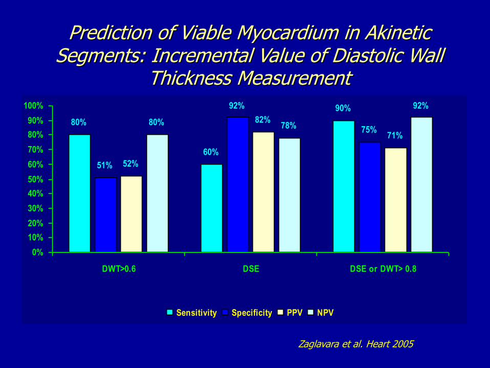
Prediction of Viable Myocardium in AkineticSegments: Incremental Value of Diastolic Wall
Thickness Measurement
Zaglavara et al. Heart 2005
80%
60%
90%
51%
92%
75%
52%
82%
71%
80% 78%
92%
0%
10%
20%
30%
40%
50%
60%
70%
80%
90%
100%
DWT>0.6 DSE DSE or DWT> 0.8
Sensitivity Specificity PPV NPV

Algorithm of Management of Patients with Ischaemic LV dysfunction
Rahimtoola SH, et al. JACC Cardiovascular Imaging 2008


Ionizing Radiation in Cardiac Imaging.
American Heart Association Recommendations:
Cardiac imaging studies that expose patients to ionizing radiation should be ordered only after thoughtful consideration of the potential benefit to the patient and in keeping with established appropriatness criteria (Class I)
Considerations should include options for answering the clinical question at hand by means that do not use ionizing radiation or choosing the type of study that exposes the patient to the lowest amount of radiation (Class I)
Routine surveillance radionuclide stress tests or cardiac CTs in asymptomatic patients at low risk for ischaemic heart disease are not recommended (Class I)
Healthcare providers should discuss the risks and benefits of planned imaging procedures with patients whenever practical or appropriate (Class I)
Circulation 2009


Multimodality Cardiovascular Imaging in CAD

Trends in the Use of Cardiac Imaging up to the Year 2020
(British Cardiovascular Society Working Group)

The right test for the right patient at the right time!

First Do Not Harm!“Ωφελέειν, ή Μη Βλάπτειν”
The art (medicine) consists in three things: the disease, the patient and the physician.
Hippocrates, Epidemics, 5th century B.C