cardiovascular magnetic resonance in patients...
TRANSCRIPT
Mwscphsfiwdbcammbioa
FMDdc
a
Journal of the American College of Cardiology Vol. 55, No. 1, 2010© 2010 by the American College of Cardiology Foundation ISSN 0735-1097/10/$36.00P
STATE-OF-THE-ART PAPERS
Cardiovascular Magnetic Resonancein Patients With Myocardial InfarctionCurrent and Emerging Applications
Han W. Kim, MD,*† Afshin Farzaneh-Far, MD, PHD,*† Raymond J. Kim, MD*†‡
Durham, North Carolina
In patients with known or suspected myocardial infarction (MI), cardiovascular magnetic resonance (CMR) pro-vides a comprehensive, multifaceted view of the heart. The data, including that from a recent multicenter clini-cal trial, indicate that delayed-enhancement cardiac magnetic resonance imaging (DE-CMR) is a well-validated,robust technique that can be easily implemented on scanners that are commonly available worldwide, with aneffectiveness that clearly rivals the best available imaging techniques for the detection and assessment of acuteand chronic MI. When patients present outside the diagnostic window of cardiac troponins, DE-CMR may be es-pecially useful. Moreover, because DE-CMR can uniquely differentiate between ischemic and various nonisch-emic forms of myocardial injury, it may be helpful in cases of diagnostic uncertainty, such as in patients withclassical features of MI in whom coronary angiography does not show a culprit lesion. Even after the diagnosisof MI has been made, CMR provides clinically relevant information by identifying residual viability, microvasculardamage, stunning, and right ventricular infarction. In addition, post-MI sequelae, including left ventricular throm-bus and pericarditis, are easily identified. Given that quantification of infarct size by DE-CMR is highly reproduc-ible, this technique may provide a useful surrogate end point for clinical trials with appreciable reductions insample size compared with alternative methods. (J Am Coll Cardiol 2010;55:1–16) © 2010 by the AmericanCollege of Cardiology Foundation
ublished by Elsevier Inc. doi:10.1016/j.jacc.2009.06.059
nas
MtddnicidbrpofiaCevit
yocardial infarction (MI) is a leading cause of deathorldwide (1). Accordingly, preventative and therapeutic
trategies are aimed at reducing its occurrence and adverseonsequences. New serological biomarkers, such as tro-onins, have radically improved the diagnosis of MI andave enabled the recognition of a group of patients withmall infarcts, many of whom would not have been identi-ed in earlier eras (2). Reclassifying this group as patientsith MI has significant implications (2–4). From an epi-emiological perspective, a substantial increase in the num-er of patients diagnosed with MI creates difficulties inomparing the results of new trials with those of older onesnd confounds efforts to monitor the impact of public healtheasures and treatments. In some clinical trials, the resultsay be significantly altered because the diagnosis of MI can
e used as an entry criterion, an end point, or both. Forndividual patients, the label of MI can affect a wide rangef issues, including employment, disability claims, insur-nce, and psychological well-being. Importantly, the diag-
rom the *Duke Cardiovascular Magnetic Resonance Center, †Department ofedicine, and the ‡Department of Radiology, Duke University Medical Center,urham, North Carolina. Dr. R. J. Kim is an inventor of a U.S. patent on
elayed-enhancement CMR that is owned by Northwestern University, and is aofounder of HeartIT, LLC.
fManuscript received March 9, 2009; revised manuscript received May 26, 2009,
ccepted June 18, 2009.
osis of MI also impacts clinical management, and thevailable data indicate that even small infarcts portend worsehort- and long-term prognosis (5).
Despite the use of improved biomarkers, the diagnosis ofI can still be difficult. There is marked heterogeneity in
he presentation of MI and significant overlap with otherisorders that result in myocardial injury, such as myocar-itis and Takotsubo cardiomyopathy. In this context, it isoteworthy that the latest consensus guidelines defining MI
nclude noninvasive imaging because it may be helpful inases of uncertainty (6). The new criteria incorporatingmaging evidence of MI involve not only established mo-alities such as echocardiography and radionuclide imaging,ut also the newer modalities, cardiovascular magneticesonance (CMR) and cardiac computed tomography. Inarticular, delayed enhancement (DE)-CMR appears toffer advantages in detecting small or subendocardial in-arcts with high accuracy and is well validated (7–12). Thus,t is timely to review the role of CMR in the diagnosis andssessment of MI. In this article, we examine the utility ofMR in patients with known or suspected MI with
mphasis on the additive clinical information it may pro-ide. Additionally, there has been growing interest in usingnfarct size measured by CMR as an end point for clinicalrials, and we discuss operational and other relevant issues
or this application.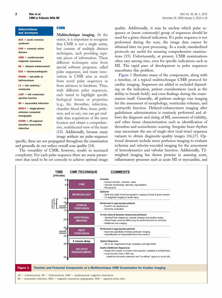
sa
ce
qqupoptoMe
aciaifcglatmvtiowi
2 Kim et al. JACC Vol. 55, No. 1, 2010CMR in Patients With MI December 29, 2009/January 5, 2010:1–16
CMR
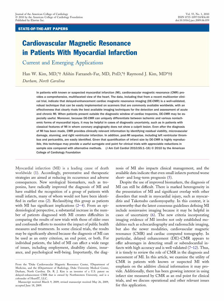
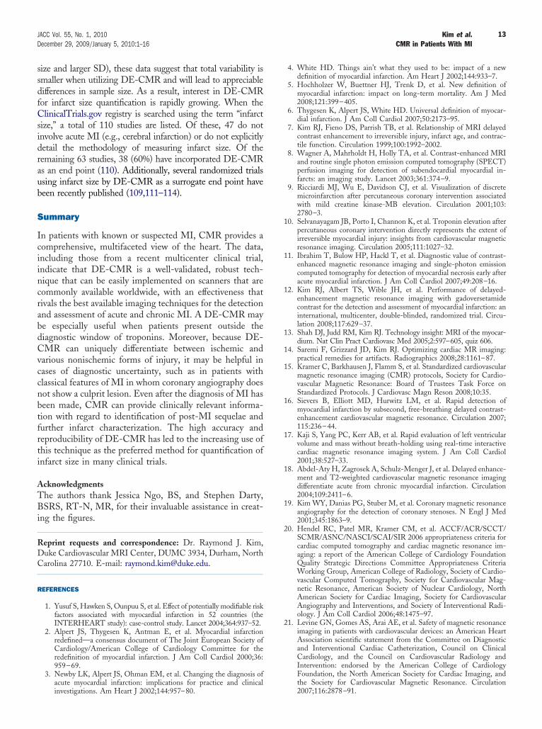
Multitechnique imaging. At theoutset, it is important to recognizethat CMR is not a single entity,but consists of multiple distincttechniques, each providing sepa-rate pieces of information. Thesedifferent techniques arise fromspecial software programs, calledpulse sequences, and many inno-vations in CMR arise as muchfrom novel pulse sequences asfrom advances in hardware. Thus,with different pulse sequences,each tuned to highlight specificbiological tissues or properties(e.g., fat, thrombus, infarction,chamber blood flow, tissue perfu-sion, and so on), one can get mul-tiple data acquisitions of the samelocation and obtain a comprehen-sive, multifaceted view of the heart(13). Additionally, because manyimage artifacts are pulse-sequence
pecific, these are not propagated throughout the examinationnd generally do not reduce overall scan quality (14).
The versatility of CMR, however, results in increasedomplexity. For each pulse sequence there are many param-ters that need to be set correctly to achieve optimal image
Figure 1 Timeline and Potential Components of a Multitechniqu
2D � 2-dimensional; 3D � 3-dimensional; CMR � cardiovascular magnetic resonaMI � myocardial infarction; MRA � magnetic resonance angiography; SNR � signa
Abbreviationsand Acronyms
ACS � acute coronarysyndrome
CAD � coronary arterydisease
CMR � cardiovascularmagnetic resonance
DE � delayed enhancement
ECG � electrocardiogram
FWHM � full-width athalf-maximum
LV � left ventricle/ventricular
LVEF � left ventricularejection fraction
MI � myocardial infarction
SPECT � single-photonemission computedtomography
STEMI � ST-segmentelevation myocardialinfarction
uality. Additionally, it may be unclear which pulse se-uence or (more commonly) group of sequences should besed for a given clinical indication. If a pulse sequence is noterformed during the scan, the image data cannot bebtained later via post-processing. As a result, standardizedrotocols are useful for ensuring comprehensive examina-ions (15). Unfortunately, at present, CMR examinationsften vary among sites, even for specific indications such asI. The rapid pace of development in pulse sequences
xacerbates this problem.Figure 1 illustrates many of the components, along withtimeline, of a typical multitechnique CMR protocol for
ardiac imaging. Sequences are added or excluded depend-ng on the indication, patient considerations (such as thebility to breath-hold), and even findings during the exam-nation itself. Generally, all patients undergo cine imagingor the assessment of morphology, ventricular volumes, andontractile function. Delayed-enhancement imaging afteradolinium administration is routinely performed and al-ows the diagnosis and sizing of MI, assessment of viability,nd other tissue characterization such as identification ofhrombus and nonischemic scarring. Irregular heart rhythmay necessitate the use of single-shot (real-time) sequence
ariants to obtain diagnostic-quality images (16,17). Op-ional elements include stress perfusion imaging to evaluateschemia and velocity-encoded imaging for the assessmentf hemodynamics and valvular function. Additionally, T2-eighted imaging has shown promise in assessing acute,
nflammatory processes such as acute MI or myocarditis, and
R Examination for Cardiac Imaging
ise ratio.
e CM
nce;l-to-no
moroo
ltUsialrneanpbopi[DtDioWbhntbpgctcammtmtthsanvsonh
iCoc
niotiaiwmacdFels
3JACC Vol. 55, No. 1, 2010 Kim et al.December 29, 2009/January 5, 2010:1–16 CMR in Patients With MI
ay prove useful in distinguishing chronic lesions from thosef recent onset (18). At experienced centers, coronary magneticesonance angiography may be performed to exclude left mainr 3-vessel coronary artery disease (CAD) in selected patientsr to evaluate coronary anomalies (19,20).
Even in a 30- to 45-min examination, there are often aarge number of data acquisitions (�50) using differentechniques across multiple spatial locations. The followingRL (http://dcmrc.duhs.duke.edu/figure/, Online Fig. 1)
hows images from a comprehensive study and highlights themportance of side-by-side viewing of different techniques tollow efficient and accurate interpretation. The strengths andimitations of each of the different CMR techniques should beecognized when considering which to use during the exami-ation as well as during the interpretation. The levels ofxperimental and clinical validation vary among techniques,nd some are still undergoing development or lack the robust-ess necessary for general clinical use. Finally, as with anyrocedure, the risks of CMR must be weighed against theenefits, and patients with contraindications to magnetic res-nance imaging or gadolinium contrast, such as those withacemakers/defibrillators (21) or advanced kidney disease (ow-ng to the risk of developing nephrogenic systemic fibrosis22]) are not infrequently encountered.
E. Although several CMR techniques could be used forhe diagnosis of MI, the most accurate and best validated isE-CMR. The technique is straightforward. It involves
nversion-recovery imaging after intravenous administrationf gadolinium contrast and a 5- to 10-min delay (23,24).
ith appropriate settings, normal myocardium appearslack or nulled, whereas nonviable regions appear bright oryperenhanced. The mechanism of hyperenhancement hasot been fully elucidated, but one has been proposed (25)hat is based on 2 simple facts. First, in normal myocardium,ecause myocytes are densely packed, tissue volume isredominately intracellular (�75% to 80%) (26). Second,adolinium chelates are extracellular agents that cannotross intact sarcolemmal membranes (27). It then followshat gadolinium distribution volume is small and tissueoncentration is low in a voxel of normal myocardium. Withcute necrosis (acute MI, myocarditis, and so on), there isembrane rupture, which allows gadolinium to diffuse intoyocytes. This results in increased gadolinium concentra-
ion (28), shortened T1 relaxation, and thus hyperenhance-ent. In the chronic setting, scar has replaced necrotic
issue and the interstitial space is expanded. This again leadso increased gadolinium concentration (28) and hyperen-ancement. In both acute and chronic settings (and alltages in between), one can consider viable myocytes asctively excluding gadolinium. Thus, the unifying mecha-ism of hyperenhancement appears to be the absence ofiable myocytes rather than any inherent properties that arepecific for acute necrosis, collagenous scar, or other formsf nonviable myocardium (25). In the literature, the tech-ique of DE imaging is also known as delayed hyperen-
ancement, myocardial delayed enhancement, late gadolin-um enhancement, and simply contrast-enhanced CMR.oncerning late gadolinium enhancement, we note thatther T1-shortening contrast media aside from gadoliniumould show late enhancement.
In animal models, extensive comparisons have shown aearly exact relationship between the size and shape of
nfarcted myocardium by DE-CMR to that of histopathol-gy (Fig. 2A) (7,8,28–33). Additionally, these studies showhat DE-CMR can distinguish between reversible andrreversible injury independent of wall motion, infarct age,nd reperfusion status. Studies in humans have shown thatnfarct size measured by DE-CMR is closely associatedith peak cardiac enzyme release (10,34–38) and measure-ents by positron emission tomography (39,40). DE-CMR
lso appears to be superior to single-photon emissionomputed tomography (SPECT) in detecting subendocar-ial infarcts and infarcts in nonanterior locations (8,11).urthermore, the high spatial resolution of DE-CMRnables visualization of even microinfarctions, involving asittle as 1 g of tissue, which may occur during otherwiseuccessful percutaneous coronary intervention (Fig. 2B) (9,10).
A
B
HISTOLOGY DE-CMR SPECT
Patient 1 Patient 2
Figure 2 MI Visualized by DE-CMR
(A) Comparison of delayed-enhancement (DE)-CMR with histopathology and sin-gle-photon emission computed tomography (SPECT) in 2 animals with subendo-cardial MI. Note that the size and shape of hyperenhanced regions by DE-CMR(blue arrows) match the size and shape of infarcted regions delineated by his-tological vital staining. No infarcts are evident by SPECT. Modified, with permis-sion, from Wagner et al. (8). (B) Microinfarction (blue arrows) associated withsuccessful percutaneous coronary intervention is shown in 2 patients. Redarrows point to the coronary stents. Modified, with permission, from Ricciardiet al. (9). Guidelines for DE-CMR parameter adjustments may be found else-where (14,23–25). Abbreviations as in Figure 1.
oIfiUtoMFw9mIdAmtndfatndmtttatd
D
ICc
cehinestiitisMMaoao
tvacaestdMvsbNma
4 Kim et al. JACC Vol. 55, No. 1, 2010CMR in Patients With MI December 29, 2009/January 5, 2010:1–16
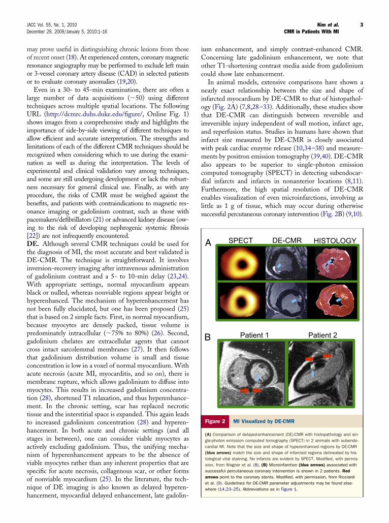
Recently, the performance of DE-CMR for the detectionf MI was tested in an international multicenter trial (12).n total, 282 patients with acute and 284 with chronicrst-time MI were scanned in 26 centers throughout the.S., Europe, and South America. The study showed that
he sensitivity of DE-CMR increased with increasing gad-linium dose, reaching 99% and 94% in acute and chronicI, respectively, with the 0.3-mmol/kg dose (Fig. 3).
urthermore, with doses 0.2 mmol/kg or higher, when MIas identified, it was in the correct location in more than7% of patients (i.e., the location of hyperenhancementatched the perfusion territory of the infarct-related artery).
mportantly, this study represents the first multicenter trialesigned to evaluate an imaging approach for detecting MI.lthough several multicenter trials have used infarct sizeeasurements by SPECT as a surrogate end point to assess
he efficacy of an investigative therapy (41), these trials wereot designed to evaluate SPECT, and limited multicenterata on the sensitivity or accuracy of radionuclide imagingor detecting or localizing MI have been reported. Inddition, the multicenter DE-CMR trial tested the sensi-ivity of imaging for both acute and chronic MI. This isotable because there are far fewer clinical trial data on theetection of chronic MI, and chronic infarcts are generallyore difficult to detect than acute infarcts because substan-
ial shrinkage can occur during healing (7). Thus, in sum,he data indicate that DE-CMR is a well-validated, robustechnique that can be easily implemented on scanners thatre commonly available worldwide, with an effectivenesshat rivals the best available imaging techniques for theetection and assessment of MI.
iagnosis and Assessment of MI
ssues in clinical diagnosis. According to the Americanollege of Cardiology/European Society of Cardiology
onsensus document on the universal definition of MI, the
14 (6-22)
99 (97-100) 95
(90-100) 84 (76-93)
48 (36-59)
13 (5-21)
17 (8-26)
15 (7-24)
45 (34-56)
80 (70-89)
92 (86-98)
99 (98-100)
100
80
60
40
20
0 Pre-Contrast Post-10 Mins Post-30 Mins
Figure 3 Sensitivity of DE-CMR for Acute and Chronic MI
The diagnostic sensitivity of detecting MI is summarized according to gadoversetaNumbers in parentheses are 95% confidence intervals. Modified, with permission,
ornerstone tests for the diagnosis are troponins and thelectrocardiogram (ECG) (6). Unfortunately, these testsave some limitations. Although troponin assays are exquis-
tely sensitive and can detect minute amounts of myocardialecrosis, levels are elevated for only a few days after an acutevent (42). Importantly, many patients do not have classicymptoms and do not seek medical attention within theime window when troponins are elevated. The consequences that many subacute and all chronic infarcts will not bedentified by troponins. The ECG is helpful in this situa-ion, and incident Q waves have been the basis for diagnos-ng silent or clinically unrecognized MI in populationtudies (43). These surveys have shown that unrecognized
Is are common, comprising as many as 40% to 60% of allIs. By definition, however, all unrecognized infarcts that
re non–Q-wave will be missed (44). Additionally, Q wavesften occur in the setting of nonischemic cardiomyopathy,nd the specificity of the ECG may be poor in the setting ofther cardiopulmonary disorders (45).In these circumstances, cardiac imaging has the potential
o provide corroborative diagnostic information. The uni-ersal definition indicates that new regional wall motionbnormalities or a loss of viable myocardium could beonsidered evidence of MI (6). However, wall motionbnormalities may not occur unless the infarcted regionxceeds 20% to 50% of the myocardial wall (2,46). Similarly,cintigraphic defects may not be apparent until �10 g ofissue is infarcted (2). Thus, because a sizable threshold ofamage is required, echocardiography or SPECT may missI, particularly when it is small or subendocardial. Con-
ersely, when abnormalities in regional function or perfu-ion are present, they do not always indicate MI. Both maye abnormal in the setting of ischemia without infarction.onischemic conditions, such as cardiomyopathy or inflam-atory or infiltrative diseases, can also lead to wall motion
bnormalities or loss of viable myocardium. Hence, the
6 (1-12)
3 (0-7)
8 (2-15)
10 (3-17)
52 (40-63)
83 (74-91)
87 (79-95)
94 (89-100)
44 (33-52)
73 (62-83)
84 (76-93)
92 (86-99)
Pre-Contrast Post-10 Mins Post-30 Mins
0.05 0.1 0.2 0.3
Dose (mmol/kg) Gadoversetamide
Legend
ose group and imaging time point.im et al. (12). Abbreviations as in Figures 1 and 2.
mide dfrom K
ph
MadiedtpnpciS3Awlq
ItDcAcphspthr3Qrd
t13
P
*e
S
5JACC Vol. 55, No. 1, 2010 Kim et al.December 29, 2009/January 5, 2010:1–16 CMR in Patients With MI
ositive predictive value of these imaging findings is notigh unless these conditions can be excluded (6).Even some patients who have all of the classic features ofI—for example, chest pain, new ST-segment changes,
nd an increase in troponins—and fulfill the universalefinition, may ultimately prove not to have had an MI. For
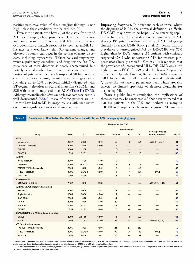
nstance, it is well known that ST-segment changes andlevated troponins can occur in the setting of many disor-ers, including myocarditis, Takotsubo cardiomyopathy,rauma, pulmonary embolism, and drug toxicity (6). Therevalence of these disorders is poorly characterized, butotably, several studies have shown that a nontrivial pro-ortion of patients with clinically suspected MI have normaloronary arteries or insignificant disease at angiography,ncluding up to 10% of patients initially diagnosed withT-segment elevation myocardial infarction (STEMI) and2% with acute coronary syndrome (ACS) (Table 1) (47–62).lthough recanalization after an occlusive coronary event isell documented (63,64), many of these patients are un-
ikely to have had an MI, leaving clinicians with unanswereduestions regarding diagnosis and management.
revalence of Nonobstructive CAD in Patients With MI or ACS UndTable 1 Prevalence of Nonobstructive CAD in Patients With MI
Study Year n*
STEMI
Larson et al. 2007 1,335
DANAMI-2 substudy 2007 516
CAPTIM 2002 405
GUSTO IIb 1999 2,251
NSTEMI
ICTUS substudy 2007 599
CRUSADE 2006 38,301
TACTICS–TIMI 18 substudy 2005 542
FRISC II substudy 2001 1,142‡
GUSTO IIb 1999 1,749
Non–Q-wave MI
VANQWISH substudy 2002 350
NSTEMI and ACS (negative biomarker)
SYNERGY 2007 7,005
Bugiardini et al. 2006 7,656
ISAR-COOL 2003 410
RITA-3 2002 865
PURSUIT 2000 5,767
TIMI IIIB 1994 1,193
STEMI, NSTEMI, and ACS (negative biomarker)
GRACE 2009 26,755
MATE 1998 163
ACS (negative biomarker)
TACTICS–TIMI 18 substudy 2005 353
FRISC II substudy 2001 1,142‡
GUSTO IIb 1999 2,406
Patients who underwent angiography and had data available. †Estimated from patients in angixplanation provided. ‡Values reflect the data from the combined group of NSTEMI and ACS with
— � data not available; ACS � acute coronary syndrome; CAD � coronary artery disease; F � female; MTEMI � ST-segment elevation myocardial infarction.
mproving diagnosis. In situations such as these, wherehe diagnosis of MI by the universal definition is difficult,E-CMR may prove to be helpful. One emerging appli-
ation has been the identification of unrecognized MI.mong 195 patients without a history of MI undergoing
linically indicated CMR, Kwong et al. (65) found that therevalence of unrecognized MI by DE-CMR was 76%igher than by ECG. Among 185 patients with clinicallyuspected CAD who underwent CMR for research pur-oses (not clinically ordered), Kim et al. (44) reported thathe prevalence of unrecognized MI by DE-CMR was 313%igher than by ECG. In 259 randomly chosen 70-year-oldesidents of Uppsala, Sweden, Barbier et al. (66) observed a90% higher rate. In all 3 studies, several patients with
waves did not have hyperenhancement, which probablyeflects the limited specificity of electrocardiography foriagnosing MI.From a public health standpoint, the implications of
hese studies may be considerable. It has been estimated that90,000 patients in the U.S. and perhaps as many as00,000 in Europe suffer from unrecognized MI annually
ng AngiographyCS Undergoing Angiography
Nonobstructive CAD
No Single Culprit(None, Multiple) Ref. #tion
Prevalence (%)
Overall M F
% 10 8 14 15% (14%, 1%) 61
% 4 — — — 47
10† — — — 48
8 7 10 — 49
% 9 — — — 50
% 9 6 12 — 51
% 6 4 10 — 52
% 7 4 14 44%‡ 53
5 4 9 — 49
% 6 — — 51% (37%, 14%) 54
8 — — — 55
% 9 — — — 56
% 11 — — — 57
% 22 — — — 58
% 12 — — — 59
% 16 — — — 60
% 8 6 12 — 61
% 25 — — 45% (44%, 1%) 62
% 21 17 28 — 52
% 32 26 43 44%‡ 53
20 14 31 — 49
arm not undergoing percutaneous coronary intervention because of normal coronary flow or noe biomarker.
ergoior A
Defini
�50
�50
—
—
�70
�50
�50
�50
—
�50
—
—
�50
�50
�70
�50
�60
�50
�70
�50
�50
—
oplastynegativ
� male; MI � myocardial infarction; NSTEMI � non–ST-segment elevation myocardial infarction;
(tailtpCeDataw
ewnsCmspm
Cnitrtcecseis(mtiosuSifbp
dcpud
mI
6 Kim et al. JACC Vol. 55, No. 1, 2010CMR in Patients With MI December 29, 2009/January 5, 2010:1–16
67,68). Because these estimates reflect only patients iden-ified by ECG, the DE-CMR studies suggest that thectual incidence may be 3-fold higher. However, infarct sizes larger and left ventricular ejection fraction (LVEF) isower in patients with unrecognized MI with Q waves thanhose without (44), and one might question whether therognosis of individuals with unrecognized MI by DE-MR is relatively benign. On this point, we note that Kwong
t al. (65) reported that the presence of unrecognized MI byE-CMR conferred nearly a 6-fold increased risk for major
dverse cardiac events. Likewise, Kim et al. (44) reported thathe presence of unrecognized non–Q-wave MI predictedn 11-fold higher risk of all-cause mortality than thoseithout MI.Initial studies suggest that CMR may also be useful in the
mergency department setting for the evaluation of patientsith chest pain. Kwong et al. (69) showed that a multitech-ique CMR examination can be performed rapidly andafely in emergency department patients, and reported thatMR added diagnostic value over standard clinical assess-ent for the diagnosis of ACS. Cury et al. (70) reported
imilar findings, but also showed that CMR has theotential to diagnose acute MI even when the first troponineasurement is negative.Similar to troponins, the detection of injury by DE-
MR is specific for irreversible myocardial damage but isot specific for MI. One potential advantage of DE-CMR
s that the pattern of hyperenhancement, rather than simplyhe presence or extent, may offer important informationegarding the etiology of myocardial damage (71–73). Forhis purpose, the concept that ischemic myonecrosis pro-eeds as a wavefront (74) from the subendocardium to thepicardium with increasing coronary occlusion time is cru-ial. Correspondingly, hyperenhancement patterns thatpare the subendocardium and are limited to the middle orpicardial portion of the left ventricular (LV) wall arenvariably nonischemic in origin because damage in theetting of CAD almost always involves the subendocardium71–73). Moreover, certain nonischemic disorders, such asyocarditis, have characteristic hyperenhancement patterns
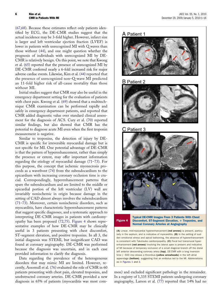
hat suggest specific diagnoses, and a systematic approach tonterpreting DE-CMR images in patients with cardiomy-pathy has been proposed (72,75). Figure 4 shows repre-entative examples of how DE-CMR may be clinicallyseful in 3 patients presenting with chest discomfort,T-segment elevation, and positive troponins. In all 3, the
nitial diagnosis was STEMI, but insignificant CAD wasound at coronary angiography. DE-CMR was performedecause the diagnosis was uncertain, and in each caserovided information to clarify the diagnosis.Data regarding the prevalence of the heterogeneous
isorders that may mimic MI are limited. However, re-ently, Assomull et al. (76) evaluated the role of CMR in 60atients presenting with chest pain, elevated troponins, andnobstructed coronary arteries. DE-CMR provided a new
iagnosis in 65% of patients (myocarditis was most com- aon) and excluded significant pathology in the remainder.n a registry of 1,335 STEMI patients undergoing coronary
A Patient 1
B Patient 2
C Patient 3
Figure 4Typical DE-CMR Images From 3 Patients With ChestDiscomfort, ST-Segment Elevation, � Troponins, andNormal Coronary Arteries at Angiography
(A) Linear, mid-myocardial hyperenhancement (red arrows) is present, particu-larly in the septum, and is indicative of myocarditis. (B) In the setting of sud-den emotional stress and apical ballooning, the absence of hyperenhancementis consistent with Takotsubo cardiomyopathy. (C) Focal but transmural hyper-enhancement (red arrows) involving the lateral apex is present and indicativeof MI because of temporary occlusion of a small diagonal branch off the distalleft anterior descending coronary artery (top). DE-CMR with a long inversiontime (�600 ms) shows a thrombus (yellow arrowheads) in the left atrialappendage (bottom), suggesting that an embolus led to the MI. Abbreviationsas in Figures 1 and 2.
ngiography, Larson et al. (77) reported that 14% had no
cgttgtcamcMiIsoshso
oit(diCmdapcnaa
mat
7JACC Vol. 55, No. 1, 2010 Kim et al.December 29, 2009/January 5, 2010:1–16 CMR in Patients With MI
ulprit artery and 9.5% did not have significant CAD. In theroup without a clear culprit artery, CMR established thathe most common diagnoses were myocarditis (31%), Tako-subo cardiomyopathy (31%), and STEMI without an angio-raphic lesion (29%). Concerning the latter, Table 1 showshat in many patients with acute MI or ACS, the culprit arteryannot be identified because of the absence of CAD or thebsence of typical angiographic characteristics, or becauseultivessel CAD is present and more than 1 artery/culprit
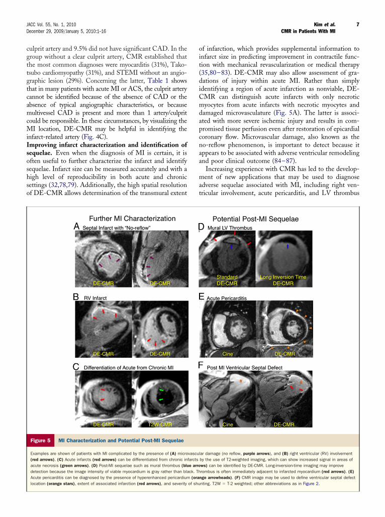
ould be responsible. In these circumstances, by visualizing theI location, DE-CMR may be helpful in identifying the
nfarct-related artery (Fig. 4C).mproving infarct characterization and identification ofequelae. Even when the diagnosis of MI is certain, it isften useful to further characterize the infarct and identifyequelae. Infarct size can be measured accurately and with aigh level of reproducibility in both acute and chronicettings (32,78,79). Additionally, the high spatial resolutionf DE-CMR allows determination of the transmural extent
Figure 5 MI Characterization and Potential Post-MI Sequelae
Examples are shown of patients with MI complicated by the presence of (A) micro(red arrows). (C) Acute infarcts (red arrows) can be differentiated from chronic inacute necrosis (green arrows). (D) Post-MI sequelae such as mural thrombus (bludetection because the image intensity of viable myocardium is gray rather than blaAcute pericarditis can be diagnosed by the presence of hyperenhanced pericardiumlocation (orange stars), extent of associated infarction (red arrows), and severity
f infarction, which provides supplemental information tonfarct size in predicting improvement in contractile func-ion with mechanical revascularization or medical therapy35,80–83). DE-CMR may also allow assessment of gra-ations of injury within acute MI. Rather than simplydentifying a region of acute infarction as nonviable, DE-MR can distinguish acute infarcts with only necroticyocytes from acute infarcts with necrotic myocytes and
amaged microvasculature (Fig. 5A). The latter is associ-ted with more severe ischemic injury and results in com-romised tissue perfusion even after restoration of epicardialoronary flow. Microvascular damage, also known as theo-reflow phenomenon, is important to detect because itppears to be associated with adverse ventricular remodelingnd poor clinical outcome (84–87).
Increasing experience with CMR has led to the develop-ent of new applications that may be used to diagnose
dverse sequelae associated with MI, including right ven-ricular involvement, acute pericarditis, and LV thrombus
ar damage (no reflow, purple arrows), and (B) right ventricular (RV) involvementby the use of T2-weighted imaging, which can show increased signal in areas ofws) can be identified by DE-CMR. Long-inversion-time imaging may improverombus is often immediately adjacent to infarcted myocardium (red arrows). (E)
nge arrowheads). (F) CMR image may be used to define ventricular septal defectnting. T2W � T-2 weighted; other abbreviations as in Figure 2.
vasculfarctse arrock. Th(ora
of shu
(eEwiutmmpsp(seacpiwoi9WbspfwsawAt
tlcueivatqaWustbiptsc
stm
CS
TrMrdTbiApsrtitmliinppssoWsaitvcmsIvw
vs2idcrnv
8 Kim et al. JACC Vol. 55, No. 1, 2010CMR in Patients With MI December 29, 2009/January 5, 2010:1–16
Figs. 5B to 5F). In patients with acute inferior MI, Kumart al. (88) showed that even when physical examination,CG with right precordial leads, and echocardiographyere negative, DE-CMR could detect right ventricular
nvolvement in nearly 25% of patients. Taylor et al. (89)sed a multitechnique CMR examination to evaluate pa-ients with pericardial disease. Pericardial hyperenhance-ent appeared to be both sensitive and specific for inflam-atory pericarditis, as verified by histopathology, whereas
ericardial thickening without hyperenhancement was con-istent with a noninflammatory fibrotic pericardium. Inatients with MI or ischemic cardiomyopathy, Mollet et al.90) reported that DE-CMR identified LV thrombus inubstantially more patients than cine-CMR or transthoracicchocardiography; however, a reference standard was notvailable. Srichai et al. (91) evaluated a protocol combiningine- and DE-CMR for the diagnosis of LV thrombus inatients with advanced ischemic cardiomyopathy undergo-ng surgical LV reconstruction. Among 160 patients (inhom there was surgical and/or pathological confirmationf thrombus), CMR showed higher sensitivity and specific-ty (88% and 99%, respectively) than transthoracic (23%,6%) and transesophageal (40%, 96%) echocardiography.
einsaft et al. (92) assessed the prevalence of LV thrombusy cine and DE-CMR in 784 consecutive patients withystolic dysfunction. The DE-CMR detected a higherrevalence than cine-CMR (7.0% vs. 4.7%, p � 0.005), andollow-up for embolic events or pathological confirmationas consistent with DE-CMR as the better reference
tandard. Interestingly, patients with ischemic cardiomyop-thy were 5 times more likely to have thrombus than thoseith nonischemic cardiomyopathy despite similar LVEF.dditionally, myocardial scarring by DE-CMR was iden-
ified as a novel risk factor for thrombus.The ability of DE-CMR to identify thrombus based on
issue characteristics rather than just anatomical appearanceikely explains its improved performance compared withine-CMR or noncontrast echocardiography. The basicnderlying principle is that thrombi are avascular and havessentially no gadolinium uptake. Thus, thrombus can bedentified as a nonenhancing defect surrounded by brightentricular blood and contrast-enhanced myocardium. Im-ge intensity differences between normal myocardium andhrombus can be accentuated by using a DE-CMR se-uence in which the inversion time is increased to nullvascular tissue such as thrombus (500 to 600 ms) (92).
ith long-inversion-time imaging, regions with contrastptake such as viable myocardium increase in image inten-ity, whereas thrombus appears homogeneously black, andhere is improved delineation, particularly of mural throm-us (Fig. 5D). The concept that contrast uptake, albeit low,s not zero in normal myocardium, is one reason why werefer to describe nonviable regions as hyperenhanced ratherhan simply enhanced because depending on the DE-CMRettings, viable myocardium can show contrast enhancement
ompared with avascular tissue. DFigure 6 outlines possible CMR findings in patients withuspected MI. Furthermore, it shows the potential of CMRo provide additive diagnostic information and how CMRay be incorporated with traditional clinical assessment.
MR Infarct Size as aurrogate End Point for Clinical Trials
he ultimate goal of a new therapy for acute MI is aeduction in mortality. In the current era, treatment of acute
I is quite effective; therefore, demonstrating a furthereduction in mortality with novel treatments is increasinglyifficult and necessitates studies with large sample sizes.his requirement imposes significant logistical and financialarriers on testing potential new therapies, and correspond-ngly limits the number of treatments that can be evaluated.s such, there is considerable interest in using surrogate endoints to assess the efficacy of acute MI therapies. Infarctize is a particularly attractive surrogate end point for severaleasons (41,93). First, it is useful in early screening studieso test whether a new therapy is biologically active. Second,t can serve as an end point for phase II dose-ranging studieso test efficacy and/or safety. Third, it may indicate a lateortality benefit, and thus rationale for performing a
onger-term study, even if an early benefit is not seen. Fornstance, a reduction in infarct size may lead to a long-termmprovement in ventricular remodeling, a benefit that mayot be manifest on 30-day mortality rates. Fourth, it canrovide a mechanism for improvement in outcome, becauserognosis after acute MI is strongly determined by infarctize (94,95). Several investigations have reported that infarctize measured by DE-CMR is a stronger predictor ofutcome than LVEF and LV volumes (96–98).
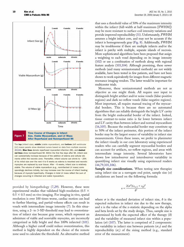
hen to measure infarct size. Measurements of infarctize in the first few weeks after infarction need to take intoccount what Reimer and Jennings (74) termed the chang-ng anatomic reference base of evolving myocardial infarc-ion. In their pioneering studies, they showed that infarctolume can almost double during the first few days afteroronary artery occlusion, even in the absence of additionalyocyte death via lethal reperfusion injury or infarct exten-
ion, because of the addition of edema and cellular elements.n contrast, infarct volume may shrink to 25% of its initialolume as necrotic muscle is replaced by scar over 4 to 6eeks.Using DE-CMR, similar findings have been observed in
ivo in a canine model (Fig. 7) (99,100). Fieno et al. (99)howed that terminal infarct size at 4 to 8 weeks averaged4% of that found at 3 days, and reperfusion acceleratednfarct resorption. Moreover, the mass of viable myocar-ium increased systematically with time, although the timeourse of hypertrophy was different from that of infarctesorption. Importantly, measurements of total LV mass didot reflect the changes occurring separately in infarcted andiable regions. These results highlight the capability of
E-CMR to improve the assessment of post-infarction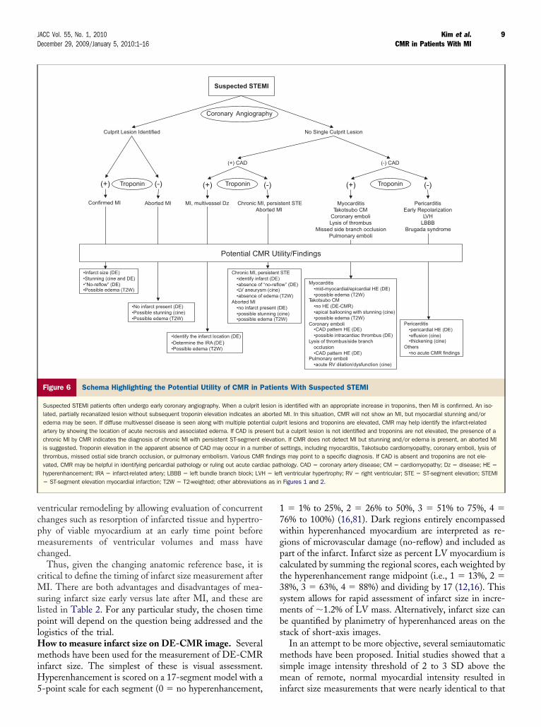
vcpmc
cMslplHmiH5
17wgpct3smbs
msm
9JACC Vol. 55, No. 1, 2010 Kim et al.December 29, 2009/January 5, 2010:1–16 CMR in Patients With MI
entricular remodeling by allowing evaluation of concurrenthanges such as resorption of infarcted tissue and hypertro-hy of viable myocardium at an early time point beforeeasurements of ventricular volumes and mass have
hanged.Thus, given the changing anatomic reference base, it is
ritical to define the timing of infarct size measurement afterI. There are both advantages and disadvantages of mea-
uring infarct size early versus late after MI, and these areisted in Table 2. For any particular study, the chosen timeoint will depend on the question being addressed and the
ogistics of the trial.ow to measure infarct size on DE-CMR image. Severalethods have been used for the measurement of DE-CMR
nfarct size. The simplest of these is visual assessment.yperenhancement is scored on a 17-segment model with a
Suspected STEM
Culprit Lesion Identified
(+) CAD
Coronary Angiograp
(+) (-)
Aborted MI
(+)
MI, multivessel Dz Chronic MIAbo
Confirmed MI
•Infarct size (DE) •Stunning (cine and DE) •“No-reflow” (DE) •Possible edema (T2W)
Potential CM
•No infarct present (DE) •Possible stunning (cine) •Possible edema (T2W)
•Identify the infarct location (DE) •Determine the IRA (DE) •Possible edema (T2W)
Chronic MI, pe•identify infar•absence of •LV aneurysm•absence of
Aborted MI •no infarct pr•possible stu•possible ede
Troponin Troponin
Figure 6 Schema Highlighting the Potential Utility of CMR in P
Suspected STEMI patients often undergo early coronary angiography. When a culprit lelated, partially recanalized lesion without subsequent troponin elevation indicates an aedema may be seen. If diffuse multivessel disease is seen along with multiple potentartery by showing the location of acute necrosis and associated edema. If CAD is preschronic MI by CMR indicates the diagnosis of chronic MI with persistent ST-segment eis suggested. Troponin elevation in the apparent absence of CAD may occur in a numthrombus, missed ostial side branch occlusion, or pulmonary embolism. Various CMRvated, CMR may be helpful in identifying pericardial pathology or ruling out acute cardihyperenhancement; IRA � infarct-related artery; LBBB � left bundle branch block; LVH� ST-segment elevation myocardial infarction; T2W � T2-weighted; other abbreviation
-point scale for each segment (0 � no hyperenhancement, i
� 1% to 25%, 2 � 26% to 50%, 3 � 51% to 75%, 4 �6% to 100%) (16,81). Dark regions entirely encompassedithin hyperenhanced myocardium are interpreted as re-ions of microvascular damage (no-reflow) and included asart of the infarct. Infarct size as percent LV myocardium isalculated by summing the regional scores, each weighted byhe hyperenhancement range midpoint (i.e., 1 � 13%, 2 �8%, 3 � 63%, 4 � 88%) and dividing by 17 (12,16). Thisystem allows for rapid assessment of infarct size in incre-ents of �1.2% of LV mass. Alternatively, infarct size can
e quantified by planimetry of hyperenhanced areas on thetack of short-axis images.
In an attempt to be more objective, several semiautomaticethods have been proposed. Initial studies showed that a
imple image intensity threshold of 2 to 3 SD above theean of remote, normal myocardial intensity resulted in
No Single Culprit Lesion
(-) CAD
(+) (-)
tent STE I
Myocarditis Takotsubo CM
Coronary emboli Lysis of thrombus
Missed side branch occlusion Pulmonary emboli
Pericarditis Early Repolarization
LVH LBBB
Brugada syndrome
lity/Findings
Pericarditis •pericardial HE (DE) •effusion (cine) •thickening (cine)
Others •no acute CMR findings
STE
w” (DE) T2W)
E) ine) W)
Myocarditis •mid-myocardial/epicardial HE (DE) •possible edema (T2W)
Takotsubo CM •no HE (DE-CMR) •apical ballooning with stunning (cine) •possible edema (T2W)
Coronary emboli •CAD pattern HE (DE) •possible intracardiac thrombus (DE)
Lysis of thrombus/side branch occlusion •CAD pattern HE (DE)
Pulmonary emboli •acute RV dilation/dysfunction (cine)
Troponin
ts With Suspected STEMI
identified with an appropriate increase in troponins, then MI is confirmed. An iso-MI. In this situation, CMR will not show an MI, but myocardial stunning and/orrit lesions and troponins are elevated, CMR may help identify the infarct-relatedt a culprit lesion is not identified and troponins are not elevated, the presence of an. If CMR does not detect MI but stunning and/or edema is present, an aborted MIsettings, including myocarditis, Takotsubo cardiomyopathy, coronary emboli, lysis ofs may point to a specific diagnosis. If CAD is absent and troponins are not ele-
hology. CAD � coronary artery disease; CM � cardiomyopathy; Dz � disease; HE �
t ventricular hypertrophy; RV � right ventricular; STE � ST-segment elevation; STEMIFigures 1 and 2.
I
hy
(-)
, persisrted M
R Uti
rsistent ct (DE) “no-reflo
(cine) edema (
esent (Dnning (cma (T2
atien
sion isbortedial culpent bulevatiober offindingac pat� lef
s as in
nfarct size measurements that were nearly identical to that
pe0rtvttacAmz
twmpaimiMa(fmasrm
odrMdafta(tbmtrcisq(Suc
weaadaltr
10 Kim et al. JACC Vol. 55, No. 1, 2010CMR in Patients With MI December 29, 2009/January 5, 2010:1–16
rovided by histopathology (7,29). However, these werexperimental studies that validated high-resolution (0.5 �.5 � 0.5 mm) ex vivo imaging. For imaging in vivo, spatialesolution is over 100 times worse, cardiac motion can leado further blurring, and partial-volume effects can result inoxels with intermediate image intensity (7). In this situa-ion, using a 2- to 3-SD threshold may lead to overestima-ion of infarct size because gray zones, which represent andmixture of viable and nonviable myocytes, are incorrectlyategorized as fully bright and thus 100% infarcted (101).lthough a higher cutoff could reduce overestimation, thisethod is highly dependent on the choice of the remote
Figure 7Time Course of Changes in InfarctSize, Viable Myocardium, and LV MassAfter Reperfused and Nonreperfused MI
The top (infarct size), middle (viable myocardium), and bottom (left ventricular[LV] mass) panels show idealized curves based on data from multiple sources(83,84). Blue lines denote reperfused myocardial infarction (MI), and dashedred lines show nonreperfused MI. Within the first few days after MI, infarct sizecan substantially increase because of the addition of edema and cellular ele-ments within the necrotic zone. Thereafter, infarct volume can shrink to �25%of its initial size over the next 4 to 6 weeks as edema is resorbed and necroticmyocytes are replaced by scar tissue. After �6 weeks, infarct size is relativelystable. The volume of viable myocardium (e.g., noninfarcted LV mass) corre-spondingly declines initially, but may increase over the course of infarct healingbecause of myocyte hypertrophy. Changes in total LV mass reflect the sum ofchanges occurring in infarcted and viable myocardium.
one used to calculate the threshold. An alternative method e
hat uses a threshold value of 50% of the maximum intensityithin the infarct (full-width at half-maximum [FWHM])ay be more resistant to surface-coil intensity variations and
rovide improved reproducibility (31). Unfortunately, FWHMssumes a bright infarct core, and may not be accurate if thenfarct is homogeneously gray (Fig. 8). Additionally, FWHM
ay be troublesome if there are multiple infarcts and/or thenfarct is patchy with multiple, separate islands of necrosis.
ore sophisticated algorithms have been proposed that assignweighting to each voxel depending on its image intensity
102) or use a combination of methods along with regionaleature analysis (103,104). Although promising, these newerethods (and many semiautomated methods) are not widely
vailable, have been tested in few patients, and have not beenhown to work equivalently for images from different magneticesonance imaging vendors. The latter would be important forulticenter trials.Moreover, these semiautomated methods are not as
bjective as one might think. All require user input toistinguish bright artifact and/or noise voxels (false-positiveegions) and dark no-reflow voxels (false-negative regions).
ost important, all require manual tracing of the myocar-ial borders. This is because there are no automatedlgorithms that can reliably distinguish the bright LV cavityrom the bright endocardial border of the infarct. Indeed,issue contrast-to-noise ratio is far lower between infarctnd LV cavity than between infarct and normal myocardium16,105). Because the endocardial border may constitute upo 50% of the infarct perimeter, this portion of the infarctorder may be the largest source of variability in infarct sizeeasurements. Given these issues, we prefer to planimeter
he infarct visually in our core laboratory using experiencedeaders who can carefully segment myocardial borders andan account for artifacts, no-reflow regions, and areas withntermediate image intensity. Several laboratories havehown low intraobserver and interobserver variability inuantifying infarct size visually using experienced readers44,79,101,106).ample size considerations. When testing new therapiessing infarct size as a surrogate end point, sample size (n)alculations are based on the following formula:
n �2(�2)(z�⁄2 � z�)2
�2
here � is the standard deviation of infarct size, � is thexpected reduction in infarct size due to the new therapy,nd z is the value of the z-statistic depending on the alphand beta levels set by the study design. Thus, sample size isetermined by both the expected effect of the therapy (�)nd the variability of measured infarct size within a popu-ation (�) (107). The latter is comprised of 2 components:he variability in infarct size between patients (�T) and theeproducibility (�e) of the sizing method (e.g., standard
rror of the measurement).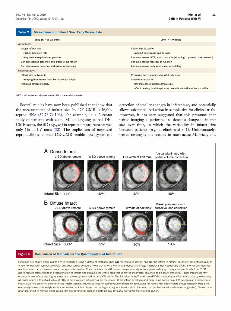
trsCor
daHpsbp
M
L
11JACC Vol. 55, No. 1, 2010 Kim et al.December 29, 2009/January 5, 2010:1–16 CMR in Patients With MI
Several studies have now been published that show thathe measurement of infarct size by DE-CMR is highlyeproducible (32,78,79,106). For example, in a 3-centertudy of patients with acute MI undergoing paired DE-MR scans, the SD (e.g., �e) in repeated measurements wasnly 1% of LV mass (32). The implication of improvedeproducibility is that DE-CMR enables the systematic
A Dense Infarct 2 SD above remote 5 SD above remo
44%* 42%*Infarct Size
2 SD above remote 5 SD above remoB Diffuse Infarct
33%* 5%*Infarct Size
Figure 8 Comparison of Methods for the Quantification of Infar
Examples are shown when infarct size is quantified using 4 different methods whea user to manually contour epicardial and endocardial contours. Note that when thresult in infarct size measurements that are quite similar. When the infarct is diffuabove remote often results in overestimation of infarct size because the entire areunderestimate infarct size if gray zones are incorrectly assumed to be 100% viableall pixels above a threshold value of 50% of the maximum intensity within the infainfarct size. We prefer to planimeter the infarct visually, but will correct for partialume analysis methods weight each voxel within the infarct based on the highest safter user input to remove noise pixels that are beyond the chosen cutoff but are
easurement of Infarct Size: Early Versus LateTable 2 Measurement of Infarct Size: Early Versus Late
Early (<7 to 10 Days)
Advantages
Larger infarct size
Higher detection rate
May reduce required sample size
Can also assess presence and extent of no reflow
Can also assess presence and extent of stunning
Disadvantages
Infarct size is dynamic
Imaging time frame must be narrow (�3 days)
Requires patient stability
VEF � left ventricular ejection fraction; MI � myocardial infarction.
etection of smaller changes in infarct size, and potentiallyllows substantial reduction in sample size for clinical trials.owever, it has been suggested that this presumes that
aired imaging is performed to detect a change in infarctize over time, in which the variability in infarct sizeetween patients (�T) is eliminated (41). Unfortunately,aired testing is not feasible in most acute MI trials, and
Full width at half maxVisual planimetry with
partial volume correction
40%42%
Full width at half max
30%
Visual planimetry with partial volume correction
18%
e
the infarct is dense, and (B) the infarct is diffuse. Currently, all methods requirerct is dense and image intensity is homogeneously bright, the various methodsd image intensity is homogeneously gray, using a simple threshold of 2 SDis gray is incorrectly assumed to be 100% infarcted. Higher thresholds may
full width at half maximum (FWHM) method quantifies infarct size by measuringthe infarct is diffuse and there is no dense core, FWHM can also overestimate
effects by accounting for voxels with intermediate image intensity. Partial vol-ntensity within the infarct or the blood cavity (whichever is greater). *Infarct sizesly not within the infarcted region.
Late (>4 Weeks)
Infarct size is stable
Imaging time frame can be wide
Can also assess LVEF, which is stable (stunning, if present, has resolved)
Can also assess recovery of function
Can also assess early ventricular remodeling
Presumes survival and successful follow-up
Smaller infarct size
May increase required sample size
Infarct healing (shrinkage) may preclude detection of very small MI
te
te
ct Siz
n (A)e infase ana that. The
rct. Ifvolumeignal iobviou
ihtv�Ds
oifmascstFr
sathDM(s(1(ibeDrc
12 Kim et al. JACC Vol. 55, No. 1, 2010CMR in Patients With MI December 29, 2009/January 5, 2010:1–16
nfarct size is only assessed after the treatment being testedas been given (unpaired). Thus, because the variability inrue infarct size among patients is typically larger than theariability added by the infarct sizing method (e.g.,T � �e), it is possible that the improved reproducibility ofE-CMR may not translate into significant reductions in
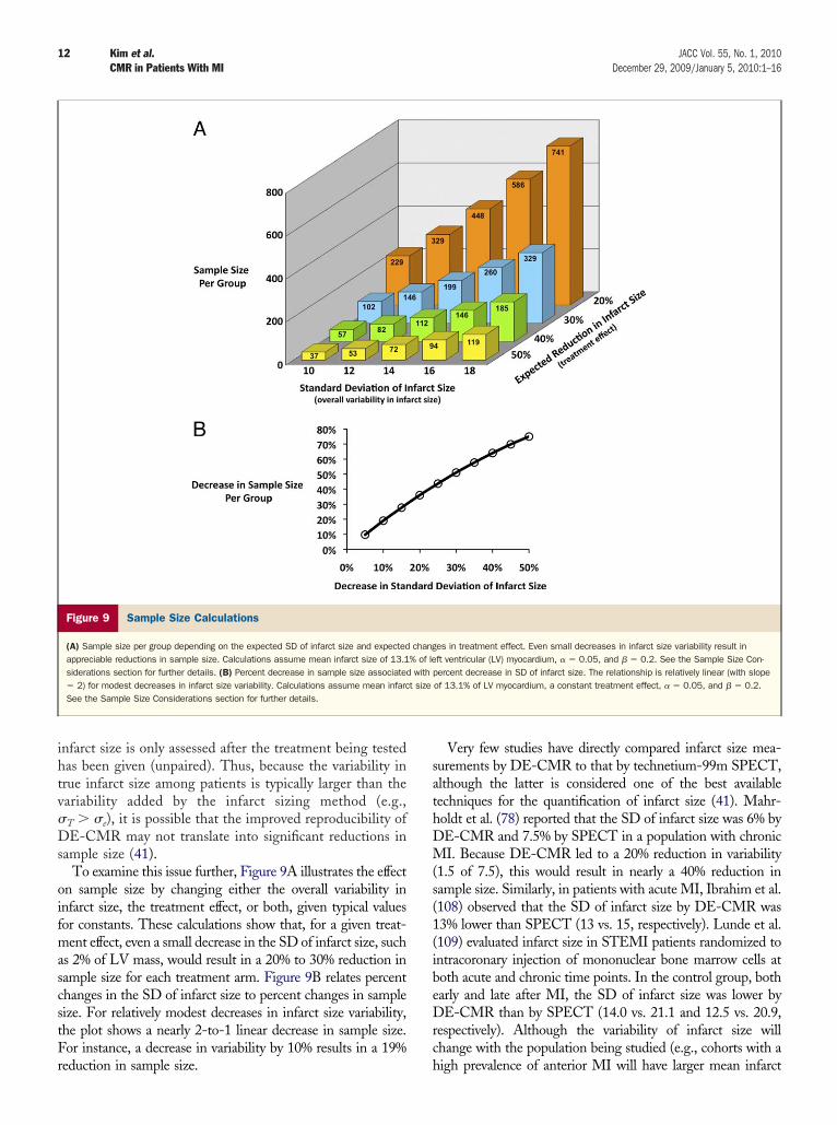
ample size (41).To examine this issue further, Figure 9A illustrates the effect
n sample size by changing either the overall variability innfarct size, the treatment effect, or both, given typical valuesor constants. These calculations show that, for a given treat-ent effect, even a small decrease in the SD of infarct size, such
s 2% of LV mass, would result in a 20% to 30% reduction inample size for each treatment arm. Figure 9B relates percenthanges in the SD of infarct size to percent changes in sampleize. For relatively modest decreases in infarct size variability,he plot shows a nearly 2-to-1 linear decrease in sample size.or instance, a decrease in variability by 10% results in a 19%
Figure 9 Sample Size Calculations
(A) Sample size per group depending on the expected SD of infarct size and expectedappreciable reductions in sample size. Calculations assume mean infarct size of 13.1siderations section for further details. (B) Percent decrease in sample size associated� 2) for modest decreases in infarct size variability. Calculations assume mean infarcSee the Sample Size Considerations section for further details.
eduction in sample size. h
Very few studies have directly compared infarct size mea-urements by DE-CMR to that by technetium-99m SPECT,lthough the latter is considered one of the best availableechniques for the quantification of infarct size (41). Mahr-oldt et al. (78) reported that the SD of infarct size was 6% byE-CMR and 7.5% by SPECT in a population with chronicI. Because DE-CMR led to a 20% reduction in variability
1.5 of 7.5), this would result in nearly a 40% reduction inample size. Similarly, in patients with acute MI, Ibrahim et al.108) observed that the SD of infarct size by DE-CMR was3% lower than SPECT (13 vs. 15, respectively). Lunde et al.109) evaluated infarct size in STEMI patients randomized tontracoronary injection of mononuclear bone marrow cells atoth acute and chronic time points. In the control group, botharly and late after MI, the SD of infarct size was lower byE-CMR than by SPECT (14.0 vs. 21.1 and 12.5 vs. 20.9,
espectively). Although the variability of infarct size willhange with the population being studied (e.g., cohorts with a
es in treatment effect. Even small decreases in infarct size variability result inft ventricular (LV) myocardium, � � 0.05, and � � 0.2. See the Sample Size Con-ercent decrease in SD of infarct size. The relationship is relatively linear (with slope
of 13.1% of LV myocardium, a constant treatment effect, � � 0.05, and � � 0.2.
chang% of lewith p
t size
igh prevalence of anterior MI will have larger mean infarct
ssdfCsidraub
S
IciincrabdCvccnbtfrti
ATBi
RDC
R
13JACC Vol. 55, No. 1, 2010 Kim et al.December 29, 2009/January 5, 2010:1–16 CMR in Patients With MI
ize and larger SD), these data suggest that total variability ismaller when utilizing DE-CMR and will lead to appreciableifferences in sample size. As a result, interest in DE-CMRor infarct size quantification is rapidly growing. When thelinicalTrials.gov registry is searched using the term “infarct
ize,” a total of 110 studies are listed. Of these, 47 do notnvolve acute MI (e.g., cerebral infarction) or do not explicitlyetail the methodology of measuring infarct size. Of theemaining 63 studies, 38 (60%) have incorporated DE-CMRs an end point (110). Additionally, several randomized trialssing infarct size by DE-CMR as a surrogate end point haveeen recently published (109,111–114).
ummary
n patients with known or suspected MI, CMR provides aomprehensive, multifaceted view of the heart. The data,ncluding those from a recent multicenter clinical trial,ndicate that DE-CMR is a well-validated, robust tech-ique that can be easily implemented on scanners that areommonly available worldwide, with an effectiveness thativals the best available imaging techniques for the detectionnd assessment of acute and chronic MI. A DE-CMR maye especially useful when patients present outside theiagnostic window of troponins. Moreover, because DE-MR can uniquely differentiate between ischemic and
arious nonischemic forms of injury, it may be helpful inases of diagnostic uncertainty, such as in patients withlassical features of MI in whom coronary angiography doesot show a culprit lesion. Even after the diagnosis of MI haseen made, CMR can provide clinically relevant informa-ion with regard to identification of post-MI sequelae andurther infarct characterization. The high accuracy andeproducibility of DE-CMR has led to the increasing use ofhis technique as the preferred method for quantification ofnfarct size in many clinical trials.
cknowledgmentshe authors thank Jessica Ngo, BS, and Stephen Darty,SRS, RT-N, MR, for their invaluable assistance in creat-
ng the figures.
eprint requests and correspondence: Dr. Raymond J. Kim,uke Cardiovascular MRI Center, DUMC 3934, Durham, Northarolina 27710. E-mail: [email protected].
EFERENCES
1. Yusuf S, Hawken S, Ounpuu S, et al. Effect of potentially modifiable riskfactors associated with myocardial infarction in 52 countries (theINTERHEART study): case-control study. Lancet 2004;364:937–52.
2. Alpert JS, Thygesen K, Antman E, et al. Myocardial infarctionredefined—a consensus document of The Joint European Society ofCardiology/American College of Cardiology Committee for theredefinition of myocardial infarction. J Am Coll Cardiol 2000;36:959–69.
3. Newby LK, Alpert JS, Ohman EM, et al. Changing the diagnosis of
acute myocardial infarction: implications for practice and clinicalinvestigations. Am Heart J 2002;144:957–80.4. White HD. Things ain’t what they used to be: impact of a newdefinition of myocardial infarction. Am Heart J 2002;144:933–7.
5. Hochholzer W, Buettner HJ, Trenk D, et al. New definition ofmyocardial infarction: impact on long-term mortality. Am J Med2008;121:399–405.
6. Thygesen K, Alpert JS, White HD. Universal definition of myocar-dial infarction. J Am Coll Cardiol 2007;50:2173–95.
7. Kim RJ, Fieno DS, Parrish TB, et al. Relationship of MRI delayedcontrast enhancement to irreversible injury, infarct age, and contrac-tile function. Circulation 1999;100:1992–2002.
8. Wagner A, Mahrholdt H, Holly TA, et al. Contrast-enhanced MRIand routine single photon emission computed tomography (SPECT)perfusion imaging for detection of subendocardial myocardial in-farcts: an imaging study. Lancet 2003;361:374–9.
9. Ricciardi MJ, Wu E, Davidson CJ, et al. Visualization of discretemicroinfarction after percutaneous coronary intervention associatedwith mild creatine kinase-MB elevation. Circulation 2001;103:2780–3.
10. Selvanayagam JB, Porto I, Channon K, et al. Troponin elevation afterpercutaneous coronary intervention directly represents the extent ofirreversible myocardial injury: insights from cardiovascular magneticresonance imaging. Circulation 2005;111:1027–32.
11. Ibrahim T, Bulow HP, Hackl T, et al. Diagnostic value of contrast-enhanced magnetic resonance imaging and single-photon emissioncomputed tomography for detection of myocardial necrosis early afteracute myocardial infarction. J Am Coll Cardiol 2007;49:208–16.
12. Kim RJ, Albert TS, Wible JH, et al. Performance of delayed-enhancement magnetic resonance imaging with gadoversetamidecontrast for the detection and assessment of myocardial infarction: aninternational, multicenter, double-blinded, randomized trial. Circu-lation 2008;117:629–37.
13. Shah DJ, Judd RM, Kim RJ. Technology insight: MRI of the myocar-dium. Nat Clin Pract Cardiovasc Med 2005;2:597–605, quiz 606.
14. Saremi F, Grizzard JD, Kim RJ. Optimizing cardiac MR imaging:practical remedies for artifacts. Radiographics 2008;28:1161–87.
15. Kramer C, Barkhausen J, Flamm S, et al. Standardized cardiovascularmagnetic resonance imaging (CMR) protocols, Society for Cardio-vascular Magnetic Resonance: Board of Trustees Task Force onStandardized Protocols. J Cardiovasc Magn Reson 2008;10:35.
16. Sievers B, Elliott MD, Hurwitz LM, et al. Rapid detection ofmyocardial infarction by subsecond, free-breathing delayed contrast-enhancement cardiovascular magnetic resonance. Circulation 2007;115:236–44.
17. Kaji S, Yang PC, Kerr AB, et al. Rapid evaluation of left ventricularvolume and mass without breath-holding using real-time interactivecardiac magnetic resonance imaging system. J Am Coll Cardiol2001;38:527–33.
18. Abdel-Aty H, Zagrosek A, Schulz-Menger J, et al. Delayed enhance-ment and T2-weighted cardiovascular magnetic resonance imagingdifferentiate acute from chronic myocardial infarction. Circulation2004;109:2411–6.
19. Kim WY, Danias PG, Stuber M, et al. Coronary magnetic resonanceangiography for the detection of coronary stenoses. N Engl J Med2001;345:1863–9.
20. Hendel RC, Patel MR, Kramer CM, et al. ACCF/ACR/SCCT/SCMR/ASNC/NASCI/SCAI/SIR 2006 appropriateness criteria forcardiac computed tomography and cardiac magnetic resonance im-aging: a report of the American College of Cardiology FoundationQuality Strategic Directions Committee Appropriateness CriteriaWorking Group, American College of Radiology, Society of Cardio-vascular Computed Tomography, Society for Cardiovascular Mag-netic Resonance, American Society of Nuclear Cardiology, NorthAmerican Society for Cardiac Imaging, Society for CardiovascularAngiography and Interventions, and Society of Interventional Radi-ology. J Am Coll Cardiol 2006;48:1475–97.
21. Levine GN, Gomes AS, Arai AE, et al. Safety of magnetic resonanceimaging in patients with cardiovascular devices: an American HeartAssociation scientific statement from the Committee on Diagnosticand Interventional Cardiac Catheterization, Council on ClinicalCardiology, and the Council on Cardiovascular Radiology andIntervention: endorsed by the American College of CardiologyFoundation, the North American Society for Cardiac Imaging, andthe Society for Cardiovascular Magnetic Resonance. Circulation
2007;116:2878–91.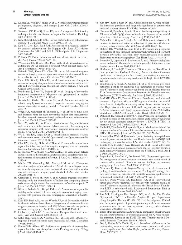
14 Kim et al. JACC Vol. 55, No. 1, 2010CMR in Patients With MI December 29, 2009/January 5, 2010:1–16
22. Kribben A, Witzke O, Hillen U, et al. Nephrogenic systemic fibrosis:pathogenesis, diagnosis, and therapy. J Am Coll Cardiol 2009;53:1621–8.
23. Simonetti OP, Kim RJ, Fieno DS, et al. An improved MR imagingtechnique for the visualization of myocardial infarction. Radiology2001;218:215–23.
24. Kim RJ, Shah DJ, Judd RM. How we perform delayed enhancementimaging. J Cardiovasc Magn Reson 2003;5:505–14.
25. Kim RJ, Choi KM, Judd RM. Assessment of myocardial viabilityby contrast enhancement. In: Higgins CB, Roos AD, editors.Cardiovascular MRI and MRA. Philadelphia, PA: Lippincott,2003:209 –37.
26. Polimeni PI. Extracellular space and ionic distribution in rat ventri-cle. Am J Physiol 1974;227:676–83.
27. Weinmann HJ, Brasch RC, Press WR, et al. Characteristics ofgadolinium-DTPA complex: a potential NMR contrast agent. AJRAm J Roentgenol 1984;142:619–24.
28. Rehwald WG, Fieno DS, Chen EL, et al. Myocardial magneticresonance imaging contrast agent concentrations after reversible andirreversible ischemic injury. Circulation 2002;105:224–9.
29. Fieno DS, Kim RJ, Chen EL, et al. Contrast-enhanced magneticresonance imaging of myocardium at risk: distinction between revers-ible and irreversible injury throughout infarct healing. J Am CollCardiol 2000;36:1985–91.
30. Barkhausen J, Ebert W, Debatin JF, et al. Imaging of myocardialinfarction: comparison of Magnevist and gadophrin-3 in rabbits.J Am Coll Cardiol 2002;39:1392–8.
31. Amado LC, Gerber BL, Gupta SN, et al. Accurate and objectiveinfarct sizing by contrast-enhanced magnetic resonance imaging in acanine myocardial infarction model. J Am Coll Cardiol 2004;44:2383–9.
32. Wagner A, Mahrholdt H, Thomson L, et al. Effects of time, dose,and inversion time for acute myocardial infarct size measurementsbased on magnetic resonance imaging-delayed contrast enhancement.J Am Coll Cardiol 2006;47:2027–33.
33. Saeed M, Weber O, Lee R, et al. Discrimination of myocardial acuteand chronic (scar) infarctions on delayed contrast enhanced magneticresonance imaging with intravascular magnetic resonance contrastmedia. J Am Coll Cardiol 2006;48:1961–8.
34. Wu E, Judd RM, Vargas JD, et al. Visualisation of presence, location,and transmural extent of healed Q-wave and non-Q-wave myocardialinfarction. Lancet 2001;357:21–8.
35. Choi KM, Kim RJ, Gubernikoff G, et al. Transmural extent of acutemyocardial infarction predicts long-term improvement in contractilefunction. Circulation 2001;104:1101–7.
36. Ingkanisorn WP, Rhoads KL, Aletras AH, et al. Gadolinium delayedenhancement cardiovascular magnetic resonance correlates with clin-ical measures of myocardial infarction. J Am Coll Cardiol 2004;43:2253–9.
37. Martin TN, Groenning BA, Murray HM, et al. ST-segmentdeviation analysis of the admission 12-lead electrocardiogram as anaid to early diagnosis of acute myocardial infarction with a cardiacmagnetic resonance imaging gold standard. J Am Coll Cardiol2007;50:1021–8.
38. Giannitsis E, Steen H, Kurz K, et al. Cardiac magnetic resonanceimaging study for quantification of infarct size comparing directlyserial versus single time-point measurements of cardiac troponin T.J Am Coll Cardiol 2008;51:307–14.
39. Klein C, Nekolla SG, Bengel FM, et al. Assessment of myocardialviability with contrast-enhanced magnetic resonance imaging: com-parison with positron emission tomography. Circulation 2002;105:162–7.
40. Kuhl HP, Beek AM, van der Weerdt AP, et al. Myocardial viabilityin chronic ischemic heart disease: comparison of contrast-enhancedmagnetic resonance imaging with (18)F-fluorodeoxyglucose positronemission tomography. J Am Coll Cardiol 2003;41:1341–8.
41. Gibbons RJ, Valeti US, Araoz PA, et al. The quantification of infarctsize. J Am Coll Cardiol 2004;44:1533–42.
42. Katus HA, Remppis A, Neumann FJ, et al. Diagnostic efficiency oftroponin T measurements in acute myocardial infarction. Circulation1991;83:902–12.
43. Kannel WB, Abbott RD. Incidence and prognosis of unrecognizedmyocardial infarction. An update on the Framingham study. N Engl
J Med 1984;311:1144–7.44. Kim HW, Klem I, Shah DJ, et al. Unrecognized non-Q-wave myocar-dial infarction: prevalence and prognostic significance in patients withsuspected coronary disease. PLoS Med 2009;6:e1000057.
45. Uusitupa M, Pyorala K, Raunio H, et al. Sensitivity and specificity ofMinnesota Code Q-QS abnormalities in the diagnosis of myocardialinfarction verified at autopsy. Am Heart J 1983;106:753–7.
46. Mahrholdt H, Wagner A, Parker M, et al. Relationship of contractilefunction to transmural extent of infarction in patients with chroniccoronary artery disease. J Am Coll Cardiol 2003;42:505–12.
47. Hofsten DE, Wachtell K, Lund B, et al. Prevalence and prognosticimplications of non-sustained ventricular tachycardia in ST-segmentelevation myocardial infarction after revascularization with eitherfibrinolysis or primary angioplasty. Eur Heart J 2007;28:407–14.
48. Bonnefoy E, Lapostolle F, Leizorovicz A, et al. Primary angioplastyversus prehospital fibrinolysis in acute myocardial infarction: a ran-domised study. Lancet 2002;360:825–9.
49. Hochman JS, Tamis JE, Thompson TD, et al., for the Global Use ofStrategies to Open Occluded Coronary Arteries in Acute CoronarySyndromes IIb Investigators. Sex, clinical presentation, and outcomein patients with acute coronary syndromes. N Engl J Med 1999;341:226–32.
50. Windhausen F, Hirsch A, Sanders GT, et al. N-terminal pro-brainnatriuretic peptide for additional risk stratification in patients withnon-ST-elevation acute coronary syndrome and an elevated troponinT: an Invasive versus Conservative Treatment in Unstable coronarySyndromes (ICTUS) substudy. Am Heart J 2007;153:485–92.
51. Patel MR, Chen AY, Peterson ED, et al. Prevalence, predictors, andoutcomes of patients with non-ST-segment elevation myocardialinfarction and insignificant coronary artery disease: results from theCan Rapid risk stratification of Unstable angina patients SuppressADverse outcomes with Early implementation of the ACC/AHAGuidelines (CRUSADE) initiative. Am Heart J 2006;152:641–7.
52. Dokainish H, Pillai M, Murphy SA, et al. Prognostic implications ofelevated troponin in patients with suspected acute coronary syndromebut no critical epicardial coronary disease: a TACTICS-TIMI-18substudy. J Am Coll Cardiol 2005;45:19–24.
53. Lindahl B, Diderholm E, Lagerqvist B, et al. Mechanisms behind theprognostic value of troponin T in unstable coronary artery disease: aFRISC II substudy. J Am Coll Cardiol 2001;38:979–86.
54. Kerensky RA, Wade M, Deedwania P, et al. Revisiting the culprit lesionin non-Q-wave myocardial infarction. Results from the VANQWISHtrial angiographic core laboratory. J Am Coll Cardiol 2002;39:1456–63.
55. Echols MR, Mahaffey KW, Banerjee A, et al. Racial differencesamong high-risk patients presenting with non-ST-segment elevationacute coronary syndromes (results from the SYNERGY trial). Am JCardiol 2007;99:315–21.
56. Bugiardini R, Manfrini O, De Ferrari GM. Unanswered questionsfor management of acute coronary syndrome: risk stratification ofpatients with minimal disease or normal findings on coronaryangiography. Arch Intern Med 2006;166:1391–5.
57. Neumann FJ, Kastrati A, Pogatsa-Murray G, et al. Evaluation ofprolonged antithrombotic pretreatment (“cooling-off” strategy) be-fore intervention in patients with unstable coronary syndromes: arandomized controlled trial. JAMA 2003;290:1593–9.
58. Fox KA, Poole-Wilson PA, Henderson RA, et al. Interventionalversus conservative treatment for patients with unstable angina ornon-ST-elevation myocardial infarction: the British Heart Founda-tion RITA 3 randomised trial. Randomized Intervention Trial ofunstable Angina. Lancet 2002;360:743–51.
59. Roe MT, Harrington RA, Prosper DM, et al., for The PlateletGlycoprotein IIb/IIIa in Unstable Angina: Receptor SuppressionUsing Integrilin Therapy (PURSUIT) Trial Investigators. Clinicaland therapeutic profile of patients presenting with acute coronarysyndromes who do not have significant coronary artery disease.Circulation 2000;102:1101–6.
60. Effects of tissue plasminogen activator and a comparison of early invasiveand conservative strategies in unstable angina and non-Q-wave myocar-dial infarction. Results of the TIMI IIIB trial. Thrombolysis in Myo-cardial Ischemia. Circulation 1994;89:1545–56.
61. Dey S, Flather MD, Devlin G, et al. Sex-related differences in thepresentation, treatment and outcomes among patients with acutecoronary syndromes: the Global Registry of Acute Coronary Events.
Heart 2009;95:20–6.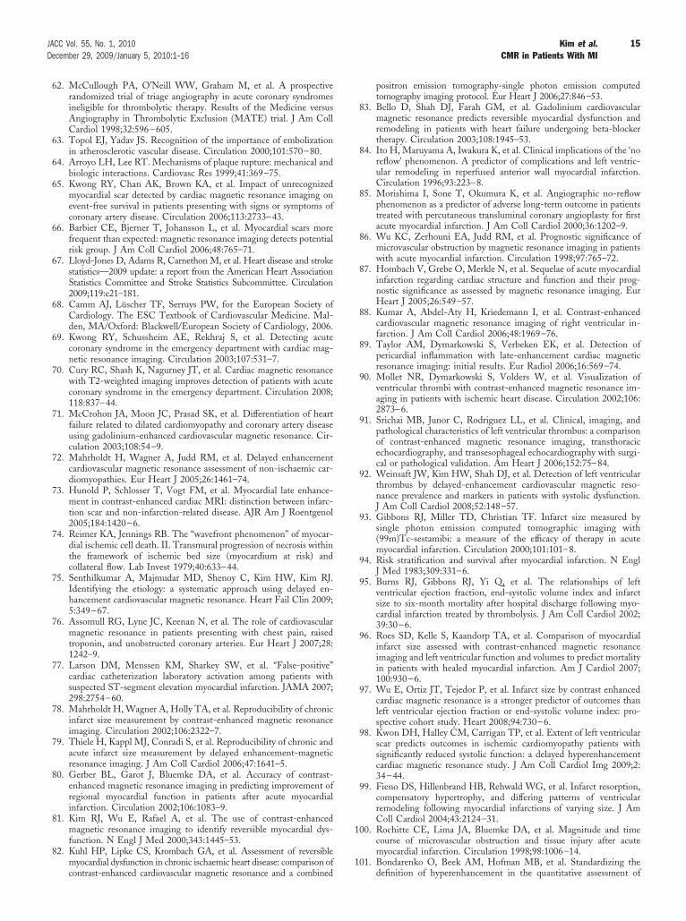
1
1
15JACC Vol. 55, No. 1, 2010 Kim et al.December 29, 2009/January 5, 2010:1–16 CMR in Patients With MI
62. McCullough PA, O’Neill WW, Graham M, et al. A prospectiverandomized trial of triage angiography in acute coronary syndromesineligible for thrombolytic therapy. Results of the Medicine versusAngiography in Thrombolytic Exclusion (MATE) trial. J Am CollCardiol 1998;32:596–605.
63. Topol EJ, Yadav JS. Recognition of the importance of embolizationin atherosclerotic vascular disease. Circulation 2000;101:570–80.
64. Arroyo LH, Lee RT. Mechanisms of plaque rupture: mechanical andbiologic interactions. Cardiovasc Res 1999;41:369–75.
65. Kwong RY, Chan AK, Brown KA, et al. Impact of unrecognizedmyocardial scar detected by cardiac magnetic resonance imaging onevent-free survival in patients presenting with signs or symptoms ofcoronary artery disease. Circulation 2006;113:2733–43.
66. Barbier CE, Bjerner T, Johansson L, et al. Myocardial scars morefrequent than expected: magnetic resonance imaging detects potentialrisk group. J Am Coll Cardiol 2006;48:765–71.
67. Lloyd-Jones D, Adams R, Carnethon M, et al. Heart disease and strokestatistics—2009 update: a report from the American Heart AssociationStatistics Committee and Stroke Statistics Subcommittee. Circulation2009;119:e21–181.
68. Camm AJ, Lüscher TF, Serruys PW, for the European Society ofCardiology. The ESC Textbook of Cardiovascular Medicine. Mal-den, MA/Oxford: Blackwell/European Society of Cardiology, 2006.
69. Kwong RY, Schussheim AE, Rekhraj S, et al. Detecting acutecoronary syndrome in the emergency department with cardiac mag-netic resonance imaging. Circulation 2003;107:531–7.
70. Cury RC, Shash K, Nagurney JT, et al. Cardiac magnetic resonancewith T2-weighted imaging improves detection of patients with acutecoronary syndrome in the emergency department. Circulation 2008;118:837–44.
71. McCrohon JA, Moon JC, Prasad SK, et al. Differentiation of heartfailure related to dilated cardiomyopathy and coronary artery diseaseusing gadolinium-enhanced cardiovascular magnetic resonance. Cir-culation 2003;108:54–9.
72. Mahrholdt H, Wagner A, Judd RM, et al. Delayed enhancementcardiovascular magnetic resonance assessment of non-ischaemic car-diomyopathies. Eur Heart J 2005;26:1461–74.
73. Hunold P, Schlosser T, Vogt FM, et al. Myocardial late enhance-ment in contrast-enhanced cardiac MRI: distinction between infarc-tion scar and non-infarction-related disease. AJR Am J Roentgenol2005;184:1420–6.
74. Reimer KA, Jennings RB. The “wavefront phenomenon” of myocar-dial ischemic cell death. II. Transmural progression of necrosis withinthe framework of ischemic bed size (myocardium at risk) andcollateral flow. Lab Invest 1979;40:633–44.
75. Senthilkumar A, Majmudar MD, Shenoy C, Kim HW, Kim RJ.Identifying the etiology: a systematic approach using delayed en-hancement cardiovascular magnetic resonance. Heart Fail Clin 2009;5:349–67.
76. Assomull RG, Lyne JC, Keenan N, et al. The role of cardiovascularmagnetic resonance in patients presenting with chest pain, raisedtroponin, and unobstructed coronary arteries. Eur Heart J 2007;28:1242–9.
77. Larson DM, Menssen KM, Sharkey SW, et al. “False-positive”cardiac catheterization laboratory activation among patients withsuspected ST-segment elevation myocardial infarction. JAMA 2007;298:2754–60.
78. Mahrholdt H, Wagner A, Holly TA, et al. Reproducibility of chronicinfarct size measurement by contrast-enhanced magnetic resonanceimaging. Circulation 2002;106:2322–7.
79. Thiele H, Kappl MJ, Conradi S, et al. Reproducibility of chronic andacute infarct size measurement by delayed enhancement-magneticresonance imaging. J Am Coll Cardiol 2006;47:1641–5.
80. Gerber BL, Garot J, Bluemke DA, et al. Accuracy of contrast-enhanced magnetic resonance imaging in predicting improvement ofregional myocardial function in patients after acute myocardialinfarction. Circulation 2002;106:1083–9.
81. Kim RJ, Wu E, Rafael A, et al. The use of contrast-enhancedmagnetic resonance imaging to identify reversible myocardial dys-function. N Engl J Med 2000;343:1445–53.
82. Kuhl HP, Lipke CS, Krombach GA, et al. Assessment of reversiblemyocardial dysfunction in chronic ischaemic heart disease: comparison of
contrast-enhanced cardiovascular magnetic resonance and a combinedpositron emission tomography-single photon emission computedtomography imaging protocol. Eur Heart J 2006;27:846–53.
83. Bello D, Shah DJ, Farah GM, et al. Gadolinium cardiovascularmagnetic resonance predicts reversible myocardial dysfunction andremodeling in patients with heart failure undergoing beta-blockertherapy. Circulation 2003;108:1945–53.
84. Ito H, Maruyama A, Iwakura K, et al. Clinical implications of the ‘noreflow’ phenomenon. A predictor of complications and left ventric-ular remodeling in reperfused anterior wall myocardial infarction.Circulation 1996;93:223–8.
85. Morishima I, Sone T, Okumura K, et al. Angiographic no-reflowphenomenon as a predictor of adverse long-term outcome in patientstreated with percutaneous transluminal coronary angioplasty for firstacute myocardial infarction. J Am Coll Cardiol 2000;36:1202–9.
86. Wu KC, Zerhouni EA, Judd RM, et al. Prognostic significance ofmicrovascular obstruction by magnetic resonance imaging in patientswith acute myocardial infarction. Circulation 1998;97:765–72.
87. Hombach V, Grebe O, Merkle N, et al. Sequelae of acute myocardialinfarction regarding cardiac structure and function and their prog-nostic significance as assessed by magnetic resonance imaging. EurHeart J 2005;26:549–57.
88. Kumar A, Abdel-Aty H, Kriedemann I, et al. Contrast-enhancedcardiovascular magnetic resonance imaging of right ventricular in-farction. J Am Coll Cardiol 2006;48:1969–76.
89. Taylor AM, Dymarkowski S, Verbeken EK, et al. Detection ofpericardial inflammation with late-enhancement cardiac magneticresonance imaging: initial results. Eur Radiol 2006;16:569–74.
90. Mollet NR, Dymarkowski S, Volders W, et al. Visualization ofventricular thrombi with contrast-enhanced magnetic resonance im-aging in patients with ischemic heart disease. Circulation 2002;106:2873–6.
91. Srichai MB, Junor C, Rodriguez LL, et al. Clinical, imaging, andpathological characteristics of left ventricular thrombus: a comparisonof contrast-enhanced magnetic resonance imaging, transthoracicechocardiography, and transesophageal echocardiography with surgi-cal or pathological validation. Am Heart J 2006;152:75–84.
92. Weinsaft JW, Kim HW, Shah DJ, et al. Detection of left ventricularthrombus by delayed-enhancement cardiovascular magnetic reso-nance prevalence and markers in patients with systolic dysfunction.J Am Coll Cardiol 2008;52:148–57.
93. Gibbons RJ, Miller TD, Christian TF. Infarct size measured bysingle photon emission computed tomographic imaging with(99m)Tc-sestamibi: a measure of the efficacy of therapy in acutemyocardial infarction. Circulation 2000;101:101–8.
94. Risk stratification and survival after myocardial infarction. N EnglJ Med 1983;309:331–6.
95. Burns RJ, Gibbons RJ, Yi Q, et al. The relationships of leftventricular ejection fraction, end-systolic volume index and infarctsize to six-month mortality after hospital discharge following myo-cardial infarction treated by thrombolysis. J Am Coll Cardiol 2002;39:30–6.
96. Roes SD, Kelle S, Kaandorp TA, et al. Comparison of myocardialinfarct size assessed with contrast-enhanced magnetic resonanceimaging and left ventricular function and volumes to predict mortalityin patients with healed myocardial infarction. Am J Cardiol 2007;100:930–6.
97. Wu E, Ortiz JT, Tejedor P, et al. Infarct size by contrast enhancedcardiac magnetic resonance is a stronger predictor of outcomes thanleft ventricular ejection fraction or end-systolic volume index: pro-spective cohort study. Heart 2008;94:730–6.
98. Kwon DH, Halley CM, Carrigan TP, et al. Extent of left ventricularscar predicts outcomes in ischemic cardiomyopathy patients withsignificantly reduced systolic function: a delayed hyperenhancementcardiac magnetic resonance study. J Am Coll Cardiol Img 2009;2:34–44.
99. Fieno DS, Hillenbrand HB, Rehwald WG, et al. Infarct resorption,compensatory hypertrophy, and differing patterns of ventricularremodeling following myocardial infarctions of varying size. J AmColl Cardiol 2004;43:2124–31.
00. Rochitte CE, Lima JA, Bluemke DA, et al. Magnitude and timecourse of microvascular obstruction and tissue injury after acutemyocardial infarction. Circulation 1998;98:1006–14.
01. Bondarenko O, Beek AM, Hofman MB, et al. Standardizing the
definition of hyperenhancement in the quantitative assessment of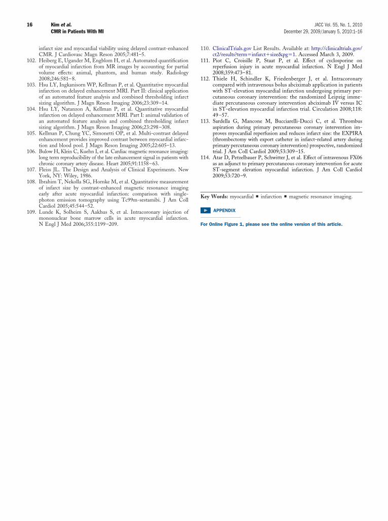
1
1
1
1
1
1
1
1
1
1
1
1
1
K
16 Kim et al. JACC Vol. 55, No. 1, 2010CMR in Patients With MI December 29, 2009/January 5, 2010:1–16
infarct size and myocardial viability using delayed contrast-enhancedCMR. J Cardiovasc Magn Reson 2005;7:481–5.
02. Heiberg E, Ugander M, Engblom H, et al. Automated quantificationof myocardial infarction from MR images by accounting for partialvolume effects: animal, phantom, and human study. Radiology2008;246:581–8.
03. Hsu LY, Ingkanisorn WP, Kellman P, et al. Quantitative myocardialinfarction on delayed enhancement MRI. Part II: clinical applicationof an automated feature analysis and combined thresholding infarctsizing algorithm. J Magn Reson Imaging 2006;23:309–14.
04. Hsu LY, Natanzon A, Kellman P, et al. Quantitative myocardialinfarction on delayed enhancement MRI. Part I: animal validation ofan automated feature analysis and combined thresholding infarctsizing algorithm. J Magn Reson Imaging 2006;23:298–308.
05. Kellman P, Chung YC, Simonetti OP, et al. Multi-contrast delayedenhancement provides improved contrast between myocardial infarc-tion and blood pool. J Magn Reson Imaging 2005;22:605–13.
06. Bulow H, Klein C, Kuehn I, et al. Cardiac magnetic resonance imaging:long term reproducibility of the late enhancement signal in patients withchronic coronary artery disease. Heart 2005;91:1158–63.
07. Fleiss JL. The Design and Analysis of Clinical Experiments. NewYork, NY: Wiley, 1986.
08. Ibrahim T, Nekolla SG, Hornke M, et al. Quantitative measurementof infarct size by contrast-enhanced magnetic resonance imagingearly after acute myocardial infarction: comparison with single-photon emission tomography using Tc99m-sestamibi. J Am CollCardiol 2005;45:544–52.
09. Lunde K, Solheim S, Aakhus S, et al. Intracoronary injection ofmononuclear bone marrow cells in acute myocardial infarction.
N Engl J Med 2006;355:1199–209. F10. ClinicalTrials.gov List Results. Available at: http://clinicaltrials.gov/ct2/results?term�infarct�size&pg�1. Accessed March 3, 2009.
11. Piot C, Croisille P, Staat P, et al. Effect of cyclosporine onreperfusion injury in acute myocardial infarction. N Engl J Med2008;359:473–81.
12. Thiele H, Schindler K, Friedenberger J, et al. Intracoronarycompared with intravenous bolus abciximab application in patientswith ST-elevation myocardial infarction undergoing primary per-cutaneous coronary intervention: the randomized Leipzig imme-diate percutaneous coronary intervention abciximab IV versus ICin ST-elevation myocardial infarction trial. Circulation 2008;118:49 –57.
13. Sardella G, Mancone M, Bucciarelli-Ducci C, et al. Thrombusaspiration during primary percutaneous coronary intervention im-proves myocardial reperfusion and reduces infarct size: the EXPIRA(thrombectomy with export catheter in infarct-related artery duringprimary percutaneous coronary intervention) prospective, randomizedtrial. J Am Coll Cardiol 2009;53:309–15.
14. Atar D, Petzelbauer P, Schwitter J, et al. Effect of intravenous FX06as an adjunct to primary percutaneous coronary intervention for acuteST-segment elevation myocardial infarction. J Am Coll Cardiol2009;53:720–9.
ey Words: myocardial y infarction y magnetic resonance imaging.
APPENDIX
or Online Figure 1, please see the online version of this article.