cardiovascular pathology - semmelweis...
TRANSCRIPT

Cardiovascular
Pathology
- coronary sclerosis,
diseases of the
myocardium -
Semmelweis University
2nd Department of Pathology
_______ _______
2017/2018 – Autumn Semester
Tibor Glasz MD PhD
_______ _______

Pathology of the
coronary arteries ____________ ____________

The system of coronary arteries - Anatomy -
- subepicardial main branches
- epicardial side branches
- intramural small vessels
- collateral arteries
Positional variations: intramurally running segments of the
subepicardial large coronary arteries through formation of so-called
muscle-bridges

http://images.md
Collateral vessel communications - experimental corrosion specimen of a canine heart -

Coronary stenosis or occlusion - Causes -
- up to 80-90% atherosclerosis, atherosclerotic plaque
- less frequently: plaque hemorrhage
- thrombosis
- embolus
- very rarely: congenital developmental anomalies (coronary
hypoplasia, anomalies of coronary anatomy – e.g. right coronary
artery going out from the left side of the aortic wall)
- inflammations
- autoimmune diseases
- muscular spasms of the coronary wall (Prinzmetal’s ‘variant’
angina)
- coronary kinking
- muscle bridges

- central coronary artery disease
- very good bypassing results
- peripheral coronary artery disease
- small-vessel-disease, no bypassing possible
- diffuse coronary artery disease
- the most frequent form, bypassing offers only limited
success
Coronary arteriosclerosis - according to extension within a coronary artery -

- Arteriosclerosis on a single coronary artery: one-vessel-disease
- clinically anginal pain frequently localized to the thorax,
with a history of not longer than 3 years
- Arteriosclerosis on two coronary arteries: two-vessel-disease
- Arteriosclerosis on three coronary arteries: three-vessel-disease
- clinically anginal pain frequently radiating into the arms,
neck and dorsal parts, with a history of longer than 3 years
Coronary arteriosclerosis - according to coronary artery topography -

- low-grade coronary arteriosclerosis: stenosis < 50% of the native
lumen
- intermediate coronary arteriosclerosis: stenosis > 50%, but <75%
of the native lumen
- ECG in rest yet normal
- high-grade coronary arteriosclerosis: stenosis >75% of the native
lumen
- ECG on exercise is abnormal: coronary heart failure
Coronary arteriosclerosis - according to severeness of the disease: grades -

fibrous
plaque
residual
lumen


Coronary plaques, the problem of instability - Bernoulli’s law and the ‘steal’-phenomenon -
- stable plaques: high %-age of connective tissue: fibrous plaque
- unstable plaques: high %-age of lipid substances: atheroma
- at stenotic segments: increased blood flow speed causes decreased
intraluminary pressure (Bernoulli’s phenomenon) – suction effect on
stenosed vessel parts
- poststenotically: slower blood flow, higher hydrostatic pressure
- and yet: flexibility differences between sclerotic-rigid and
neighbouring elastic wall segments (so-called wind-kettle function
of arteries)
- mechanic overload on plaque periphery: dissection, plaque rupture
and hemorrhage
Restitution of the coronary flow
- conservative (medication based) therapy
- invasive:
- angioplasty,
- endarterectomy
- stent implantation
- bypass operation (open heart intervention: CABG – coronary
artery bypass grafting)
} PCI – percutaneous coronary
interventions


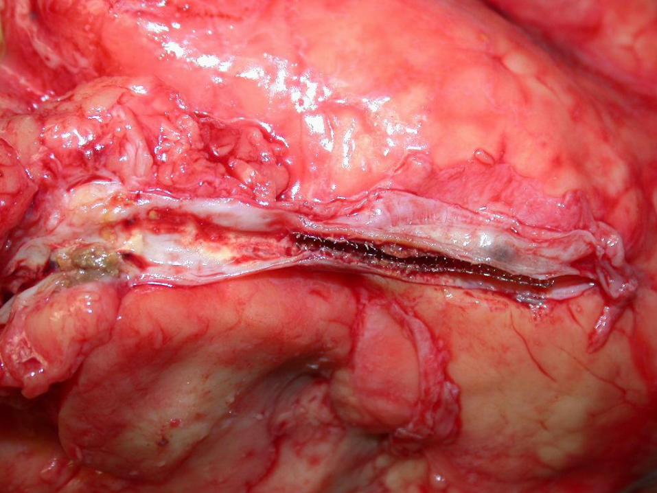
Photography by T. Glasz MD;
Semmelweis University, 2nd Dept. of Pathology

Photography by T. Glasz MD;
Semmelweis University, 2nd Dept. of Pathology

Photography by T. Glasz MD;
Semmelweis University, 2nd Dept. of Pathology

Ischemic heart disease ______________ _______________
Ischemic heart disease - Definition -
Ischemic heart disease is the general term for clinico-pathologic
appearances that develop on insufficient oxygen supply to
myocardium and consequent myocardial damage.
Formerly known as coronary heart disease.

Causes of ischemic heart disease - Causes of insufficient myocardial oxygen supply -
A. Alteration of coronary arteries
1. Diseases of the subepicardial main branches
- stenosing atherosclerotic plaque (over 90%!)
- complicated plaque (hemorrhage or usuration+thrombosis)
- arterial dissection
- embolus
- so-called ‘steal’-syndrome (e.g. myocardial arteriovenous malfor-
mations; coronary-subclavian-steal-syndrom after coronary bypass
grafting /CABG/ by means of the internal mammary artery /IMA/)
2. Diseases of the intramyocardial small vessels
- so-called small-vessel-disease (diabetes mellitus, amyloidosis, etc.)
- DIC, thrombocyte aggregation
- arterial spasm of small vessels
- perivascular fibrosis

B. Extracoronary causes of a relative myocardial oxygen deficiency
(relative coronary insufficiencies)
- muscular hypertrophy (heart weight > 500g)
- aortal or aortic valve stenosis
- anemia
- shock
- diminished pO2 in the air
- pneumonia
- enhanced myocardial oxygen demand of any kind (e.g.
physical excerise)
Causes of ischemic heart disease - Causes of insufficient myocardial oxygen supply -

Clinico-pathologic syndroms
of ischemic heart disease
1. Angina pectoris
2. Myocardial infarction (‘heart attack’)
3. Sudden cardiac death
4. Chronic ischemic heart disease
(so-called ischemic cardiomyopathy)

Angina pectoris
- first described by Heberden in 1768
- definition: variably strong, thoracally based attacks of pain of
cardial origin
- primarily a clinical syndrome without corresponding macro- o.
micromorphologic alterations
- stable angina: pain attacks on body exercise; generally there is a
high-grade stenosing subepicardial coronary arteriosclerosis, where
well developped intercoronary collaterals can yet prevent an
infarction
- unstable angina: precursor of infarction; pain attacks also in rest
- Prinzmetal’s angina: described by Prinzmetal in 1959; pain attacks
on muscular spasms of variably sclerotic coronary vessels;
complaints are experienced in rest and are limited in time
Sudden cardiac death
- definition: death within 1 hour after onset of symptoms in an appa-
rently healthy or chronicly diseased individual
- incidency: 30 deaths / week / 1 million persons in the western world
- in the background there is very often a high-grade coronary arteri-
osclerosis (75%)
- further causes: extracoronary functional heart disease (cca. 20%)
such as electromechanic instability (sick sinus disease, AV-node
disease, WPW-syndrome, ventricular flutter, bradycardia) or structural
abnormalities (vitia, cardiomyopathies, infectiv endocarditides,
myocarditides, dysfunction of valvular prostheses, heart wall rupture).
In 5% of the cases no detectable cause of death.
- histologically coagulation myocytolyses in up to 67% >> pathogene-
tic role of sympatico-adrenal hyperactivity, stress situations, physical
exercise

Adams-Stokes syndrome
- definition: sudden cardiac arrest
- symptomatically quick onset of weakness, dizziness (vertigo), col-
lapse
- physically: no systole, no pulse, no heart sounds, circulatory arrest,
quick loss of blood pressure, tonal-clonal cramps >> exitus letalis
- chances of survival depend on length of the critical situation
- a few seconds: ‘forme fruste’ with slight vertigo
- 10 seconds: facial paleness, collapse, muscular clonus, pupils
dilated
- 20-40 seconds: tonal cramps, cyanosis, loss of control on
sphincter function (miction, defecation)
- over 3 minutes: chances of survival very thin

- cause: primarily ischemic heart disease, especially acute myocardial
infarction >> ventricular fibrillation
- further causes: cardiac (rheumatic myocarditis, cardiomyopathy,
cardiac tumours) and non-cardiac (metabolic and/or electrolite
alterations, medicaments, thyreotoxicosis, carotic sinus-hyperesthesia)
- arrhythmias causing an Adams-Stokes syndrome (so-called electric
catastrophies):
- hypodynamic alterations: complete AV-block, sinus bradycardia
with partial AV-block
- hyperdynamic alterations: ventricular fibrillation (a dreaded
complication of a myocardial infarction), paroxysmal ventricular
tachycardia
Adams-Stokes syndrome

Chronic ischemic heart disease
(so-called ischemic cardiomyopathy)
- direct cause of death in 40% of all ischemic forms of heart disease
- definition: chronic ischemic damage of myocardium
- causes: severe coronary arteriosclerosis, muscular cardiac
hypertrophy (>500g), aortic hypoplasia, coronary hypoplasia >>
long-term myocardial hypoxia
- macroscopy: atrophy, normotrophy, hypertrophy equally possible
- microscopy: variable picture with atrophic and compensationally
hypertrophic muscle cells, microinfarctions (< 10mm), focal fibroses,
interstitial fibrosis, myocytolytic foci – especially subendocardially

- Forms of cellular damage of cardiac muscle : coagulation necrosis
and single cell necroses (so-called myocytolyses)
- Coagulation necrosis : seen in myocardial infarction; atonic death of
muscle fibers (irreversible relaxation) >> fiber lengthening and
meandering reactive to intraventricular pressure: so-called wavy fibers
- Liquefaction myocytolysis: disappearance of the sarcoplasm with
retained reticular skeleton of the myocardium >> no inflammatory
reaction >> scarring through collapse and condensation of residual
sarcolemmata and reticular fibers
- Coagulation myocytolysis: tetanic cell death on metabolic basis;
cellular necrosis in irreversible contraction; in pheochromocytoma,
after heart transplantation, electric shock
Chronic ischemic heart disease
(so-called ischemic cardiomyopathy)

Myocardial infarction ______________ _______________

Myocardial infarction
Definition
- acute: myocardial necrosis localized to a circumscribed area of
coronary blood supply, resulting from an acute ischemic insult
exceeding the ischemic reserve capacities of the heart muscle tissue.
- chronic: cardiac muscular scarring over 1 cm in diameter
Classification according to extension
- regional extension
- transmural versus subendocardial
Epidemiology
- in the western world myocardial infarction is typically a
disease of the 8th-9th decade of life
- dramatic difference in Hungary: myocardial infarction is
typical under the age of 65! (hungarian data for the
prevalence and mortality by myocardial infarction are 2.5x
greater than those in the EU)
- women suffer a myocardial infarction 8 years later than
men

Myocardial infarction – Causes –
Causes of infarction are causes of the acute ischemic insult
- high-grade coronary arteriosclerosis (especially the so-called
unstable plaques with complication: rupture, hemorrhage)
- coronary arterial dissection
- coronary embolism (very rarely)
- intermediate coronary arteriosclerosis with relative hypoxic periods
(pneumonia, body exercise, low pO2 of air)
- extreme stress

Photography by T. Glasz MD;
Semmelweis University, 2nd Dept. of Pathology

Transmural myocardial infarction
- Infarction through the entire thickness of the myocardial wall: from
endocardium to epicardium/subepicardial fat tissue
- 5 possible localisations:
- anterior
- posterior
- lateral
- septal
- circular

Anterior transmural myocardial infarction
- cause is an occlusive insult in the left anterior descending (LAD)
branch
- 40-50% of all infarctions
- most frequent localisation: apical part of the anterior wall and
ventral two-thirds of the septum

Posterior transmural myocardial infarction
- cause is an occlusive insult in the right coronary artery (RCA), or
in the circumflex artery (RCX)
- 20-30% of all infarctions
- infarctions are especially large, if the dominant artery is the RCA
- topographical extension: posterior wall of the left ventricle, dorsal
third of the septum, sometimes the paraseptal part of the right
ventricular posterior wall

Lateral transmural myocardial infarction
- cause is an occlusive insult in the circumflex artery (RCX),or in the
marginal artery
- 15-20% of all infarctions
Circular transmural myocardial infarction
- cause is synchronic or subsequent occlusive insults in possibly 2-3
main branches within the time frame of a few hours
- very rare
- topographical extension: 60-75% of the left ventricular
myocardium

Septal transmural myocardial infarction
- cause is an occlusive insult in the coronary artery branch
responsible for the supply of the affected myocardial area
- very rare
- yet a minute focus of an infarction in this area can be lethal, if
localized on the innervation conducting system (esp. upper third of
the muscular septum) >> complete electro-mechanical block >> so-
called malignant arrhythmias

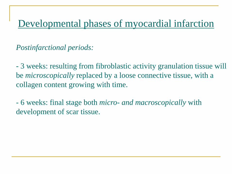
Developmental phases of myocardial infarction
Postinfarctional periods:
- 5-6 hours: microscopically swelling of fibers with intact striation.
Nuclei pale, swollen, ruptured, lobulated. Interstitium yet intact.
Macroscopically no visible alterations. Clinically rescue manoeuvers
for saving the necrobiotic (dying) myocardium yet possible (e.g.
thrombolytic therapy)
- 15 hours: microscopically lengthening, thinning and meandering of
necrotic muscle fibers. Macroscopically the first alterations appear :
Paling and slight swelling of the affected myocardial area.


Postinfarctional periods:
- 36 hours: macroscopically are the central infarction areas already
slightly yellowish. Peripherally develops a hemorrhagic zone of
demarcation.
- 3-4 days: macroscopically is the infarcted area clayish yellow with
geographically irregular periphery and hemorrhagic demarcation.
Synchronously (1-5. postinfarction day) the cytoplasmic striation of
the myocytes disappear under the microscope with accompanying
homogenesation and hypereosinophilia. Nuclear decoloration
progresses. The interstitial connective tissue fibers undergo
fragmentation. Infiltration of growing numbers of granulocytic
inflammatory cell elements.
Developmental phases of myocardial infarction


Postinfarctional periods:
- 1 week: microscopically phagocytosis of the necrotic tissue begins
with gathering of macrophages and fibroblasts. Highest danger of a
myocardial rupture!
- 2 weeks: microscopical signs of reparation develop with laterally
beginning granulation. Granulocytes disappear to give place for
lymphocytes, plasmacells and eosinophils. At the same time (10.
postinfarction day) the infarction zone turns macroscopically greyish
with fading demarcation and signs of shrinking.
Developmental phases of myocardial infarction

Postinfarctional periods:
- 3 weeks: resulting from fibroblastic activity granulation tissue will
be microscopically replaced by a loose connective tissue, with a
collagen content growing with time.
- 6 weeks: final stage both micro- and macroscopically with
development of scar tissue.
Developmental phases of myocardial infarction


Paradox myocardial infarction
- Definition: acute myocardial infarction, that with respect to its age
does not correspond to the occlusion of its relevant supplying artery
(supplying artery with chronic occlusion, whereas acute infarction in
the corresponding myocardial area)
- Prerequisite anatomy: well functioning collateral communications
between supplied myocardial areas.
- Mechanism of pathogenesis: blood supply of a myocardial area of a
coronary artery with chronic occlusion is done by collaterals from
one of the neighbouring areas >> acute occlusion (e.g. thrombosis)
of the supplying artery of the neighbouring area >> acute
myocardial infarction of the area with chronic coronary occlusion

Progressiv myocardial infarction
- Definition: acute myocardial infarction, developping peripheral to
a chronic infarction area
- Mechanism of pathogenesis : derangement of the perifocal
(periinfarctional) microcirculation, or backward growing of the
coronary thrombosis >> growing extension of the infarction with
always newly involved infarction zones >> so-called wavefront-
phenomenon

Subendocardial myocardial infarction
- Definition: a myocardial infarction limited to the inner (subendo-
cardial) third of the ventricular wall
- the subendocardium represents a ‘strategic zone’ of the
myocardium: highest interstitial pressure within the whole
myocardium >> microcirculation is periodical: only possible during
diastole, whereas capillary blood supply ceases during systole >>
limited oxygenisation reserves of subendocardium
- in coronary heart disease the subendocardium is the most
vulnerable area

- microscopically: patchy picture with microinfarctions as large as
10 mm, liquefaction- and coagulation myocytolyses beside normal
myocytes
- macroscopically: geographically irregular, partially fading
peripheral zones
- reparation already in the 1. postinfarction week and from the richly
vascularised granulation tissue quickly develops the subepicardial
scarring
- by means of a wavefront-mechanism out of a subendocardial
infarction can develop a transmural one.
Subendocardial myocardial infarction
htt
p:/
/im
ag
es.m
d
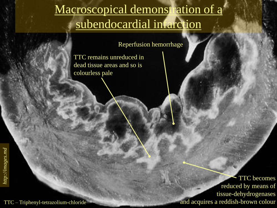
Macroscopical demonstration of a
subendocardial infarction
TTC becomes
reduced by means of
tissue-dehydrogenases
and acquires a reddish-brown colour
TTC remains unreduced in
dead tissue areas and so is
colourless pale
Reperfusion hemorrhage
TTC – Triphenyl-tetrazolium-chloride

Complications of a myocardial infarction:
left heart failure and cardiogenic shock
- failure of the left ventricular pump function from a slight
congestion through severe circulatory insufficiency to a cardiogenic
shock
- background: electro-mechanic dissociation (electric catastrophy –
yet with a minute infarction in the upper septum!); ventricular
fibrillation; with widely extended infarction there remains only an
insufficient quantity of working musculature >> an infarction
involving at least 40% of the left ventricular myocardium causes
cardiogenic shock


- vitious circle of cardiogenic shock: pump insufficiency with lower
circulatory volume >> enhanced sympathetic tonus >> systemic
vasoconstriction >> further hemodynamic overload of the heart
- clinical appearance: dyspnoe; pulmonary edema to the extreme of
an asthma cardiale; drop of blood pressure; skin paleness with ‘cold
sweat’; acrocyanosis; oliguria; derangement of cerebral functions
- cardiogenic shock develops in some 10% of patients with infarc-
tion and is generally a complication of the 2. or 3. subsequent heart
attack
- mortality of cardiogenic shock is dramatically high: 80% with, and
90-100% without therapy
Complications of a myocardial infarction:
left heart failure and cardiogenic shock

- Definition: variable degree of ventricular wall thinning and
sacculation
- Frequency: develops in 15% of patients with infarction
- Background: infarcerated myocardium or postinfarctional scar
tissue represents low mechanic resistence >> gradual outbulging
(sacculation) of the affected area >> ventricular aneurysm
- acute ventricular aneurysm: in the acute phase of infarction
(during the 1. postinfarctional week, in the phase of dead tissue
degradation by means of macrophages) >> often gives rise to a
ventricular wall rupture!
- chronic ventricular aneurysm: after scarring of the infarcerated
myocardial area (after months to years) – practically doesn’t rupture
Complications of a myocardial infarction:
left ventricular aneurysm

- derangements of myocardial motion capacities after infarction:
- hypokinesis (impairment of muscular wall motility)
- akinesis (complete arrest of myocardial movements in the
affected area)
- paradox pulsation – typical for ventricular wall aneurysms
with large cavity comperable to the remaining lumen of the
ventricle and a passive motion reversed to normal parietal
pulsation >> blood volume oscillating between ventricular
space and aneurysmal lumen >> enhanced work overload of
the already damaged myocardium and further limitation of
coronary blood flow >> left ventricular decompensation
- in the aneurysmal sack: thrombosis (50% of the cases) >> danger
of embolisation (in 5% of cases)
Complications of a myocardial infarction:
left ventricular aneurysm


Chronic left ventricular aneurysm
Scarred
aneurysmal
sack

Chronic left ventricular
aneurysm
Left
ventricular
space
Residual
myocardium
Scarred, thin,
transparent
wall of the
aneurysmal
sack

- frequency: 1-2% of all infarctions; in the 2-4. postinfarction day
- drasticly progressing clinical picture: anginal pain attack,
hypotension, acute heart failure (possibly cardiogenic shock)
- hemodynamically: septal defect with a left-to-right shunt >> the
left sided heart failure is quickly followed by a right heart failure.
Furthermore, part of the oxygenated blood escapes to the right heart
without supplying the coronary system. In complicated cases the
pump function is further compromized by a damage of the
conduction system >> severe arrhythmias or ventricular fibrillation.
Complications of a myocardial infarction:
septal rupture and defect


- papillary muscles are from the metabolic, pathophysiologic and
pathologic point of view part of the subendocardium
- functional derangement of the papillary muscles is to be detected
already during attacks of angina pectoris
- in infarction: definitive damage of papillary muscle function, most
frequently on the 2-7. postinfarction day
- rupture of papillary muscle or that of cordae is a life threatening
situation >> sudden, severe valvular vitium and cardiac failure >>
emergency implantation of a valve prosthesis is mandatory
Complications of a myocardial infarction:
papillary muscle dysfunction, rupture of cordae


- frequency: 10-20% of all lethal infarction cases; most frequently on
the 3-5. postinfarction day
- complications: pericardial tamponade >> electro-mechanic
dissociation >> sudden cardiac death
- small rupture results in slowly progressive development of a
tamponade >> emergency surgery is yet possible
- sometimes introductory event is the formation of an acute myo-
cardial aneurysm
- predisposing factors for cardiac wall rupture: transmural
infarction; high age; female sex; steroide therapy; hypertension;
few collaterals; myocardial fibrosis
Complications of a myocardial infarction:
muscular wall rupture, pericardial tamponade


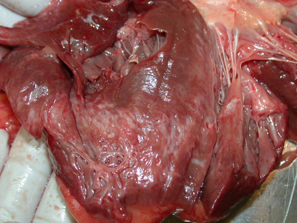
Myocardial rupture,
pericardial
tamponade

Rupture
line
Pericardial
hemorrhage
Myocardial
rupture,
pericardial
tamponade

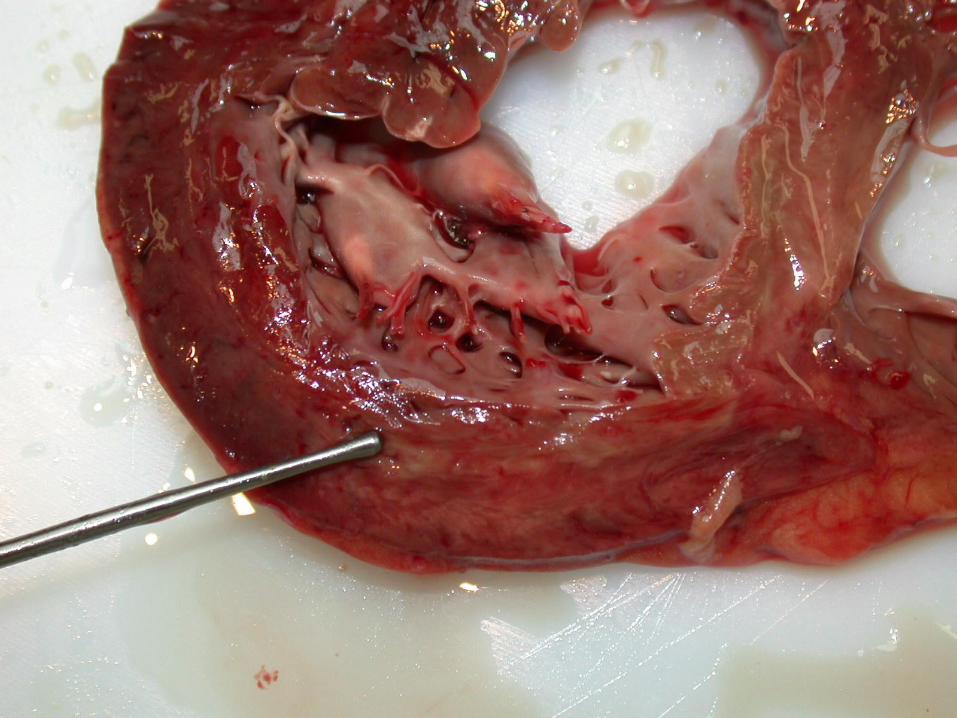
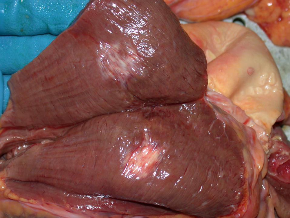
Acute myocardial infarction. Myocardial rupture.
Pale zones of
infarction
Ruptured
areas


Cardiomyopathies ____________ ____________

Cardiomyopathies
Definitions:
- primary (idiopathic) cardiomyopathies: progressive chronic myocardial
failure of unknown origin that after variously long periods lead to a therapy
resistant circulatory insufficiency.
- cardiopathies or secondary cardiomyopathies: progressive, diffuse
myocardial diseases, that may be identical to the idiopathic forms in their
clinical and pathologi-cal presentation, yet can be derived from a detectable
origin.
Therapy is possible only by heart transplantation.

Primary (idiopathic) cardiomyopathies
The following 3 groups are defined according to basic clinico-
pathological differences:
(a) dilatative (congestive) cardiomyopathy
(b) hypertrophic (obstructive) cardiomyopathy
(c) restrictive (obliterative) cardiomyopathy

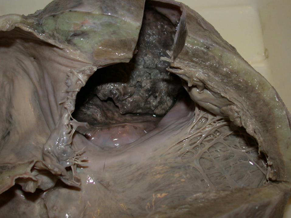
Dilatative/congestive cardiomyopathy (DCM) - Morphology -
- morphologic criteria: severely enlarged heart (weight sometimes 3 times
the normal – cor bovinum) with extremely dilated, ball-shaped ventricles,
rounded apex, from basis to apex progressively thinning wall and parietal
thrombi.
- further macroscopic alterations: atrial thrombosis; myocardium loose,
patchy-fibrotic, pale; valves secondarily and relatively insufficient.
Coronary arteries and valves morphologically intact!
- microscopy: no diagnostic alterations, only signes of a muscular
hypertrophy (enlarged muscle fibers and nuclei) and secondary signes of a
relative coronary insufficiency (myocytolysis, microinfarctions-microscars,
interstitial fibrosis, single fiber necroses)

Postmortem Photo Archive of the 2nd Dept. of Pathology;
Semmelweis University
Dilatative
cardiomyopathy.
Note the rounded
ventricle with local
endocardial
thickenings
representing
organized remnants
of former parietal
thromboses.

- presentation in all age-groups, yet, most frequently in the young
- appearance sporadic, only seldom familiar (here genetic background
possible), sometimes molecular biologic traces of enteroviral genom
detectable (viral myocarditis in the anamnesis?)
- the clinical picture is that of a slowly developping, therapy resistant
circulatory insufficiency
- begins slowly, lingering over the years with atypical complaints, the
diagnosis is established generally in the stage of the circulatory
insufficiency
- the end-diastolic volume increases progressively, the ejection fraction
decreases
- leads in 5-10 years to death
Dilatative/congestive cardiomyopathy (DCM) - Clinical aspects -
- cardiac muscle contractility is secured by the strength of the sarcomeric
contraction as well as by its transmission from sarcomer to sarcolemma and further
to the extracellular matrix
- the connection between sarcomer and sarcolemma is given by the dystrophin-
sarcoglycane proteincomplex
- certain mutations of the dystrophin gene (on the X-chromosome) lead to selective
absence of the dystrophin in the myocardium (>>DCM) but not in the skeletal
musculature (e.g. no Duchenne-Becker’s muscle dystrophy)
- mutation of the δ-sarcoglycane gene >> DCM
- mutation of the distal part of the myocardium-specific actin (contacting part
between actin and dystrophin with the help of a protein named desmin) and
mutations of desmin >> DCM
- further mutations of e.g. binding structures between neighbouring muscle cells, or
that of the energy production can lead to DCM
Dilatative/congestive cardiomyopathy (DCM) - Molecular characteristics -

Hypertrophic/obstructive cardiomyopathy (HCM) - Morphology -
- macroscopically: severely enlarged heart (weight sometimes 1000g – cor
bovinum) with a disproportionate left heart hypertrophy especially at the
septum >> decreased ability to dilatation (‘compliance’) and stenosis of the
way leading out from the ventricle with cardiac insufficiency >> hence
synonym terms: ‘asymmetrical septal hypertrophy’ (ASH); ‘idiopathic
hypertrophic subaortic stenosis’ (IHSS)
- microscopically: a diagnostic picture: (a) extreme hypertrophy of the
muscle fibers; (b) enlarged, bizarr nuclei with pale perinuclear rim (halo);
(c) very typically irregular-chaotic, syntitially woven fiber connections:
beside normal end-to-end connections there are end-to-side and side-to-side
fiber connections. This chaotic micromorphology explaines the clinicallly
often experienced cardiac arrhythmias.

- ethiology and pathogenesis unknown, the genetic background is
however proven (HCM is an inherited disease)
- clinical symptomes appear only around the 30th year of life
- first angina and dyspnoe on body excersize
- conduction abnormalities are often seen (arrhythmias)
- sudden cardiac death is possible
- with the disease at end stage, there is a therapy resistant cardiac
insufficiency
Hypertrophic/obstructive cardiomyopathy (HCM) - Clinical aspects -

- generally it is a familiar disease with autosomal dominant
inheritence and varying penetrance
- rarely sporadic appearance through de novo mutations is
possible
- HCM is a disease of the sarcomer: as well the thick
(myosin) as the thin (actin, tropomyosin, etc.) filament
genes may be affected
Hypertrophic/obstructive cardiomyopathy (HCM) - Molecular characteristics -

Restrictive/obliterative cardiomyopathy (RCM) - General comments -
- a rare disease
- important is the restricted ability of the heart ventricle to dilate (reduced
diastolic filling)
- the ventricle is capable neither of contracting nor of expanding to the
desirable degree
- the combined systolic and diastolic derangement leads to cardiac insuffi-
ciency
- the disease is generally detected very late, in the stage of cardiac insuffi-
ciency
- according to classic understanding basis of the disease lies in the parietal
endocardium, namely (a) an endocarditis parietalis fibroplastica secundum
Loeffler; or (b) an endomyocardial fibrosis

- in the background there is a severe peripheral and interstitial
eosinophilia (sometimes even an eosinophilic leukemia)
- it is a lethal disease
- the atypical, degranulated, circulating eosinophils cause endo-
myocardial necrosis by their toxic substances >> thickening and
scarring of the endocardium and the subendocardium >> formation
of parietal thrombi >> organisation of thrombi >> the very rigid
endocardium leads to myocardial motility derangements
Restrictive/obliterative cardiomyopathy (RCM) - Endocarditis parietalis fibroplastica secundum Loeffler -

- endocardial changes as with Loeffler’s endocarditis, yet without an
eosinophilia
- it is most frequently seen in the first 2 years of life, in adults rare
- the proliferating connective tissue that thickens the parietal
endocardium infiltrates also into the subendocardial myocardium
- prognosis depends on dimensions of the disease: focal endocardial
thickenings can remain symptomless, whereas a diffuse disease leads
quickly to cardial decompensation and death
Restrictive/obliterative cardiomyopathy (RCM) - Endomyocardial fibrosis -

Museum of Pathology;
2nd Dept. of Pathology; Semmelweis University
Restrictive
cardiomyopathy of a
new-born. Note
thickened left-
ventricular
endocardium.

- diffuse myocardial diseases of known origin >> important, that with the
therapy of the causative circumstances also the cardiac status gets relief or
will even be cured
- (a) Alcoholic cardiomyopathy – the most frequent cause, that leads to a
dilatative type cardiac disease. No coronary sclerosis. First symptoms are
arrhythmias without congestive signes. Beside a normal coronarogram angina
pectoris is possible. At the beginning the developping heart insufficiency can
be reversed by alcohol abstinence and specific supportive cardiotherapy. With
continued alcohol abuse an irreversible circulatory decompensation will
follow. Cause of death is often embolisation from parietal thrombi.
- (b) Peripartal (pregnancy-linked) cardiomyopathy – in the 3rd trimester of
pregnancy or within 6 weeks after birth. Disease characteristics as with a
dilatative type cardiopathy. Specific therapy makes a complete recovery
possible.
Secondary cardiomyopathies: cardiopathies

- (c) Hemochromatosis – genetic derangement of iron uptake and -stockage. Iron
reserves appear pathologically also in parenchymal cells causing functional
alterations in many organs: liver, pancreas, heart, skin, etc. The clinical
appearance of the heart disease that of a DCM. Later the myocardium develops
progressiv rigidity through accumulating iron contents, so the clinical picture turns
into one resembling a RCM. Macroscopically the myocardium is stiffened, dark
coloured. Microscopically the muscle fibers are massively overloaded with iron
containing hemosiderin pigment (positive Prussian-blue reaction).
- (d) Amyloidosis – the amyloid protein is deposited in the myocardial interstitium
and in small vessels >> thickening of myocardium all over the heart (especially in
the left ventricle). A cardiac insufficiency with lung edema and systolic functional
decrease develops typically in an unexpected, abrupt manner. Myocardium stif-
fened and rigid with a waxy-glassy hue on the cut surface >> the clinical presen-
tation is that of a RCM. Microscopically amyloid is seen as a homogenous
eosinophilic material (congo-red staining positive).
Secondary cardiomyopathies: cardiopathies

- (e) Sarcoidosis – in 8% of patients with sarcoidosis also cardiac disease
develops. Sarcoidotic granulomas appear in the pericardium and
myocardium (most frequently in the upper third of the interventricular
septum and in the papillary muscles). The clinical picture is dominated by
arrhythmias. The overall picture as with RCM. Arrhythmias may lead to
sudden cardiac death.
Secondary cardiomyopathies: cardiopathies

Cardiac decompensation _________________ _________________

Cardiac decompensation - General comments -
- it is the end stage of severe heart diseases; prognosis bad
- the clinical progression is defined by the basic disease, age, overall status and
other factors (e.g. social status of the patient)
- acute cardiac decompensation: as with myocardial infarction, valve rupture in
destructive endocarditis
- chronic cardiac decompensation: as with non-treated hypertension, chronic
valvular endocarditis, cardiomyopathies
- pump failure (forward failure) – disturbance of contractility
- filling failure (backward failure) – disturbance of dilatation
- at the beginning cardiac decompensations are generally one sided: either left-
or right heart failure, which can later combine

- causes are:
- ischemic heart disease
- hypertension
- vitia (other than a mitral stenosis)
- diseases of the myocardium (cardiomyopathies)
- backward failure: congestion of the lungs with chronic pulmonary edema,
‘heart failure cells’, brown stiffening of the lungs (induratio brunea
pulmonum)
- clinically: dyspnoe; orthopnoe; nocturnal respiratory complaints; frequent
and blood-stained coughs; (when combined with right heart failure:)
hydrothorax with compression and atelectasis of the lungs (atelectasia e
compressione); cerebral hypoxia with sleepiness (stupor) and rarely
hypoxic encephalopathy up to coma; decreased renal filtration, salt and
water retention, peripheral edemas
Cardiac decompensation - Left heart failure -

- most frequently in combination with a left heart failure – congestive
cardiac decompensation
- isolated right heart failure develops in only 15% of the cases, especially
with
- mitral stenosis
- some congenital vitia
- cor pulmonale
- pulmonary fibrosis
- clinically: congestion of the superficial jugular veins; lower limb edema
(anasarca); sometimes hydrothorax; hypoxic encephalopathy as with left
heart failure; liver congestion with development of a severe nutmeg liver
and a so-called cardiac cirrhosis; hepato-splenomegaly; congestive
gastroenteritis; ascites
Cardiac decompensation - Right heart failure -

Museum of Pathology;
2nd Dept. of Pathology; Semmelweis University
Cardiac decompensation. Cavities of both sides are tremendously dilated.
Tumors of the heart _____________ _____________

Primary cardiac tumors
- Benign
- Myxoma 25%
- Lipoma 8%
- Papillary fibroelastoma 8%
- Rhabdomyoma 7%
- Mesothelioma of the AV-Nodule 2%
- Malignant
- Angiosarcoma 7%
- Rhabdomyosarcoma 5%
- Mesothelioma 4%
- Fibrosarcoma 3%

Secondary cardiac tumors
- in 5% of all malignancy-related death cases cardiac metastases can
be found
- primary tumor locations in order of frequency are
- pulmonary carcinoma
- mammary carcinoma
- renal cell carcinoma
- malignant melanoma
- lymphoma / leukemia

Myxoma - most frequent tumor of the heart
- originates from the parietal endocardium
- macroscopy: a soft, greyish-reddish, sessile or steeled, varyingly large tumor
- microscopy: very loose, myxoid stroma with disseminated small vessels, on the
surface a covering layer endothelium
- danger of complication in approx. 50% of the cases is systemic embolisation from
fragmented tumor particles
- the lesion can unequivocally be detected radiologically
- therapy: operative resection; healing rate high; recurrences infrequent
- familiar appearance as a so-called ‘Carney-syndrome’ possible: multiple cardiac
myxomas, sometimes extracardiac (e.g. cutaneous) myxomas, patchy dermal
pigmentation, endocrine hyperfunction >> in case of a myxoma, echocardiography
of closer relatives is indicated
- differential diagnosis against an organized parietal thrombus is both macro- and
micro-scopically often probematic

Papillary fibroelastoma
- it is probably a residuum of an organized thrombus
- a bunch-like formation at the semilunar and cuspidal valves with hairy, repeatedly
bifurcating, thin branches and endothelial lining on the surface, usually measuring
cca. 1cm
- most frequent localisation: aortic valves >> danger of complication: stenosis or
occlusion of the coronary ostia with angina pectoris or even sudden cardiac death
Rhabdomyoma
- most frequent in new-borns and small children
- possible presentation with tuberous sclerosis
- no real tumor, but a hamartoma*
- in the left ventricular myocardium multiple nodules, sometimes with elevation of
the endocardial inner surface
*Hamartoma – a tumor-like lesion with tissue components, that are also present under normal conditions of the
presenting localisation, the morphologic composition and percentage relations of which being however abnormal.

Mesothelioma of the AV-nodule
- a typically cystic tumor in the location of the AV-nodule
measuring from microscopically small up to even 3 cm
- the tumor is connatal, that develops during the embryonal
period, primarily in females
- danger of complication: recurrent fits of Adams-Stokes’
syndrome already in childhood; complete AV-blockage;
sudden cardiac death of unknown origin in a young person
>> often makes the implantation of a pacemaker necessary