cardiovascular risk factors after childhood cancer...
TRANSCRIPT
Research ArticleCardiovascular Risk Factors after Childhood Cancer TreatmentAre Independent of the FTO Gene Polymorphism?
Małgorzata Sawicka-Żukowska , Maryna Krawczuk-Rybak , Paweł Bernatowicz,Katarzyna Muszyńska-Rosłan, Jerzy Konstantynowicz, and Włodzimierz Łuczyński
Department of Pediatric Oncology and Hematology, Medical University of Bialystok, Ul. Waszyngtona 17, 15-274 Bialystok, Poland
Correspondence should be addressed to Małgorzata Sawicka-Żukowska; [email protected]
Received 17 July 2017; Revised 5 December 2017; Accepted 17 December 2017; Published 20 February 2018
Academic Editor: Andrea Tura
Copyright © 2018 Małgorzata Sawicka-Żukowska et al. This is an open access article distributed under the Creative CommonsAttribution License, which permits unrestricted use, distribution, and reproduction in any medium, provided the original workis properly cited.
The study objective was to assess the prevalence of cardiovascular disease risk factors in patients treated for childhood cancer(N = 101) and to determine the involvement of clinical (cancer type and therapy) and/or genetic (FTO gene rs9939609polymorphism) factors. Anthropometric features, laboratory findings, and standardized osteodensitometric indices (fat andlean mass) were considered. Overweight/obesity was found in 17.82% of the patients; however, central adiposity was foundin as many as 42.5%. At least one abnormality in lipid metabolism was observed in 35.6%. Densitometry revealed elevatedlevels of fat mass in 44.55% of the patients. None of the parameters studied were associated with the FTO genepolymorphism. Standardized waist circumference was significantly higher in patients treated for leukemia than thosetreated for solid tumors (p = 0 04). Our findings indicate a high rate of central adiposity among childhood cancersurvivors, especially leukemia patients. The prevalence of risk factors of cardiovascular disease after anticancer therapy isnot FTO gene polymorphism-dependent.
1. Introduction
Childhood cancer survivors are at risk of developing over-weight/obesity or cardiovascular disease [1], which can bedue to central nervous system (CNS)/total body irradiation(TBI), administration of high steroid doses, reduced physicalexercise (immobility), and changes in lifestyle/eating habitsduring and after treatment [2]. Obesity is a multifactorialphenomenon that, in the general population, is mainlycaused by consumption of excessive amounts of energy andlow physical activity. Thanks are due to novel diagnosticcapabilities where risk factors of cardiovascular disease canbe assessed using not only anthropometric parameters andlipid profiles but also by densitometry (including percentageof fat mass) [3]. The location of disease, disease advance-ment, and diverse therapeutic modes (including surgery,chemotherapy, and radiotherapy) are all factors that affectbody mass in patients treated for childhood cancer. However,
due to family histories of obesity, its genetic background is alsoinvestigated [4]. In recent years, themost commonly analyzedgenetic factor is the fat-mass and obesity-associated (FTO)gene. The FTO gene rs9939609 polymorphism shows correla-tions with body mass index (BMI), overweight, obesity, andother risk factors of cardiovascular disease [5, 6]. While themechanism of the FTO gene product has not been fully eluci-dated, it is assumed to play a role in the regulation of energyexpenditure in the hypothalamus [7].
The aim of this study was to assess the prevalence of riskfactors of cardiovascular disease in patients treated for child-hood cancer and to determine the involvement of clinical(cancer type or therapy) and/or genetic (FTO gene polymor-phism) factors. Anthropometric features (standardized: BMIand waist circumference), laboratory findings (lipid profile),and standardized osteodensitometric indices (fat and leanmass) were considered. The results were compared with thestandards defined recently for Polish children [8–10].
HindawiInternational Journal of EndocrinologyVolume 2018, Article ID 7495234, 6 pageshttps://doi.org/10.1155/2018/7495234
2. Patients and Methods (Full Version inSupplemental Material)
A total of 101 childhood cancer survivors from theDepartment of Pediatric Hematology and Oncology of theMedical University of Bialystok (part of the Polish PediatricGroup for the Treatment of Leukemias/Lymphomas andSolid Tumors) were qualified to take part in the studyduring standard, periodic check-ups. The following param-eters were evaluated in all subjects: anthropometric anddisease-associated data, lipid profile, densitometric data,and genotype. The anthropometric parameters includedage, sex, height, weight, BMI standardized deviation score(BMI-SDS), and waist circumference SDS. Underweightwas defined as a BMI-SDS of ≤−1, normal weight as >−1and <1, overweight as ≥1 and <2, and obesity as ≥2. Centralobesity was recognized at a waist circumference above the90th percentile [10]. The following disease-associated param-eters were evaluated in the cancer survivor group: cancertype; disease duration; type of treatment regimen; elapsedtime since therapy cessation; history of CNS radiotherapyor TBI (12 or 18Gy); history of steroid treatment, type (pred-nisone and dexamethasone), and dose (standard or high);and history of hematopoietic stem cell transplantation (TableS1 in Supplementary Materials). Using densitometry, thefollowing parameters were estimated and expressed in stan-dard deviation scores: fat mass SDS (FAT SDS) and lean bodymass SDS (LEAN SDS). Their values were compared withour own data and definitions developed by the InternationalSociety for Clinical Densitometry. Lipid profiles wereanalyzed in the hospital’s central laboratory and includedserum LDL cholesterol, HDL cholesterol, and triglyceridelevels after 8 hours of fasting. Glucose and insulin concentra-tions were also assessed (fasting and 2 hours after oralglucose tolerance test). HOMA insulin resistance index wascalculated according to the following formula: fasting insulin(microU/L) × fasting glucose (nmol/L)/22.5. All childrenwere assessed for the FTO rs9939609 polymorphism via alle-lic discrimination with ABI 7900HT Fast Real-Time PCRSystem with SDS 2.1 software.
3. Presentation of Data and Statistical Analysis
Analysis results were presented as means with a standarddeviation and rates of incidence of a given characteristic inthe study group. p < 0 05 was considered to be statisticallysignificant. Univariate analysis was performed using theStudent’s t-test in cases of continuous variables and theChi-square test for nominal ones. Correlation of the obtaineddata was performed using the Spearman’s test. Frequencies ofthe alleles observed in the control group were tested againstthe Hardy-Weinberg equilibrium using the Chi-square test.
4. Results
The study was conducted at the mean age of 12.7 years(±4.2), on average 8.2 (±3.5) years after termination of anti-cancer therapy. No differences were found between boysand girls in relation to age at examination and diagnosis or
time elapsed since treatment cessation. However, patientstreated due to lymphomas were significantly older than thosetreated for leukemias or solid tumors (p < 0 05) (see Table S2in Supplementary Materials).
4.1. Results of Anthropometric Analysis. The mean standard-ized BMI was 0.69± 1.65. Overweight was found in 12patients (11.88%), and obesity was found in 6 (5.94%). Themean standardized waist circumference was 1.07± 1.97.Central adiposity was recognized in 43 children (42.5%).
4.2. Laboratory Findings. At least one deviation in lipidmetabolism was noted in 36 patients (35.6%). LDL choles-terol fraction and triglycerides were elevated in 29 (28.7%)and 9 (8.9%) patients, respectively. The HDL cholesterolfraction was decreased in 8 patients (7.9%).
Abnormal plasma glucose levels were noted in 2 patients(1.9%) after fasting and in 1 patient (0.9%) after oral glucosetolerance test. The insulin resistance index HOMA (mean2.92± 10.4) was abnormal in 18 cases (17.8%) (HOMA> 3).Lipid and carbohydrate levels did not show any statisticallysignificant differences with respect to sex and diagnosis.Children with insulin resistance had a higher standardizedwaist circumference compared to patients without insulinresistance (data not presented), but no difference was foundin BMI-SDS. No differences were observed in other param-eters between these groups of patients.
4.3. Results of Densitometry. Standardized fat mass (FATSDS) was decreased (<−1) in 4 patients (3.96%) andincreased (>1) in as many as 45 patients (44.55%). Standard-ized lean mass (LEAN SDS) was decreased (<−1) in 12patients (11.88%) and increased (>1) in 21 patients(20.8%). No differences were noted in densitometric param-eters according to sex and diagnosis (data not presented).
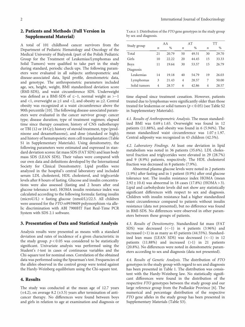
4.4. Results of Genetic Analysis. The distribution of FTOgenotypes in the study group with regard to sex and diagnosishas been presented in Table 1. The distribution was consis-tent with the Hardy-Weinberg law. No statistically signifi-cant differences were found in the distribution of therespective FTO genotypes between the study group and ourlarge reference group from the Podlaskie Province [6]. Thenumerical and percentage distribution of the respectiveFTO gene alleles in the study group has been presented inSupplementary Materials (Table S3).
Table 1: Distribution of the FTO gene genotypes in the study groupby sex and diagnosis.
Study groupAA AT TT
n % n % n %
Total 21 20.79 50 49.51 30 29.70
Girls 10 22.22 20 44.45 15 33.33
Boys 11 19.64 30 53.57 15 26.79
Diagnosis
Leukemias 14 19.18 40 54.79 19 26.03
Lymphomas 3 21.43 4 28.57 7 50.00
Solid tumors 4 28.57 6 42.86 4 28.57
2 International Journal of Endocrinology
The values of the standardized BMI according to geno-type did not differ significantly (p = 0 77) and were as fol-lows: AA, 0.94± 0.38; AT, 0.48± 0.24; and TT, 0.86± 0.31.The unfavorable FTO AA genotype was not significantlymore common in the group of patients with overweightand/or obesity (Table S4 in Supplementary Materials). Like-wise, no statistically significant differences were noted inthe analysis of AA genotypes versus AT+TT or AA+ATversus TT (data not presented). The values of the standard-ized waist circumference did not differ according to FTOgenotype (p > 0 05) and were as follows: AA, 1.07± 1.89;AT, 0.7± 1.94; and TT, 1.60± 1.88. Similarly, patientsmeeting the criteria of central adiposity did not show ahigher frequency of the FTO AA genotype and A allele (datanot presented).
Mean values of lipid profile and carbohydrate metabo-lism did not differ between groups of patients with therespective genotypes (Tables S5 and S6). Similarly, no differ-ences were noted in the frequency of FTO genotypes inpatients with abnormal lipids or carbohydrate metabolismcompared to patients with normal parameters in this field(data not presented). The analysis of densitometric findingsshowed no difference in FAT and LEAN SDS according toFTO genotype (Table S7). No differences were found in thedistribution of genotypes in patients with an elevated per-centage of fat mass assessed by densitometry (FAT SDS)(data not presented).
4.5. Analysis of Risk Factors of Cardiovascular Disease withRespect to Clinical Features
4.5.1. Primary Disease. Following the division into groupsaccording to diagnosis, waist circumference SDS was signifi-cantly higher in the group of children treated for leukemiathan in those treated for solid tumors (0.84± 0.07 versus0.8± 0.02; p = 0 04).
4.5.2. High Steroid Doses. The steroid dose applied to thebody surface had no impact on the anthropometric, labora-tory, and densitometric parameters studied (p > 0 05). Thedistribution of the respective genotypes and FTO gene allelesdid not differ statistically in either group.
4.5.3. CNS Irradiation. The anthropometric, laboratory, anddensitometric parameters did not differ significantly betweenthe group of children subjected to CNS irradiation and thosewho were not (p > 0 05). Considering the respective FTOgenotypes, no statistically significant differences were foundin anthropometric parameters, lipid profile, or densitometrybetween these groups.
4.5.4. Stem-Cell and Marrow Transplantation. Patients whohad undergone bone marrow or stem cell transplantationhad significantly lower standardized BMI (−0.44± 0.91versus 0.76± 1.67; p = 0 042) and waist circumference(−1.37± 0.80 versus 1.16± 1.95; p = 0 02) than those whohad not. We did not observe a higher frequency of the unfa-vorable FTO genotype or its effect on laboratory parameters,body mass components, densitometry, or lipid profiles in thegroup of children after bone marrow transplantation.
4.5.5. Diagnosis before, during, and after Puberty. In thegroup of children who developed cancer before puberty, nosignificant differences were found in the anthropometricparameters BMI-SDS and waist circumference SDS, apartfrom a significantly higher hip circumference expressed inSDS than patients who developed cancer during puberty orafter its termination (1.05± 1.62 versus −0.45± 1.42; p >0 05). Likewise, there were no differences in laboratoryfindings among these three groups of patients. In densi-tometry, LEAN SDS was significantly lower in childrenwho developed cancer during or after puberty than inthose who developed cancer before puberty (−1.50± 1.91versus 0.51± 1.64, resp.; p = 0 01). No differences were notedin the distribution of FTO genotypes in these groups.
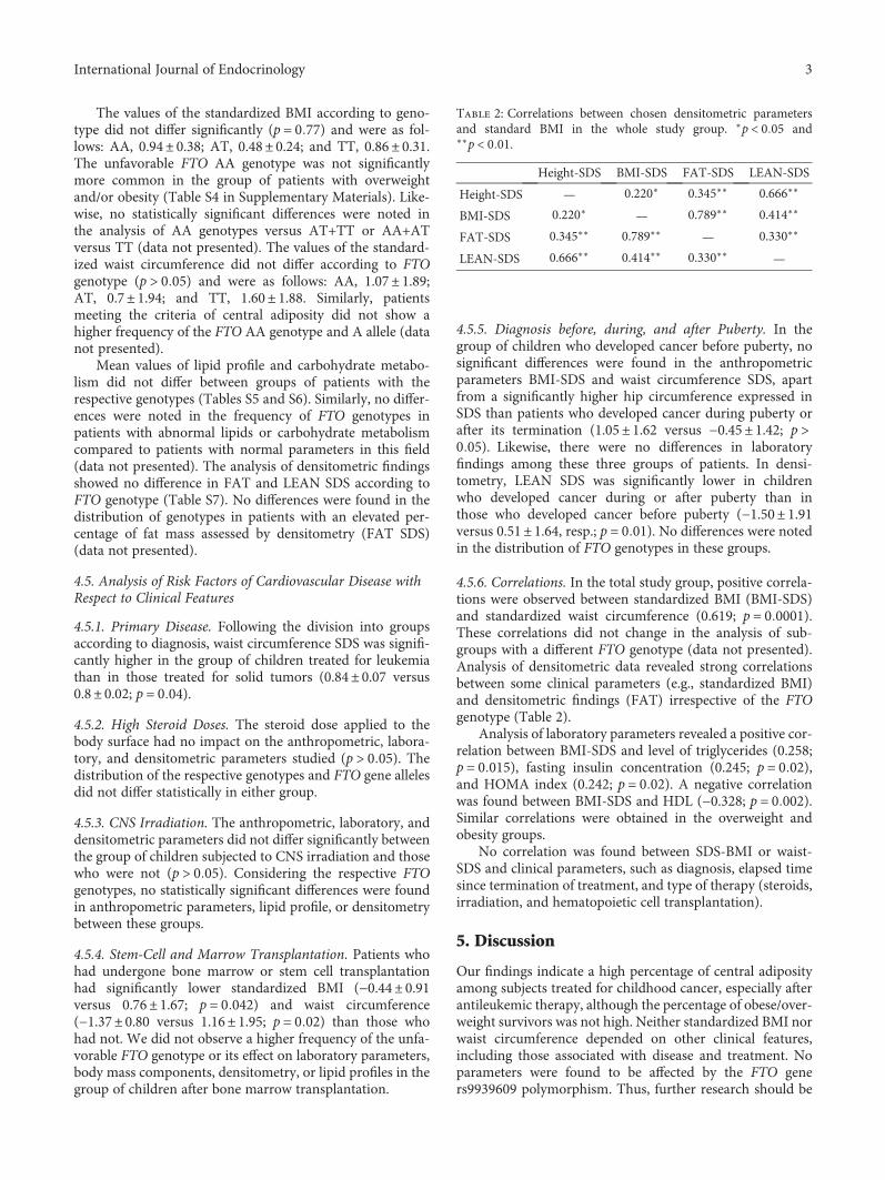
4.5.6. Correlations. In the total study group, positive correla-tions were observed between standardized BMI (BMI-SDS)and standardized waist circumference (0.619; p = 0 0001).These correlations did not change in the analysis of sub-groups with a different FTO genotype (data not presented).Analysis of densitometric data revealed strong correlationsbetween some clinical parameters (e.g., standardized BMI)and densitometric findings (FAT) irrespective of the FTOgenotype (Table 2).
Analysis of laboratory parameters revealed a positive cor-relation between BMI-SDS and level of triglycerides (0.258;p = 0 015), fasting insulin concentration (0.245; p = 0 02),and HOMA index (0.242; p = 0 02). A negative correlationwas found between BMI-SDS and HDL (−0.328; p = 0 002).Similar correlations were obtained in the overweight andobesity groups.
No correlation was found between SDS-BMI or waist-SDS and clinical parameters, such as diagnosis, elapsed timesince termination of treatment, and type of therapy (steroids,irradiation, and hematopoietic cell transplantation).
5. Discussion
Our findings indicate a high percentage of central adiposityamong subjects treated for childhood cancer, especially afterantileukemic therapy, although the percentage of obese/over-weight survivors was not high. Neither standardized BMI norwaist circumference depended on other clinical features,including those associated with disease and treatment. Noparameters were found to be affected by the FTO geners9939609 polymorphism. Thus, further research should be
Table 2: Correlations between chosen densitometric parametersand standard BMI in the whole study group. ∗p < 0 05 and∗∗p < 0 01.
Height-SDS BMI-SDS FAT-SDS LEAN-SDS
Height-SDS — 0.220∗ 0.345∗∗ 0.666∗∗
BMI-SDS 0.220∗ — 0.789∗∗ 0.414∗∗
FAT-SDS 0.345∗∗ 0.789∗∗ — 0.330∗∗
LEAN-SDS 0.666∗∗ 0.414∗∗ 0.330∗∗ —
3International Journal of Endocrinology
performed to determine the causes of central adiposity andattempts should be made for its prevention to reduce the riskof cardiovascular disease.
The reported rates of overweight and obesity amongPolish children are 15.5% and 2%, respectively, similar tothe rates found in our study [11]. Most authors observedhigher percentages of overweight and obesity among patientsafter antineoplastic treatment [12]. However, it is notcompletely clear whether it is the disease, therapy, or patientlifestyle that is responsible for the increased risk of cardiovas-cular disease in these patients. The patient population mostaffected are those treated by total body irradiation [13],which, in extreme cases, may lead to considerable insulinresistance, type 2 diabetes, dyslipidemia, and hepaticsteatosis [14]. Jarfelt et al. noted higher waist circumfer-ence in men treated for acute lymphoblastic leukemia withradiotherapy than male patients using other treatmentmethods. However, such differences were not observed inwomen [15]. Similar correlations were found in relationto body fat mass assessed using the DEXA method. Theauthors suggested the responsibility of growth factor defi-ciency due to CNS irradiation [15]. The study was con-ducted on a group of patients treated in the 1970s and1980s using a dose of 24Gy. Since that time, indicationsfor CNS irradiation have been markedly limited [16],which resulted in a small number of irradiated patientsin our study group (17 patients [16.83%] received 12Gy).However, in the Janiszewski et al.’s study, obesity and cen-tral adiposity after leukemia treatment were not associatedwith CNS irradiation [17]. In our patient group, no differ-ences were observed in the risk factors of cardiovasculardisease according to treatment, including CNS and/orTBI. This data suggests that a 12Gy dose does not havea major effect on the physical condition of patients, fattytissue location, or lipid indices.
It is difficult to assess what factors determine thedevelopment of overweight and obesity in patients. In astudy performed by Gofman and Ducore, a younger ageat the time of diagnosis and Spanish origin correlated withthe occurrence of obesity after anticancer treatment [18];on the other hand, sex, irradiation and its dose, durationof therapy, and family history had no effect on body mass.Standardized BMI at the time of diagnosis of ALL also hada strong effect on standardized BMI after treatment andthe risk of obesity [19]. Environmental factors play a keyrole in the development of obesity. In a small group ofBrazilian patients, the prevalence rate of obesity was nothigher after treatment for ALL and the role of environ-mental factors was suggested (i.e., living conditions of adeveloping country) [20].
As in other populations, the elevated BMI in patientsafter anticancer treatment is a predictor of insulin resistance[21]. However, insulin resistance after anti-ALL treatmentwas also observed in subjects with normal BMI [22]. In ourstudy, we observed a high percentage of patients meetingthe diagnostic criteria of insulin resistance (HOMA> 3). Thisobservation and a high percentage of central adiposity indi-cate the risk of insulin resistance among patients after anti-cancer treatment.
Genetic factors also play an essential role in the patho-genesis of abnormal weight. Obesity in girls after leukemiatreatment is associated with obesity in their mothers butnot fathers [23]. It is still unknown which genes are stronglyrelated to body mass. The FTO gene rs9939609 polymor-phism is associated with body mass and risk of overweightand obesity in Polish children, as well as in the PodlaskieProvince [6]. However, no relationship was found betweenthe FTO gene polymorphism and body mass or risk of over-weight/obesity in subjects after anticancer treatment. InSkoczen et al.’s study, a group of Polish children subjectedto irradiation due to ALL showed a lower frequency of theT allele at the site of the FTO gene rs9939609 in subjects withoverweight [24], which may be connected to a protectiveeffect of the T allele preventing binge eating [25]. Polymor-phisms of FTO and adiponectin genes can be used (in addi-tion to age and BMI at time of diagnosis) in the model ofobesity prediction after breast cancer treatment [26]. It is alsoknown that obesity is associated with a higher risk of certaincancers, although no relation with the FTO gene polymor-phism is known [27].
We found a high percentage of abnormal parameters oflipid metabolism, similar to other authors. In a group of 75patients after ALL treatment, Gurney et al. showed abnormallevels of HDL cholesterol, but no other cholesterol fractions[28]. They observed a higher percentage of central adiposityin women than in the reference group, but not in the wholestudy group. Features of metabolic syndrome were foundmainly in subjects undergoing CNS radiation. Likewise, agreater amount of fatty tissue, including total body fat, wasrevealed using the DEXA method in a group of individualstreated for ALL in childhood [15].
Lack of a control group is a limitation of the currentstudy. However, we refer our findings to the database ofhealthy inhabitants of the Podlaskie Province. Future studiesshould include analysis of diet and physical activity ofpatients to determine the causes of central adiposity ofsubjects cured of leukemia. Similarly, other studies are char-acterized by limitations such as a small number of qualifiedpatients, various inclusion criteria, or retrospective observa-tion. Thus, it is still unknown whether the high frequencyof risk factors of cardiovascular disease is actually caused bythe disease and anticancer treatment [29].
6. Conclusion
The results of our study, including a high percentage of chil-dren with central adiposity, features of insulin resistance,abnormal lipid profile, and excessive amount of fatty tissuein densitometry (all without the genetic background of FTOgene polymorphism), indicate the potential threat of cardio-vascular disease risk factors in childhood cancer patients.This patient population should be monitored by pediatri-cians and pediatric oncologists, especially to detect centraladiposity in patients after antileukemic treatment. Properdiet, physical exercise, changes in lifestyle, and drugs in thecase of failure should be recommended for the preventionand treatment of cardiovascular complications in this groupof patients.
4 International Journal of Endocrinology
Conflicts of Interest
The authors declare that there is no conflict of interestregarding the publication of this paper.
Supplementary Materials
Supplemental Material includes full version of “Patients andMethods” with references, 7 tables, and with additional datafor “Results” section. Table S1: type of treatment accordingto diagnosis. Table S2: study group characteristics with divi-sion into sex and type of cancer. Table S3: the frequency ofthe A and T alleles at the rs9939609 site of the FTO gene inthe study group. Table S4: numerical and percentage distri-bution of FTO genotypes according to BMI. The differenceswere not statistically significant. Table S5: mean levels of lipidmetabolism parameters with respect to the FTO genotype.Table S6: mean levels of carbohydrate metabolism parame-ters with respect to the FTO genotype. Table S7: osteodensi-tometric parameters with respect to the FTO genotype.(Supplementary Materials)
References
[1] K. C. Oeffinger, A. C. Mertens, C. A. Sklar et al., “Obesity inadult survivors of childhood acute lymphoblastic leukemia: areport from the childhood cancer survivor study,” Journal ofClinical Oncology, vol. 21, no. 7, pp. 1359–1365, 2003.
[2] D. M. Green, C. L. Cox, L. Zhu et al., “Risk factors for obesity inadult survivors of childhood cancer: a report from the child-hood cancer survivor study,” Journal of Clinical Oncology,vol. 30, no. 3, pp. 246–255, 2012.
[3] C. A. J. Brouwer, J. A. Gietema,W. A. Kamps, E. G. E. de Vries,and A. Postma, “Changes in body composition after childhoodcancer treatment: impact on future health status—a review,”Critical Reviews in Oncology/Hematology, vol. 63, no. 1,pp. 32–46, 2007.
[4] Y. F. Pei, L. Zhang, Y. Liu et al., “Meta-analysis of genome-wide association data identifies novel susceptibility loci forobesity,” Human Molecular Genetics, vol. 23, no. 3,pp. 820–830, 2014.
[5] T. M. Frayling, N. J. Timpson, M. N. Weedon et al., “A com-mon variant in the FTO gene is associated with body massindex and predisposes to childhood and adult obesity,” Science,vol. 316, no. 5826, pp. 889–894, 2007.
[6] W. Luczynski, G. Zalewski, and A. Bossowski, “The associationof the FTO rs9939609 polymorphism with obesity andmetabolic risk factors for cardiovascular diseases in Polishchildren,” Journal of Physiology and Pharmacology, vol. 63,no. 3, pp. 241–248, 2012.
[7] T. Gerken, C. A. Girard, Y.-C. L. Tung et al., “The obesity-associated FTO gene encodes a 2-oxoglutarate-dependentnucleic acid demethylase,” Science, vol. 318, no. 5855,pp. 1469–1472, 2007.
[8] Z. Kulaga, M. Litwin, M. Tkaczyk et al., “The height-, weight-,and BMI-for-age of Polish school-aged children and adoles-cents relative to international and local growth references,”BMC Public Health, vol. 10, no. 1, p. 109, 2010.
[9] J. Konstantynowicz, T. V. Nguyen, M. Kaczmarski,J. Jamiolkowski, J. Piotrowska-Jastrzebska, and E. Seeman,
“Fractures during growth: potential role of a milk-free diet,”Osteoporosis International, vol. 18, no. 12, pp. 1601–1607, 2007.
[10] Z. Kulaga, A. Krzyzaniak, I. Palczewska, and K. Barwicka,“Dynamika narastania nadwagi i otyłości dzieci i młodzieży -wybrana populacja polska na tle populacji USA,” StandardyMedyczne Pediatria, vol. 4, no. 3, pp. 267–271, 2007.
[11] M. Chrzanowska, T. Łaska-Mierzejewska, and A. Suder,“Overweight and obesity in rural girls from Poland: changesbetween 1987 and 2001,” Journal of Biosocial Science, vol. 45,no. 02, pp. 217–229, 2013.
[12] L. Iughetti, P. Bruzzi, B. Predieri, and P. Paolucci, “Obesity inpatients with acute lymphoblastic leukemia in childhood,”Italian Journal of Pediatrics, vol. 38, no. 1, article 4, 2012.
[13] R. Rajendran, E. Abu, A. Fadl, and C. D. Byrne, “Late effects ofchildhood cancer treatment: severe hypertriglyceridaemia,central obesity, non alcoholic fatty liver disease and diabetesas complications of childhood total body irradiation,” DiabeticMedicine, vol. 30, no. 8, pp. e239–e242, 2013.
[14] S. Mayson, V. Parker, M. Schutta, R. Semple, and M. Rickels,“Severe insulin resistance and hypertriglyceridemia after child-hood total body irradiation,” Endocrine Practice, vol. 19, no. 1,pp. 51–58, 2013.
[15] M. Jarfelt, B. Lannering, I. Bosaeus, G. Johannsson, andR. Bjarnason, “Body composition in young adult survivors ofchildhood acute lymphoblastic leukaemia,” European Journalof Endocrinology, vol. 153, no. 1, pp. 81–89, 2005.
[16] J. Stary, M. Zimmermann, M. Campbell et al., “Intensivechemotherapy for childhood acute lymphoblastic leukemia:results of the randomized intercontinental trial ALL IC-BFM 2002,” Journal of Clinical Oncology, vol. 32, no. 3,pp. 174–184, 2014.
[17] P. M. Janiszewski, K. C. Oeffinger, T. S. Church et al., “Abdom-inal obesity, liver fat, and muscle composition in survivors ofchildhood acute lymphoblastic leukemia,” The Journal ofClinical Endocrinology & Metabolism, vol. 92, no. 10,pp. 3816–3821, 2007.
[18] I. Gofman and J. Ducore, “Risk factors for the development ofobesity in children surviving ALL and NHL,” Journal of Pedi-atric Hematology/Oncology, vol. 31, no. 2, pp. 101–107, 2009.
[19] F. F. Zhang, A. M. Rodday, M. J. Kelly et al., “Predictors ofbeing overweight or obese in survivors of pediatric acute lym-phoblastic leukemia (ALL),” Pediatric Blood & Cancer, vol. 61,no. 7, pp. 1263–1269, 2014.
[20] C. Papadia, L. A. Naves, S. S. S. Costa, J. A. R. Vaz,L. Domingues, and L. A. Casulari, “Incidence of obesity doesnot appear to be increased after treatment of acute lympho-blastic leukemia in Brazilian children: role of leptin, insulin,and IGF-1,” Hormone Research in Pædiatrics, vol. 68, no. 4,pp. 164–170, 2007.
[21] S. Lowas, S. Malempati, and D. Marks, “Body mass index pre-dicts insulin resistance in survivors of pediatric acute lympho-blastic leukemia,” Pediatric Blood & Cancer, vol. 53, no. 1,pp. 58–63, 2009.
[22] E. S. Tonorezos, G. L. Vega, C. A. Sklar et al., “Adipokines,body fatness, and insulin resistance among survivors of child-hood leukemia,” Pediatric Blood & Cancer, vol. 58, no. 1,pp. 31–36, 2012.
[23] M. P. Shaw, L. E. Bath, J. Duff, C. J. Kelnar, andW. H.Wallace,“Obesity in leukemia survivors: the familial contribution,”Journal of Pediatric Hematology/Oncology, vol. 17, no. 3,pp. 231–237, 2000.
5International Journal of Endocrinology
[24] S. Skoczen, M. Bik-Multanowski, W. Balwierz et al.,“Homozygosity for the rs9939609T allele of the FTO genemay have protective effect on becoming overweight in survi-vors of childhood acute lymphoblastic leukaemia,” Journalof Genetics, vol. 90, no. 2, pp. 365–368, 2011.
[25] J. Wardle, C. Llewellyn, S. Sanderson, and R. Plomin, “TheFTO gene and measured food intake in children,” Interna-tional Journal of Obesity, vol. 33, no. 1, pp. 42–45, 2009.
[26] S. M. Reddy, M. Sadim, J. Li et al., “Clinical and genetic predic-tors of weight gain in patients diagnosed with breast cancer,”British Journal of Cancer, vol. 109, no. 4, pp. 872–881, 2013.
[27] G. Li, Q. Chen, L. Wang, D. Ke, and Z. Yuan, “Associationbetween FTO gene polymorphism and cancer risk: evidencefrom 16,277 cases and 31,153 controls,” Tumor Biology,vol. 33, no. 4, pp. 1237–1243, 2012.
[28] J. G. Gurney, K. K. Ness, S. D. Sibley et al., “Metabolic syn-drome and growth hormone deficiency in adult survivors ofchildhood acute lymphoblastic leukemia,” Cancer, vol. 107,no. 6, pp. 1303–1312, 2006.
[29] C. Anarollo, L. Airaghi, G. Saporiti, F. Onida, A. Cortelezzi,and G. L. Deliliers, “Metabolic syndrome in patients withhematological diseases,” Expert Review of Hematology, vol. 5,no. 4, pp. 439–458, 2012.
6 International Journal of Endocrinology

Stem Cells International
Hindawiwww.hindawi.com Volume 2018
Hindawiwww.hindawi.com Volume 2018
MEDIATORSINFLAMMATION
of
EndocrinologyInternational Journal of
Hindawiwww.hindawi.com Volume 2018
Hindawiwww.hindawi.com Volume 2018
Disease Markers
Hindawiwww.hindawi.com Volume 2018
BioMed Research International
OncologyJournal of
Hindawiwww.hindawi.com Volume 2013
Hindawiwww.hindawi.com Volume 2018
Oxidative Medicine and Cellular Longevity
Hindawiwww.hindawi.com Volume 2018
PPAR Research
Hindawi Publishing Corporation http://www.hindawi.com Volume 2013Hindawiwww.hindawi.com
The Scientific World Journal
Volume 2018
Immunology ResearchHindawiwww.hindawi.com Volume 2018
Journal of
ObesityJournal of
Hindawiwww.hindawi.com Volume 2018
Hindawiwww.hindawi.com Volume 2018
Computational and Mathematical Methods in Medicine
Hindawiwww.hindawi.com Volume 2018
Behavioural Neurology
OphthalmologyJournal of
Hindawiwww.hindawi.com Volume 2018
Diabetes ResearchJournal of
Hindawiwww.hindawi.com Volume 2018
Hindawiwww.hindawi.com Volume 2018
Research and TreatmentAIDS
Hindawiwww.hindawi.com Volume 2018
Gastroenterology Research and Practice
Hindawiwww.hindawi.com Volume 2018
Parkinson’s Disease
Evidence-Based Complementary andAlternative Medicine
Volume 2018Hindawiwww.hindawi.com
Submit your manuscripts atwww.hindawi.com