cardiovascular system module 3: heart anatomy - cnx.org2.pdf... · in order to understand how that...
TRANSCRIPT

OpenStax-CNX module: m49683 1
Cardiovascular System Module 3:
Heart Anatomy*
Donna Browne
Based on Heart Anatomy� by
OpenStax
This work is produced by OpenStax-CNX and licensed under the
Creative Commons Attribution License 4.0�
Abstract
By the end of this section, you will be able to:
• Describe the location and position of the heart within the body cavity• Describe the internal and external anatomy of the heart• Identify the tissue layers of the heart• Relate the structure of the heart to its function as a pump• Compare systemic circulation to pulmonary circulation• Identify the veins and arteries of the coronary circulation system• Trace the pathway of oxygenated and deoxygenated blood thorough the chambers of the heart
The vital importance of the heart is obvious. If one assumes an average rate of contraction of 75contractions per minute, a human heart would contract approximately 108,000 times in one day, more than39 million times in one year, and nearly 3 billion times during a 75-year lifespan. Each of the majorpumping chambers of the heart ejects approximately 70 mL blood per contraction in a resting adult. Thiswould be equal to 5.25 liters of �uid per minute and approximately 14,000 liters per day. Over one year,that would equal 10,000,000 liters or 2.6 million gallons of blood sent through roughly 60,000 miles ofvessels. In order to understand how that happens, it is necessary to understand the anatomy and physiologyof the heart.
1 Location of the Heart
The human heart is located within the thoracic cavity, medially between the lungs in the space known asthe mediastinum. Figure 1 (Position of the Heart in the Thorax ) shows the position of the heart withinthe thoracic cavity. Within the mediastinum, the heart is separated from the other mediastinal structuresby a tough membrane known as the pericardium, or pericardial sac, and sits in its own space called thepericardial cavity. The dorsal surface of the heart lies near the bodies of the vertebrae, and its anteriorsurface sits deep to the sternum. The great veins, the superior and inferior venae cavae, and the great arteries,
*Version 1.2: Mar 18, 2014 7:18 am -0500�http://cnx.org/content/m46676/1.3/�http://creativecommons.org/licenses/by/4.0/
http://cnx.org/content/m49683/1.2/

OpenStax-CNX module: m49683 2
the aorta and pulmonary trunk, are attached to the superior surface of the heart, called the base.Figure 1(Position of the Heart in the Thorax ). The inferior tip of the heart is called the apex.
Position of the Heart in the Thorax
Figure 1: The heart is located within the thoracic cavity, medially between the lungs in the mediastinum.It is about the size of a �st, is broad at the top, and tapers toward the base.
2 Shape and Size of the Heart
The shape of the heart is similar to a triangle, rather broad at the superior surface and tapering to the apex(see Figure 1 (Position of the Heart in the Thorax )). A typical heart is approximately the size of your
http://cnx.org/content/m49683/1.2/
OpenStax-CNX module: m49683 3
�st. Given the size di�erence between most members of the sexes, the weight of a female heart is smalleron average than the male's heart. The heart of a well-trained athlete, especially one specializing in aerobicsports, can be considerably larger than this. Cardiac muscle responds to exercise in a manner similar tothat of skeletal muscle. Hearts of athletes can pump blood more e�ectively at lower rates than those ofnonathletes.
3 Chambers and Circulation through the Heart
The human heart consists of four chambers: The left side and the right side each have one atrium and oneventricle. Each of the upper chambers, the right atrium (plural = atria) and the left atrium, acts as areceiving chamber and contracts to push blood into the lower chambers, the right ventricle and the leftventricle. The ventricles serve as the primary pumping chambers of the heart, propelling blood to thelungs or to the rest of the body.
There are two distinct but linked circuits in the human circulation called the pulmonary and systemiccircuits. The pulmonary circuit transports blood to and from the lungs, where it picks up oxygen anddelivers carbon dioxide for exhalation. The systemic circuit transports oxygenated blood to virtually allof the tissues of the body and returns to the heart.
The right ventricle pumps deoxygenated blood into the pulmonary trunk, which leads toward thelungs and splits into the left and right pulmonary arteries. These vessels in turn branch many timesbefore reaching the pulmonary capillaries, where gas exchange occurs: Carbon dioxide exits the bloodand oxygen enters. The pulmonary trunk arteries and their branches are the only arteries in the body thatcarry relatively deoxygenated blood. Highly oxygenated blood returning from the pulmonary capillaries inthe lungs passes through a series of vessels that join together to form the pulmonary veins�the only veinsin the body that carry highly oxygenated blood. The pulmonary veins bring blood into the left atrium,which delivers the blood into the left ventricle, which in turn pumps oxygenated blood into the aorta and onto the many branches of the systemic circuit. Eventually, these vessels will lead to the systemic capillaries,where exchange with the tissue �uid and cells of the body occurs. In this case, oxygen and nutrients exitthe systemic capillaries to be used by the cells in their metabolic processes, and carbon dioxide and wasteproducts will enter the blood.
The blood that has traveled throughout the body is lower in oxygen concentration than when it entered.The superior vena cava and the inferior vena cava return blood to the right atrium. The blood in thesuperior and inferior venae cavae �ows into the right atrium, which delivers blood into the right ventricle.This process of blood circulation continues as long as the individual remains alive. (Figure 2 (Dual Systemof the Human Blood Circulation )).
http://cnx.org/content/m49683/1.2/

OpenStax-CNX module: m49683 4
Dual System of the Human Blood Circulation
Figure 2: Blood �ows from the right atrium to the right ventricle, where it is pumped into the pulmonarycircuit. The blood in the pulmonary artery branches is low in oxygen but relatively high in carbon dioxide.Gas exchange occurs in the pulmonary capillaries (oxygen into the blood, carbon dioxide out), and bloodhigh in oxygen and low in carbon dioxide is returned to the left atrium. From here, blood enters theleft ventricle, which pumps it into the systemic circuit. Following exchange in the systemic capillaries(oxygen and nutrients out of the capillaries and carbon dioxide and wastes in), blood returns to the rightatrium and the cycle is repeated.
http://cnx.org/content/m49683/1.2/

OpenStax-CNX module: m49683 5
4 Membranes, Surface Features, and Layers
Our exploration of more in-depth heart structures begins by examining the membrane that surrounds theheart, the prominent surface features of the heart, and the layers that form the wall of the heart. Each ofthese components plays its own unique role in terms of function.
4.1 Membranes
The membrane that directly surrounds the heart and de�nes the pericardial cavity is called the pericardiumor pericardial sac. The pericardium, which literally translates as �around the heart,� consists of two distinctsublayers: the sturdy outer �brous pericardium and the inner serous pericardium. The �brous pericardiumis made of tough, dense connective tissue that protects the heart and maintains its position in the thorax.The more delicate serous pericardium consists of two layers: the parietal pericardium, which is fused to the�brous pericardium, and an inner visceral pericardium, or epicardium, which is fused to the heart and ispart of the heart wall. The pericardial cavity, �lled with lubricating serous �uid, lies between the epicardiumand the pericardium.
Pericardial Membranes and Layers of the Heart Wall
Figure 3: The pericardial membrane that surrounds the heart consists of three layers and the pericardialcavity. The heart wall also consists of three layers. The pericardial membrane and the heart wall sharethe epicardium.
http://cnx.org/content/m49683/1.2/

OpenStax-CNX module: m49683 6
4.2 Surface Features of the Heart
Inside the pericardium, the surface features of the heart are visible, including the four chambers. Alsoprominent is a series of fat-�lled grooves, each of which is known as a sulcus (plural = sulci), along thesuperior surfaces of the heart. Major coronary blood vessels are located in these sulci. The deep coronarysulcus is located between the atria and ventricles. Located between the left and right ventricles are twoadditional sulci that are not as deep as the coronary sulcus. The anterior interventricular sulcus isvisible on the anterior surface of the heart, whereas the posterior interventricular sulcus is visible onthe posterior surface of the heart. Figure 4 (External Anatomy of the Heart ) illustrates anterior andposterior views of the surface of the heart.
http://cnx.org/content/m49683/1.2/

OpenStax-CNX module: m49683 7
External Anatomy of the Heart
Figure 4: Inside the pericardium, the surface features of the heart are visible.
4.3 Layers
The wall of the heart is composed of three layers of unequal thickness. From super�cial to deep, these arethe epicardium, the myocardium, and the endocardium (see Figure 3 (Pericardial Membranes andLayers of the Heart Wall )). The outermost layer of the wall of the heart is also the innermost layer of thepericardium, the epicardium, or the visceral pericardium discussed earlier.
The middle and thickest layer is the myocardium, made largely of cardiac muscle cells. It is built upon
http://cnx.org/content/m49683/1.2/
OpenStax-CNX module: m49683 8
a framework of �bers, plus the blood vessels that supply the myocardium and the nerve �bers that helpregulate the heart. It is the contraction of the myocardium that pumps blood through the heart and intothe major arteries.
Although the ventricles on the right and left sides pump the same amount of blood per contraction, themuscle of the left ventricle is much thicker and better developed than that of the right ventricle. In orderto overcome the high resistance required to pump blood into the long body circuit, the left ventricle mustgenerate a great amount of pressure. The right ventricle does not need to generate as much pressure, sincethe route to and from the lungs is shorter and provides less resistance. illustrates the di�erences in muscularthickness needed for each of the ventricles.
The innermost layer of the heart wall, the endocardium The endocardium lines the chambers wherethe blood circulates and covers the heart valves. It is made of simple squamous epithelium.
5 Internal Structure of the Heart
Recall that the heart's contraction cycle follows a dual pattern of circulation�the pulmonary (lungs)andsystemic (body) circuits�because of the pairs of chambers that pump blood into the circulation. In orderto develop a more precise understanding of cardiac function, it is �rst necessary to explore the internalanatomical structures in more detail.
5.1 Septa of the Heart
The word septum is derived from the Latin for �something that encloses;� in this case, a septum (plural =septa) refers to a wall or partition that divides the heart into chambers. The septa are physical extensionsof the myocardium lined with endocardium. Located between the two atria is the interatrial septum.Normally in an adult heart, the interatrial septum bears an oval-shaped depression known as the fossa ovalis,a remnant of an opening in the fetal heart known as the foramen ovale. The foramen ovale allowed bloodin the fetal heart to pass directly from the right atrium to the left atrium, allowing some blood to bypass thepulmonary circuit. Between the two ventricles is a second septum known as the interventricular septum.Unlike the interatrial septum, the interventricular septum is substantially thicker than the interatrial septum,since the ventricles generate far greater pressure when they contract.Valves of the Heart
The septum between the atria and ventricles is known as the atrioventricular septum. It is markedby the presence of four openings that allow blood to move from the atria into the ventricles and from theventricles into the pulmonary trunk and aorta. Located in each of these openings between the atria andventricles is a valve, a specialized structure that ensures one-way �ow of blood. The valves between theatria and ventricles are known generically as atrioventricular valves or the tricuspid (right side)and thebicuspid (left side) valve. The valves at the openings that lead to the pulmonary trunk and aorta are knowngenerically as semilunar valves or the pulmonary and the aortic valve. The interventricular septum isvisible in Figure 8.
http://cnx.org/content/m49683/1.2/

OpenStax-CNX module: m49683 9
Internal Structures of the Heart
Figure 5: This anterior view of the heart shows the four chambers, the major vessels and their earlybranches, as well as the valves. The presence of the pulmonary trunk and aorta covers the interatrialseptum, and the atrioventricular septum is cut away to show the atrioventricular valves.
5.2 Right Atrium *Module 3B* *Module 3B* *Module 3B* *Module 3B* *Module 3B*
The right atrium serves as the receiving chamber for blood returning to the heart from the systemic cir-culation. The two major systemic veins, are the superior and inferior venae cavae. A third coronary veincalled the coronary sinus delivers blood that has nourished the heart wall and drains this blood into theright atrium. The superior vena cava drains blood from regions superior to the diaphragm: the head,neck, upper limbs, and the thoracic region. It empties into the superior and posterior portions of the rightatrium. The inferior vena cava drains blood from areas inferior to the diaphragm: the lower limbs andabdominopelvic region of the body. It, too, empties into the posterior portion of the atria, but inferior to theopening of the superior vena cava. Immediately superior and slightly medial to the opening of the inferiorvena cava on the posterior surface of the atrium is the opening of the coronary sinus.
http://cnx.org/content/m49683/1.2/

OpenStax-CNX module: m49683 10
5.3 Right Ventricle
The right ventricle receives blood from the right atrium through the tricuspid valve. Each �ap of the valveis attached to strong strands of connective tissue, the chordae tendineae, literally �tendinous cords,� orsometimes more poetically referred to as �heart strings.� They connect each of the �aps to a papillary
muscle that extends from the inferior ventricular surface. There several papillary muscles in the rightventricle.
When the myocardium of the ventricle contracts, pressure within the ventricular chamber rises. Blood,like any �uid, �ows from higher pressure to lower pressure areas, in this case, toward the pulmonary trunkand the atrium. To prevent any potential back�ow, the papillary muscles also contract, generating tension onthe chordae tendineae. This prevents the �aps of the valves from being forced into the atria and regurgitationof the blood back into the atria during ventricular contraction. Figure 6 (Chordae Tendineae and PapillaryMuscles ) shows papillary muscles and chordae tendineae attached to the tricuspid valve.
Chordae Tendineae and Papillary Muscles
Figure 6: In this frontal section, you can see papillary muscles attached to the tricuspid valve on theright as well as the mitral valve on the left via chordae tendineae. (credit: modi�cation of work by �PVKS�/�ickr.com)
The walls of the ventricle are lined with trabeculae carneae, ridges of cardiac muscle covered byendocardium. In addition to these muscular ridges, a band of cardiac muscle, also covered by endocardium,
http://cnx.org/content/m49683/1.2/

OpenStax-CNX module: m49683 11
known as the moderator band (see Figure 5 (Internal Structures of the Heart )) reinforces the thin wallsof the right ventricle and plays a crucial role in cardiac conduction.
5.4 Left Atrium
After exchange of gases in the pulmonary capillaries, blood returns to the left atrium high in oxygenvia one of the four pulmonary veins. Blood �ows nearly continuously from the pulmonary veins back intothe atrium, which acts as the receiving chamber, and from here through an opening into the left ventricle.The opening between the left atrium and ventricle is guarded by the the Left AV or bicuspid valve. Themyocardium of the thick walled left ventricle pumps strongly creating enough pressure to force blood �owthrough the entire body. As the blood leaves the left ventricle, it passes through the aortic valve into theaorta.The Left Ventricle
Recall that, although both sides of the heart will pump the same amount of blood, the muscular layer ismuch thicker in the left ventricle compared to the right (see Figure 7). Like the right ventricle, the leftalso has trabeculae carneae, but there is no moderator band. The bicuspid valve is connected to papillarymuscles via chordae tendineae. There are two papillary muscles. The left ventricle is the major pumpingchamber for the systemic circuit; it ejects blood into the aorta through the aortic semilunar valve.
5.5 Heart Valve Structure and Function
A transverse section through the heart slightly above the level of the atrioventricular septum reveals allfour heart valves along the same plane (). The valves ensure unidirectional blood �ow through the heart.Between the right atrium and the right ventricle is the right atrioventricular valve, or tricuspid valve.It typically consists of three �aps, or lea�ets, . The �aps are connected by chordae tendineae to the papillarymuscles, which control the opening and closing of the valves.
Emerging from the right ventricle at the base of the pulmonary trunk is the pulmonary semilunar valve, orthe pulmonary valve; it is also known as the pulmonic valve or the right semilunar valve. The pulmonaryvalve is comprised of three small �aps. When the ventricle relaxes, the pressure di�erential causes blood to�ow back into the ventricle from the pulmonary trunk. This �ow of blood �lls the pocket-like �aps of thepulmonary valve, causing the valve to close and producing an audible sound. Unlike the atrioventricularvalves, there are no papillary muscles or chordae tendineae associated with the pulmonary valve.
ocated at the opening between the left atrium and left ventricle is the mitral valve, also called thebicuspid valve or the left atrioventricular valve. Structurally, this valve consists of two cusps. The twocusps of the mitral valve are attached by chordae tendineae to two papillary muscles that project from thewall of the ventricle.
At the base of the aorta is the aortic semilunar valve, or the aortic valve, which prevents back�ow fromthe aorta. It normally is composed of three �aps. When the ventricle relaxes and blood attempts to �owback into the ventricle from the aorta, blood will �ll the cusps of the valve, causing it to close and producingan audible sound.
In Figure 7 (Blood Flow from the Left Atrium to the Left Ventricle )a, the two atrioventricular valves areopen and the two semilunar valves are closed. This occurs when both atria and ventricles are relaxed andwhen the atria contract to pump blood into the ventricles. Figure 7 (Blood Flow from the Left Atrium tothe Left Ventricle )b shows a frontal view. Although only the left side of the heart is illustrated, the processis virtually identical on the right.
http://cnx.org/content/m49683/1.2/

OpenStax-CNX module: m49683 12
Blood Flow from the Left Atrium to the Left Ventricle
Figure 7: (a) A transverse section through the heart illustrates the four heart valves. The two atrioven-tricular valves are open; the two semilunar valves are closed. The atria and vessels have been removed.(b) A frontal section through the heart illustrates blood �ow through the mitral valve. When the mitralvalve is open, it allows blood to move from the left atrium to the left ventricle. The aortic semilunarvalve is closed to prevent back�ow of blood from the aorta to the left ventricle.
a shows the atrioventricular valves closed while the two semilunar valves are open. This occurs when theventricles contract to eject blood into the pulmonary trunk and aorta. Closure of the two atrioventricularvalves prevents blood from being forced back into the atria. This stage can be seen from a frontal view in b.
The aortic and pulmonary semilunar valves lack the chordae tendineae and papillary muscles associated
http://cnx.org/content/m49683/1.2/
OpenStax-CNX module: m49683 13
with the atrioventricular valves. Instead, they consist of pocket-like folds of endocardium. When the ventri-cles relax and the change in pressure forces the blood toward the ventricles, the blood presses against thesecusps and seals the openings.
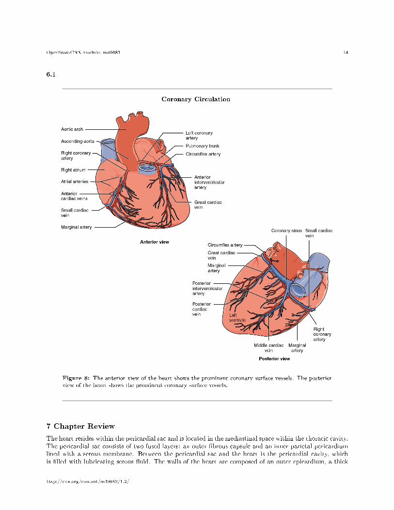
6 Coronary Circulation
You will recall that the heart is a remarkable pump composed largely of cardiac muscle cells that areincredibly active throughout life. Like all other cells, a cardiomyocyte requires a reliable supply of oxygenand nutrients, and a way to remove wastes, so it needs a dedicated, complex, and extensive coronarycirculation. And because of the critical and nearly ceaseless activity of the heart throughout life, this needfor a blood supply is even greater than for a typical cell. However, coronary circulation is not continuous;rather, it cycles, reaching a peak when the heart muscle is relaxed and nearly ceasing while it is contracting.
Coronary arteries supply blood to the myocardium and other components of the heart. The �rstportion of the aorta after it arises from the left ventricle gives rise to the coronary arteries.Coronary veins
drain the blood from the heart and generally parallel the large surface arteries (see Figure 8 (CoronaryCirculation )
http://cnx.org/content/m49683/1.2/
OpenStax-CNX module: m49683 14
6.1
Coronary Circulation
Figure 8: The anterior view of the heart shows the prominent coronary surface vessels. The posteriorview of the heart shows the prominent coronary surface vessels.
7 Chapter Review
The heart resides within the pericardial sac and is located in the mediastinal space within the thoracic cavity.The pericardial sac consists of two fused layers: an outer �brous capsule and an inner parietal pericardiumlined with a serous membrane. Between the pericardial sac and the heart is the pericardial cavity, whichis �lled with lubricating serous �uid. The walls of the heart are composed of an outer epicardium, a thick
http://cnx.org/content/m49683/1.2/

OpenStax-CNX module: m49683 15
myocardium, and an inner lining layer of endocardium. The human heart consists of a pair of atria, whichreceive blood and pump it into a pair of ventricles, which pump blood into the vessels. The right atriumreceives systemic blood relatively low in oxygen and pumps it into the right ventricle, which pumps it intothe pulmonary circuit. Exchange of oxygen and carbon dioxide occurs in the lungs, and blood high in oxygenreturns to the left atrium, which pumps blood into the left ventricle, which in turn pumps blood into theaorta and the remainder of the systemic circuit. The septa are the partitions that separate the chambersof the heart. They include the interatrial septum, the interventricular septum, and the atrioventricularseptum. Two of these openings are guarded by the atrioventricular valves, the right tricuspid valve and theleft mitral valve, which prevent the back�ow of blood. Each is attached to chordae tendineae that extend tothe papillary muscles, which are extensions of the myocardium, to prevent the valves from being blown backinto the atria. The pulmonary valve is located at the base of the pulmonary trunk, and the left semilunarvalve is located at the base of the aorta. The right and left coronary arteries are the �rst to branch o� theaorta and arise from two of the three sinuses located near the base of the aorta and are generally located inthe sulci. Cardiac veins parallel the small cardiac arteries and generally drain into the coronary sinus.
Glossary
De�nition 8: anastomosis
(plural = anastomoses) area where vessels unite to allow blood to circulate even if there may bepartial blockage in another branch
De�nition 8: anterior cardiac veins
vessels that parallel the small cardiac arteries and drain the anterior surface of the right ventricle;bypass the coronary sinus and drain directly into the right atrium
De�nition 8: anterior interventricular artery
(also, left anterior descending artery or LAD) major branch of the left coronary artery that followsthe anterior interventricular sulcus
De�nition 8: anterior interventricular sulcus
sulcus located between the left and right ventricles on the anterior surface of the heart
De�nition 8: aortic valve
(also, aortic semilunar valve) valve located at the base of the aorta
De�nition 8: atrioventricular septum
cardiac septum located between the atria and ventricles; atrioventricular valves are located here
De�nition 8: atrioventricular valves
one-way valves located between the atria and ventricles; the valve on the right is called the tricuspidvalve, and the one on the left is the mitral or bicuspid valve
De�nition 8: atrium
(plural = atria) upper or receiving chamber of the heart that pumps blood into the lower chambersjust prior to their contraction; the right atrium receives blood from the systemic circuit that �owsinto the right ventricle; the left atrium receives blood from the pulmonary circuit that �ows intothe left ventricle
De�nition 8: auricle
extension of an atrium visible on the superior surface of the heart
De�nition 8: bicuspid valve
(also, mitral valve or left atrioventricular valve) valve located between the left atrium and ventricle;consists of two �aps of tissue
De�nition 8: cardiac notch
depression in the medial surface of the inferior lobe of the left lung where the apex of the heart islocated
http://cnx.org/content/m49683/1.2/

OpenStax-CNX module: m49683 16
De�nition 8: cardiac skeleton
(also, skeleton of the heart) reinforced connective tissue located within the atrioventricular septum;includes four rings that surround the openings between the atria and ventricles, and the openingsto the pulmonary trunk and aorta; the point of attachment for the heart valves
De�nition 8: cardiomyocyte
muscle cell of the heart
De�nition 8: chordae tendineae
string-like extensions of tough connective tissue that extend from the �aps of the atrioventricularvalves to the papillary muscles
De�nition 8: circum�ex artery
branch of the left coronary artery that follows coronary sulcus
De�nition 8: coronary arteries
branches of the ascending aorta that supply blood to the heart; the left coronary artery feeds the leftside of the heart, the left atrium and ventricle, and the interventricular septum; the right coronaryartery feeds the right atrium, portions of both ventricles, and the heart conduction system
De�nition 8: coronary sinus
large, thin-walled vein on the posterior surface of the heart that lies within the atrioventricularsulcus and drains the heart myocardium directly into the right atrium
De�nition 8: coronary sulcus
sulcus that marks the boundary between the atria and ventricles
De�nition 8: coronary veins
vessels that drain the heart and generally parallel the large surface arteries
De�nition 8: endocardium
innermost layer of the heart lining the heart chambers and heart valves; composed of endotheliumreinforced with a thin layer of connective tissue that binds to the myocardium
De�nition 8: endothelium
layer of smooth, simple squamous epithelium that lines the endocardium and blood vessels
De�nition 8: epicardial coronary arteries
surface arteries of the heart that generally follow the sulci
De�nition 8: epicardium
innermost layer of the serous pericardium and the outermost layer of the heart wall
De�nition 8: foramen ovale
opening in the fetal heart that allows blood to �ow directly from the right atrium to the left atrium,bypassing the fetal pulmonary circuit
De�nition 8: fossa ovalis
oval-shaped depression in the interatrial septum that marks the former location of the foramenovale
De�nition 8: great cardiac vein
vessel that follows the interventricular sulcus on the anterior surface of the heart and �ows alongthe coronary sulcus into the coronary sinus on the posterior surface; parallels the anterior interven-tricular artery and drains the areas supplied by this vessel
De�nition 8: hypertrophic cardiomyopathy
pathological enlargement of the heart, generally for no known reason
De�nition 8: inferior vena cava
large systemic vein that returns blood to the heart from the inferior portion of the body
De�nition 8: interatrial septum
cardiac septum located between the two atria; contains the fossa ovalis after birth
http://cnx.org/content/m49683/1.2/

OpenStax-CNX module: m49683 17
De�nition 8: interventricular septum
cardiac septum located between the two ventricles
De�nition 8: left atrioventricular valve
(also, mitral valve or bicuspid valve) valve located between the left atrium and ventricle; consistsof two �aps of tissue
De�nition 8: marginal arteries
branches of the right coronary artery that supply blood to the super�cial portions of the rightventricle
De�nition 8: mesothelium
simple squamous epithelial portion of serous membranes, such as the super�cial portion of theepicardium (the visceral pericardium) and the deepest portion of the pericardium (the parietalpericardium)
De�nition 8: middle cardiac vein
vessel that parallels and drains the areas supplied by the posterior interventricular artery; drainsinto the great cardiac vein
De�nition 8: mitral valve
(also, left atrioventricular valve or bicuspid valve) valve located between the left atrium and ven-tricle; consists of two �aps of tissue
De�nition 8: moderator band
band of myocardium covered by endocardium that arises from the inferior portion of the inter-ventricular septum in the right ventricle and crosses to the anterior papillary muscle; containsconductile �bers that carry electrical signals followed by contraction of the heart
De�nition 8: myocardium
thickest layer of the heart composed of cardiac muscle cells built upon a framework of primarilycollagenous �bers and blood vessels that supply it and the nervous �bers that help to regulate it
De�nition 8: papillary muscle
extension of the myocardium in the ventricles to which the chordae tendineae attach
De�nition 8: pectinate muscles
muscular ridges seen on the anterior surface of the right atrium
De�nition 8: pericardial cavity
cavity surrounding the heart �lled with a lubricating serous �uid that reduces friction as the heartcontracts
De�nition 8: pericardial sac
(also, pericardium) membrane that separates the heart from other mediastinal structures; consistsof two distinct, fused sublayers: the �brous pericardium and the parietal pericardium
De�nition 8: pericardium
(also, pericardial sac) membrane that separates the heart from other mediastinal structures; consistsof two distinct, fused sublayers: the �brous pericardium and the parietal pericardium
De�nition 8: posterior cardiac vein
vessel that parallels and drains the areas supplied by the marginal artery branch of the circum�exartery; drains into the great cardiac vein
De�nition 8: posterior interventricular artery
(also, posterior descending artery) branch of the right coronary artery that runs along the posteriorportion of the interventricular sulcus toward the apex of the heart and gives rise to branches thatsupply the interventricular septum and portions of both ventricles
De�nition 8: posterior interventricular sulcus
sulcus located between the left and right ventricles on the anterior surface of the heart
http://cnx.org/content/m49683/1.2/

OpenStax-CNX module: m49683 18
De�nition 8: pulmonary arteries
left and right branches of the pulmonary trunk that carry deoxygenated blood from the heart toeach of the lungs
De�nition 8: pulmonary capillaries
capillaries surrounding the alveoli of the lungs where gas exchange occurs: carbon dioxide exits theblood and oxygen enters
De�nition 8: pulmonary circuit
blood �ow to and from the lungs
De�nition 8: pulmonary trunk
large arterial vessel that carries blood ejected from the right ventricle; divides into the left and rightpulmonary arteries
De�nition 8: pulmonary valve
(also, pulmonary semilunar valve, the pulmonic valve, or the right semilunar valve) valve at thebase of the pulmonary trunk that prevents back�ow of blood into the right ventricle; consists ofthree �aps
De�nition 8: pulmonary veins
veins that carry highly oxygenated blood into the left atrium, which pumps the blood into the leftventricle, which in turn pumps oxygenated blood into the aorta and to the many branches of thesystemic circuit
De�nition 8: right atrioventricular valve
(also, tricuspid valve) valve located between the right atrium and ventricle; consists of three �apsof tissue
De�nition 8: semilunar valves
valves located at the base of the pulmonary trunk and at the base of the aorta
De�nition 8: septum
(plural = septa) walls or partitions that divide the heart into chambers
De�nition 8: septum primum
�ap of tissue in the fetus that covers the foramen ovale within a few seconds after birth
De�nition 8: small cardiac vein
parallels the right coronary artery and drains blood from the posterior surfaces of the right atriumand ventricle; drains into the great cardiac vein
De�nition 8: sulcus
(plural = sulci) fat-�lled groove visible on the surface of the heart; coronary vessels are also locatedin these areas
De�nition 8: superior vena cava
large systemic vein that returns blood to the heart from the superior portion of the body
De�nition 8: systemic circuit
blood �ow to and from virtually all of the tissues of the body
De�nition 8: trabeculae carneae
ridges of muscle covered by endocardium located in the ventricles
De�nition 8: tricuspid valve
term used most often in clinical settings for the right atrioventricular valve
De�nition 8: valve
in the cardiovascular system, a specialized structure located within the heart or vessels that ensuresone-way �ow of blood
http://cnx.org/content/m49683/1.2/

OpenStax-CNX module: m49683 19
De�nition 8: ventricle
one of the primary pumping chambers of the heart located in the lower portion of the heart; the leftventricle is the major pumping chamber on the lower left side of the heart that ejects blood intothe systemic circuit via the aorta and receives blood from the left atrium; the right ventricle is themajor pumping chamber on the lower right side of the heart that ejects blood into the pulmonarycircuit via the pulmonary trunk and receives blood from the right atrium
http://cnx.org/content/m49683/1.2/