cario vascular system cario vascular system. the heart anatomy and physiology the heart anatomy and...
Post on 20-Dec-2015
224 views
TRANSCRIPT

CARIO VASCULAR CARIO VASCULAR SYSTEMSYSTEM
THE HEARTTHE HEARTAnatomy and PhysiologyAnatomy and Physiology
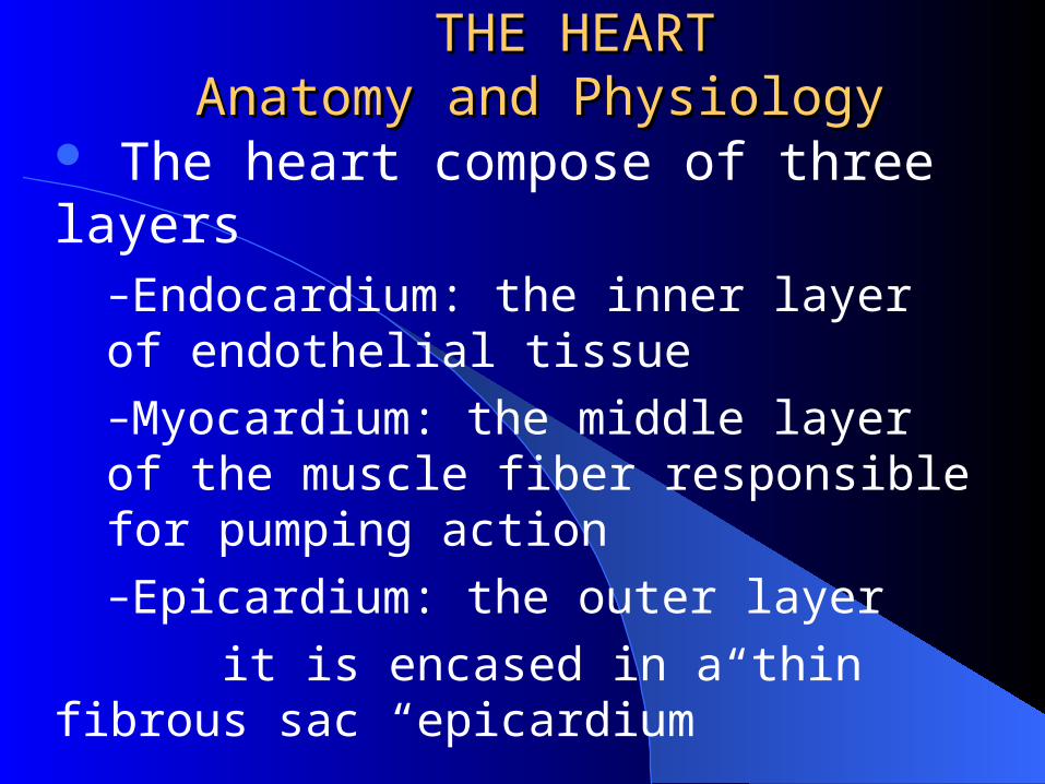
The heart compose of three layers–Endocardium: the inner layer of endothelial tissue–Myocardium: the middle layer of the muscle fiber responsible for pumping action–Epicardium: the outer layer
it is encased in a thin fibrous sac “epicardium”
THE HEARTTHE HEARTAnatomy and PhysiologyAnatomy and Physiology
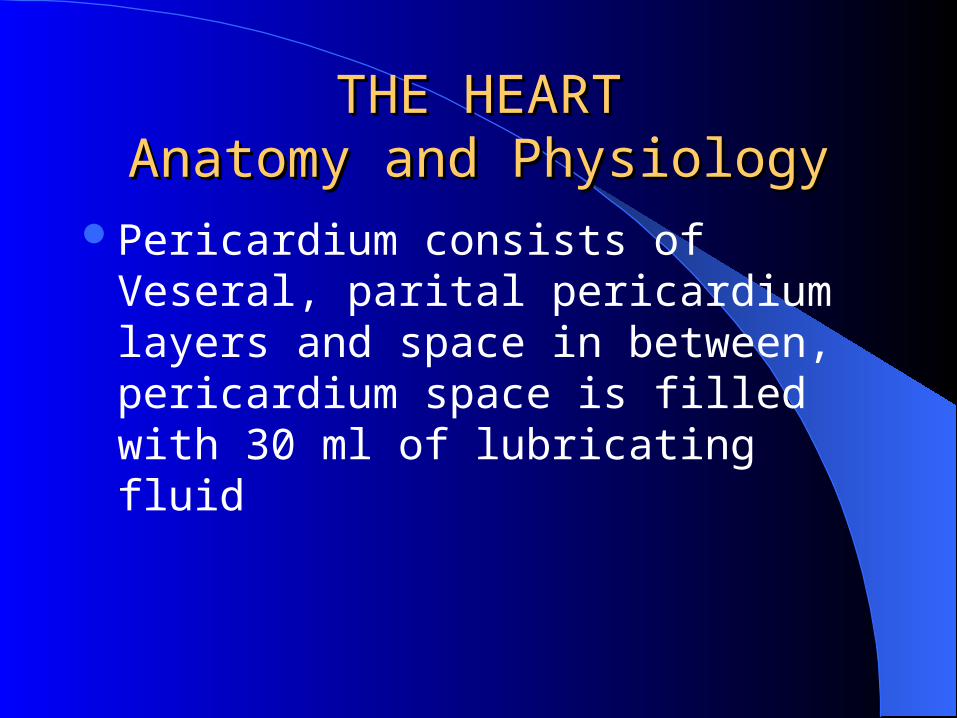
Pericardium consists of Veseral, parital pericardium layers and space in between, pericardium space is filled with 30 ml of lubricating fluid
Heart ChampersHeart Champers
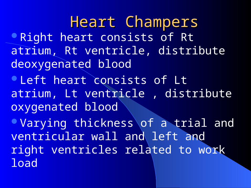
Right heart consists of Rt atrium, Rt ventricle, distribute deoxygenated bloodLeft heart consists of Lt atrium, Lt ventricle , distribute oxygenated bloodVarying thickness of a trial and ventricular wall and left and right ventricles related to work load

Heart ChampersHeart Champers
HEART VALVES: – Atrioventricular, separate atria from
ventricles– Semilunar valves ( three half-moon
leaflet

Coronary ArteriesCoronary Arteries
The heart has large metabolic requirement, 70-80% of delivered oxygenCoronary arteries are perfused during diastole Left coronary artery branches ( LAD & LCX )Right coronary artery branch ( RCA )Posterior wall received blood by ( PDCA )
CONDUCTING SYSTEMCONDUCTING SYSTEM
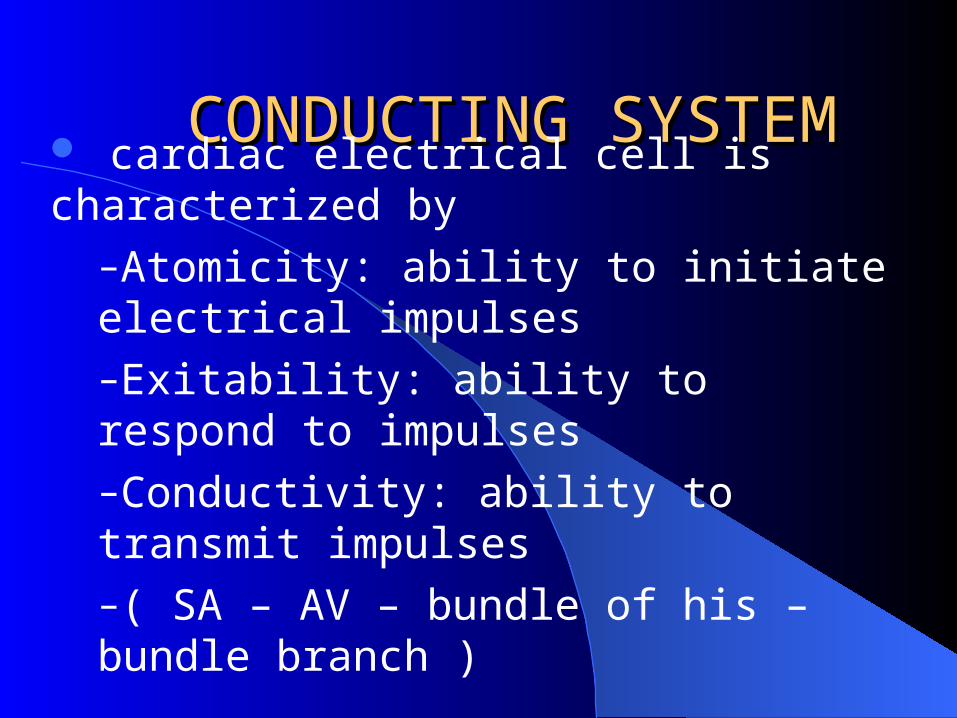
cardiac electrical cell is characterized by–Atomicity: ability to initiate electrical impulses–Exitability: ability to respond to impulses–Conductivity: ability to transmit impulses –( SA – AV – bundle of his – bundle branch )
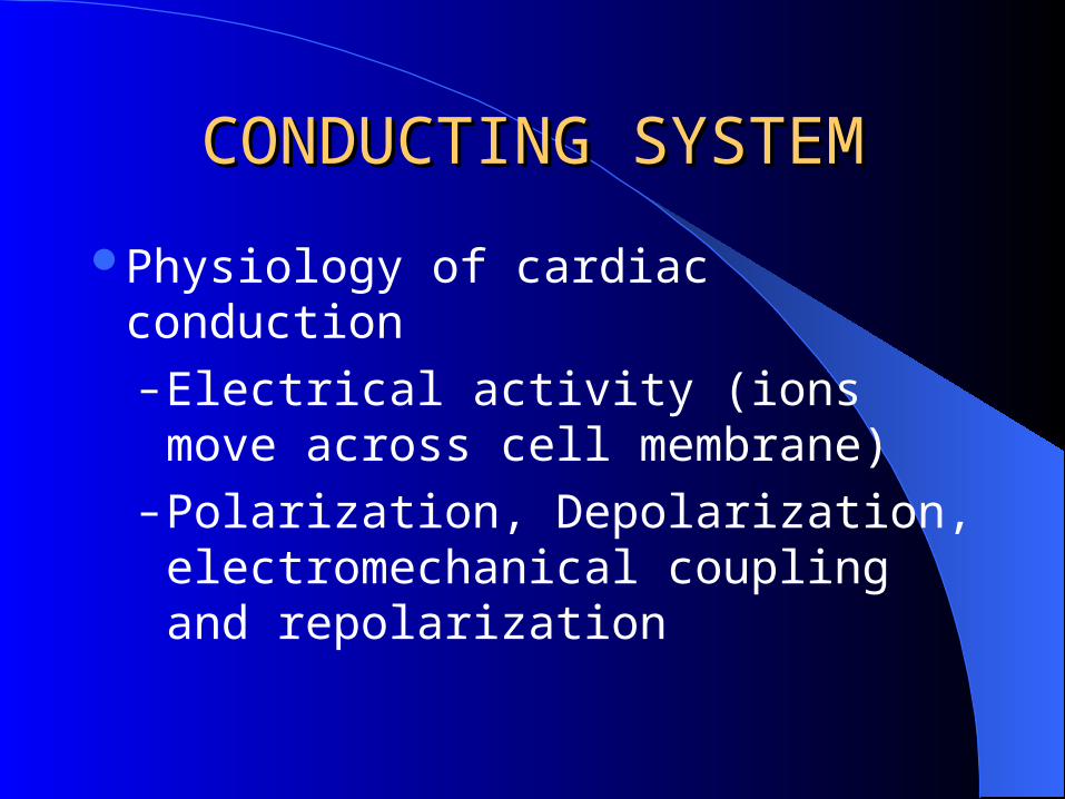
CONDUCTING SYSTEMCONDUCTING SYSTEM
Physiology of cardiac conduction– Electrical activity (ions move across
cell membrane)– Polarization, Depolarization,
electromechanical coupling and repolarization

contcont
Cardiac homodynamic – Cardiac cycle ( flow of blood )– Cardiac output ( ejection of blood )
= stroke volume x heart rate– Heart Rate: affected by central
nervous system and baroreceptors

cont.cont.
STROKE VOLUME : the amount of blood ejected by Lt ventricle in each beat which is 70 ml determined by preload, after load and contractilityEJECTION FRACTION: 42% for right heart and 50% for the left heart

ASSESSMENTASSESSMENT
The extent of assessment is determined by:–Purpose of nursing assessment–Severity of pt’s condition–Practice setting of the nurse

ASSESSMENTASSESSMENT
Health history and clinical manifestations– Acute symptoms: current medication,
allergies, general appearance, hemodynamic status
– Stable patients: complete history, spouse or partner, demographic information

ASSESSMENTASSESSMENT
Cardiac symptoms: pt will have one of the following– Shortness of breath, dizziness,
syncope, loss of consciousness, edema and weight gain, fatigue, palpitation and chest discomfort

NURSING DIAGNOSISNURSING DIAGNOSIS
Decrease cardiac output related to structural disordersActivity intolerance related to decrease cardiac output or excessive fluid volumeAnxiety related to change in health status and change in role function
COLLABRATIVE PROBLEMS
Congestive heart failure, ventricular dysrhythmias, and atrial dysrhythmias

PLANNING AND GOALSPLANNING AND GOALS
Improve and maintain cardiac outputIncrease activity toleranceReduction of anxietyAbsence of complications

NURSING IMPLEMENTATIONNURSING IMPLEMENTATION
Encourage rest, leaning back in a chairOxygen through nasal prongsCareful monitoring to correlate intervention with patient’s response to adjust treatment planDecrease sodium diet intakeChange position frequently

PHYSICAL ASSESSMENTPHYSICAL ASSESSMENT
Performed to confirm the data in health historyIt should include the following
–Effectiveness of heart as a pump, filling volume and pressure, cardiac output, compensatory mechanisms

PHYSICAL ASSESSMENTPHYSICAL ASSESSMENT
General appearance– Level of consciousness, thought
process, heart ability to perfuse brain tissue, distress and anxiety

PHYSICAL ASSESSMENTPHYSICAL ASSESSMENT
Skin– Pallor around finger nails and lips, peripheral
cyanosis, central cyanosis, xenthelasma ( yellowish indicates high cholestrol), reduce skin turgor, bruice ( anti coogulants )
Blood pressure– Normal is 120/80 mmhg ( 100/60 – 140/90 )
invasive and non invasive

Pulse pressure: –Difference between systolic and diastolic, 30-40 mmhg, reflects strock volume & ejection velocity, vascular resistance, –Less than 30 mmhg = serious reduction in output
PHYSICAL ASSESSMENTPHYSICAL ASSESSMENT
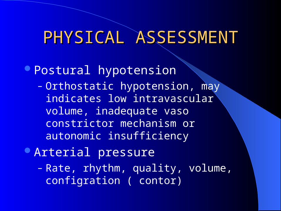
Postural hypotension– Orthostatic hypotension, may indicates low
intravascular volume, inadequate vaso constrictor mechanism or autonomic insufficiency
Arterial pressure– Rate, rhythm, quality, volume, configration
( contor)

PHYSICAL ASSESSMENTPHYSICAL ASSESSMENT
Jugular venous pressure– Reflects Rt heart function, provide estimation
of CVP, increase incase of HF decrease in FVD
Heart – Inspection, palpation, percussion , auscultation,
S1 S2

PHYSICAL ASSESSMENT
Inspection of Extremities–Capillary refill time (CHF, hypertension ), hematoma (surgery & cath ), vascular changes, peripheral edema, lower extremeties ulcer
Lungs–Tachypnea ( HF, pain, anxiety ), chynestokes breathing ( pulmonary edema ), dry cough, crackles and whezes

PHYSICAL ASSESSMENTPHYSICAL ASSESSMENT
Abdomen– Hepato jugular reflux, bladder distention
RISK FACTORS– Nonmodifiable: age, positive family history,
race &gender– Modifiable: hyperglycemia, hyperlipidemia,
hypertension, inactivity, smoking, obesity & type A personality

DIAGNOSTIC EVALUATIONDIAGNOSTIC EVALUATIONLaboratory tests
–Cardiac enzymesReleased from injured cells when ruptured the membraneMost of them are not specific for one type of cellIso enzymes are more specific, createnine kinase ( CK ) and its iso enzyme( CK-MB ), lactic dehydrogenase (LDH) , troponin I

DIAGNOSTIC EVALUATIONDIAGNOSTIC EVALUATION– Blood chemistry
Lipid profile– A- cholesterol (less than 200 mg / dl ),
required for hormonal synthesis, found mainly in brain tissue and liver
– B- triglycerides ( 40 – 150 mg / dl ) source of energy, cell wall, store in a dipose tissue
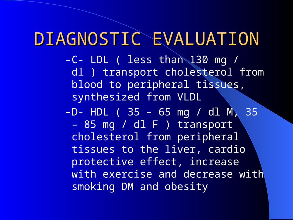
DIAGNOSTIC EVALUATIONDIAGNOSTIC EVALUATION– C- LDL ( less than 130 mg / dl ) transport
cholesterol from blood to peripheral tissues, synthesized from VLDL
– D- HDL ( 35 – 65 mg / dl M, 35 – 85 mg / dl F ) transport cholesterol from peripheral tissues to the liver, cardio protective effect, increase with exercise and decrease with smoking DM and obesity

Serum electrolytes–K, Na, Ca and other electrolytes can reflect the heart function as well as fluid & electrolyte disturbances
BUN–May indicates impaired renal function and impaired cardiac output
DIAGNOSTIC EVALUATIONDIAGNOSTIC EVALUATION
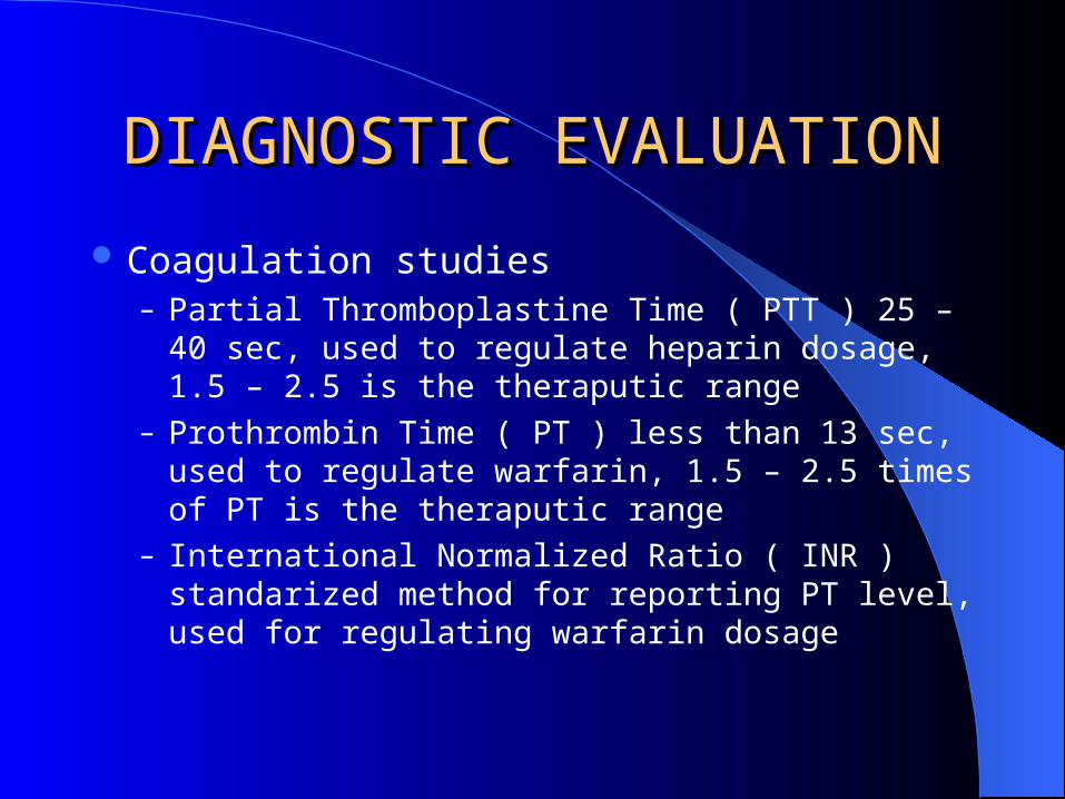
Coagulation studies– Partial Thromboplastine Time ( PTT ) 25 – 40 sec, used
to regulate heparin dosage, 1.5 – 2.5 is the theraputic range
– Prothrombin Time ( PT ) less than 13 sec, used to regulate warfarin, 1.5 – 2.5 times of PT is the theraputic range
– International Normalized Ratio ( INR ) standarized method for reporting PT level, used for regulating warfarin dosage

Chest X-Ray ( CXR )
–Assess size, position of the heart, cardiothoracic ratio ( CTR ), position of central lines
Elecrocardiography ( ECG )–Can be either on bed side or from a distance, 12 leads ECG, continuous monitoring, telemetry monitoring 2 or 3 leads monitoring )

DIAGNOSTIC EVALUATIONDIAGNOSTIC EVALUATION
Cardiac Stress TestDuring time of increased demand, abnormalities
in cardiovascular functions are more likely to be detected, used to evaluate the heart function, coronary arteries as well as the cause of chest pain

Con. –Exercise stress test: pt walk on a treadmill or pedals (stationary bicycle), the goal is to increase HR and monitored for ECH changes, arrhythmias, hypotension, pain, dyspnea and dizziness. Pt fast 4 hours before test, nurse needs to instruct pt about the test

DIAGNOSTIC EVALUATIONDIAGNOSTIC EVALUATION
Echocardiography: a non invasive ultrasound used to examine size shape and cardiac motion, used also to evaluate heart function, valves and peripheral effusion
Pharmacologic stress test: used for pts unable to achieve target HR, Dipyridamole, Adenosine & dobutamine are used for this purpose

Cardiac catheterization
–Invasive diagnostic procedure involves introduction of specific catheter into Rt & Lt side blood vessels under fluoroscopy. Its used to evaluate coronary arteries patency, heart function as a pump, vascular system and heart structure–Its considered as a highly critical procedure–Take in consideration: monitoring IV line, BP, ECG, LOC, well prepared staff to provide ACLS, revission of lab tests

CORONARY CORONARY VASCULAR VASCULAR
DISORDERSDISORDERS

ATHEROSCLEROSISATHEROSCLEROSIS
Definition: abnormal accumulation of lipid or fatty substances and fibrous tissues in vessel wall Pathophysiology
–It begins as a fatty streak, this streak develop into advanced lesion which involves inflammatory response, T.lymphocytes and monocytes ingest the lipid and form fibrous cap called Atheroma Plaque, this protrude into the lumen of the vessel narrowing and obstruct it
ATHEROSCLEROSISATHEROSCLEROSIS
Clinical manifestations– Acute onset chest pain, ECG changes,
dyarhythmias & death
Risk factors– Age, family history of non modifiable risk
factors, high cholestrol, cigarette smoking, hypertension & DM

PREVENTIONControl cholestrol level, LDL less than normalDietary control decrease fat & increase fiberMedication to decrease serum fat & cholesterolQuit smokingEarly detection & control hypertensionControl DMGender & estrogen levelBehavior pattern

ANGINA PECTORISANGINA PECTORISDefinition: it’s a clinical syndrome ch.ch by episodes of pain or pressure in the anterior chestPathophysiology
–Caused by atherosclerotic disease, associated with significant obstruction of CA and any cause that increase demand or decrease supply
ANGINA PECTORISANGINA PECTORIS
Clinical manifestation– Pain deep behind upper third of sternum
mediated to jaw neck shoulders & left arm– Chest heaviness & tightness with choking
sensation– Weakness, numbness in arms, wrist &
hands– Short breathing, pallor, dizziness, nausea &
vomiting

ANGINA PECTORISANGINA PECTORISMedical Management: the objectives are to decrease the demand and increase blood supply to the heart
–Pharmacological therapy & control risk factorsNitroglycerides, beta blockers, Ca channel blockers & antiplatlet agents ( aspirin heparin )
–Revascularization proceduresCoronary artery bipass ABGPercutaneus transluminal coronary angioplasy PTCA
–O2 administration

MYOCARDIAL INFARCTION MIMYOCARDIAL INFARCTION MI
Definition: death of heart tissue caused by ischemia, a process by which areas of the myocardial cells destroyedClinical manifestation
–Sudden sever chest pain radiated to left arm–anxious & restlessness,–cool pale moist skin, sweating–tachypnea & tachycardia

MYOCARDIAL INFARCTION MIMYOCARDIAL INFARCTION MI
Diagnostic findings and assessment– Pt history, ECG: T wave & ST
segment changes echo & lab testsMedical management: objectives are to - minimize myocardial damage, - preserve function & prevent
complications

MYOCARDIAL INFARCTION MIMYOCARDIAL INFARCTION MI
– Emergent PTCA to open occluded artery
– Pharmacologic therapy:– thrombolytic ( STK, TPA ), – Analgesics– ACE inhibitors

MITRAL STENOSISMITRAL STENOSIS
Definition: an obstruction of blood flowing from the left atrium into the left ventricle, most often caused by rheumatic endocarditisClinical manifestation
–Progressive fatigue due to low cardiac output,–hemoptesis, dyspnea–cough & repeated respiratory infections

MITRAL STENOSISMITRAL STENOSIS
Management– Antibiotic– Valvoplasty– medication in case of surgical failure– PTCA may relieve symptoms

MITRAL REGURGITATIONMITRAL REGURGITATION
Definition: blood flowing back from the left ventricle into the left atrium during systole, the margins of the mitral valves cannot close during systole

MITRAL REGURGITATIONMITRAL REGURGITATIONClinical manifestation
– Chronic often asymptomatic– Acute :severe congestive heart failure,– dyspnea, palpitation, fatigue & weakness,
shortness of breath & cough from pulmonary congestion
Management– Surgical mitral valve replacement– Valvuloplasty

AORTIC STENOSISAORTIC STENOSIS
Definition: narrowing of the orifice between the left ventricle &the aorta, congenital malformation
Clinical manifestation–Many pts are asymptomatic–Exertional dyspnea, dizziness, fainting, angina pectoris, low pulse pressure ( 30 mmhg or less )

AORTIC STENOSISAORTIC STENOSIS
Management– Prophylactic antibiotic to prevent
endocarditis– Medication as prescribed for dysrhythmias– Surgical replacement for the aortic valve– One-or-two-balloon percotaneous
valvuplasty
AORTIC REGURGITATIONAORTIC REGURGITATION
Definition: flow of blood back into the left ventricle from the aorta during diastole, congenital deformities, endocarditis, syphilis, dissecting aneurysm Clinical manifestation
–Force full heart beat ( head & neck ),– arterial pulsation– exertional dyspnea and fatigue, difficult breaths specially at night
AORTIC REGURGITATIONAORTIC REGURGITATIONManagement
– Prophylactic antibiotic to prevent endocarditis
– Treatment of heart failure and dysrhythmias– Aortic valve replacement ( treatment of
choice )– Surgery is recommended for pt with Lt
ventricular hypertrophy regardless the presence or absence of symptoms

CARDIOMYOPATHIES CARDIOMYOPATHIES
Definition: it’s a heart muscle disease of unknown cause, an inherited genetic disorderTypes
–Dilated or congestive cardiomyopathy–Hypertrophic–Restrictive or constrictive

CARDIOMYOPATHIESCARDIOMYOPATHIES Clinical manifestations
– A symptomatic for many years– Shortness of breath, noctural dyspnea, cough,
chest pain, palpitation, dizziness & fatigue Medical management
– Mange precipitating cause, correct heart failure, diet & exercise regimen, control dysrhythmias,
– Implanted electrical device– myomectomy & heart transplantation

RHEUMATIC ENDOCARDITISRHEUMATIC ENDOCARDITIS
Definition: inflammation of endocardium result from rheumatic fever caused by a group A streptococcal infection Clinical manifestation
–Tiny translucent growth, pin-head size beats of the valve flaps, serious dysrhythmias, pneumonia, valvular deformaties, murmer, thrill & palpitation
Medical management–Eradicate organism & prevent complications–Penicillin as a drug of choice ( long term therapy )
Nursing management –Teach pt about disease, prevention & treatment–Instruct about long term therapy–Instruct for prophylactic therapy
PERICARDITISPERICARDITISDefinition: inflammation of the pricardium
Clinical manifestations–Pain over pericardium, clavicle, neck & scapula–friction rub, aggravated by breathing& turning in bed, relieved by sitting up– dyspnea, low cardiac output, increase WBC, pt appears extremely ill

PERICARDITISPERICARDITISMedical management
– Determine& treat cause, bed rest,– analgesics NSAID, corticosteroids– prevent pericardial effusion
Nursing management– Medication as prescribed, gradual
increase in activity unless fever, pain and friction rub reappear
ACUTE PULMONARY EDEMAACUTE PULMONARY EDEMA Definition: abnormal accumulation of fluids in the lungs either in interstitial space or in alveoli
Clinical manifestations–Restlessness, confusion, breathlessness– sense of suffocation, rapid weak pulse– distended neck veins, cold hands, cyanosed nail beds, gray skin– cough & decrease O2 saturation

ACUTE PULMONARY EDEMAACUTE PULMONARY EDEMA
Medical management: to improve res. Exchange– O2 therapy, medication and nursing
support– Intubation and mechanical ventilator in
severe failure– PEEP, oximetry ABGs– Morphine ( 2-5 mg ) to reduce anxiety

ACUTE PULMONARY EDEMAACUTE PULMONARY EDEMA
Duretic therapy– To increase rate of urin production, decrease
ECF, thiazide & loopduretics ( dose depends on indication, clinical signs & renal function )
– Medications to increase myocardial contractility & cardiac output ( digitalis )

ACUTE PULMONARY EDEMAACUTE PULMONARY EDEMA
Nursing management
– Position pt to promote circulation– Psychological support– Monitor medication

CARDIAC FAILURE CARDIAC FAILURE congestive heart failurecongestive heart failure
Definition: inability of heart to pump sufficient blood to meat needs of tissue for O2 & nutrientClinical manifestations
–In adequate tissue perfusion, dizziness, confusion, fatigue, cool extremities,–exercise & heat intolerance– low urine output, high venous pressure, pulmonary & peripheral edema, weight gain

CARDIAC FAILURE CARDIAC FAILURE congestive heart failurecongestive heart failure
Management– Counseling & education for regular
exercise, sodium restriction, avoid excessive fluid intake, medication based on symptoms, O2 therapy intubation if needed, transplantation
CARDIAC FAILURE CARDIAC FAILURE congestive heart failurecongestive heart failure
Nursing management – I&O, daily wt, daily auscultate lung
sounds, jugular vein assessment, edema, pulse rate, BP, skin turgur & manage complications

VASCULAR VASCULAR SYSTEMSYSTEM

The vascular system consists of two interdependent systems
–Right side of the heart pump–Left side of the heart pumpBLOOD VESSELS
Arteries and arteriols–High-pressure system, thick wall, transport oxygenated blood away from the heart–Located in protected areas away from skin surface–Wall of arteries and arteriols composed of 3 layers
Intema: inner indothelial layer, contact with bloodMedia: smmoth elastic tissue, constrict & dilate vesselsAdventitia: connective tissue, anchors vessels to surrounding
Capillaries–Single layer of endothelial cells, permits rapid & effective transport of nutrients to the cells & removal of metabolic waste

Veins and venules
–Larger in diameter than arteries but the wall are thinner because there is a less muscle & elastic tissue in the tonic & media, this allow these vessels to distend more than arteries–Equipped by one-way bicusped valves that prevent blood to back flow–Valves composed of endothelial leaflets–Transport deoxygenated blood from the body to the heart

Health History and Clinical Health History and Clinical ManifestationsManifestations
A muscular cramp-type pain in the extremities reproduce with the same degree of exercise or activity & relieved by rest is experienced by patients with peripheral arterial insufficiencyIntermittent claudication: its due to inability of arterial system to provide adequate blood flow to the tissues in the face of increased demand for nutrients during exercise
–Rest pain: worse at night & may interfere with sleep–As general role, the pain of intermittent claudication occurs one joint level below the disease process

Changes in skin appearance and temperature–Inadequate blood flow cause cool & pale extremities–Redish-blue discoloration of extremities (rubor)–Additional changes resulting from chronically reduced nutrients supply like: loss of hair, brittle nails, dry skin, atrophy, ulceration, gangrene by traumatic events
Pulses–Determining the presence or absence as well as quality of peripheral pulses to assess the status of peripheral arterial circulation–Absence of pulse may indicates the size of stenosis (narrowing or constriction )
Diagnostic EvaluationDiagnostic EvaluationDoppler ultrasound
–When pulses cannot be reliably palpated, use of a microphne-like hand held doppler ultrasound device (tranceducer or prob) may be helpful in detecting and assessing peripheral flow–Procedure
Exercise testing–Used to determine how long can a pt walk & measure ankle systolic BP in response to walking–A normal response is little or no drop in systolic BP, it drops in true claudication
Angiography –Used to confirm the diagnosis of occlusive arterial disease when considering surgery or intervention–Injecting radiopaque contrast agent directly into vascular system to visualize the vessels–Pts experience temporary warmth during injection, side effects are local irritation, allergic reactions, dyspnea, N & V, sweating tachycardia, numbness–Additional risks: vessel injury, bleeding & strock
Contrast plebography ( venography )–Performed if pt is to undergo thrombolytic therapy–Contrast is injected via dorsal foot vein then X-ray image will disclos an unfilled vein–Injection may cause brief painful inflammation of the vein
MANAGEMENT OF ARTERIAL MANAGEMENT OF ARTERIAL DISORDERSDISORDERS
Arteriosclerosis & Atherosclerosis–Arteriosclerosis: the most common, its hardening of the arteries, the muscle fiber & endothelial walls of small arteries & arteriols become thickened–Atherosclerosis: affect the intema of the large & medium size arteries due to accumulation of lipids, calcium, blood components, fibrous, carbohydrates on the internal layer
Risk factors–Modifiable: diet, DM, high BP, stress, life style –Non modifiable: age, gender
Clinical manifestations–Depends on tissue or organ affected, –Coronary: (angina, MI ), cerebrovascular ( TIA, strocke), Aorta ( aneurysm ), renovascular ( hypertension )
Medical management–Modification of risk factors to improve circulation
Surgical managementRadiologic intervention

PERIPHERAL ARTERIAL PERIPHERAL ARTERIAL OCCLUSIVE DISEASEOCCLUSIVE DISEASE
Arterial Insufficiency of extremities is usually found in older than 50 yrs, most often in men, legs are most frequently affected
–Distal occlusive disease frequently seen in pts with DM & elderly pts
Clinical manifestation Intermittent claudication described as aching, cramp, fatigue, weakness in joint muscles below stenosis or occlusion areaDecrease ability to walk, increase pain with ambulationIschemic pain at rest & worse at night Bruit sound on auscultationAbsence of peripheral pulse
PERIPHERAL ARTERIAL PERIPHERAL ARTERIAL OCCLUSIVE DISEASEOCCLUSIVE DISEASE
Medical management– Exercise program with weight reduction– Smoking cessation
Surgical management
- Grafting
- Endarterectomy

Nursing ManagementNursing Management
Maintaining circulation–Check pulses of affected extremities, note symmetry, color, temp., capillary refill, sensory & motor hourly –Doppler evaluation of vessels distal to bypass graft
Monitoring & managing potential complications
–Urine output, CVP, pulse, leak, hematoma & edema
Promoting home & community based care–Discharge plan, pt education, assess ability to change life style & self care
AORTIC ANEURYSM AORTIC ANEURYSM
It’s a localized sac or dilation involving an artery formed at a weak point in vessel wallTHORASIC AORTIC ANEURYSM
–Caused by atherosclerosis in men aged ( 40-70 yrs )–Most common is dissecting aneurysm, 1/3 of cases die of rupture

AORTIC ANEURYSMAORTIC ANEURYSM
Clinical manifestation– Pain in supine position, dyspnea, hoarseness or
aphonia ( complete loss of voice ), dysphagia
Medical management– Surgical repair, control BP, correcting risk
factors

ABDOMINAL AORTIC ANEURYSMmost common cause is atherosclerosis, affect men
4 times than women, occur mostly below renal stenosis, untreated outcome is rupture & death
Clinical manifestations–Abdominal heart beat on lying position,– abdominal mass – thrombing

ABDOMINAL AORTIC ABDOMINAL AORTIC ANEURYSMANEURYSM
Medical management– Surgery is the treatment of choice
Bypass graft, endovascular graft ( suturless )
Nursing management– Assessment before surgery, post op. systematic
monitoring, neurological assessment, signs of impeding rupture ( abdominal & back pain )

ARTERIAL EMBOLISM Its acute vascular occlusion due to an embolus or acute thrombosis
Causes –Iatrogenic injury ( insertion of catheters ), trauma from fractures, crush injury, penetrating wound, thrombi development in heart champers as a result of (AF, MI, CHF)
Clinical manifestations–Cessation of distal bld flow, gradual loss of sensory & motor function, pain, pallor, cold, paresthesia, pulse lessens & paralysis

ARTERIAL EMBOLISMARTERIAL EMBOLISM
Medical management– Surgery ( embolectomy ),– Medication ( heparin bolus5000-10000 then
contentious infusion) Thrombolytic therapy (STK; streptokainase)
Nursing management– Pre-op. bed rest, warm at room temp. protection
from trauma– Post-op. encourage movement & continue
anticoogulants

VENOUS DISORDERSVENOUS DISORDERS
DVT, THROMBOPHLEBITIS and PHLEBOTHROMBITISClinical manifestations
–Massive swelling, tenderness,– warmer affected extremity,– homans sign, –heaviness & functional loss
VENOUS DISORDERSVENOUS DISORDERS
Medical management – Anti coagulants (heparin 5-7 days), low
molecular-weight heparin, thrombolytic therapy– Surgical management: thromboectomy when
anti coagulant is contraindicated or danger of pulmonary embolism is extreme
Nursing management– Monitor PT, PTT, Hb, platlets, report bleeding,
assess anti coagulant therapy, monitor & manage complications, provide comfort & apply elastic pressure stockings.

CHRONIC VENOUS INSUFFICIENCY
Obstruction of venous valves in the legs or a reflux of blood back through the valvesClinical manifestations
–Chronic venous stasis, altered pigmentation, pain, stasis dermatitis, stasis ulceration, dry skin, cracks & itches
CHRONIC VENOUS CHRONIC VENOUS INSUFFICIENCYINSUFFICIENCY
Medical & nursing management – Reducing venous stasis & prevent ulceration– Elevating legs to reduce edema & promote venous
return– Encourage walking– Compression with elastic stockings to reduce blood
pooling– Protect from trauma– Keep skin dry & soft– Immediate report for signs of ulceration
LEG ULCERS
Its an excavation of the skin surface that occurs when inflamed necrotic tissue sloughs offClinical manifestations
–Open inflamed sore, pain & edema, discharge may be present, heaviness, itching, area may covered with eschar, gangrene
Medical management–Pharmacologic therapy (antibiotics based on culture) depridement, topical therapy, stimulated healing by Epigram

VARICOSE VEINS
Abnormally dilated torturous superficial veins caused by incompetent venous valvesClinical manifestations
–Dull aches, muscle cramps, increase muscle fatigue, ankle edema, S&S of venous insufficiency if obstruct

VARICOSE VEINSVARICOSE VEINS
Medical management– Surgery ( ligation ) & sclerotherapy
Nursing management– Bed rest 1st 24 hours & start walking at 2nd day
5-10 min /2 hours, elastic pressure stockings, elevate foot, discourage standing & sitting, promote comfort (analgesia), home & community based care

NURSING PROCESSNURSING PROCESS
Assessment–Sub. (interview) & obj. (physical assessment)
Diagnosis–Alteration in peripheral tissue perfusion–Pain, risk for impaired skin integrity, knowledge deficit regarding self care activities

NURSING PROCESSNURSING PROCESS
Implementation– Lower the extremity below level of the heart– Encourage moderate amount of walking– Maintain warm & discourage nicotine use– Administer prescribed vasodilators, adrenergic block
agents Evaluation
– Goal met evidenced by: extremities warm to touch, color improved, decrease muscle pain, tolerate performing exercise 6 times / day

HYPERTENTIONHYPERTENTION
Definition: it’s a raise of blood pressure above normal range” systolic above 140 mmhg & diastolic above 90 mmhg” over sustained periodA multifactorial conditionA sign ,a risk factor , and a disease .
HYPERTENTIONHYPERTENTION Types & causes
– 1- Primary (idiopathic essential ) hypertention80-90% of cases are of unknown cause but
predisposed by: old age over 60 yrs, obesity, black race, atherosclerosis
– Benign or chronic hypertentionRise is usually slight to moderate & continue
to rise slowly often asymptomatic ( silent killer )
– Malignant (accelerated) hypertentionBP very high & continue to raise rapidly,
diastolic pressure in excess of 120 mmhg & the effects are quickly apparent

HYPERTENTIONHYPERTENTION
2- Secondary hypertention: Increase BP from an identified cause resulting from other disease
Causes – Renal disease, endocrine disorders, age & sex,
stressful occupation & situation, family tendency, DM, dyslipidemia, smoking & alcohol

HYPERTENTIONHYPERTENTION
Clinical manifestations– High blood pressure reading– Headache, epistaxis, angina,– dizziness, dyspnea, ringing in ears– Retinal changes “may be papilledema”– “ a common consequence M.I. & CAD

HYPERTENTIONHYPERTENTION
Medical management – Treatment of underlying cause– Life stile modification :-– Management of predisposing factors ( low
salt diet, decease weight, stop smoking, decrease stress level)
– Pharmacologic therapy (diuretics, vasoconstrictive agents & agents to decrease cardiac output )
Nursing ProcessNursing ProcessNursing Diagnosis
–Knowledge deficit ( medication & disease process )–Non compliance with therapeutic regimen
E.O–Pt will understand disease process & its treatment–Pt will participate in self care program
Implementation–Allow pt to rest & relax, medication as prescribed, report side effects, educate pt about rebound hypertention that occurs if therapy suddenly stopped–Measure BP routinely, follow up appointments, –Educate pt about arthostatic hypotention so to stand gradually –Life style modification