case-based approach to the incidental liver...
TRANSCRIPT
Managing Incidental Findings 1
Case-Based Approach to the Incidental Liver LesionRajan T. Gupta1,2
How can we, as radiologists, ensure that the correct im-aging studies are being performed as we transition to val-ue-based care? One way is by critically looking at our rec-ommendations for incidental findings on CT. Too often, the recommendations for an identical lesion vary widely by radi-ologist, practice type, and region. The purpose of this chap-ter is to show a case-based approach to the incidental liver lesion using the American College of Radiology (ACR) White Paper [1] as a guide.
As the use of MDCT has increased, there has been a con-comitant increase in the frequency of detecting incidental find-ings [2–5]. These incidental findings are defined as findings that are unrelated to the clinical indication for the imaging examination. Although it can be helpful to detect these find-ings, detection of these lesions can also potentially confound physicians and patients with regard to the optimal means by which to manage them [1]. One major issue is that the workup of these incidental lesions can lead to increased utilization of cross-sectional imaging and, subsequently, increased health care costs [1]. Given that workup and follow-up recommenda-tions for these lesions can vary widely by physician, hospital, and region, some potential advantages of standardizing the ap-proach would be to attempt to limit costs and reduce risk to patients from unnecessary imaging [1, 6]. A full discussion of costs and the conundrum associated with reducing costs while maintaining quality is beyond the scope of this chapter, but the ACR White Paper [1] does begin to address this issue.
It is difficult to quantify how often incidental liver lesions are encountered in clinical practice, but some estimates suggest that more than 50% of patients without a history of malignancy had benign hepatic lesions at autopsy [7, 8] and that up to 15% of CT studies show incidental findings in the liver [8]. The ACR White Paper on Incidental Findings [1] has provided an outstanding evidence- and expert consensus–based approach for incidental lesions that are commonly encountered in prac-tice. This document is comprehensive in that it deals with inci-dental findings in the kidneys, adrenal glands, pancreas, liver, and so on. This chapter will be a case-based approach to the
incidental liver lesion with a focus on pertinent management and important lessons learned in clinical practice.
First Things FirstWhen an incidental focal liver lesion is identified, the
ACR Incidental Findings Committee [1] suggests that there are three key questions that should be asked to determine the next best steps. First, does the incidental liver lesion put the patient at risk for an adverse outcome? Next, is it possible to differentiate a benign cause from a malignant cause with confidence? Finally, if the lesion is benign, are there any po-tential complications that could arise from it (e.g., bleeding or hemorrhage from hepatic adenoma) [1]? To ascertain the importance of the incidental liver lesion, one must be able to separate individual patients with these findings into clinical risk categories. There are three risk categories as defined in the ACR White Paper [1]: low risk, average risk, and high risk. Low-risk individuals are 40 years old or younger with no history of malignancy, no risk factors, and no symptoms attributable to hepatic disease. Average-risk individuals are differentiated from low-risk individuals by age alone (> 40 years old). Finally, high-risk individuals are those who have a known primary malignancy that can metastasize to the liver, hepatic risk factors (detailed in the ACR White Paper), or a known hepatic disease such as cirrhosis.
Establishing the clinical risk category helps to determine the next steps in management—that is, additional imaging, bi-opsy, or no further workup [1]. Although biopsy of focal liver lesions is generally a safe procedure, it is not without risk, morbidity, or controversy [9]. The postprocedural morbidity of percutaneous liver biopsy is estimated to range from 2.0% to 4.8% with a mortality of 0.05% [10–12]. In clinical prac-tice, my colleagues and I use percutaneous liver lesion biopsy as the final step if we are unable to reach a conclusion about the cause of an incidental liver lesion with noninvasive cross-sectional modalities; therefore, we believe that biopsy remains an important part of the workup of a small number of inciden-tal liver lesions similar to the way in which it is used by other authors [13].
1Department of Radiology, Duke University Medical Center, Box 3808, Durham, NC 27710. Address correspondence to R. T. Gupta ([email protected]).
2Duke Cancer Institute, Durham, NC.

2 Managing Incidental Findings
Gupta
The FlowchartYou have now identified an incidental
liver lesion, measured its size, and have placed the patient in a risk category, so what’s next? The ACR White Paper [1] has a flowchart with color coding to lead you through the next steps (Fig. 1). The key numbers that you need to know are 0.5 and 1.5 cm because these values are the key size thresholds that the ACR White Paper uses in the flowchart. The yellow boxes identify the imaging features and risk category needed to lead you to pink boxes, indicating that the workup can stop because the finding is likely to be benign and does not require additional follow-up, or to green boxes, indicating the need to pursue further diagnostic testing, biopsy, or follow-up [1]. With this flowchart in mind, I will discuss some case examples.
Case ExamplesCase 1
Low-attenuation lesions between 0.5 and 1.5 cm are commonly encountered on MDCT. The key factor that deter-mines how to follow these lesions is whether the lesion shows benign or sus-picious imaging features. This delinea-tion is critical because the risk category does not impact recommendations.
Benign imaging features include well-circumscribed margins, attenuation val-ues of less than about 20 HU, homogene-ous appearance, and findings suggestive of typical hemangiomas [1]. In contrast, suspicious imaging features include ill-defined margins, enhancement with at-tenuation values of greater than 20 HU, heterogeneous appearance, and enlarge-ment [1] (Fig. 2).
Case 2The recommendations for flash-filling
lesions between 0.5 and 1.5 cm depend on the risk category of the patient. For patients who are low or average risk, these lesions are considered to be benign findings according to the ACR White Paper [1] and no further follow-up is recommended (Fig. 3). In high-risk in-dividuals, if the lesion shows suspicious features, multiphase MRI is recommend-ed; otherwise, follow-up CT or MRI in 6 months is advised.
Case 3Although the flowchart in the ACR
White Paper [1] is generally intended for use on incidental liver lesions iden-tified on MDCT, it can be used at times for lesions identified on abdominal sono-graphy. In these cases, my colleagues and I find it helpful to use lesion size and patient risk category to determine the next steps because some of the imaging features on the flowchart are not as eas-ily assessed on sonography (Fig. 4).
Case 4For a flash-filling lesion larger than
1.5 cm, the key factor in determining the next steps in management is the presence or absence of benign features on diagnos-tic imaging [1]; the patient risk category does not play a significant role in deter-mining the next steps (Fig. 1) for these lesions. If there are benign diagnostic im-aging features, the differential diagnosis for these lesions is typically focal nodular hyperplasia (FNH) or adenoma. Multi-phase MRI is recommended to make this distinction. The use of hepatobiliary MRI contrast agents to assist with this distinc-tion, especially for the diagnosis of FNH, has been well documented [14–16]. This recommendation is related to the fact that hepatobiliary MRI contrast agents have a dual route of excretion through the biliary system and kidneys, whereas conventional extracellular MRI contrast agents have a single route of excretion through the kidneys. This unique prop-erty allows hepatobiliary contrast agents to be taken up by hepatocytes during the dynamic phases of imaging and then to give FNH a characteristic appearance in the delayed hepatocyte phase, which can lead to a confident diagnosis. This look of heterogeneous, popcornlike re-tention of contrast agent is related to the presence of malformed biliary elements within FNH (Fig. 5) and its “liver-within-a-liver” appearance [15, 16].
Case 5In patients with low-attenuation le-
sions larger than 1.5 cm and suspicious imaging features, accurate clinical risk stratification is critical to determining the next steps. Again, suspicious imag-
ing features include ill-defined margins, enhancement with attenuation values of greater than about 20 HU, heterogeneous appearance, and enlargement [1]. For the low-risk individual, follow-up imaging with CT or MRI in 6 months is recom-mended. For the average-risk individual, evaluation with multiphase MRI is ad-vised (Fig. 6). Finally, for the high-risk individual, biopsy—ideally, core biop-sy—is preferred to establish the cause of the lesion. This scenario is the one in which biopsy is advised in the ACR White Paper for incidental liver lesions; the other scenario that leads to a recom-mendation of biopsy is a flash-filling le-sion that is larger than 1.5 cm and has no benign features on diagnostic imaging.
Case 6Appropriate workup of the inciden-
tal liver lesion requires accurate assess-ment of benign and suspicious imaging features. Hemangiomas are the most common benign liver tumor, with an estimated incidence of 5–20% in the general population [17, 18]. On contrast-enhanced CT, hemangiomas classically show peripheral, discontinuous, nodular enhancement; centripetal progression of lesion enhancement; and puddling of contrast material within the lesion [19]. A critical element for the accurate diag-nosis of hemangiomas is to ensure that the enhancing areas of hemangiomas are isoattenuating to the aorta during the he-patic arterial phase and to the blood pool during the hepatic venous and delayed phases [20, 21]; hemangiomas should not be isointense to the liver parenchyma in the hepatic venous and delayed phases [1]. Failure to recognize this key differ-ence can lead to the incorrect diagnosis of a hemangioma (Fig. 7) and potentially to an adverse outcome for the patient from a delay in the appropriate workup or treatment of the lesion.
ConclusionIncidental lesions in the liver are a
common occurrence in clinical prac-tice. Knowing how to deal with these lesions using an evidence- and expert consensus–based approach is a key step to standardizing the workup of these le-

Managing Incidental Findings 3
Incidental Liver Lesions
sions across physicians, practices, and regions. This standardization is criti-cal to minimize unnecessary diagnostic testing, decrease worry and angst felt by patients and family members, and po-tentially reduce costs as we move to a value-based approach to imaging. The ACR White Paper [1] is an outstand-ing resource to help radiologists achieve these goals.REFERENCES 1. Berland LL, Silverman SG, Gore RM, et al. Managing
incidental findings on abdominal CT: white paper of the ACR Incidental Findings Committee. J Am Coll Radiol 2010; 7:754–773
2. Pickhardt PJ, Hanson ME, Vanness DJ, et al. Unsus-pected extracolonic findings at screening CT colo-nography: clinical and economic impact. Radiology 2008; 249:151–159
3. Bovio S, Cataldi A, Reimondo G, et al. Prevalence of adrenal incidentaloma in a contemporary comput-erized tomography series. J Endocrinol Invest 2006; 29:298–302
4. Wagner SC, Morrison WB, Carrino JA, Schweitzer ME, Nothnagel H. Picture archiving and commu-nication system: effect on reporting of incidental findings. Radiology 2002; 225:500–505
5. Yee J, Kumar NN, Godara S, et al. Extracolonic abnormalities discovered incidentally at CT colo-nography in a male population. Radiology 2005; 236:519–526
6. Hitzeman N, Cotton E. Incidentalomas: initial man-agement. Am Fam Physician 2014; 90:784–789
7. Washington K. Masses of the liver. In: Odze RD, Goldblum JR, eds. Surgical pathology of the GI tract, liver, biliary tract, and pancreas. New York, NY: Else-vier Health Sciences, 2009:657–789
8. Ehrl D, Rothaug K, Herzog P, Hofer B, Rau HG. “In-cidentaloma” of the liver: management of a diag-nostic and therapeutic dilemma. HPB Surg 2012; 2012:891797
9. Dunne DF, Jones RP, Malik H, Fenwick S, White D, Poston G. Biopsy is not appropriate for incidentally detected liver lesions. (letter) BMJ 2012; 344:e2559; author reply, e2561
10. Padia SA, Baker ME, Schaeffer CJ, et al. Safety and efficacy of sonographic‐guided random real‐time core needle biopsy of the liver. J Clin Ultrasound 2009; 37:138–143
11. Appelbaum L, Kane RA, Kruskal JB, Romero J, Sosna J. Focal hepatic lesions: US-guided biopsy—les-sons from review of cytologic and pathologic ex-amination results. Radiology 2009; 250:453–458
12. Whitmire L, Galambos J, Phillips V, et al. Imaging guided percutaneous hepatic biopsy: diagnos-tic accuracy and safety. J Clin Gastroenterol 1985; 7:511–515
13. Patel DV, Scott V, Pilcher J. Investigating focal liver
lesions. BMJ 2012; 344:e657 14. Gupta RT, Iseman CM, Leyendecker JR, Shyknevsky
I, Merkle EM, Taouli B. Diagnosis of focal nodular hyperplasia with MRI: multicenter retrospective study comparing gadobenate dimeglumine to gadoxetate disodium. AJR 2012; 199:35–43
15. Grazioli L, Morana G, Kirchin MA, Schneider G. Ac-curate differentiation of focal nodular hyperplasia from hepatic adenoma at gadobenate dimeglu-mine–enhanced MR imaging: prospective study. Radiology 2005; 236:166–177
16. Purysko AS, Remer EM, Coppa CP, Obuchowski NA, Schneider E, Veniero JC. Characteristics and distinguishing features of hepatocellular adeno-ma and focal nodular hyperplasia on gadoxetate disodium–enhanced MRI. AJR 2012; 198:115–123
17. Karhunen P. Benign hepatic tumours and tumour like conditions in men. J Clin Pathol 1986; 39:183–188
18. Jang H, Kim T, Lim H, et al. Hepatic hemangioma: atypical appearances on CT, MR imaging, and sonography. AJR 2003; 180:135–141
19. Gupta RT, Marin D. Multimodality approach to detec-tion and characterization of hepatic hemangiomas. In: El-Baz A, Saba L, Suri JS, eds. Abdomen and thoracic imaging: a combined engineering and clinical perspec-tive. New York, NY: Springer Healthcare, 2014:123–144
20. Nelson RC, Chezmar JL. Diagnostic approach to he-patic hemangiomas. Radiology 1990; 176:11–13
21. Quinn SF, Benjamin G. Hepatic cavernous heman-giomas: simple diagnostic sign with dynamic bolus CT. Radiology 1992; 182:542–548
Figures begin on next page .

4 Managing Incidental Findings
Gupta
High risk3Low risk1 Average risk2
Biopsy, core preferredFollow-up4 Evaluate7
Any risklevel1,2,3
Any risklevel1,2,3 High risk3Low or average
risk1,2
Low attenuation,suspicious
imaging features6
Flash filling(robustly
enhancing)
Low attenuation,benign imaging
features5
Follow-up4Benign, nofurther follow-up
Benign, nofurther follow-up6
Follow-up4
Evaluate7 Follow-up4
Benign, nofurther follow-up6
Benign, nofurther follow-up8,9
Incidental Liver MassDetected on CT
0.5-1.5 cm >1.5 cm<0.5 cm
High risk3Low or average risk1,2
Low attenuation,suspicious
imaging features7
Flash filing(robustly
enhancing)
Low attenuation,benign imaging
features5
Benigndiagnostic
imaging features8,9
Benign, evaluateif possible FNH,
adenoma8,9
Follow-up4,evaluate7 or biopsy, core
preferred
No benigndiagnostic
imaging features10
Fig . 1—Flowchart from American College of Radiology White Paper on incidentalomas [1]. Yellow boxes identify imaging features and risk category that are needed to lead to pink boxes, which indicate that workup can stop because finding is likely to be benign and does not require additional follow-up, or imaging features and risk category that are needed to lead to green boxes, which indicate need to pursue further diagnostic testing, biopsy, or follow-up. (Reprinted from J Am Coll Radiol, Vol. 7/edition 10, Berland LL, Silverman SG, Gore RM, et al. Managing incidental findings on abdominal CT: white paper of the ACR Incidental Findings Committee. Pages 754–773, Copyright 2010, with permission from Elsevier)Definitions of numbers in flowchart are as follows:
1 Low risk individuals: Young patient (≤ 40 years old), with no known malignancy, hepatic dysfunction, hepatic malignant risk factors, or symptoms attributable to the liver.
2 Average risk individuals: Patient >40 years old, with no known malignancy, hepatic dysfunction, abnormal liver function tests or hepatic malignant risk factors or symptoms attributable to the liver.
3 High risk individuals: Known primary malignancy with a propensity to metastasize to the liver, cirrhosis, and/or other hepatic risk factors. Hepatic risk factors include hepatitis, chronic active hepatitis, sclerosing cholangitis, primary biliary cirrhosis, hemochromatosis, hemosiderosis, oral contraceptive use, anabolic steroid use.
4 Follow-up CT or MRI in 6 months. May need more frequent follow-up in some situations, such as a cirrhotic patient who is a liver transplant candidate.5 Benign imaging features: Typical hemangioma (see below), sharply marginated, homogeneous low attenuation (up to about 20 HU), no
enhancement. May have sharp, but irregular margins.6 Benign low attenuation masses: Cyst, hemangioma, hamartoma, Von Meyenberg complex (bile duct hamartomas). 7 Suspicious imaging features: Ill-defined margins, enhancement (more than about 20 HU), heterogeneous, enlargement. To evaluate, prefer multiphasic MRI.8 Hemangioma features: Nodular discontinuous peripheral enhancement with progressive enlargement of enhancing foci on subsequent phases.
Nodule isodense with vessels, not parenchyma.9 Small robustly enhancing lesion in average risk, young patient: hemangioma, focal nodular hyperplasia (FNH), transient hepatic attenuation difference
(THAD) flow artifact, and in average risk, older patient: hemangioma, THAD flow artifact. Other possible diagnoses: adenoma, arterio-venous malformation (AVM), nodular regenerative hyperplasia. Differentiation of FNH from adenoma important especially if larger than 4 cm and subcapsular.
10 Hepatocellular or common metastatic enhancing malignancy: islet cell, neuroendocrine, carcinoid, renal cell carcinoma, melanoma, choriocarcinoma, sarcoma, breast, some pancreatic lesions.

Managing Incidental Findings 5
Incidental Liver Lesions
AFig . 2—Case 1: 50-year-old woman with remote history of melanoma who presented for contrast-enhanced CT (CECT) of abdomen and pelvis. (Courtesy of Sandrasegaran K, Indiana University School of Medicine, Indianapolis, IN)A, CECT image reveals 0.8-cm low-attenuation lesion (asterisk) in right hepatic lobe. Based on lesion attenuation of approximately 58 HU, ill-defined margins of lesion, and high-risk category of patient, recommendation is follow-up imaging. Ideally, follow-up imaging will be with multiphase MRI. M = mean attenuation of lesion.B, Follow-up CECT image obtained 6 months after A reveals stability of lesion in size and appearance. These findings suggest that lesion is unlikely to be metastasis from remote history of melanoma.
B
AFig . 3—Case 2: 21-year-old woman who presented to emergency department with abdominal pain. Contrast-enhanced CT (CECT) was performed.A, CECT image shows 0.8-cm flash-filling lesion in posterior segment of right hepatic lobe (arrow). Patient was low risk. Therefore, no further follow-up is recommended because lesion is considered to be benign.B, CECT image obtained for unrelated indication 5 years after A reveals interval enlargement of lesion (arrow) to 2.5 cm. Because of size of lesion on current CT image, different section of flowchart (Fig. 1) should be used to determine next steps. Lesion has generally benign-appearing features, and recommendation was made to consider multiphase MRI to ascertain if lesion is focal nodular hyperplasia or adenoma. Patient did not undergo MRI and was lost to follow-up.
B
AFig . 4—Case 3: 31-year-old woman who presented to primary care physician with right upper quadrant and epigastric abdominal pain and bloating. Abdominal sonography was performed.A and B, Gray-scale (A) and color Doppler (B) sonography images of left hepatic lobe reveal isoechoic mass (≈ 6 cm) with some internal blood flow (arrows). Multiphase MRI was ordered for this low-risk individual to further evaluate lesion because lesion lacks benign features.
(Fig . 4 continues on the next page)
B
6 Managing Incidental Findings
Gupta
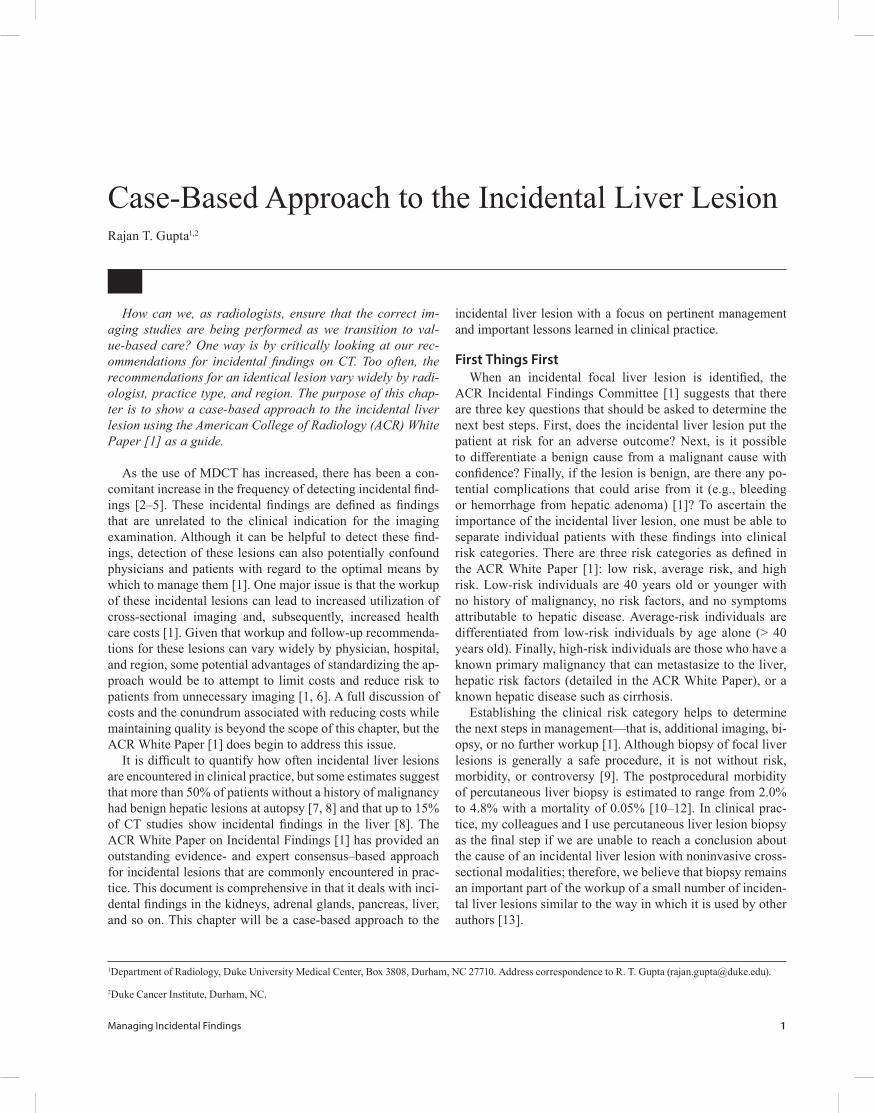
DCFig . 4 (continued)—Case 3: 31-year-old woman who presented to primary care physician with right upper quadrant and epigastric abdominal pain and bloating. Abdominal sonography was performed.C, Axial T2-weighted image with fat suppression reveals stealthy-appearing lesion in left hepatic lobe (arrow) that corresponds to lesion seen on sonography (A and B).D, Axial T1-weighted image with fat suppression in arterial phase after administration of extracellular MRI contrast agent shows homogeneous hyperenhancement of lesion (arrow).E, Axial T1-weighted image with fat suppression in equilibrium phase after administration of extracellular MRI contrast agent shows lesion (arrow) is similar in appearance to remainder of liver. There is suggestion of increased signal intensity in central portion of lesion, presumably within central scar. Diagnosis was focal nodular hyperplasia. This diagnosis was confirmed at surgical pathology. Patient underwent resection of lesion because she was deemed to be symptomatic from lesion by her surgeon.
E
C
A
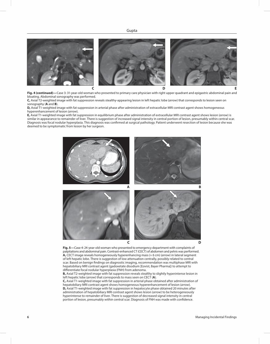
Fig . 5—Case 4: 24-year-old woman who presented to emergency department with complaints of palpitations and abdominal pain. Contrast-enhanced CT (CECT) of abdomen and pelvis was performed.A, CECT image reveals homogeneously hyperenhancing mass (≈ 6 cm) (arrow) in lateral segment of left hepatic lobe. There is suggestion of low attenuation centrally, possibly related to central scar. Based on benign findings on diagnostic imaging, recommendation was multiphase MRI with hepatobiliary MRI contrast agent (gadoxetate disodium [Eovist, Bayer Pharma]) to attempt to differentiate focal nodular hyperplasia (FNH) from adenoma.B, Axial T2-weighted image with fat suppression reveals stealthy to slightly hyperintense lesion in left hepatic lobe (arrow) that corresponds to mass seen on CECT (A).C, Axial T1-weighted image with fat suppression in arterial phase obtained after administration of hepatobiliary MRI contrast agent shows homogeneous hyperenhancement of lesion (arrow).D, Axial T1-weighted image with fat suppression in hepatocyte phase obtained 20 minutes after administration of hepatobiliary MRI contrast agent shows lesion (arrow) to be heterogeneously hyperintense to remainder of liver. There is suggestion of decreased signal intensity in central portion of lesion, presumably within central scar. Diagnosis of FNH was made with confidence.
D
B

Managing Incidental Findings 7
Incidental Liver Lesions
D
A
F
C
Fig . 6—Case 5: 66-year-old average-risk woman with no cirrhosis risk factors who presented with complaints of left lower quadrant pain. Contrast-enhanced CT (CECT) of abdomen and pelvis was performed.A, CECT image reveals two low-attenuation lesions measuring approximately 2 cm in right hepatic lobe. Both medial (black arrow) and lateral (white arrow) lesions show suspicious imaging features: relatively ill-defined margins, enhancement with attenuation values greater than about 20 HU, and heterogeneous appearance. Based on these findings and average-risk category of patient, recommendation is evaluation with multiphase MRI.B, Axial T2-weighted image with fat suppression reveals that two lesions have different signal intensities on T2-weighted imaging. Medial lesion (black arrow) is hyperintense to background liver, and lateral lesion (white arrow) is more intermediate bright to background liver. These findings suggest that differential diagnosis for medial lesion includes hemangioma and differential diagnosis for lateral lesion includes malignancy such as hepatocellular carcinoma (HCC) or solitary liver metastasis.C–E, Axial T1-weighted images with fat suppression obtained in arterial (C), portal venous (D), and late venous (E) phases show classic centripetal nodular enhancement with puddling in medial lesion (black arrows); these findings are compatible with hemangioma. Lateral lesion (white arrows) shows rapid wash-in and rapid washout with capsule. Based on this appearance, presumptive diagnosis of HCC was made. Because of high suspicion of malignancy, α-fetoprotein (AFP) level was checked. AFP value was more than 1480 ng/mL; based on imaging and laboratory results, recommendation was partial liver resection.F, Photograph of gross pathologic specimen reveals lateral lesion (arrow) is moderately differentiated HCC. Medial lesion was not resected, but its appearance during surgical exploration was compatible with benign hemangioma. (Courtesy of Zani SC, Duke University Medical Center, Durham, NC)
E
B

8 Managing Incidental Findings
Gupta
D
A
CFig . 7—Case 6: 58-year-old average-risk man who presented with abdominal pain 3 years after initial contrast-enhanced CT (CECT) at outside institution showed a liver lesion.A and B, CECT images of abdomen and pelvis obtained in portal venous (A) and delayed (B) phases at outside institution reveal 4-cm enhancing lesion (arrows) in posterior segment of right hepatic lobe. Lesion is heterogeneously enhancing on portal venous phase image and is isodense to hypodense to liver parenchyma on delayed phase image. Note that the attenuation of lesion does not follow the attenuation of blood pool. This lesion was interpreted to be hemangioma on initial study performed at outside institution.C–E, Multiphase MRI performed 3 years after CECT (A and B). T2-weighted image with fat suppression (C) and T1-weighted images in arterial (D) and portal venous (E) phases reveal marked interval enlargement of lesion (arrows) in right hepatic lobe. Lesion is heterogeneously hyperintense on T2-weighted image, shows rapid wash-in with hypervascularity on arterial phase image, and shows concomitant washout on portal venous phase image. This lesion was pathologically proven to be hepatocellular carcinoma. Key teaching point of this case is that hemangioma must prove itself to be hemangioma—that is, diagnosis of hemangioma cannot be diagnosis of exclusion.
E
B