case note keratocystic odontogenic tumour mimicking ... odontogenic tumour mimicking lateral...
TRANSCRIPT
VOL. 14 | NO. 4 | ISSUE 56 | OCT.-DEC. 2016
Page 365
Keratocystic Odontogenic Tumour Mimicking Lateral Periodontal Cyst: A Diagnostic DilemmaManohar B,1 Baidya D,1 Bhuvaneswari S,2 Rai AB3
1Department of Periodontics
2Department of Oral Medicine and Radiology
3Department of Oral and Maxillofacial Surgery
Pacific Dental College and Hospital, Debari.
Udaipur, Rajasthan, Pin. - 313024.
Corresponding Author
Manohar Balaji
Department of Periodontics
Pacific Dental College and Hospital, Debari,
Udaipur, Rajasthan, Pin. - 313024.
E-mail: [email protected]
Citation
Balaji M, Dhritiman B, Bhuvaneswari S, Rai BA. Keratocystic Odontogenic Tumour Mimicking Lateral Periodontal Cyst: A Diagnostic Dilemma. Kathmandu Univ Med J 2016; 56(4):365-7.
ABSTRACTThe Keratocystic Odontogenic Tumor is a developmental cyst derived from the enamel organ or from the dental lamina. It is a benign, multicystic, intraosseous tumor of odontogenic origin, with a characteristic lining of parakeratinized stratified squamous epithelium and has a potential for aggressive, infiltrative behavior and recurrence. Keratocystic Odontogenic Tumors have a predilection for males and occurs mainly in the second and third decade of life, most commonly in the mandible, mostly in the posterior body, the angle and the ascending ramus. It extends in the intramedullary space making it difficult to diagnose at an early stage. It is regarded as a distinctive entity because of its characteristic histology, proliferation kinetics and behavior. Main in 1970 described, collateral variant of Keratocystic Odontogenic Tumor, which presents adjacent to the roots of the teeth usually in the mandibular premolar region and radiologically is indistinguishable from the lateral periodontal cyst and gingival cyst.
KEY WORDS Collateral variant, developmental cyst, keratocystic odontogenic tumor, lateral periodontal cyst
INTRODUCTIONThe odontogenic keratocyst was first described by Philipsen in 1956, in a publication in Danish language.1 WHO Working Group recommends the term Keratocystic Odontogenic Tumor (KCOT), as it better reflects its neoplastic nature. KCOT occur from the first to the ninth decades with a peak in the second and third decades.2 Main in 1970 described collateral variant of KCOT as the one, which presents adjacent to the roots of the teeth usually in the mandibular premolar region and radiologically indistinguishable from the lateral periodontal cyst. Lateral periodontal cysts are developmental odontogenic cysts and can be clearly differentiated from KCOT histopathologically. It is a very aggressive lesion with a high recurrence rate. Here, we present a case of KCOT mimicking a lateral periodontal
cyst in a 72 year old male patient, which was provisionally diagnosed by clinical and radiographical findings, later on confirmed by histopathology.
CASE REPORTSA 72 year old male patient reported to the private dental establishment with a complaint of a swelling in the lower right back tooth region for the past two months. The swelling gradually increased in size, but no extra-oral facial changes were seen. The patient did not present with any systemic illness. Dental examination revealed edentulous region in the mandibular right and left posterior region and maxillary right posterior region. The extractions were done due to caries.
Case Note
KATHMANDU UNIVERSITY MEDICAL JOURNAL
Page 366
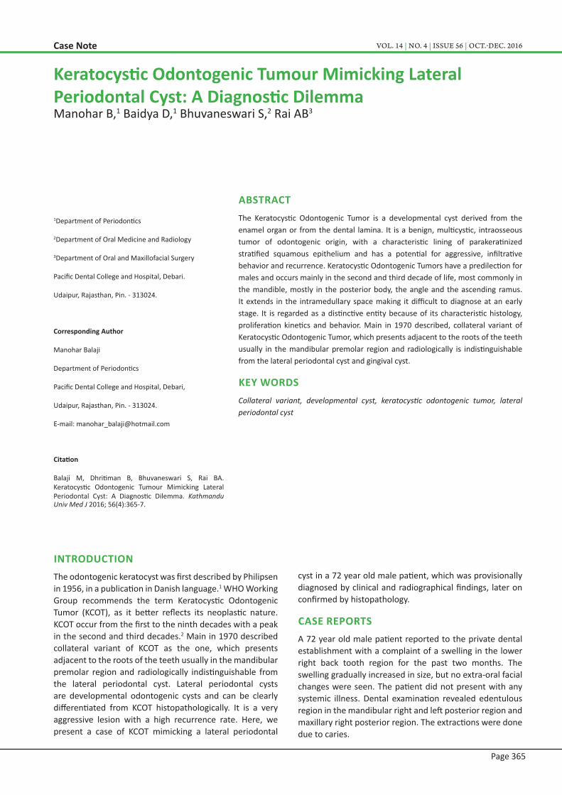
On clinical examination of the mandibular right premolar region, a diffuse, round swelling measuring approximately 1.5cm X 1.5cm was present between the premolars (Fig. 1). On palpation, the swelling was tender and seen to be extending from the attached gingiva to the vestibule. The overlying gingiva and oral mucosa were seen to be normal. There was no mobility of the premolar teeth and were vital. The conventional and the panoramic radiographs revealed a unilocular, ovoid lesion in between the roots of the premolars. A slight distal drifting of the root of the 2nd
premolar was observed (Fig. 2a, 2b). Based on the clinical and radiographic features, a differential diagnosis of lateral periodontal cyst and KCOT was made.
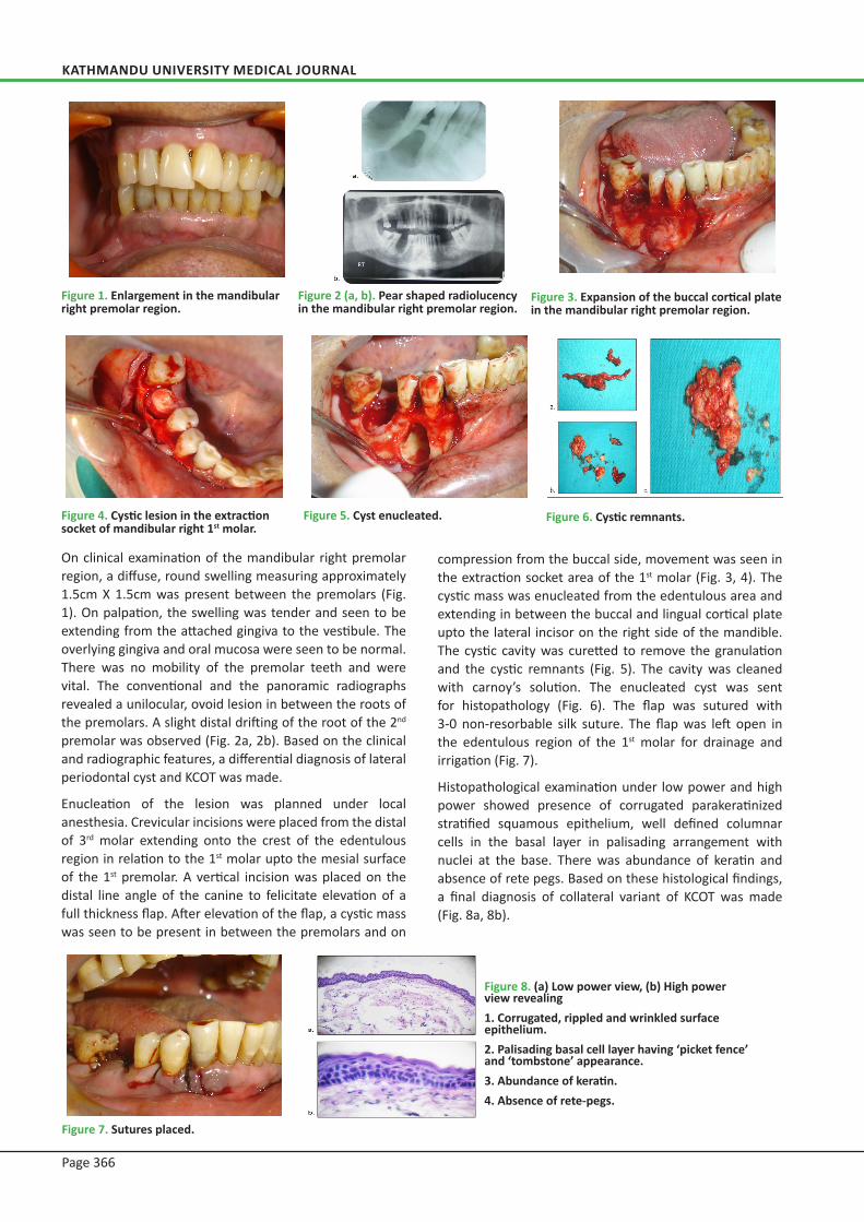
Enucleation of the lesion was planned under local anesthesia. Crevicular incisions were placed from the distal of 3rd molar extending onto the crest of the edentulous region in relation to the 1st molar upto the mesial surface of the 1st premolar. A vertical incision was placed on the distal line angle of the canine to felicitate elevation of a full thickness flap. After elevation of the flap, a cystic mass was seen to be present in between the premolars and on
Figure 1. Enlargement in the mandibular right premolar region.
Figure 4. Cystic lesion in the extraction socket of mandibular right 1st molar.
Figure 7. Sutures placed.
Figure 5. Cyst enucleated.
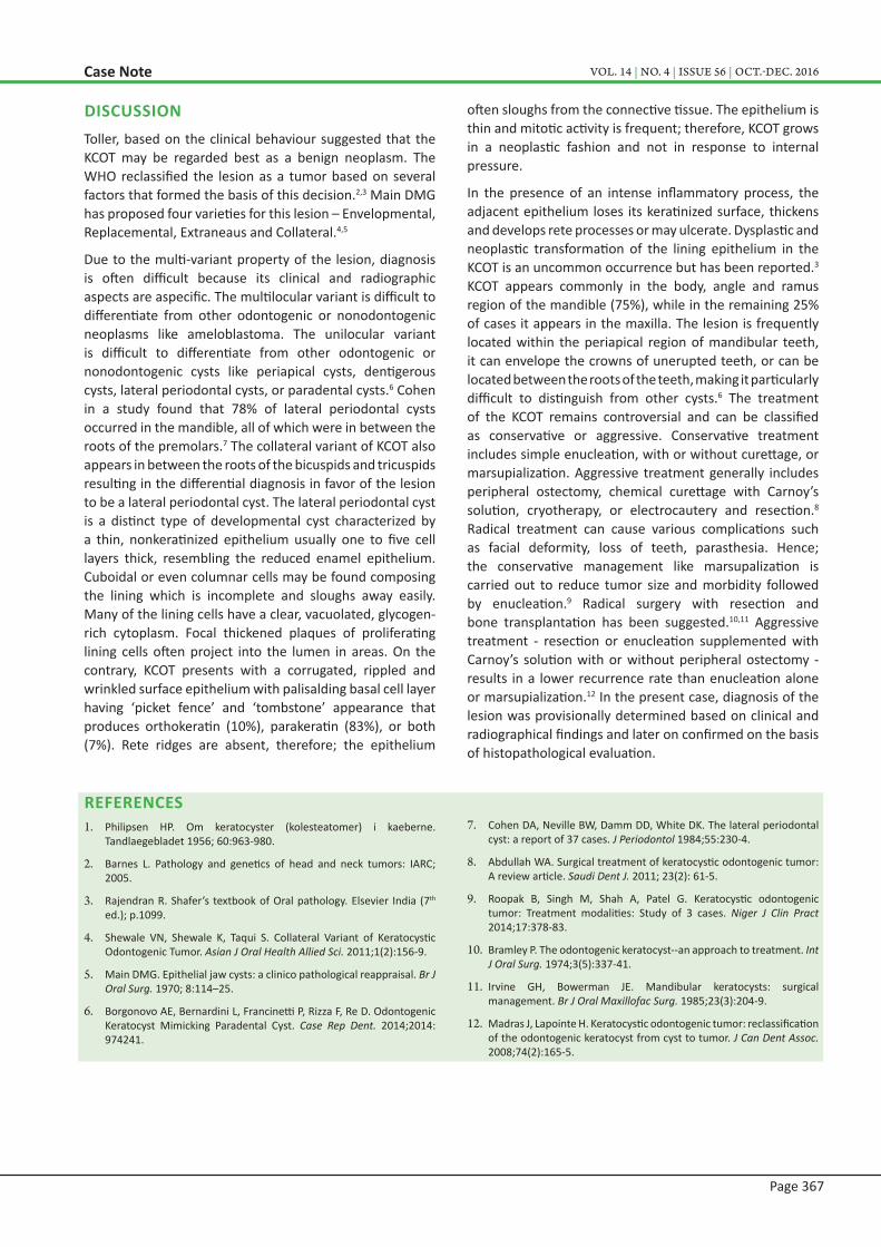
Figure 8. (a) Low power view, (b) High power view revealing 1. Corrugated, rippled and wrinkled surface epithelium.2. Palisading basal cell layer having ‘picket fence’ and ‘tombstone’ appearance.3. Abundance of keratin.4. Absence of rete-pegs.
Figure 6. Cystic remnants.
Figure 3. Expansion of the buccal cortical plate in the mandibular right premolar region.
Figure 2 (a, b). Pear shaped radiolucency in the mandibular right premolar region.
compression from the buccal side, movement was seen in the extraction socket area of the 1st molar (Fig. 3, 4). The cystic mass was enucleated from the edentulous area and extending in between the buccal and lingual cortical plate upto the lateral incisor on the right side of the mandible. The cystic cavity was curetted to remove the granulation and the cystic remnants (Fig. 5). The cavity was cleaned with carnoy’s solution. The enucleated cyst was sent for histopathology (Fig. 6). The flap was sutured with 3-0 non-resorbable silk suture. The flap was left open in the edentulous region of the 1st molar for drainage and irrigation (Fig. 7).
Histopathological examination under low power and high power showed presence of corrugated parakeratinized stratified squamous epithelium, well defined columnar cells in the basal layer in palisading arrangement with nuclei at the base. There was abundance of keratin and absence of rete pegs. Based on these histological findings, a final diagnosis of collateral variant of KCOT was made (Fig. 8a, 8b).
VOL. 14 | NO. 4 | ISSUE 56 | OCT.-DEC. 2016
Page 367
DISCUSSIONToller, based on the clinical behaviour suggested that the KCOT may be regarded best as a benign neoplasm. The WHO reclassified the lesion as a tumor based on several factors that formed the basis of this decision.2,3 Main DMG has proposed four varieties for this lesion – Envelopmental, Replacemental, Extraneaus and Collateral.4,5
Due to the multi-variant property of the lesion, diagnosis is often difficult because its clinical and radiographic aspects are aspecific. The multilocular variant is difficult to differentiate from other odontogenic or nonodontogenic neoplasms like ameloblastoma. The unilocular variant is difficult to differentiate from other odontogenic or nonodontogenic cysts like periapical cysts, dentigerous cysts, lateral periodontal cysts, or paradental cysts.6 Cohen in a study found that 78% of lateral periodontal cysts occurred in the mandible, all of which were in between the roots of the premolars.7 The collateral variant of KCOT also appears in between the roots of the bicuspids and tricuspids resulting in the differential diagnosis in favor of the lesion to be a lateral periodontal cyst. The lateral periodontal cyst is a distinct type of developmental cyst characterized by a thin, nonkeratinized epithelium usually one to five cell layers thick, resembling the reduced enamel epithelium. Cuboidal or even columnar cells may be found composing the lining which is incomplete and sloughs away easily. Many of the lining cells have a clear, vacuolated, glycogen-rich cytoplasm. Focal thickened plaques of proliferating lining cells often project into the lumen in areas. On the contrary, KCOT presents with a corrugated, rippled and wrinkled surface epithelium with palisalding basal cell layer having ‘picket fence’ and ‘tombstone’ appearance that produces orthokeratin (10%), parakeratin (83%), or both (7%). Rete ridges are absent, therefore; the epithelium
often sloughs from the connective tissue. The epithelium is thin and mitotic activity is frequent; therefore, KCOT grows in a neoplastic fashion and not in response to internal pressure.
In the presence of an intense inflammatory process, the adjacent epithelium loses its keratinized surface, thickens and develops rete processes or may ulcerate. Dysplastic and neoplastic transformation of the lining epithelium in the KCOT is an uncommon occurrence but has been reported.3 KCOT appears commonly in the body, angle and ramus region of the mandible (75%), while in the remaining 25% of cases it appears in the maxilla. The lesion is frequently located within the periapical region of mandibular teeth, it can envelope the crowns of unerupted teeth, or can be located between the roots of the teeth, making it particularly difficult to distinguish from other cysts.6 The treatment of the KCOT remains controversial and can be classified as conservative or aggressive. Conservative treatment includes simple enucleation, with or without curettage, or marsupialization. Aggressive treatment generally includes peripheral ostectomy, chemical curettage with Carnoy’s solution, cryotherapy, or electrocautery and resection.8 Radical treatment can cause various complications such as facial deformity, loss of teeth, parasthesia. Hence; the conservative management like marsupalization is carried out to reduce tumor size and morbidity followed by enucleation.9 Radical surgery with resection and bone transplantation has been suggested.10,11 Aggressive treatment - resection or enucleation supplemented with Carnoy’s solution with or without peripheral ostectomy - results in a lower recurrence rate than enucleation alone or marsupialization.12 In the present case, diagnosis of the lesion was provisionally determined based on clinical and radiographical findings and later on confirmed on the basis of histopathological evaluation.
REFERENCES1. Philipsen HP. Om keratocyster (kolesteatomer) i kaeberne.
Tandlaegebladet 1956; 60:963-980.
2. Barnes L. Pathology and genetics of head and neck tumors: IARC; 2005.
3. Rajendran R. Shafer’s textbook of Oral pathology. Elsevier India (7th
ed.); p.1099.
4. Shewale VN, Shewale K, Taqui S. Collateral Variant of Keratocystic Odontogenic Tumor. Asian J Oral Health Allied Sci. 2011;1(2):156-9.
5. Main DMG. Epithelial jaw cysts: a clinico pathological reappraisal. Br J Oral Surg. 1970; 8:114–25.
6. Borgonovo AE, Bernardini L, Francinetti P, Rizza F, Re D. Odontogenic Keratocyst Mimicking Paradental Cyst. Case Rep Dent. 2014;2014: 974241.
7. Cohen DA, Neville BW, Damm DD, White DK. The lateral periodontal cyst: a report of 37 cases. J Periodontol 1984;55:230-4.
8. Abdullah WA. Surgical treatment of keratocystic odontogenic tumor: A review article. Saudi Dent J. 2011; 23(2): 61-5.
9. Roopak B, Singh M, Shah A, Patel G. Keratocystic odontogenic tumor: Treatment modalities: Study of 3 cases. Niger J Clin Pract 2014;17:378-83.
10. Bramley P. The odontogenic keratocyst--an approach to treatment. Int J Oral Surg. 1974;3(5):337-41.
11. Irvine GH, Bowerman JE. Mandibular keratocysts: surgical management. Br J Oral Maxillofac Surg. 1985;23(3):204-9.
12. Madras J, Lapointe H. Keratocystic odontogenic tumor: reclassification of the odontogenic keratocyst from cyst to tumor. J Can Dent Assoc. 2008;74(2):165-5.
Case Note