case report a differential diagnosis of haematuria...
TRANSCRIPT

Case ReportA Differential Diagnosis of Haematuria following a MotorVehicle Collision: Nutcracker Syndrome
Gary Sharp1 and Derek Glenn2
1Trauma Department at St George Hospital, Kogarah, Sydney, NSW 2217, Australia2St George Hospital, Kogarah, Sydney, NSW 2217, Australia
Correspondence should be addressed to Gary Sharp; [email protected]
Received 2 October 2014; Accepted 15 December 2014
Academic Editor: Christophoros Foroulis
Copyright © 2015 G. Sharp and D. Glenn. This is an open access article distributed under the Creative Commons AttributionLicense, which permits unrestricted use, distribution, and reproduction in any medium, provided the original work is properlycited.
A young female presented to the emergency department following a motor vehicle collision. She complained of left flank pain andwas found to have haematuria. After investigation no trauma related injuries were identified. However, an incidental finding ofnutcracker phenomenon/syndrome was made. Nutcracker phenomenon is a rare cause of haematuria resulting from nontraumaticcompression of the left renal vein between the superior mesenteric artery and the aorta. It affects females more than males and itspresentation can range from asymptomatic to debilitating haematuria, pelvic congestion in females, varicosities in males, and pain.No validated diagnostic criteria exist and treatment is usually surgical in those with debilitating symptoms or refractory anaemia.
1. Introduction
Nutcracker syndrome (NCS) was first described by Grant [1],who likened the impingement of the left renal vein (LRV) bythe superiormesenteric artery (SMA) against the aorta to thatof a nut within the jaws of a nutcracker. The first clinical caseof NCS was acknowledged by El-Sadr and Mina in 1950 [2]and its management was first documented in 1974 [3].
A clear distinction between NCS, the clinical manifes-tation of mesoaortic compression of the LRV, and nutcrackerphenomenon (NCP), the anatomical identification ofmesoaortic compression of the LRV, exists [4–6]. Its rarity isrepresented through scant evidence with no validateddiagnostic or therapeutic guidelines [5]. As such there is nodata quantifying prevalence or incidence [4, 5]. NCP is subdi-vided into anterior and posterior subtypes. Anterior NCPrefers to mesoaortic compression of the LRV whilst the rarerposterior NCP denotes compression of a retroaortic LRVbetween the aorta and vertebrae [4–6]. Only anterior NCPwill be addressed hereafter.
Females are most often affected in bimodal fashion [5, 7]with the first peak at 20–30 years and the second inmiddle age[5, 7]. It must be stressed however that both sexes and all agescan be affected [4, 5, 7]. Low body mass index is regarded as
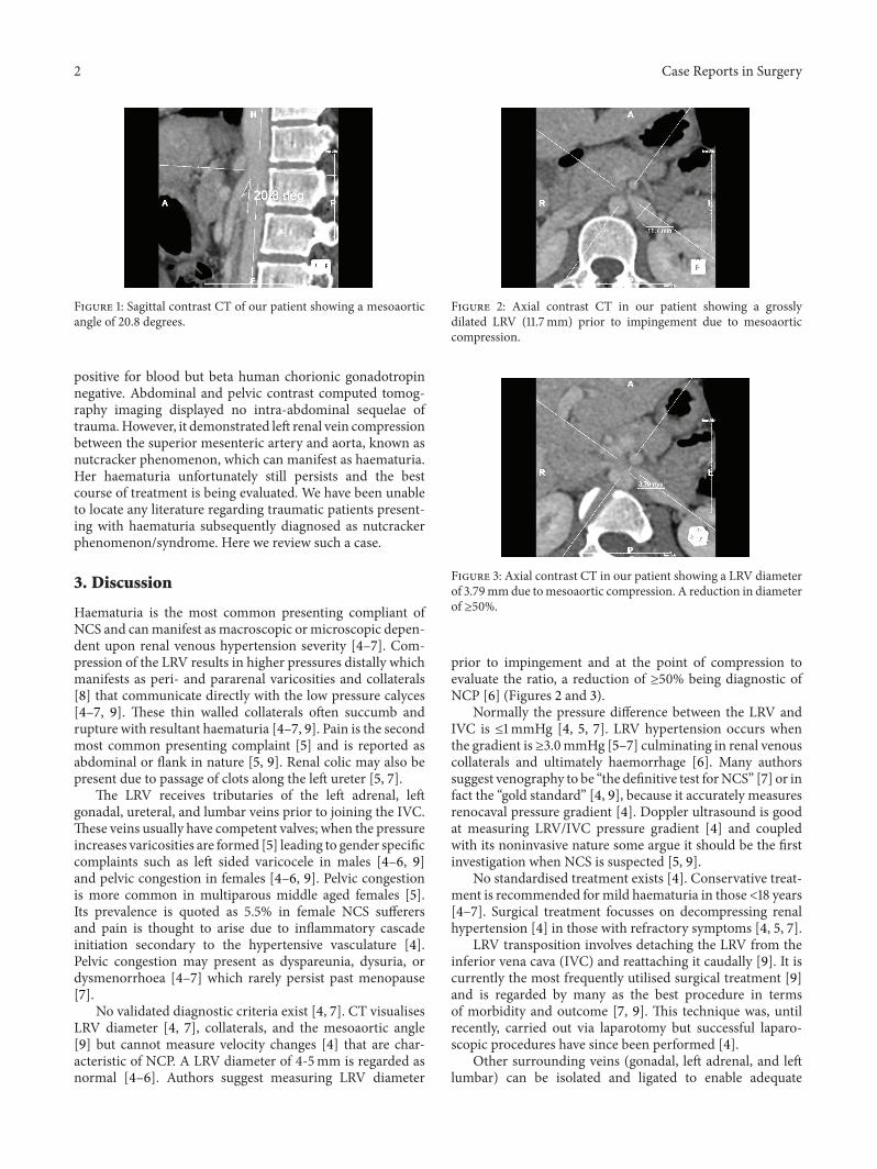
a risk factor ofNCP [4, 5, 7] due to a paucity of retroperitonealadipose tissue reducing the mesoaortic angle and/or causingposterior renal ptosis [5, 7]. Posterior renal ptosis refers todorsal migration of the kidney and renal pelvis due to theaforementioned retroperitoneal adipose tissue paucity [4, 6,7]. This posterior displacement stretches and compresses theLRV [6]. NCP may also arise due to anatomical variationssuch as a LRV that is more cephalad upon union with theinferior vena cava and as such is immediately inferior tothe SMA or an SMA that instantly descends [5]. In healthyindividuals themesoaortic angle is reportedly between 38 and90 degrees; in NCP it is suggested that this angle is greaterthan halved [5] (Figure 1). Other causes of LRV compressioninclude pancreatic neoplasms, para-aortic lymphadenopathy,retroperitoneal tumours, aortic aneurysms, or fibrolymphatictissue between the SMA and aorta [4, 5]. No genetic link hasbeen identified [5–7].
2. Case Report
A 31-year-old female presented to the emergency depart-ment following a motor vehicle collision. On examinationshe complained of left flank tenderness. Her urine was
Hindawi Publishing CorporationCase Reports in SurgeryVolume 2015, Article ID 749182, 3 pageshttp://dx.doi.org/10.1155/2015/749182
2 Case Reports in Surgery
Figure 1: Sagittal contrast CT of our patient showing a mesoaorticangle of 20.8 degrees.
positive for blood but beta human chorionic gonadotropinnegative. Abdominal and pelvic contrast computed tomog-raphy imaging displayed no intra-abdominal sequelae oftrauma.However, it demonstrated left renal vein compressionbetween the superior mesenteric artery and aorta, known asnutcracker phenomenon, which can manifest as haematuria.Her haematuria unfortunately still persists and the bestcourse of treatment is being evaluated. We have been unableto locate any literature regarding traumatic patients present-ing with haematuria subsequently diagnosed as nutcrackerphenomenon/syndrome. Here we review such a case.
3. Discussion
Haematuria is the most common presenting compliant ofNCS and canmanifest as macroscopic ormicroscopic depen-dent upon renal venous hypertension severity [4–7]. Com-pression of the LRV results in higher pressures distally whichmanifests as peri- and pararenal varicosities and collaterals[8] that communicate directly with the low pressure calyces[4–7, 9]. These thin walled collaterals often succumb andrupture with resultant haematuria [4–7, 9]. Pain is the secondmost common presenting complaint [5] and is reported asabdominal or flank in nature [5, 9]. Renal colic may also bepresent due to passage of clots along the left ureter [5, 7].
The LRV receives tributaries of the left adrenal, leftgonadal, ureteral, and lumbar veins prior to joining the IVC.These veins usually have competent valves; when the pressureincreases varicosities are formed [5] leading to gender specificcomplaints such as left sided varicocele in males [4–6, 9]and pelvic congestion in females [4–6, 9]. Pelvic congestionis more common in multiparous middle aged females [5].Its prevalence is quoted as 5.5% in female NCS sufferersand pain is thought to arise due to inflammatory cascadeinitiation secondary to the hypertensive vasculature [4].Pelvic congestion may present as dyspareunia, dysuria, ordysmenorrhoea [4–7] which rarely persist past menopause[7].
No validated diagnostic criteria exist [4, 7]. CT visualisesLRV diameter [4, 7], collaterals, and the mesoaortic angle[9] but cannot measure velocity changes [4] that are char-acteristic of NCP. A LRV diameter of 4-5mm is regarded asnormal [4–6]. Authors suggest measuring LRV diameter
Figure 2: Axial contrast CT in our patient showing a grosslydilated LRV (11.7mm) prior to impingement due to mesoaorticcompression.
Figure 3: Axial contrast CT in our patient showing a LRV diameterof 3.79mmdue tomesoaortic compression. A reduction in diameterof ≥50%.
prior to impingement and at the point of compression toevaluate the ratio, a reduction of ≥50% being diagnostic ofNCP [6] (Figures 2 and 3).
Normally the pressure difference between the LRV andIVC is ≤1mmHg [4, 5, 7]. LRV hypertension occurs whenthe gradient is ≥3.0mmHg [5–7] culminating in renal venouscollaterals and ultimately haemorrhage [6]. Many authorssuggest venography to be “the definitive test forNCS” [7] or infact the “gold standard” [4, 9], because it accurately measuresrenocaval pressure gradient [4]. Doppler ultrasound is goodat measuring LRV/IVC pressure gradient [4] and coupledwith its noninvasive nature some argue it should be the firstinvestigation when NCS is suspected [5, 9].
No standardised treatment exists [4]. Conservative treat-ment is recommended formild haematuria in those<18 years[4–7]. Surgical treatment focusses on decompressing renalhypertension [4] in those with refractory symptoms [4, 5, 7].
LRV transposition involves detaching the LRV from theinferior vena cava (IVC) and reattaching it caudally [9]. It iscurrently the most frequently utilised surgical treatment [9]and is regarded by many as the best procedure in termsof morbidity and outcome [7, 9]. This technique was, untilrecently, carried out via laparotomy but successful laparo-scopic procedures have since been performed [4].
Other surrounding veins (gonadal, left adrenal, and leftlumbar) can be isolated and ligated to enable adequate

Case Reports in Surgery 3
LRV mobilisation and tension-free anastomoses [9]. Theoriginal LRV attachment to the IVC is oversewn and a newanastomosis is fashioned inferiorly [9].The addition of a greatsaphenous venous cuff may be utilised in the presence ofinadequate LRV length to ensure a tension-free transposition[9].
A recent study suggested 59 out of 61 patients treated byendovascular stenting had resolution of symptoms [4]. Over-sized, self-expanding stents carry the least migration risk[9]. Regardless, migration, restenosis, thrombosis, and pul-monary embolism have all been reported [7, 9]. No long-term data regarding such complications in a usually youngpopulation exist [9]. Stent insertion must be followed by pro-longed anticoagulation and/or antiplatelet therapy [4] whichin themselves carry risk.
Conflict of Interests
The authors declare that there is no conflict of interestsregarding the publication of this paper.
References
[1] J. Grant,Methods of Anatomy,Williams andWilkens, Baltimore,Md, USA, 1937.
[2] A. R. El-Sadr and E. Mina, “Anatomical and surgical aspects inthe operative management of varicocele,” The Urologic andCutaneous Review, vol. 54, no. 5, pp. 257–262, 1950.
[3] S. P. Pastershank, “Left renal vein obstruction by a superiormesenteric artery,”CanadianAssociation of Radiologists Journal,vol. 25, no. 1, pp. 52–54, 1974.
[4] Y. He, Z. Wu, S. Chen et al., “Nutcracker syndrome—how welldo we know it?” Urology, vol. 83, no. 1, pp. 12–17, 2014.
[5] A. K. Kurklinsky and T. W. Rooke, “Nutcracker phenomenonand nutcracker syndrome,”Mayo Clinic Proceedings, vol. 85, no.6, pp. 552–559, 2010.
[6] M. Polguj, M. Topol, and A. Majos, “An unusual case of leftvenous renal entrapment syndrome: a new type of nutcrackerphenomenon?” Surgical and Radiologic Anatomy, vol. 35, no. 3,pp. 263–267, 2013.
[7] M. T.Menard, “Nutcracker syndrome: when should it be treatedand how?” Perspectives in Vascular Surgery and EndovascularTherapy, vol. 21, no. 2, pp. 117–124, 2009.
[8] U. Rudloff, R. J. Holmes, J. T. Prem, G. R. Faust, R. Moldwin,and D. Siegel, “Mesoaortic compression of the left renal vein(nutcracker syndrome): case reports and review of the litera-ture,” Annals of Vascular Surgery, vol. 20, no. 1, pp. 120–129,2006.
[9] S. M. Said, P. Gloviczki, M. Kalra et al., “Renal nutcracker syn-drome: surgical options,” Seminars in Vascular Surgery, vol. 26,no. 1, pp. 35–42, 2013.

Submit your manuscripts athttp://www.hindawi.com
Stem CellsInternational
Hindawi Publishing Corporationhttp://www.hindawi.com Volume 2014
Hindawi Publishing Corporationhttp://www.hindawi.com Volume 2014
MEDIATORSINFLAMMATION
of
Hindawi Publishing Corporationhttp://www.hindawi.com Volume 2014
Behavioural Neurology
EndocrinologyInternational Journal of
Hindawi Publishing Corporationhttp://www.hindawi.com Volume 2014
Hindawi Publishing Corporationhttp://www.hindawi.com Volume 2014
Disease Markers
Hindawi Publishing Corporationhttp://www.hindawi.com Volume 2014
BioMed Research International
OncologyJournal of
Hindawi Publishing Corporationhttp://www.hindawi.com Volume 2014
Hindawi Publishing Corporationhttp://www.hindawi.com Volume 2014
Oxidative Medicine and Cellular Longevity
Hindawi Publishing Corporationhttp://www.hindawi.com Volume 2014
PPAR Research
The Scientific World JournalHindawi Publishing Corporation http://www.hindawi.com Volume 2014
Immunology ResearchHindawi Publishing Corporationhttp://www.hindawi.com Volume 2014
Journal of
ObesityJournal of
Hindawi Publishing Corporationhttp://www.hindawi.com Volume 2014
Hindawi Publishing Corporationhttp://www.hindawi.com Volume 2014
Computational and Mathematical Methods in Medicine
OphthalmologyJournal of
Hindawi Publishing Corporationhttp://www.hindawi.com Volume 2014
Diabetes ResearchJournal of
Hindawi Publishing Corporationhttp://www.hindawi.com Volume 2014
Hindawi Publishing Corporationhttp://www.hindawi.com Volume 2014
Research and TreatmentAIDS
Hindawi Publishing Corporationhttp://www.hindawi.com Volume 2014
Gastroenterology Research and Practice
Hindawi Publishing Corporationhttp://www.hindawi.com Volume 2014
Parkinson’s Disease
Evidence-Based Complementary and Alternative Medicine
Volume 2014Hindawi Publishing Corporationhttp://www.hindawi.com