case report: abdominal cocoon syndrome in a child with
TRANSCRIPT
195
July 2020, Volume 8, Issue 3, Number 19
Nidhi Mahajan1 , Mitali Agarwal1, Arti Khatri1* , Yousuf Mohsin Bari Siddiqui2, Mamta Sengar2
Case Report: Abdominal Cocoon Syndrome in a Child With Intestinal Obstruction: A Case Report and Literature Review
Abdominal cocoon syndrome or idiopathic Sclerosing Encapsulating Peritonitis (SEP) is an extremely uncommon cause of intestinal obstruction. Its etiology is explainable through numerous theories.
An eleven-year-old girl referred to the pediatric surgery OPD with complaints of abdominal pain for the past two weeks and vomiting for two days. Her family history for tuberculosis was positive. On examination, the abdomen was distended and slightly firm on palpation. The X-ray of her abdomen revealed multiple air-fluid levels. The CECT of the abdomen indicated dilated abdomen, duodenum, and proximal bowel loops. Some trapped inter bowel free fluid was also observed. The clinical presentation of subacute intestinal obstruction and the radiological features suggested a differential diagnosis of tubercular peritonitis versus pseudomyxoma peritonei. The obstructive symptoms demonstrated that the patient underwent an explorative laparotomy. Preoperatively, a thin membranous sac was identified enclosing multiple dilated small bowel loops. The sac was released by blunt dissection and part of the sac was provided for histopathological examination. Based on clinical, histopathological, and radiological findings, a diagnosis of abdominal cocoon syndrome was determined. The postoperative follow-up period of 6 months was uneventful.
We presented a rare case of primary sclerosing encapsulating peritonitis, also reported as abdominal cocoon syndrome. It is among the rare potentially devastating causes of intestinal obstruction in children. A very high index of suspicion is imperative to arrive at its pre-operative diagnosis alone by clinical and radiological findings.
A B S T R A C T
Keywords:Cocoon, Encapsulating, Paediatric, Peritonitis
Article info: Received: 12 Feb 2020First Revision: 25 Feb 2020Accepted: 18 Mar 2020Published: 01 July 2020
1. Department of Pathology, Chacha Nehru Bal Chikitsalaya, Geeta Colony, Delhi, India.2. Department of Pediatric Surgery, Chacha Nehru Bal Chikitsalaya, Geeta Colony, Delhi, India.
* Corresponding Author:Arti Khatri, PhD.Address: Department of Pathology, Chacha Nehru Bal Chikitsalaya, Geeta Colony, Delhi, India.Tel: +91 (870) 0226322E-mail: [email protected]
Citation Mahajan N, Agarwal M, Khatri A, Mohsin Bari Siddiqui Y, Sengar M. Abdominal Cocoon Syndrome in a Child With Intesti-nal Obstruction: A Case Report and Literature Review. Journal of Pediatrics Review. 2020; 8(3):195-200. http://dx.doi.org/10.32598/jpr.8.3.845.2
: http://dx.doi.org/10.32598/jpr.8.3.845.2
Use your device to scan and read the article online
196
July 2020, Volume 8, Issue 3, Number 19
1. Introduction
he abdominal cocoon syndrome is an ex-tremely rare cause of intestinal obstruc-tion. It is also recognized as Sclerosing Encapsulating Peritonitis (SEP), chronic fibrosa encystica, and idiopathic encapsu-lating sclerosing peritonitis. The condition is often reported in adolescent females of tropical and subtropical countries; how-
ever, elderly and male cases are also reported. The etiology of this condition remains unclear; however, idiopathic and cases secondary to localized or systemic processes have been reported in this regard (1). We re-ported a rare case of idiopathic SEP in a young girl who presented with recurrent spasmodic abdominal pain. Furthermore, this case highlights a rare cause of intes-tinal obstruction, to preoperatively diagnose it, and its further management. Informed consent was provided by the parents of the patient for reporting their case.
2. Case Presentation
An eleven-year-old girl presented to the paediatric sur-gery outpatient ward. she had complaints of abdominal pain for the past two weeks and vomiting for two days. The pain was generalized, spasmodic, and progressively increasing in intensity. She also reported that pain was associated with the passage of hard stools, constipation, and abdominal distension. Her family history for tubercu-losis was positive. There was no significant history of the
loss of appetite or weight. On examination, the patient was afebrile and pale, but hemodynamically stable with a blood pressure of 114.78mmHg and a pulse rate of 90/min. No lymphadenopathy was seen. On auscultation, the chest was clear. The abdomen was distended and slightly firm on palpation. Hemogram revealed mild ane-mia (hemoglobin: 9.5 g/dL), and mild neutrophilic leuco-cytosis (total leucocyte count: 15,000/m3). Moreover, C-reactive protein was raised and Mantoux read 6 mm. The patient’s chest X-ray was clear. The x-ray of the abdomen revealed multiple air-fluid levels. The Contrast-Enhanced Computed Tomography (CECT) of the abdomen indicated dilated stomach, duodenum, and proximal bowel loops.
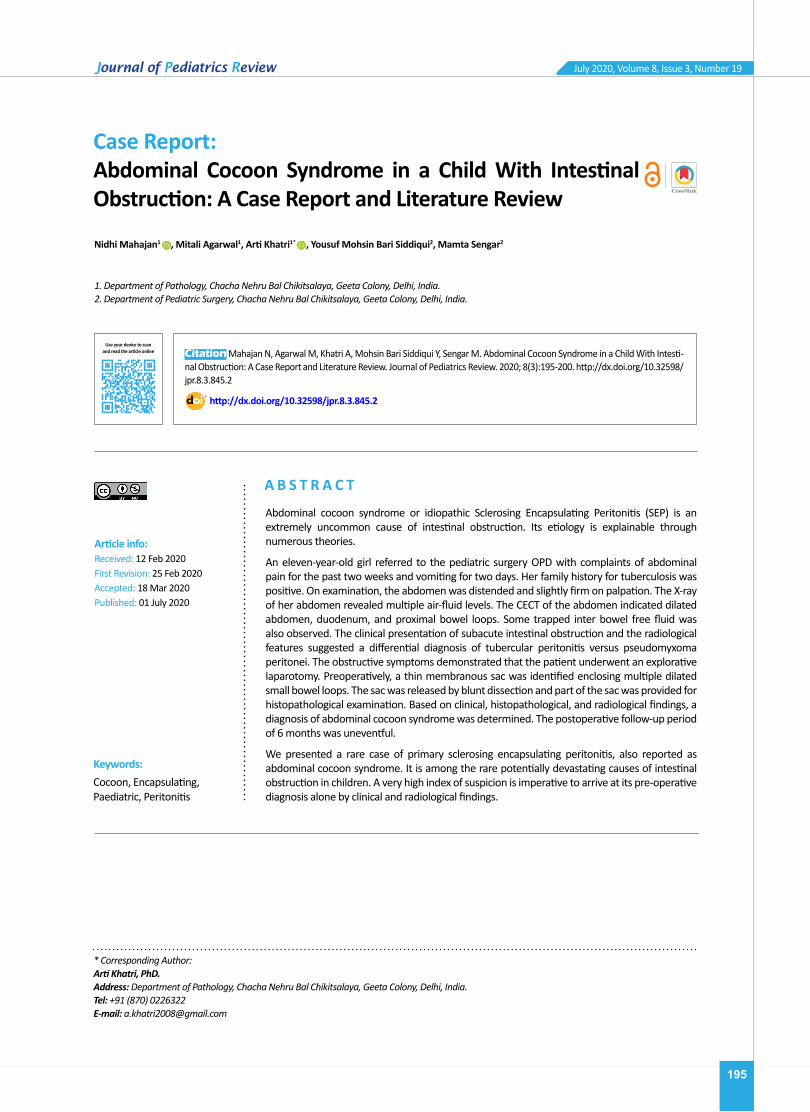
Some trapped inter bowel free fluid was also observed in the patient. The clinical presentation of subacute in-testinal obstruction and the radiological features sug-gested a differential diagnosis of tubercular peritonitis versus pseudomyxoma peritonei. The patient’s condi-tion aggravated with severe pain. The obstructive symp-toms revealed that the patient underwent an explorative laparotomy. Preoperatively, a fluid-filled thin membra-nous sac was identified enclosing multiple dilated small bowel loops (Figure 1a). The sac was released by blunt dissection and the bowel loops (Figure 1b) were decom-pressed by adhesiolysis along the avascular planes.
The post-operative period was uneventful. Part of the sac excision was provided for histopathological examina-tion. Grossly, we received a flattened grey white soft tis-sue measuring 2x1.5x0.5 cm. The histopathology of the
T
Figure 1. 1a: Intra-operative image showing a membranous sac.1b: showing healthy bowel after release from the sac
Mahajan N. et al. Abdominal Cocoon Syndrome in a Child With Intestinal Obstruction. Abdominal Cocoon Syndrome.J Pediatr Rev. 2020; 8(3):195-200.
197
July 2020, Volume 8, Issue 3, Number 19
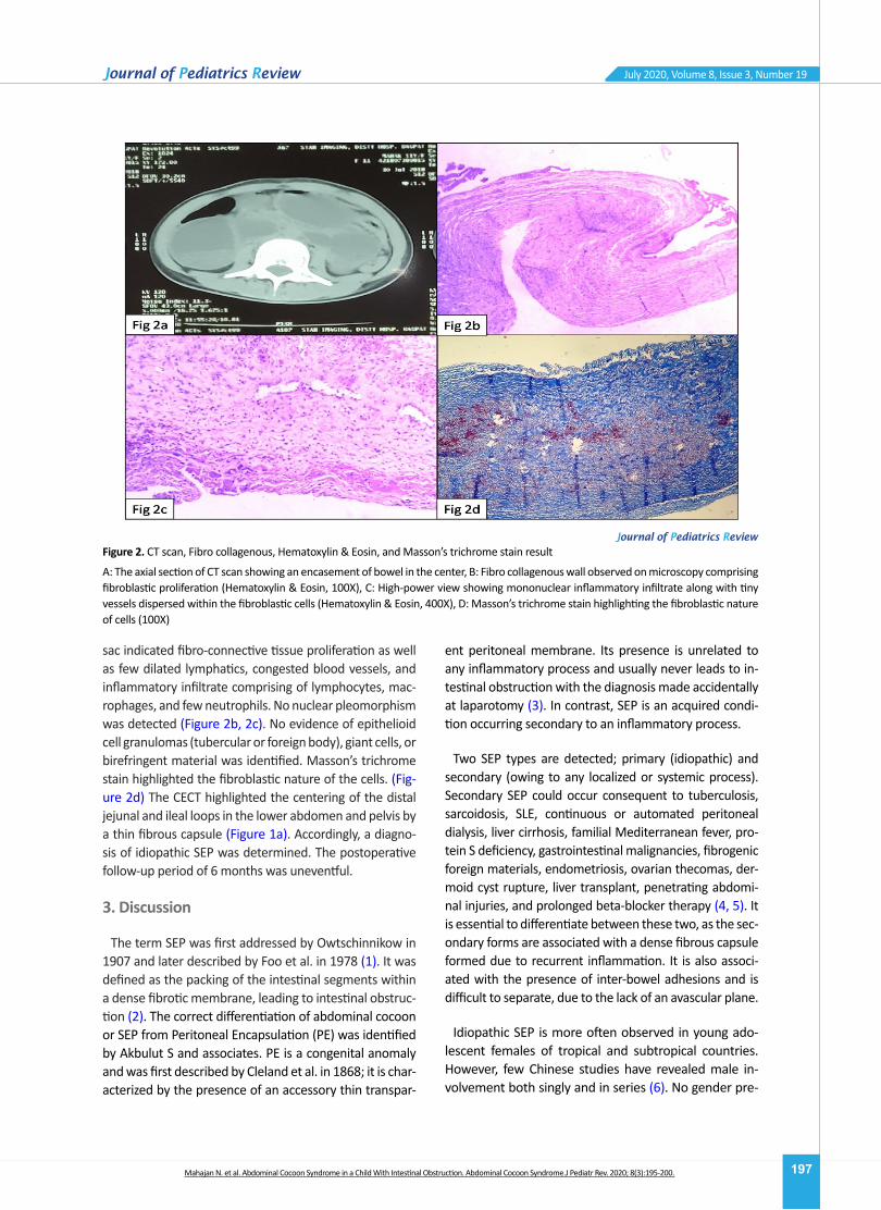
sac indicated fibro-connective tissue proliferation as well as few dilated lymphatics, congested blood vessels, and inflammatory infiltrate comprising of lymphocytes, mac-rophages, and few neutrophils. No nuclear pleomorphism was detected (Figure 2b, 2c). No evidence of epithelioid cell granulomas (tubercular or foreign body), giant cells, or birefringent material was identified. Masson’s trichrome stain highlighted the fibroblastic nature of the cells. (Fig-ure 2d) The CECT highlighted the centering of the distal jejunal and ileal loops in the lower abdomen and pelvis by a thin fibrous capsule (Figure 1a). Accordingly, a diagno-sis of idiopathic SEP was determined. The postoperative follow-up period of 6 months was uneventful.
3. Discussion
The term SEP was first addressed by Owtschinnikow in 1907 and later described by Foo et al. in 1978 (1). It was defined as the packing of the intestinal segments within a dense fibrotic membrane, leading to intestinal obstruc-tion (2). The correct differentiation of abdominal cocoon or SEP from Peritoneal Encapsulation (PE) was identified by Akbulut S and associates. PE is a congenital anomaly and was first described by Cleland et al. in 1868; it is char-acterized by the presence of an accessory thin transpar-
ent peritoneal membrane. Its presence is unrelated to any inflammatory process and usually never leads to in-testinal obstruction with the diagnosis made accidentally at laparotomy (3). In contrast, SEP is an acquired condi-tion occurring secondary to an inflammatory process.
Two SEP types are detected; primary (idiopathic) and secondary (owing to any localized or systemic process). Secondary SEP could occur consequent to tuberculosis, sarcoidosis, SLE, continuous or automated peritoneal dialysis, liver cirrhosis, familial Mediterranean fever, pro-tein S deficiency, gastrointestinal malignancies, fibrogenic foreign materials, endometriosis, ovarian thecomas, der-moid cyst rupture, liver transplant, penetrating abdomi-nal injuries, and prolonged beta-blocker therapy (4, 5). It is essential to differentiate between these two, as the sec-ondary forms are associated with a dense fibrous capsule formed due to recurrent inflammation. It is also associ-ated with the presence of inter-bowel adhesions and is difficult to separate, due to the lack of an avascular plane.
Idiopathic SEP is more often observed in young ado-lescent females of tropical and subtropical countries. However, few Chinese studies have revealed male in-volvement both singly and in series (6). No gender pre-
Figure 2. CT scan, Fibro collagenous, Hematoxylin & Eosin, and Masson’s trichrome stain result
A: The axial section of CT scan showing an encasement of bowel in the center, B: Fibro collagenous wall observed on microscopy comprising fibroblastic proliferation (Hematoxylin & Eosin, 100X), C: High-power view showing mononuclear inflammatory infiltrate along with tiny vessels dispersed within the fibroblastic cells (Hematoxylin & Eosin, 400X), D: Masson’s trichrome stain highlighting the fibroblastic nature of cells (100X)
Mahajan N. et al. Abdominal Cocoon Syndrome in a Child With Intestinal Obstruction. Abdominal Cocoon Syndrome.J Pediatr Rev. 2020; 8(3):195-200.
198
July 2020, Volume 8, Issue 3, Number 19
dilection is observed in this condition. The etiology of it remains debatable; however, the most common theory behind the idiopathic forms is recurrent subclinical peri-tonitis causing sclerosis and membrane formation. An-other hypothesis is the retrograde menstruation theory with superimposed viral infection; however, this theory fails to explain its occurrence in males and children (6). Some other hypotheses include cell-mediated immuno-logical tissue damage secondary to gynecological infec-tions. No useful biomarker has been found for predict-ing the occurrence of SEP. Based on the extent to which the intestine is covered, idiopathic SEP is categorized into three types. Type 1 indicates that only a part of the intestine is encased, type 2 suggests that complete in-testine is covered, and type 3 occurs in intestine condi-tions along with the involvement of other body organs.
Achieving a correct diagnosis of SEP preoperatively is extremely difficult, although not impossible. The clinical presentation of these cases could vary from asymptom-atic to sub-acute or acute intestinal obstruction. Usually, the symptoms are nonspecific with multiple attacks of pain, constipation, vomiting, the loss of appetite, and weight. The presentation as an abdominal mass was first reported by Basu and colleagues (7).
Radiological investigations for diagnosis include ab-dominal x-ray, barium studies, ultrasound, CECT, and occasionally contrast-enhanced Magnetic Resonance Imaging (MRI). X-ray is non-specific; however, barium studies revealed conglomerated loops in the center of the abdomen, i.e., recognized as the ‘cauliflower’ sign (3, 4). This sign, along with delay in the transit time are essential clues to SEP. Barium studies may not be pos-sible in patients presenting with acute abdomen; thus, CECT is more promising for the diagnosis. The latter presents the centering of the bowel loops in the mid-line and encasement by a dense fibrous capsule with a contrast free periphery. The histological features are not pathognomonic; however, when present in conjunction with radiological features and preoperational findings, they are diagnostic of idiopathic SEP (4).
The differential diagnosis of SEP includes congenital peritoneal encapsulation, and encapsulation second-ary to the previously mentioned causes (4). A significant history of dialysis, prolonged medication, abdominal surgery, and tuberculosis could rule out secondary SEP. Sarcoidosis and endometriosis are associated with oth-er systemic complaints. Radiological investigations may hint the diagnosis in these cases and histopathology sig-nificantly impacts delineating all the secondary causes of the abdominal cocoon. The reported patient had no
significant medical history, which would have acted as a trigger for peritoneal inflammation.
The significance of knowing the etiology cannot be un-dermined and defines the correct management of the cases. The cases not presenting obstruction could be managed by conservative methods. Such techniques in-clude nasogastric decompression, proper nutritional sup-port, and medications, like corticosteroids, tamoxifen, and azathioprine (6, 8). Enterolysis surgery, however, remains the treatment of choice in acutely symptomatic cases and patients with failed medical management. The laparo-scopic approach in these cases is beneficial, as it is both diagnostic and therapeutic. Bowel resection is usually not recommended until finding gross evidence of bowel isch-emia. Complications are rare but include adhesions, infec-tions, and the formation of enterocutaneous fistula. Idio-pathic SEP has an excellent prognosis without reportable recurrences. The secondary forms of SEP, however, have a poorer outcome, compared to primary types (8). Recur-rence is uncommon, although few patients present with adhesive intestinal obstruction in the view of the dissec-tion, i.e., made during the surgical intervention (6). One study documented only a 6% recurrence rate (9).
The diagnosis of SEP usually surprises the clinicians on the operative table and is confirmed on biopsy; how-ever, with radiological advances, like High-Resolution Computed Tomography (HRCT) scan, a pre-operative diagnosis is possible. Therefore, it directs the clinician to instigate conservative medical management for cases, not in obstruction, and avoid unnecessary surgical inter-ventions.
4. Conclusion
The current case report aimed at increasing the aware-ness of this rare, potentially devastating but completely treatable entity as a cause of intestinal obstruction in chil-dren. A strong clinical and radiological suspicion could help reach a pre-operative definitive diagnosis. Careful dissec-tion and excision of the sac with the release of the intestinal contents in primary SEP could lead to complete recovery.
Ethical Considerations
Compliance with ethical guidelines
All ethical principles are considered in this article. The participants were informed about the purpose of the research and its implementation stages; they were also assured about the confidentiality of their information; moreover, they were free to leave the study whenever
Mahajan N. et al. Abdominal Cocoon Syndrome in a Child With Intestinal Obstruction. Abdominal Cocoon Syndrome.J Pediatr Rev. 2020; 8(3):195-200.
199
July 2020, Volume 8, Issue 3, Number 19
they wished, and if desired, the research results would be available to them.
Funding
This research did not receive any specific grant from funding agencies in the public, commercial, or non-profit sectors.
Authors contributions
Conceptualization, design: Nidhi Mahajan; Manuscript preparation: Nidhi Mahajan, Mitali Agarwa, Arti Khatri; Literature search: Mitali Agarwal; Editing, and review of the manuscript: Arti Khatri; Manuscript review, treating surgeon: Yousuf Mohsin Bari Siddiqui, Mama Sengar.
Conflicts of interest
The authors declared no conflicts of interest.
References
1. Danford CJ, Lin SC, Smith MP, Wolf JL. Encapsulating perito-neal sclerosis. World Journal of Gastroenterology. 2018; 24(28):3101. [DOI:10.3748/wjg.v24.i28.3101] [PMID] [PM-CID]
2. Serter A, Kocakoç E, Çipe G. Supposed to be rare cause of intestinal obstruction; abdominal cocoon: Report of two cases. Clinical Imaging. 2013; 37(3):586-9. [DOI:10.1016/j.clinimag.2012.08.010] [PMID]
3. Akbulut S. Accurate definition and management of idiopath-ic sclerosing encapsulating peritonitis. World Journal of Gastroenterology (WJG). 2015; 21(2):675. [DOI:10.3748/wjg.v21.i2.675] [PMID] [PMCID]
4. Bothra MJ, Sjharma C, Sandlas SG, Shah HS. Idiopathic scle-rosing encapsulating peritonitis in a child. Tropical Gastro-enterology 2017; 38(1):68-70. [DOI:10.7869/tg.401]
5. Lim MC, Chotai NC, Giron DM. Idiopathic sclerosing encapsu-lating peritonitis: a rare cause of subacute intestinal obstruc-tion. Case Reports in Medicine. 2016; 2016; 2016:8206894. [DOI:10.1155/2016/8206894] [PMID] [PMCID]
6. Al-Azzawi M, Al-Alawi R. Idiopathic abdominal cocoon: A rare presentation of small bowel obstruction in a virgin abdomen. How much do we know? Case reports. 2017; 2017:bcr-2017. [DOI:10.1136/bcr-2017-219918] [PMID] [PMCID]
7. Basu A, Sukumar R, Chandra Sistla S, JagdishBasu A S, Sukumar R, Sistla SC, et al. “Idiopathic” abdominal co-coon. Surgery. 2007; 141(2):277-8. [DOI:10.1016/j.surg.2005.12.004] [PMID]
8. Xia J, Xie W, Chen L, Liu D. Abdominal cocoon with early post-operative small bowel obstruction: A case report and re-view of literature in China. Medicine. 2018; 97(25):e11102. [DOI:10.1097/MD.0000000000011102] [PMID] [PMCID]
9. Li N, Zhu W, Li Y, Gong J, Gu L, Li M, et al. Surgical treatment and perioperative management of idiopathic abdominal cocoon: Single-center review of 65 cases. World Journal of Surgery. 2014; 38(7):1860-7. [DOI:10.1007/s00268-014-2458-6] [PMID]
Mahajan N. et al. Abdominal Cocoon Syndrome in a Child With Intestinal Obstruction. Abdominal Cocoon Syndrome.J Pediatr Rev. 2020; 8(3):195-200.

This Page Intentionally Left Blank