case report child with deletion 9p syndrome presenting...
TRANSCRIPT

Hindawi Publishing CorporationCase Reports in GeneticsVolume 2013, Article ID 785830, 4 pageshttp://dx.doi.org/10.1155/2013/785830
Case ReportChild with Deletion 9p Syndrome Presenting withCraniofacial Dysmorphism, Developmental Delay, andMultiple Congenital Malformations
Nirmala D. Sirisena,1 U. Kalpani S. Wijetunge,2
Ramya de Silva,3 and Vajira H. W. Dissanayake1,2
1 Human Genetics Unit, Faculty of Medicine, University of Colombo, Kynsey Road, Colombo 08, 00800, Sri Lanka2 Asiri Center for Genomic and Regenerative Medicine, Asiri Surgical Hospital, Colombo 05, 00500, Sri Lanka3 Lady Ridgeway Children’s Hospital, Colombo 08, 00800, Sri Lanka
Correspondence should be addressed to Vajira H. W. Dissanayake; [email protected]
Received 21 June 2013; Accepted 9 July 2013
Academic Editors: D. J. Bunyan, D. M. Iovannisci, A. Sazci, and G. Vogt
Copyright © 2013 Nirmala D. Sirisena et al. This is an open access article distributed under the Creative Commons AttributionLicense, which permits unrestricted use, distribution, and reproduction in any medium, provided the original work is properlycited.
A 4-month-old Sri Lankanmale child case with a de novo terminal deletion in the p22→ pter region of chromosome 9 is described.The child presented with craniofacial dysmorphism, developmental delay, and congenital malformations in agreement with theconsensus phenotype. A distinctive feature observed in this child was complete collapse of the left lung due tomalformation of lungtissue. Cytogenetic studies confirmed terminal deletion of the short arm of chromosome 9 distal to band p22 [46,XY,del(9)(p22→pter)]. This is the first reported case of a de novo deletion 9p syndrome associated with pulmonary hypoplasia. This findingcontributes to the widening of the spectrum of phenotypic features associated with deletion 9p syndrome.
1. Introduction
Deletion 9p syndrome is a rare structural chromosomaldisorder characterized by craniofacial dysmorphism, variouscongenital malformations, and psychomotor delay. The con-sensus phenotype consists of trigonocephaly with prominentforehead, small palpebral fissures, flat nasal bridge, low-set dysplastic ears, anteverted nostrils, long philtrum, anddisproportionately long phalanges [1]. Congenital malforma-tions include cardiac defects, inguinal hernia, omphalocele,and abnormal external genitalia [2].These features are knownto be characteristically associated with deletion 9p syndrome,and knowledge of these associations aids in the early clinicalrecognition and cytogenetic diagnosis of this syndrome [3].The breakpoints usually occur in bands from 9p22 to 24, andmost patients have either pure terminal deletions involving9p or unbalanced chromosomal rearrangements involvingchromosome 9p and another chromosome. The deletion isde novo (sporadic) in two-thirds of cases arising duringeither paternal or maternal meiosis and familial in the
remaining one-third arising from unbalanced chromosomerearrangements inherited from a parent with a balancedtranslocation [1, 4]. The first case was reported by Alfi etal. in 1973, and since then, more than 150 cases have so farbeen reported worldwide [1, 3]. This paper describes the firstreported case of a de novo deletion 9p syndrome associatedwith pulmonary hypoplasia.
2. Case Presentation
A 4-month-old Sri Lankan male child with dysmorphicfeatures and congenital malformations was referred with aclinical suspicion of Down syndrome to our centre for chro-mosomal analysis and genetic counseling. He was the firstchild born to healthy, nonconsanguineous parents. Familyhistory was unremarkable. The father was aged 35 years andthe mother 30 years at the time of baby’s birth. All theantenatal scans were reported to be normal. The motherpresented with premature rupture of membranes and pyrexia

2 Case Reports in Genetics
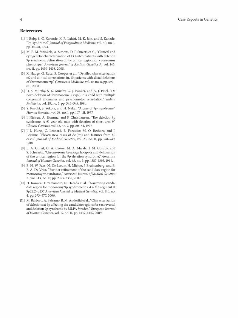
Figure 1: Frontal and lateral views of the face along with the feet showing long toes and square nails at the age of 4 months.
at 33 weeks of gestation, and the baby was delivered pretermby an emergency lower segment caesarean section due tofetal distress. At birth, he weighed 2.175 kg (50th centile)and was found to have craniofacial dysmorphism along withan omphalocele. His Apgar score was 8, 9, and 10 at 1, 5,and 10 minutes, respectively. Surgical repair of omphalocelewas performed on the second day of life, and the babywas managed postoperatively at the surgical intensive careunit on a ventilator for 48 days due to respiratory distressand repeated failed extubations. He was also concurrentlytreated for neonatal sepsis and was discharged home after fullrecovery.
On examination at the age of 4months, the child’s weight,length, and occipitofrontal circumference (OFC) were 3.4 kg(<3rd centile), 57 cm (<3rd centile), and 37 cm (<3rd centile).His gross motor and social milestones were delayed. Speechdevelopment could not be assessed due to his age. He hadmultiple craniofacial dysmorphic features such as prominentmetopic ridge with trigonocephalic head and flat occiput,midfacial hypoplasia, bilateral epicanthal folds, high archedeyebrows, downslanting palpebral fissures, ocular hyper-telorism, hypoplastic supraorbital ridges, flat nasal bridge,anteverted nostrils, low set ears with malformed auricles,long smooth philtrum with thin upper lip, downturned cor-ners of the mouth, micrognathia, short neck, asymmetricallyflattened chest wall on the left side with wide-set nipples, andlong fingers and toes due to increased length of the middlephalanges. The nails were square in shape (Figure 1).
Echocardiogram showed osteum secundum atrial septaldefect, and high resolution computed tomography scan(HRCT) of the lungs showed a complete collapse of theleft lung with almost absent lung tissue. The right lung washyperexpanded with midline shift to the left side. There wasright lower lobe atelectasis, mild interstitial thickening, andherniation of part of the right lobe to the left side but noevidence of brochopulmonary dysplasia or chronic lung dis-ease. Trachea and major bronchi were normal in caliber, and
pleural space was clear. Additional laboratory investigations,including blood urea, serum electrolytes, creatinine, alkalinephosphatase, calcium, phosphorus, and thyroid profiles, wereall normal.
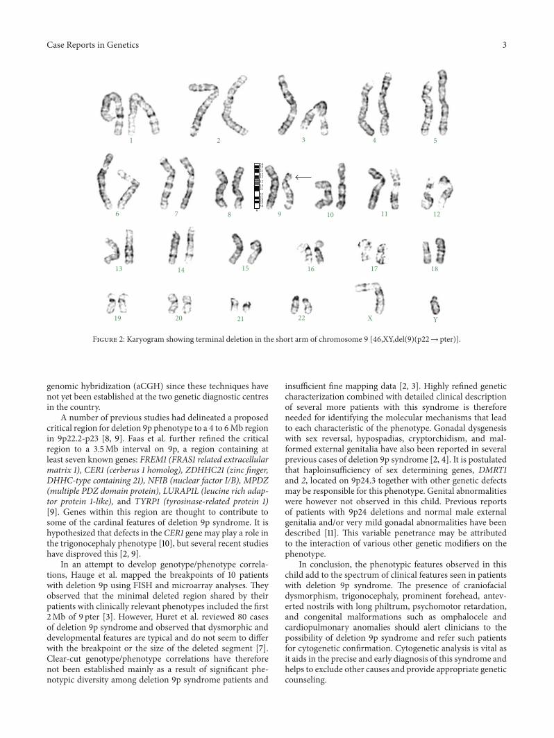
Chromosome culture and karyotyping of the child andthe parents were performed on routinely cultured peripheralblood lymphocytes. A total of 30 metaphase spreads wereanalyzed in each case according to conventional GTG band-ing technique. The maximum banding resolution achievedwas 500 bands. All the metaphases from the child showedterminal deletion of the short arm of chromosome 9 distalto band p22 as the sole abnormality. His karyotype was46,XY,del(9)(p22→ pter) shown in Figure 2, and normalkaryotypes were seen in both parents.
3. Discussion
The phenotypic features observed in this child are in linewith the consensus phenotype observed in other deletion 9psyndromepatients reported in the scientific literature [1, 4–6].In addition, this child had features of pulmonary hypoplasiawhich has not previously been reported in patients with denovo 9p deletion syndrome. It is assumed that this featuremay be a novel phenotype associated with the disruption ofyet unidentified genes in this region which may be involvedin pulmonary development.
Numerous reports in the scientific literature haveobserved that deletion 9p syndrome is caused by a partialmonosomy due to deletion of genetic material from the shortarm of chromosome 9 with breakpoint sites mainly in theregions 9p21 to 9p24 [1, 4, 5, 7, 8].The 9p22→ pter breakpointobserved in this child is consistent with that described inthe literature. High resolution mapping of the breakpoints todelineate the exact size of the deleted segment and exclusionof cryptic chromosome rearrangements however couldnot be performed in this case using fluorescence in situhybridization (FISH) or microarray-based comparative
Case Reports in Genetics 3
1 2 3 4 5
6 7 8 9 10 11 12
13 14 15 16 17 18
19 20 21 22 X Y
24232113
13
1212
212231323334
9
Figure 2: Karyogram showing terminal deletion in the short arm of chromosome 9 [46,XY,del(9)(p22→ pter)].
genomic hybridization (aCGH) since these techniques havenot yet been established at the two genetic diagnostic centresin the country.
A number of previous studies had delineated a proposedcritical region for deletion 9p phenotype to a 4 to 6Mb regionin 9p22.2-p23 [8, 9]. Faas et al. further refined the criticalregion to a 3.5Mb interval on 9p, a region containing atleast seven known genes: FREM1 (FRAS1 related extracellularmatrix 1), CER1 (cerberus 1 homolog), ZDHHC21 (zinc finger,DHHC-type containing 21), NFIB (nuclear factor I/B), MPDZ(multiple PDZ domain protein), LURAP1L (leucine rich adap-tor protein 1-like), and TYRP1 (tyrosinase-related protein 1)[9]. Genes within this region are thought to contribute tosome of the cardinal features of deletion 9p syndrome. It ishypothesized that defects in the CER1 gene may play a role inthe trigonocephaly phenotype [10], but several recent studieshave disproved this [2, 9].
In an attempt to develop genotype/phenotype correla-tions, Hauge et al. mapped the breakpoints of 10 patientswith deletion 9p using FISH and microarray analyses. Theyobserved that the minimal deleted region shared by theirpatients with clinically relevant phenotypes included the first2Mb of 9 pter [3]. However, Huret et al. reviewed 80 casesof deletion 9p syndrome and observed that dysmorphic anddevelopmental features are typical and do not seem to differwith the breakpoint or the size of the deleted segment [7].Clear-cut genotype/phenotype correlations have thereforenot been established mainly as a result of significant phe-notypic diversity among deletion 9p syndrome patients and
insufficient fine mapping data [2, 3]. Highly refined geneticcharacterization combined with detailed clinical descriptionof several more patients with this syndrome is thereforeneeded for identifying the molecular mechanisms that leadto each characteristic of the phenotype. Gonadal dysgenesiswith sex reversal, hypospadias, cryptorchidism, and mal-formed external genitalia have also been reported in severalprevious cases of deletion 9p syndrome [2, 4]. It is postulatedthat haploinsufficiency of sex determining genes, DMRT1and 2, located on 9p24.3 together with other genetic defectsmay be responsible for this phenotype. Genital abnormalitieswere however not observed in this child. Previous reportsof patients with 9p24 deletions and normal male externalgenitalia and/or very mild gonadal abnormalities have beendescribed [11]. This variable penetrance may be attributedto the interaction of various other genetic modifiers on thephenotype.
In conclusion, the phenotypic features observed in thischild add to the spectrum of clinical features seen in patientswith deletion 9p syndrome. The presence of craniofacialdysmorphism, trigonocephaly, prominent forehead, antev-erted nostrils with long philtrum, psychomotor retardation,and congenital malformations such as omphalocele andcardiopulmonary anomalies should alert clinicians to thepossibility of deletion 9p syndrome and refer such patientsfor cytogenetic confirmation. Cytogenetic analysis is vital asit aids in the precise and early diagnosis of this syndrome andhelps to exclude other causes and provide appropriate geneticcounseling.
4 Case Reports in Genetics
References
[1] J. Boby, S. C. Karande, K. R. Lahiri, M. K. Jain, and S. Kanade,“9p-syndrome,” Journal of Postgraduate Medicine, vol. 40, no. 1,pp. 40–41, 1994.
[2] M. E. M. Swinkels, A. Simons, D. F. Smeets et al., “Clinical andcytogenetic characterization of 13 Dutch patients with deletion9p syndrome: delineation of the critical region for a consensusphenotype,” American Journal of Medical Genetics A, vol. 146,no. 11, pp. 1430–1438, 2008.
[3] X. Hauge, G. Raca, S. Cooper et al., “Detailed characterizationof, and clinical correlations in, 10 patients with distal deletionsof chromosome 9p,”Genetics inMedicine, vol. 10, no. 8, pp. 599–611, 2008.
[4] D. S. Murthy, S. K. Murthy, G. J. Banker, and A. J. Patel, “Denovo deletion of chromosome 9 (9p-) in a child with multiplecongenital anomalies and psychomotor retardation,” IndianPediatrics, vol. 28, no. 5, pp. 546–549, 1991.
[5] Y. Kuroki, S. Yokota, and H. Nakai, “A case of 9p- syndrome,”Human Genetics, vol. 38, no. 1, pp. 107–111, 1977.
[6] J. Nielsen, A. Homma, and F. Christiansen, “The deletion 9psyndrome. A 61 year old man with deletion of short arm 9,”Clinical Genetics, vol. 12, no. 2, pp. 80–84, 1977.
[7] J. L. Huret, C. Leonard, B. Forestier, M. O. Rethore, and J.Lejeune, “Eleven new cases of del(9p) and features from 80cases,” Journal of Medical Genetics, vol. 25, no. 11, pp. 741–749,1988.
[8] L. A. Christ, C. A. Crowe, M. A. Micale, J. M. Conroy, andS. Schwartz, “Chromosome breakage hotspots and delineationof the critical region for the 9p-deletion syndrome,” AmericanJournal of Human Genetics, vol. 65, no. 5, pp. 1387–1395, 1999.
[9] B. H. W. Faas, N. De Leeuw, H. Mieloo, J. Bruinenberg, and B.B. A. De Vries, “Further refinement of the candidate region formonosomy 9p syndrome,”American Journal ofMedical GeneticsA, vol. 143, no. 19, pp. 2353–2356, 2007.
[10] H. Kawara, T. Yamamoto, N. Harada et al., “Narrowing candi-date region for monosomy 9p syndrome to a 4.7-Mb segment at9p22.2–p23,”American Journal of Medical Genetics, vol. 140, no.4, pp. 373–377, 2006.
[11] M. Barbaro, A. Balsamo, B.M.Anderlid et al., “Characterizationof deletions at 9p affecting the candidate regions for sex reversaland deletion 9p syndrome byMLPA Sweden,” European Journalof Human Genetics, vol. 17, no. 11, pp. 1439–1447, 2009.

Submit your manuscripts athttp://www.hindawi.com
Stem CellsInternational
Hindawi Publishing Corporationhttp://www.hindawi.com Volume 2014
Hindawi Publishing Corporationhttp://www.hindawi.com Volume 2014
MEDIATORSINFLAMMATION
of
Hindawi Publishing Corporationhttp://www.hindawi.com Volume 2014
Behavioural Neurology
EndocrinologyInternational Journal of
Hindawi Publishing Corporationhttp://www.hindawi.com Volume 2014
Hindawi Publishing Corporationhttp://www.hindawi.com Volume 2014
Disease Markers
Hindawi Publishing Corporationhttp://www.hindawi.com Volume 2014
BioMed Research International
OncologyJournal of
Hindawi Publishing Corporationhttp://www.hindawi.com Volume 2014
Hindawi Publishing Corporationhttp://www.hindawi.com Volume 2014
Oxidative Medicine and Cellular Longevity
Hindawi Publishing Corporationhttp://www.hindawi.com Volume 2014
PPAR Research
The Scientific World JournalHindawi Publishing Corporation http://www.hindawi.com Volume 2014
Immunology ResearchHindawi Publishing Corporationhttp://www.hindawi.com Volume 2014
Journal of
ObesityJournal of
Hindawi Publishing Corporationhttp://www.hindawi.com Volume 2014
Hindawi Publishing Corporationhttp://www.hindawi.com Volume 2014
Computational and Mathematical Methods in Medicine
OphthalmologyJournal of
Hindawi Publishing Corporationhttp://www.hindawi.com Volume 2014
Diabetes ResearchJournal of
Hindawi Publishing Corporationhttp://www.hindawi.com Volume 2014
Hindawi Publishing Corporationhttp://www.hindawi.com Volume 2014
Research and TreatmentAIDS
Hindawi Publishing Corporationhttp://www.hindawi.com Volume 2014
Gastroenterology Research and Practice
Hindawi Publishing Corporationhttp://www.hindawi.com Volume 2014
Parkinson’s Disease
Evidence-Based Complementary and Alternative Medicine
Volume 2014Hindawi Publishing Corporationhttp://www.hindawi.com