case report chronic acquired demyelinating polyneuropathy
TRANSCRIPT
Hindawi Publishing CorporationCase Reports in Neurological MedicineVolume 2013, Article ID 360454, 2 pageshttp://dx.doi.org/10.1155/2013/360454
Case ReportChronic Acquired Demyelinating Polyneuropathy followingRenal Transplantation
D. S. Younger1,2,3 and Stuart Orsher2
1 Department of Neurology, NYU School of Medicine, New York University Langone Medical Center, 550 First Avenue,New York, NY 10016, USA
2Department of Medicine, Lenox Hill Hospital, New York, NY 10021, USA3 Section of Neurology, Lenox Hill Hospital, New York, NY 10021, USA
Correspondence should be addressed to D. S. Younger; [email protected]
Received 5 June 2013; Accepted 16 August 2013
Academic Editors: J. L. Gonzalez-Gutierrez and D. J. Rivet
Copyright © 2013 D. S. Younger and S. Orsher. This is an open access article distributed under the Creative Commons AttributionLicense, which permits unrestricted use, distribution, and reproduction in any medium, provided the original work is properlycited.
The clinical, laboratory, and treatment findings of a patient with chronic acquired demyelinating polyneuropathy (CADP) inassociation with renal transplantation are described. Like the present case, many such patients have been described under therubric of chronic inflammatory demyelinating polyradiculoneuropathy (CIDP).
1. Introduction
The chronic acquired demyelinating polyneuropathies (CADP)are treatable heterogeneous disorders. Electrodiagnosis, whichdistinguishes primary demyelination from primary axonalneuropathy, achieves 95% sensitivity in CIDP [1]. Numerousconditions have been associated with CADP, among themsolid organ transplantation [2]. Only ten heterogeneous pa-tients [3] with preexisting axonal polyneuropathy met defi-nite demyelinating electrodiagnostic criteria after solid organtransplantation [2]. We present the detailed clinical findingsof another patient with progressive demyelinating neuropa-thy suggested by serial electrodiagnostic studies, diagnosticfeatures on cutaneous nerve biopsy, and a positive responseto intravenous immune globulin (IVIg), a preliminary reportof which has been published [4].
2. Patient Report
A previously healthy 74-year-old man with solitary kidneydeveloped renal insufficiency in 2008 prompting hemodial-ysis and eventual donor transplantation in 2009. Afterward,he was maintained on tacrolimus. This was followed shortlyafterward by glycemic intolerancemanagedwith diet. In 2011,he noted numbness, tingling sensation, and gait instability.
Neurological examination showed tandem gait imbalanceand symmetrical stocking vibratory and cold temperaturesensory loss to below the knees, with slight weakness ofthe tibialis anterior muscles versus firm resistance that wasgraded Medical Research Council (MRC) 4/5 bilaterally.There was generalized hyporeflexia with intact limb strength,cognition, and cranial nerve function. Electrodiagnosticstudies (Table 1) showed low amplitude compound muscleaction potentials (CMAP), slow motor nerve conductionvelocities, prolonged distal motor latencies, prolonged orabsent F wave latencies, and abnormal temporal disper-sion without active or chronic spontaneous activity at reston concentric needle electromyography (EMG). Epidermalnerve fiber (ENF) studies of the left calf and thigh showedsignificantly low ENF density: distal leg 0.06 and thigh5.4 ENF/mm (5th percentile reference values, resp., 5.0 and8.0 ENF/mm 9) without histologic abnormalities or amyloiddeposition. The serum creatine kinase was 172U/L (nor-mal < 150). Left sural nerve biopsy showed chronic andactive peripheral neuropathy with primary demyelinatingfeatures and mild secondary axonal loss evidenced on teasednerve fiber studies in which 27 (54%) showed segmen-tal remyelination, 23 (46%) were normal in configuration,and none showed Wallerian degeneration. Semithin epoxy
2 Case Reports in Neurological Medicine
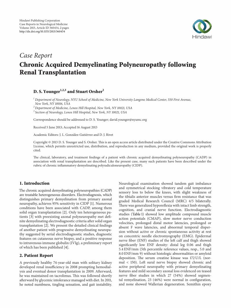
Table 1: Nerve conduction findings in a patient with progressive demyelinating neuropathy after renal transplantation.
Motor nerve conductions Sensory nerve conductionsCMAP (mV) DL (ms) Velocity (m/s) F-response (ms) SNAP (𝜇V) DL (ms) Velocity (m/s)
Nerve/study 1 1 2 1 2 1 2 1 2 1 2 1 2 1 2R. fibular 3.8 4.1 4.4 4.9 37 39 NR 65R. tibial 1.0 3.3 7.3 6.6 31 33 76 69R. sural NR 3.0 — 3.5 — 28R. sup. fibular NR NRL. fibular 1.5 3.1 6.9 5.4 37 42 NR 63L. sural NR NRL. sup. fibular 1.7 4.0 7.6 6.3 32 36 73 611: study 1, November 2011; 2: study 2, June 2012. Abbreviations: L.: left; R.: right; CMAP: compound muscle action potential; DL: distal latency; mV: millivolt;ms: milliseconds; m/s: meters per second; SNAP: sensory nerve action potential; NR: no response; sup.: superficial.
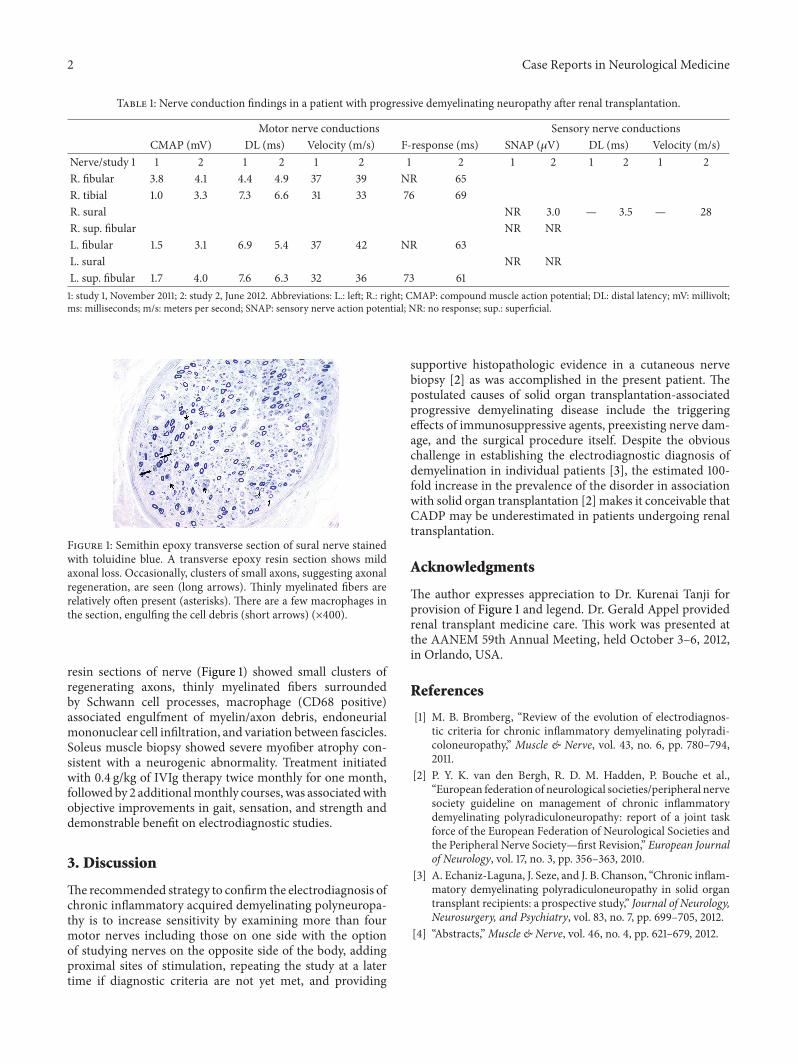
Figure 1: Semithin epoxy transverse section of sural nerve stainedwith toluidine blue. A transverse epoxy resin section shows mildaxonal loss. Occasionally, clusters of small axons, suggesting axonalregeneration, are seen (long arrows). Thinly myelinated fibers arerelatively often present (asterisks). There are a few macrophages inthe section, engulfing the cell debris (short arrows) (×400).
resin sections of nerve (Figure 1) showed small clusters ofregenerating axons, thinly myelinated fibers surroundedby Schwann cell processes, macrophage (CD68 positive)associated engulfment of myelin/axon debris, endoneurialmononuclear cell infiltration, and variation between fascicles.Soleus muscle biopsy showed severe myofiber atrophy con-sistent with a neurogenic abnormality. Treatment initiatedwith 0.4 g/kg of IVIg therapy twice monthly for one month,followed by 2 additionalmonthly courses, was associatedwithobjective improvements in gait, sensation, and strength anddemonstrable benefit on electrodiagnostic studies.
3. Discussion
Therecommended strategy to confirm the electrodiagnosis ofchronic inflammatory acquired demyelinating polyneuropa-thy is to increase sensitivity by examining more than fourmotor nerves including those on one side with the optionof studying nerves on the opposite side of the body, addingproximal sites of stimulation, repeating the study at a latertime if diagnostic criteria are not yet met, and providing
supportive histopathologic evidence in a cutaneous nervebiopsy [2] as was accomplished in the present patient. Thepostulated causes of solid organ transplantation-associatedprogressive demyelinating disease include the triggeringeffects of immunosuppressive agents, preexisting nerve dam-age, and the surgical procedure itself. Despite the obviouschallenge in establishing the electrodiagnostic diagnosis ofdemyelination in individual patients [3], the estimated 100-fold increase in the prevalence of the disorder in associationwith solid organ transplantation [2] makes it conceivable thatCADP may be underestimated in patients undergoing renaltransplantation.
Acknowledgments
The author expresses appreciation to Dr. Kurenai Tanji forprovision of Figure 1 and legend. Dr. Gerald Appel providedrenal transplant medicine care. This work was presented atthe AANEM 59th Annual Meeting, held October 3–6, 2012,in Orlando, USA.
References
[1] M. B. Bromberg, “Review of the evolution of electrodiagnos-tic criteria for chronic inflammatory demyelinating polyradi-coloneuropathy,” Muscle & Nerve, vol. 43, no. 6, pp. 780–794,2011.
[2] P. Y. K. van den Bergh, R. D. M. Hadden, P. Bouche et al.,“European federation of neurological societies/peripheral nervesociety guideline on management of chronic inflammatorydemyelinating polyradiculoneuropathy: report of a joint taskforce of the European Federation of Neurological Societies andthe Peripheral Nerve Society—first Revision,” European Journalof Neurology, vol. 17, no. 3, pp. 356–363, 2010.
[3] A. Echaniz-Laguna, J. Seze, and J. B. Chanson, “Chronic inflam-matory demyelinating polyradiculoneuropathy in solid organtransplant recipients: a prospective study,” Journal of Neurology,Neurosurgery, and Psychiatry, vol. 83, no. 7, pp. 699–705, 2012.
[4] “Abstracts,”Muscle & Nerve, vol. 46, no. 4, pp. 621–679, 2012.

Submit your manuscripts athttp://www.hindawi.com
Stem CellsInternational
Hindawi Publishing Corporationhttp://www.hindawi.com Volume 2014
Hindawi Publishing Corporationhttp://www.hindawi.com Volume 2014
MEDIATORSINFLAMMATION
of
Hindawi Publishing Corporationhttp://www.hindawi.com Volume 2014
Behavioural Neurology
EndocrinologyInternational Journal of
Hindawi Publishing Corporationhttp://www.hindawi.com Volume 2014
Hindawi Publishing Corporationhttp://www.hindawi.com Volume 2014
Disease Markers
Hindawi Publishing Corporationhttp://www.hindawi.com Volume 2014
BioMed Research International
OncologyJournal of
Hindawi Publishing Corporationhttp://www.hindawi.com Volume 2014
Hindawi Publishing Corporationhttp://www.hindawi.com Volume 2014
Oxidative Medicine and Cellular Longevity
Hindawi Publishing Corporationhttp://www.hindawi.com Volume 2014
PPAR Research
The Scientific World JournalHindawi Publishing Corporation http://www.hindawi.com Volume 2014
Immunology ResearchHindawi Publishing Corporationhttp://www.hindawi.com Volume 2014
Journal of
ObesityJournal of
Hindawi Publishing Corporationhttp://www.hindawi.com Volume 2014
Hindawi Publishing Corporationhttp://www.hindawi.com Volume 2014
Computational and Mathematical Methods in Medicine
OphthalmologyJournal of
Hindawi Publishing Corporationhttp://www.hindawi.com Volume 2014
Diabetes ResearchJournal of
Hindawi Publishing Corporationhttp://www.hindawi.com Volume 2014
Hindawi Publishing Corporationhttp://www.hindawi.com Volume 2014
Research and TreatmentAIDS
Hindawi Publishing Corporationhttp://www.hindawi.com Volume 2014
Gastroenterology Research and Practice
Hindawi Publishing Corporationhttp://www.hindawi.com Volume 2014
Parkinson’s Disease
Evidence-Based Complementary and Alternative Medicine
Volume 2014Hindawi Publishing Corporationhttp://www.hindawi.com