case report computed tomography appearance of inflammatory
TRANSCRIPT

Int J Clin Exp Med 2015;8(9):16745-16755www.ijcem.com /ISSN:1940-5901/IJCEM0008226
Case ReportComputed tomography appearance of inflammatory myofibroblastic tumor in the abdomen: CT features and pathologic correlation
Bo Liu1*, Junlong Xu2*, Jiaxin Wang3*, Hongguang Fan4, Xuan Ang4, Wenming Liu5
1Department of X-Ray, Chinese Medicine Hospital of Henan Province, Zhengzhou 450002, China; 2Department of Pathology, Liao Cheng People’s Hospital, Shandong Province, China; 3Department of Head and Neck Cancer, Tianjin Medical University Cancer Institute and Hospital, National Clinical Research Center for Cancer, Key Laboratory of Cancer Prevention and Therapy, Tianjin, 300060, China; 4Department of X-Ray, The People’s Hospital of Zhengzhou University, Zhengzhou 450003, China; 5Department of Burn and Plastic Surgery, Binzhou Medical University Hospital, Binzhou 256603, Shandong Province, China. *Equal contributors.
Received March 19, 2015; Accepted September 6, 2015; Epub September 15, 2015; Published September 30, 2015
Abstract: Objective: To evaluate CT findings of abdominal inflammatory myofibroblastic tumor (IMT) and the rela-tionship with morphological character. Materials and Methods: CT examinations and pathological findings of ten intra-abdominal IMTs were retrospectively analyzed. The histopathological characteristics of the IMTs were con-firmed by two pathologists and two radiologists evaluated CT findings of the lesion, with emphasis on the imaging features compared with the corresponding histopathology. Results: The most common imaging characteristics were presence of heterogeneity, all tumors showed varying degrees of contrast enhancement. Two major different CT patterns were individualized. In type one, the tumor had a distinct boundary without a lobular appearance and dis-played hypo-enhanced enhancement after administration of contrast in correlated with the mainly histopathologic findings of spindle cells myxoid and hypocellular fibrous (6/10; 60%). In type two, the lesions exhibited indistinct boundaries or complete capsule, ill-defined growth patterns or low intralesional attenuation with marked heteroge-neous or circumferential enhancement, which correlated well with the presence of abundance of micromodule and inflammatory cell infiltration (4/10; 40%). Conclusions: Two major different contrast enhancement CT patterns were individualized can help to determine the relationships with histopathologic findings, while cannot be reliably differ-entiated from other solid lesions based solely on the CT appearance, combined with diagnostic biopsy may facilitate to achieve a correct diagnosis and treatment.
Keywords: Inflammatory myofibroblastic tumor, abdomen, computed tomography, histology
Introduction
Inflammatory myofibroblastic tumor is a rare mesenchymal neoplasm with uncertain etio- logy. Originally, it was termed “inflammatory pseudotumor”, “postoperative spindle cell nod-ule”, inflammatory myofibrohistiocytic prolifera-tion” [1]. Subsequently, further studies have identified its true nature as a neoplasm which may recur and rarely metastasize. Its classical features were spindle cells proliferation inter-mixed with inflammatory cells which were thought to reflect diverse entities [2]. Moreover, it was classified as a tumor which has a ten-dency for local recurrence and a risk of distant metastasis malignant transformation accord-ing to World Health Organization classification.
IMT in the abdomen can occur at any location including the stomach, intestine, mesentery, peritoneum, retroperitoneum, pancreas, liver, and so on. The purpose of our retrospective study was to bring forth some evidence that dif-ferent enhanced CT imaging may be derived from the subtype of histological component and delineated the variable clinicopathologic fea-tures and corresponding CT imaging, in effort to establish an appropriate CT diagnosis approach with the use of surgical and histopathological findings as the reference standard.
Materials and methods
We retrospectively reviewed ten consecutive IMT patients and obtained informed consent

CT appearance of inflammatory myofibroblastic tumor
16746 Int J Clin Exp Med 2015;8(9):16745-16755
Table 1. Clinical, demographic and laboratory data of abdominal IMT
NO. Sex/Age (years) Symptoms/signs Location Maximum
Diameter (cm) Laboratory Abnormalities
1 F/18 Stomachache Mesentery 3.5 Normal2 M/14 Epigastric discomfort Omentum Stomach 4.5 Anemia, ESR↑3 M/23 Pain Colon 11 Leukocytosis, ESR↑, CRP↑4 M/12 Vomiting Mesentery 6.5 ESR↑5 M/38 Abdominal distention Mesentery Intestines 6.5, 9 Anemia, ESR↑6 M/54 Incidentally detected Intestines 7 Hyperglobulinemia, CRP↑7 F/18 Fever Colon retroperitoneum 12.5 eESR↑, Anemia, Leukocytosis8 F/16 Vomiting Intestines 7.5 ESR↑9 M/24 Pain Intestines 15 ESR↑10 F/26 Fever Anemia, Behind liver 9 CRP↑, Leukocytosis, ESR↑F: Female, M: Male, Pain: Abdominal pain, ESR↑: Elevated erythrocyte sedimentation rate, CRP↑: Elevated C-reactive protein.
from all patients at our department from January 2006 to August 2011. This study was approved by our institutional ethics committee. Radiology databases were searched to identify patients who had confirmed IMT histopatholog-ically and had undergone abdominal CT scan-ning. All the samples enrolled in this study were kept anonymously after retrieval of follow-up information. Previous comprehensive medi-cal records of every patient were evaluated especially the history of abdomen inflammation or trauma. All medical details were supple- mented by the out-patient and past hospital records. Demography, clinical and radiological presentation, pathological outcome were docu-mented. All patients had full physical examina-
tion, including episodes of abdominal pain and antibiotics use. All cases had undergone abdominal unenhanced and contrast-enhanced CT scanning which was performed using a SOMATOM Definition double-source helical scanner CT (Siemens, Medical Systems, Ger- many). Associated imaging findings were also evaluated, including location, lesion number, diameter, contour and border of the lesion, the growth pattern characteristics, attenuation before and after contrast enhancement pat-terns. Standard parameters for spiral CT were 120 kVp, 120 mA s, the effective slice thick-ness was 5 mm. Arterial, venous and delayed-phase CT was performed after initiation of intravenous contrast medium injection of 80 ml
Figure 1. Multicenter IMT. A. Non-enhanced CT images showed two lesions of homogeneously intensity (white ar-rows). B. Contrast-enhanced CT images showed the absence of enhancing components (White arrows).

CT appearance of inflammatory myofibroblastic tumor
16747 Int J Clin Exp Med 2015;8(9):16745-16755
intravenous contrast material with a flow of 4 ml/s. The degree and pattern of enhancement of tumor spreading patterns were evaluated by two radiologists. All cases underwent laparoto-my or performed imaging-guided biopsy and the final diagnosis of IMT was made after evalu-ation of specimen by two pathologists. All the histological material was evaluated accord- ing to the current WHO pathological criteria. Continuous variables with a normal distribution were expressed as mean ± standard deviation (Std). A P-value less than 0.05 was considered
statistically significant. all statistical tests were carried out utilizing SPSS, version 17.
Results
Clinical data
Ten patients were enrolled in the investigation, the male to female ratio was 3:2, with a mean age of 25.4 years (range, 12 to 54 years). Patients presented with alimentary tract obstructive symptoms (n=5), abdominal pain (n=2), fever (n=2), and found incidentally (n=1).
Figure 2. An unenhanced abdominal CT scan shows (A) a well-defined 13.5×15×14 cm large mass with scattered low-density areas in the center which suggestive of necrosis. (B) Contrast-enhanced CT images noted that the le-sion was in heterogeneous enhancement patterns. There is a clear plane between the mass and the adjacent liver.
Figure 3. Abdominal CT scan showing the mass was well-defined and with multiply dotted calcification in the center (white arrows).
Figure 4. Non-enhanced abdominal CT image show-ing massive calcification changes extending into the mass (white arrow).
CT appearance of inflammatory myofibroblastic tumor
16748 Int J Clin Exp Med 2015;8(9):16745-16755
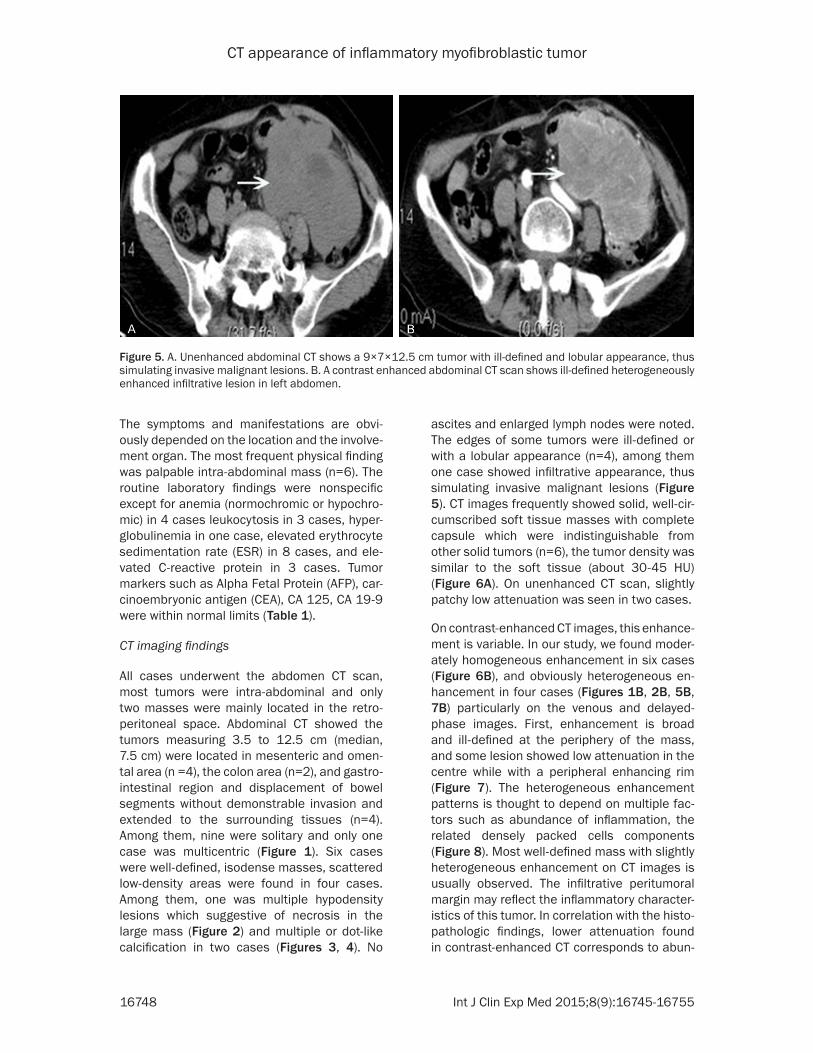
Figure 5. A. Unenhanced abdominal CT shows a 9×7×12.5 cm tumor with ill-defined and lobular appearance, thus simulating invasive malignant lesions. B. A contrast enhanced abdominal CT scan shows ill-defined heterogeneously enhanced infiltrative lesion in left abdomen.
The symptoms and manifestations are obvi-ously depended on the location and the involve-ment organ. The most frequent physical finding was palpable intra-abdominal mass (n=6). The routine laboratory findings were nonspecific except for anemia (normochromic or hypochro-mic) in 4 cases leukocytosis in 3 cases, hyper-globulinemia in one case, elevated erythrocyte sedimentation rate (ESR) in 8 cases, and ele-vated C-reactive protein in 3 cases. Tumor markers such as Alpha Fetal Protein (AFP), car-cinoembryonic antigen (CEA), CA 125, CA 19-9 were within normal limits (Table 1).
CT imaging findings
All cases underwent the abdomen CT scan, most tumors were intra-abdominal and only two masses were mainly located in the retro-peritoneal space. Abdominal CT showed the tumors measuring 3.5 to 12.5 cm (median, 7.5 cm) were located in mesenteric and omen-tal area (n =4), the colon area (n=2), and gastro-intestinal region and displacement of bowel segments without demonstrable invasion and extended to the surrounding tissues (n=4).Among them, nine were solitary and only one case was multicentric (Figure 1). Six cases were well-defined, isodense masses, scattered low-density areas were found in four cases. Among them, one was multiple hypodensity lesions which suggestive of necrosis in the large mass (Figure 2) and multiple or dot-like calcification in two cases (Figures 3, 4). No
ascites and enlarged lymph nodes were noted. The edges of some tumors were ill-defined or with a lobular appearance (n=4), among them one case showed infiltrative appearance, thus simulating invasive malignant lesions (Figure 5). CT images frequently showed solid, well-cir-cumscribed soft tissue masses with complete capsule which were indistinguishable from other solid tumors (n=6), the tumor density was similar to the soft tissue (about 30-45 HU) (Figure 6A). On unenhanced CT scan, slightly patchy low attenuation was seen in two cases.
On contrast-enhanced CT images, this enhance-ment is variable. In our study, we found moder-ately homogeneous enhancement in six cases (Figure 6B), and obviously heterogeneous en- hancement in four cases (Figures 1B, 2B, 5B, 7B) particularly on the venous and delayed-phase images. First, enhancement is broad and ill-defined at the periphery of the mass, and some lesion showed low attenuation in the centre while with a peripheral enhancing rim (Figure 7). The heterogeneous enhancement patterns is thought to depend on multiple fac-tors such as abundance of inflammation, the related densely packed cells components (Figure 8). Most well-defined mass with slightly heterogeneous enhancement on CT images is usually observed. The infiltrative peritumoral margin may reflect the inflammatory character-istics of this tumor. In correlation with the histo-pathologic findings, lower attenuation found in contrast-enhanced CT corresponds to abun-

CT appearance of inflammatory myofibroblastic tumor
16749 Int J Clin Exp Med 2015;8(9):16745-16755
dance of fibrous tissue (Figure 9), while the hyperattenuating area is corresponding to the predominantly inflammation cells infiltration and hypervascularity density pattern or micro-vascular hyperplasia (Figure 10). These images and pathological results corresponded to the findings in this present study.
Pathology and immunohistochemistry
Grossly, six specimens had complete capsule and indistinct capsule was found in four cases. The cut surface showed a fleshy texture with
regions of grey-red appearance. Patchy necro-sis areas and haemorrhage were noted in two cases, obvious calcification was found only in one case. The specimen section demonstrated the tumor cells were mainly arranged in fusi-form pattern and the characteristic features were the presence of spindle cells with an infil-tration of lymphocytes and eosinophils, occa-sional atypical cells with mitotic nuclei Figure ureures can be seen. The spindle cells showed bland, vesicular, round to oval shaped nuclei and possessed abundant eosinophiliccyto-plasms imparting myofibroblasts. The mesen-
Figure 6. A. Non-enhanced CT scan showed a solid, well-circumscribed soft tissue mass with complete capsule. B. A contrast enhanced abdominal CT scan revealed moderately homogeneous enhancement.
Figure 7. A. Non-enhanced abdominal CT imaging showed a well-defined hypoattenuating mass adjacent to the left kidney. B. On contrast enhancement CT, the lesion was with a peripheral enhancing rim while poorly enhancing in the center.

CT appearance of inflammatory myofibroblastic tumor
16750 Int J Clin Exp Med 2015;8(9):16745-16755
chymal component, generally regarded as hav-ing morphological features consistent with mucoid degeneration. Immunohistochemically, the lesions expressed smooth muscle act in (8/10, 80%), desmin (9/10, 90%), vimentin (9/10, 90%) (Figure 11A), and anaplastic lym-phoma kinase-1 (7/10, 70%) (Figure 11B), neg-ative for CD 117, CD 34 and S 100. On the basis of histology and immunohistochemistry, the pathologic diagnosis was inflammatory myofi-broblastic tumor. In our cases, IMTs can be classified into two different subtype entities based on major morphologies findings. The lesions may range from fibrosing inflammatory lesions to a myofibroblastic appearance with heavy infiltration of inflammatory cells, accom-
panied with various vessels. The most common type was abundant in compact spindle cells with inflammatory and vascular areas. (Figures 8, 10) The other subtype was a desmoid-like pattern with myxoidfibrer and hypocellular area (Figure 9). There was no evidence of purulent material or acute inflammatory cells such as neutrophils within the lesion. The pathological assessment and further subclassifications of the lesion was listed as follow (Table 2).
Discussion
Originally, IMT is proposed as a post inflamma-tory reactive process, occurring after surgery or trauma. Several terms have been used to describe this entity including “inflammatory pseudotumor”, “inflammatory myofibrohistio-cytic proliferation”, “inflammatory fibrosarco-ma” and atypical myofibroblastic tumor. To avoid ambiguity, these designations are best avoided according to WHO which IMT is classi-fied as an intermediate biological potential tumor with distinct clinicopathological entities and different from inflammatory pseudotumor [3]. Previous studies have attributed etiology to an immunological response to viral -8 or bacte-rium infection [4, 5]. But there is no evidence of a proven relationship between IMT and any specific infectious agent in our investigation.
Demographic data and clinical history have lit-tle help in the diagnosis. Children and adoles-cents constitute the majority of IMT, there is no sex predominance. IMT can occur in any ana-tomical location and predominantly arise in the
Figure 8. Microphotographs of hematoxylin & eosin-stained sections showing abundance of spindle cells arranged in storiform pattern, admixed with inflam-matory cells (×200).
Figure 9. Microphotographs of hematoxylin & eosin-stained sections showing sparsely cellular areas with abundant fibrous stroma and scattered inflammatory cells (×100).
Figure 10. High-power view showing those spindle cells with abundant inflammation cells infiltration and the hyper vascularity density pattern was con-sisted of microvascular hyperplasia (×400).

CT appearance of inflammatory myofibroblastic tumor
16751 Int J Clin Exp Med 2015;8(9):16745-16755
lung, occasionally in head and neck, extremi-ties, urinary tract, pelvis, retroperitoneal region [6-8]. The abdomen is the most extrapulmo-nary region and the lesion can be solitary or multicentric. Those occurring in the mesentery, omentum, retroperitoneum and pelvis tend to be of large dimensions. The symptoms and manifestations varied according to the locat- ion and dimension of the lesions, abdominal distention is the most common symptoms. According to the literature, 19% were accompa-nied by systemic symptom, such as fever, gas-trointestinal symptom, and weight loss or can be found occasionally. The lesions revealed dif-ferent growth patterns, leading to obstruction or displacement of bowel segments. Rapid growth and multicentre appearance of these masses simulated malignancy. Some abdomi-nal IMTs were associated with inflammatory signs and symptoms related to their local spread. A palpable mass may be the clinical presentation in abdomen that often mistaken for malignant neoplasms, such as sarcomas, lymphomas or gastrointestinal stromal tumor (GIST). Demographic data and clinical history have little help in diagnosis. Raised serum lev-els of IL-6 reported to be found in IMT patients and returned to normal postoperatively [9]. The elevated IL-6 and ALK gene rearrangements may have a potential role in diagnosis. Positive
ALK status is more frequent in aggressive tumors, whereas ALK-negative IMTs were asso-ciated with metastases [10]. But in our study, there was no examination of IL-6, and ALK was positive in about 80% in all cases.
Although CT may offer suggestive features, while the entity in the abdomen is exceedingly rare and difficult to obtain a definitive diagno-sis. The radiologic features of intra-abdominal IMTs are non-specific, radiological investiga-tions are often inconclusive owing to absence of pathognomonic features. Occasionally, some had an infiltrative appearance and ill-defined margin features and indistinguishable from malignant lesions [11]. If a spontaneous or slight regression is noted, then IMT should be considered. Confirmed diagnose is usually obtained only by biopsy or surgical intervention. Radiologically, IMT is rarely suspected before operation due to the low incidence of this tumor. Nevertheless, imaging plays its role on pre-operative planning by delineating the extent of the disease and decision of surgical scheme [12]. Considerable similarities between IMT and malignant tumor make the differentia-tion difficult before operation. All our cases were misdiagnosed as malignant or gastroin-testinal stromal tumor on preoperative CT evaluation.
Figure 11. Microphotographs of immunohistochemical stains showing: A. Diffuse cytoplasmic immunopositivity for SMA in tumor cells. (×100); B. ALK is expressed in tumor cells (×200).
CT appearance of inflammatory myofibroblastic tumor
16752 Int J Clin Exp Med 2015;8(9):16745-16755
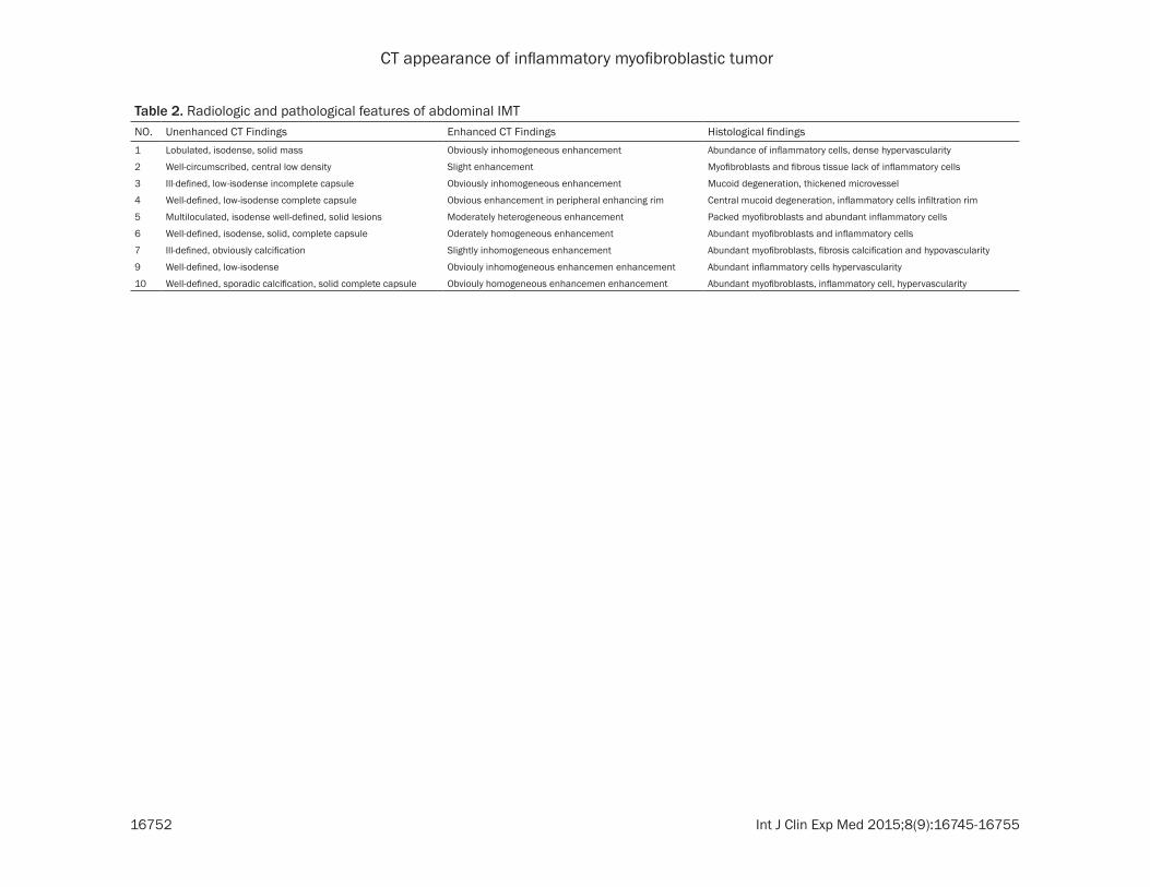
Table 2. Radiologic and pathological features of abdominal IMTNO. Unenhanced CT Findings Enhanced CT Findings Histological findings1 Lobulated, isodense, solid mass Obviously inhomogeneous enhancement Abundance of inflammatory cells, dense hypervascularity
2 Well-circumscribed, central low density Slight enhancement Myofibroblasts and fibrous tissue lack of inflammatory cells
3 Ill-defined, low-isodense incomplete capsule Obviously inhomogeneous enhancement Mucoid degeneration, thickened microvessel
4 Well-defined, low-isodense complete capsule Obvious enhancement in peripheral enhancing rim Central mucoid degeneration, inflammatory cells infiltration rim
5 Multiloculated, isodense well-defined, solid lesions Moderately heterogeneous enhancement Packed myofibroblasts and abundant inflammatory cells
6 Well-defined, isodense, solid, complete capsule Oderately homogeneous enhancement Abundant myofibroblasts and inflammatory cells
7 Ill-defined, obviously calcification Slightly inhomogeneous enhancement Abundant myofibroblasts, fibrosis calcification and hypovascularity
9 Well-defined, low-isodense Obviouly inhomogeneous enhancemen enhancement Abundant inflammatory cells hypervascularity
10 Well-defined, sporadic calcification, solid complete capsule Obviouly homogeneous enhancemen enhancement Abundant myofibroblasts, inflammatory cell, hypervascularity

CT appearance of inflammatory myofibroblastic tumor
16753 Int J Clin Exp Med 2015;8(9):16745-16755
The literature on radiological appearance is less, and there are many conflicting opinions regarding the CT imaging. In previous study, owing to the mixed histologic character, imag-ing characteristics of this tumor’s subtypes by correlation with the pathology was not possible [13]. While in our series, most lesions were homogeneous on unenhanced CT imaging and contrast-enhanced CT imaging is variable. With reference to the limited radiological descrip-tion, IMT appeared as heterogeneous solid mass with well-circumscribed margin, calcifica-tion and central necrosis [14]. In previous study, CT demonstration of prominent enhancement was suggestive of regional inflammatory chang-es, supporting the diagnosis of IMT [15]. When correlating the CT features with the histological findings, the heterogeneous enhancement in agreement with the components of the cellular and fibrous tissue especially the vascular den-sity. The hypo-enhanced areas correlated with the fibrous or desmoid-like tissue, while the hyper-enhanced areas corresponded predomi-nantly with the inflammatory cells and prolifera-tive micromodule. In addition, a blur margin of this unencapsulated tumor may reflect an inflammatory characteristic.
Recognition of this rare entity is important because the clinical manifestations and radio-logical features may be indistinguishable from a malignant disorder. Histopathologically, calci-fication, hemorrhage, necrosis, and aggressive features can be found in a minority of cases. The tumor cells were mainly arranged in fusi-form pattern with an inflammatory infiltrate and occasionally mitotic Figure ureures and atypical cells with large nuclei can be seen. IMT has a wide variation in histological appearance including three major subtypes: fibromyxoid and vascular pattern, proliferating pattern, and sclerosing pattern [10]. While in our radiologi-cal and correspondingly histological study, there are two major subtypes were available for classification. Immunohistochemically, the tumor was positive for smooth muscle actin, desmin, vimentin, and anaplastic lymphoma kinase-1 (ALK-1), negative for CD 117, CD 34 and S100. Molecularly, approximately 50% of IMTs show ALK gene rearrangement. Rare IMTs with a nuclear membrane or perinuclear pat-tern of ALK staining, suggesting that such pat-terns may predict malignant behavior [16]. IMT should be differentiated from diagnosis of a
malignant tumor because of its local invasive-ness and tendency to recur. The aggressive course and the malignancy were related to dif-ferent biologic and morphologic tumor features remains controversial. Pathologic features did not appear to be related to an unfavorable clini-cal course, do not correlate well with clinical behavior.
Although most IMT is benign, its behavior may be unpredictable. Multifocal and atypical lesions are prone to recurrences or metastasis. The lesions can be ill-defined and with exten-sive adhesion to adjacent structures that need radical excision. Complete surgical resection appears to be the most appropriate manage-ment in current option. Computed tomography examination can help to determine the areas involved by lesions which facilitate the predic-tion of the likely surgical requirements. How- ever, spontaneous regression or after nonspe-cific medical treatment such as anti-inflamma-tory or antibiotics drug has been reported [17, 18]. Anti-inflammatory drugs may eradicate large IMTs or shrink them to a resectable size [19]. When surgical management is not possi-ble, chemotherapy and radiotherapy have been attempted. Chemotherapy may be considered for in the treatment of advanced and unresect-able IMT [2, 20]. Nevertheless, there is no con-sensus on adjuvant therapy agent and no ran-domized controlled trial evidence is available to support routine use of chemotherapy or radio-therapy in complete resection currently. Meta- stasectomy may be enough to treat relapse and metastatic lesions [21, 22]. Additionally, ALK tyrosine kinase inhibitors represent a potential promising modality for targeted adju-vant therapy for incompletely resected and unresectable tumors [23].
There is no proven role with variable success and reliable prognosis reappraises the biologic behavior of IMT. According to the literature, abdominal and pelvic IMTs lesions had a recur-rence rate of 22%~85%, a tendency for distant metastasis in <5%, The cure rate following exci-sion is about 67%, 1.8% of the patients have died due to metastatic disease [24]. The 5-year and 10-year survival rates were 87.4% and 72.8%, the cure rate following complete exci-sion is about 67% [25]. In all, the optimal medi-cal management are controversial and should be decided individually and require further studies.

CT appearance of inflammatory myofibroblastic tumor
16754 Int J Clin Exp Med 2015;8(9):16745-16755
Conclusions
No pathognomonic radiology character was noted in abdominal IMT cases, it cannot be dif-ferentiated from a malignant tumor and other soft-tissue tumors based on radiographic appearance alone. CT imaging findings espe-cially enhancement may reflect the histological composition of the tumors. Variable CT features among the tumor concerning the degree of enhancement should suggest the possibility of coexisting subtypes of IMT. Awareness of these different radiological findings and histopatho-logical features may help improve the diagnosis and prevent unnecessarily aggressive therapy.
Disclosure of conflict of interest
None.
Address correspondence to: Dr. Wenming Liu, De- partment of Burn and Plastic Surgery, Binzhou Medi- cal University Hospital, Binzhou 256603, Shandong Province, China. E-mail: [email protected]
References
[1] Mergan F, Jaubert F, Sauvat F, Hartmann O, Lortat-Jacob S, Revillon Y, Nihoul-Fekete C and Sarnacki S. Inflammatory myofibroblastic tu-mor in children: clinical review with anaplastic lymphoma kinase, Epstein-Barr virus, and human herpesvirus 8 detection analysis. J Pediatr Surg 2005; 40: 1581-1586.
[2] Kovach SJ, Fischer AC, Katzman PJ, Salloum RM, Ettinghausen SE, Madeb R and Koniaris LG. Inflammatory myofibroblastic tumors. J Surg Oncol 2006; 94: 385-391.
[3] Gleason BC and Hornick JL. Inflammatory myo-fibroblastic tumours: where are we now? J Clin Pathol 2008; 61: 428-437.
[4] Cheuk W, Woo PC, Yuen KY, Yu PH and Chan JK. Intestinal inflammatory pseudotumour with regional lymph node involvement: identifica-tion of a new bacterium as the aetiological agent. J Pathol 2000; 192: 289-292.
[5] Gomez-Roman JJ, Ocejo-Vinyals G, Sanchez-Velasco P, Nieto EH, Leyva-Cobian F and Val-Bernal JF. Presence of human herpesvirus-8 DNA sequences and overexpression of human IL-6 and cyclin D1 in inflammatory myofibro-blastic tumor (inflammatory pseudotumor). Lab Invest 2000; 80: 1121-1126.
[6] Attili SV, Chandra CR, Hemant DK, Bapsy PP, RamaRao C and Anupama G. Retroperitoneal inflammatory myofibroblastic tumor. World J Surg Oncol 2005; 3: 66.
[7] Coffin CM, Watterson J, Priest JR and Dehner LP. Extrapulmonary inflammatory myofibro-blastic tumor (inflammatory pseudotumor). A clinicopathologic and immunohistochemical study of 84 cases. Am J Surg Pathol 1995; 19: 859-872.
[8] Mali VP, Tan HC, Loh D and Prabhakaran K. Inflammatory tumour of the retroperitoneum--a case report. Ann Acad Med Singapore 2005; 34: 632-635.
[9] Azuno Y, Yaga K, Suehiro Y, Ariyama S and Oga A. Inflammatory myoblastic tumor of the uterus and interleukin-6. Am J Obstet Gynecol 2003; 189: 890-891.
[10] Coffin CM, Hornick JL and Fletcher CD. Inflammatory myofibroblastic tumor: compari-son of clinicopathologic, histologic, and immu-nohistochemical features including ALK ex-pression in atypical and aggressive cases. Am J Surg Pathol 2007; 31: 509-520.
[11] Rasalkar DD, Chu WC, To KF, Cheng FW and Li CK. Radiological appearance of inflammatory myofibroblastic tumour. Pediatr Blood Cancer 2010; 54: 1029-1031.
[12] Pungpapong S, Geiger XJ and Raimondo M. Inflammatory myofibroblastic tumor present-ing as a pancreatic mass: a case report and review of the literature. JOP 2004; 5: 360-367.
[13] Horger M, Pfannenberg C, Bitzer M, Wehrmann M and Claussen CD. Synchronous gastrointes-tinal and musculoskeletal manifestations of different subtypes of inflammatory myofibro-blastic tumor: CT, MRI and pathological fea-tures. Eur Radiol 2005; 15: 1713-1716.
[14] Riedel BD, Wong RC and Ey EH. Gastric inflam-matory myofibroblastic tumor (inflammatory pseudotumor) in infancy: case report and re-view of the literature. J Pediatr Gastroenterol Nutr 1994; 19: 437-443.
[15] Narla LD, Newman B, Spottswood SS, Narla S and Kolli R. Inflammatory pseudotumor. Radiographics 2003; 23: 719-729.
[16] Marino-Enriquez A, Wang WL, Roy A, Lopez-Terrada D, Lazar AJ, Fletcher CD, Coffin CM and Hornick JL. Epithelioid inflammatory myofi-broblastic sarcoma: An aggressive intra-ab-dominal variant of inflammatory myofibroblas-tic tumor with nuclear membrane or perinucle-ar ALK. Am J Surg Pathol 2011; 35: 135-144.
[17] Doski JJ, Priebe CJ Jr, Driessnack M, Smith T, Kane P and Romero J. Corticosteroids in the management of unresected plasma cell granu-loma (inflammatory pseudotumor) of the lung. J Pediatr Surg 1991; 26: 1064-1066.
[18] Su W, Ko A, O’Connell T and Applebaum H. Treatment of pseudotumors with nonsteroidal antiinflammatory drugs. J Pediatr Surg 2000; 35: 1635-1637.

CT appearance of inflammatory myofibroblastic tumor
16755 Int J Clin Exp Med 2015;8(9):16745-16755
[19] Applebaum H, Kieran MW, Cripe TP, Coffin CM, Collins MH, Kaipainen A, Laforme A and Shamberger RC. The rationale for nonsteroidal anti-inflammatory drug therapy for inflam- matory myofibroblastic tumors: a Children’s Oncology Group study. J Pediatr Surg 2005; 40: 999-1003; discussion 1003.
[20] Kubo N, Harada T, Anai S, Otsubo K, Yoneshima Y, Ijichi K, Koga T, Takayama K and Nakanishi Y. Carboplatin plus paclitaxel in the successful treatment of advanced inflammatory myofibro-blastic tumor. Intern Med 2012; 51: 2399-2401.
[21] Saleem MI, Ben-Hamida MA, Barrett AM, Bunn SK, Huntley L, Wood KM and Yelbuz TM. Lower abdominal inflammatory myofibroblastic tu-mor -an unusual presentation- a case report and brief literature review. Eur J Pediatr 2007; 166: 679-683.
[22] Sodhi KS, Virmani V, Bal A, Saxena AK, Samujh R and Khandelwa N. Inflammatory pseudotu-mor of the omentum. Indian J Pediatr 2010; 77: 687-688.
[23] Butrynski JE, D’Adamo DR, Hornick JL, Dal Cin P, Antonescu CR, Jhanwar SC, Ladanyi M, Capelletti M, Rodig SJ, Ramaiya N, Kwak EL, Clark JW, Wilner KD, Christensen JG, Janne PA, Maki RG, Demetri GD and Shapiro GI. Crizotinib in ALK-rearranged inflammatory myofibroblas-tic tumor. N Engl J Med 2010; 363: 1727-1733.
[24] Nonaka D, Birbe R and Rosai J. So-called in-flammatory myofibroblastic tumour: a prolifer-ative lesion of fibroblastic reticulum cells? Histopathology 2005; 46: 604-613.
[25] Alaggio R, Cecchetto G, Bisogno G, Gambini C, Calabrò ML, Inserra A, Boldrini R, De Salvo GL, G d’Amore ES, Dall’igna P. Inflammatory myofi-broblastic tumors in childhood: a report from the Italian Cooperative Group studies. Cancer 2010; 116: 216-226.