case report fine needle aspiration cell blocks in the ... · these techniques have certain...
TRANSCRIPT

Int J Clin Exp Pathol 2016;9(4):4455-4461www.ijcep.com /ISSN:1936-2625/IJCEP0022713
Case Report Fine needle aspiration cell blocks in the diagnosis of medullary thyroid cancer: report of four cases and review of the literature
Lili Zhang, Yujiang Liu, Xiaoqu Tan, Linxue Qian
Department of Ultrasound, Beijing Friendship Hospital, Capital Medical University, Beijing, China
Received December 25, 2016; Accepted March 8, 2016; Epub April 1, 2016; Published April 15, 2016
Abstract: US-Fine needle aspiration biopsy (US-FNAB) is a simple diagnostic procedure to find malignant thyroid nodules. However, few papers have evaluated the specific value of FNAB to detect medullary thyroid cancer (MTC).This study aimed to appraise the value of FNAB cell blocks in the diagnosis of MTC by a novel cell block preparation method. We retrospectively reviewed four typical cases of MTC that were diagnosed with cytomorphology and im-munohistochemistry of cell blocks.
Keywords: Medullary thyroid cancer, fine needle aspiration biopsy, cell blocks
Introduction
US-Fine needle aspiration biopsy is a relatively simple, inexpensive, less traumatic and fast diagnostic procedure with high accuracy and efficacy [1, 2]. However, it has some important limitations. For example, in some specimens are not sufficient for definite diagnosis [3, 4]. Ultrasound-guided fine needle aspiration biop-sy is able to detect approximately 45-63% med-ullary thyroid cancer, which indicates that mis-diagnosis often occurs with this procedure [5].
Medullary thyroid cancer is a rare, yet clinically significant thyroid carcinoma. It accounts for approximately 5% of thyroid malignancies [1]. It is a well differentiated neuroendocrine carci-noma that arises from parafollicular thyroid cells and produces calcitonin in abundance, leading to a moderate rise in serum level. Although 50% of cases treated with surgical excision recurred, it is so far the primary cura-tive modality [6]. Thus it is important to diag-nose MTC before surgery. However, most MTC cases were diagnosed after operation which had missed the optimal treatment time. As a consequence, delay in the diagnosis or incom-plete surgical treatment leads to a poor progno-sis. New techniques have recently appeared
such as the measurement of calcitonin (CT) in the needle washout (FNAB-CT). Further studies [7-9] showed that FNAB-CT had high sensitivity and specificity in the diagnosis of MTC. However, there is still no unequivocal method for FNAB-CT sampling, or an established cut-off of FNAB-CT for the diagnosis of MTC. FNAB-CT should be considered complementary to FNAB, not as a substitute.
Based on the cytology features of MTC, we pro-cessed residual materials from cytological smears in cell blocks to identify MTC. The speci-mens were composed of random cells and tis-sue fragments, which can be sectioned for immunohistochemical staining. We have suc-cessfully diagnosed four cases of MTC through the morphology and partial history structures. We then follow up their clinical data relevant to MTC.
Case reports
We selected 4 cases of MTC preoperatively diagnosed by cell blocks from the database of Beijing Friendship Hospital, Department of Ultrasound, between August 2011 and April 2015. They were diagnosed of primary MTC, who had histopathology confirmed MTC and 3 of them accepted thyroidectomy and lymph

Four cases of fine needle aspiration cell blocks in the diagnosis of medullary thyroid cancer
4456 Int J Clin Exp Pathol 2016;9(4):4455-4461
node dissection. The remaining one had multi-ple foci when MTC was first discovered. However, the histopathology of bone metasta-sis offered diagnostic basis.
Ultrasound examination
Ultrasonography was performed using an iU22 ultrasound scanner (Phillips medical Systems Corporation, Netherlands). A complete neck ultrasonographic mapping, including the thy-roid, central and lateral neck node compart-ments, with a high-frequency (10-12 MHz) probe was performed in all patients. According to 2009 ATA guidelines [1], the suspicious malignancy features by ultrasound are: solid aspect, hypoechogenicity, microcalcification, irregular margins.
Cell block preparation procedure
FNAB was routinely performed under US guid-ance, using a 21G aspirator. Each lesion was aspirated 20-30 times. FNA specimens were collected by direct smear and hematoxylin and eosin (HE) staining. The remaining cells and tis-sue fragments were processed into cell blocks using 95% ethanol coagulation and formalde-hyde fixation. Then the sample was submitted to Department of Pathology. The procedure details were referred to their routine cell blocks procedure [10].
US examinations and US-FNAB were performed by an experienced ultrasound doctor.
Clinical data
Collected clinical data included clinical presen-tations, ultrasound thyroid examination, CT exam-
ination, laboratory examination (TSH, Tg, TPO, CEA, CT), general data (Tables 1, 2). All the nodes are characterized by isolated low density and presence of reinforcement by CT findings. Finally, patients definitely diagnosed of MTC were followed up to include their prognosis, the level of related markers, and with or without metastasis.
Patient number 1 presented with a swollen neck, no facial flushing and no diarrhea, and had right total thyroidectomy and neck dissec-tion in 2011. Based on the histology and spe-cific stain results, they were diagnosed with definite MTC accompanied with central lymph node resection. During the next four years, she had no local recurrence and metastasis. Likewise, the serum calcitonin, TSH and TG also kept at a relative low level.
Patient number 2 presented with a thyroid neo-plasm in the right lobe, no pain, and any clinical symptoms. He had total thyroidectomy and lymph node dissection of central group. The postoperative histology biopsy confirmed of MTC and central lymph node metastasis. During the two years of follow-up, she survived with good shape and no abnormal indicators of recurrence.
Patient number 3 initiated with left thyroid neo-plasm. He had right total thyroidectomy and neck lymph dissection in 2013. The postopera-tive pathology confirmed the presence of MTC without lymph node metastasis. His serum cal-cium is below the abnormal level. Nevertheless, the level of CT and CEA is far beyond the normal range. He had good prognosis, with no recur-rence to now.
Table 1. Demographic, clinical and biochemical dataPatient Age Gender TSH (μIU/ml) Tg (IU/ml) TPO (IU/ml) CT (pg/ml) CEA (ng/ml) Ca (mmol/L)1 60 F 4.60 N N >1000 40.03 N2 49 F N N N >1000 129.98 N3 37 M N N N >1000 >1000 1.744 66 M N N N >1000 260.33 N
Table 2. Ultrasound features of the nodulesSize (cm2) Location Nature Echo Calcification Margin CDFI
1 4.3×2.8 Left Solid Hypoechogenicity Absence Clear Absence2 1.8×1.5 Right Solid Hypoechogenicity Absence Clear Absence3 2.8×1.9 Left Solid Hypoechogenicity Microcalcification Clear Presence4 2.8×1.3 Left Solid Hypoechogenicity Bulky calcification Unclear Presence

Four cases of fine needle aspiration cell blocks in the diagnosis of medullary thyroid cancer
4457 Int J Clin Exp Pathol 2016;9(4):4455-4461
Patient number 4 presented with multiple bone pain and couldn’t walk at onset. Magnetic reso-nance imaging (MRI) of the spine revealed abnormal signal from C2-T12 vertebral body and part of the attachment (Figure 1). This was suspicious of metastatic tumor or multiple myeloma. Through a series of examinations, a thyroid nodule of the right lobe was discovered. The lesion had some malignant features, such as hypoecho-genicity, presence of irregular
margin and bulky calcifications (Figure 2). Moreover, there is multiple cervical lymph nodes enlargement combined with calcification (Figure 3). We performed an US-FNA procedure to obtain enough specimens to diagnose the disease. Finally, the patient was diagnosed of MTC by the cell blocks immunohistochemistry. Intraoperative thoracic spine lamina and myeloid tissue biopsy supported the conclu-
Figure 1. Presence of MRI findings in patient number 4. A. The region from C2 to C7 demonstrated long T1 sig-nal on MRI from sagittal view. B. The region from C2 to C7 demonstrated long T2 signal on MRI from sagittal view. C. The region from T1 to T12 demonstrated short T2 signal on MRI from sagittal view. D. The region from T1 to T12 demonstrated long T1 sig-nal on MRI from sagittal view. E. The region from T1 to T12 demonstrated mixed signal on T2WI + fat suppres-sion (FS) from sagittal view.
Figure 2. Presence of hypoechogenicity, irregular margin and bulky calcification.
Figure 3. Presence of multiple dotted high echoes in lymph node, indicating the presence of microcalcifi-cations.

Four cases of fine needle aspiration cell blocks in the diagnosis of medullary thyroid cancer
4458 Int J Clin Exp Pathol 2016;9(4):4455-4461
sion. CD38 and CD138 are negatively expressed that potently exclude the diagnosis of multiple myeloma. His clinical symptoms partly relieved after spinal surgery.
Main diagnostic criteria for morphology and immunohistochemistry
The main diagnostic criteria for cytological smears and cell blocks were as follows: 1. high cellularity with single cells or small clusters, absent colloid. 2. A mixture of cell types, such as round or oval, spindle-shaped, and polygonal cells. 3. Amyloidal substance (positive at Congo-red staining), homogeneous, in rods or spheres [11]. Previous studies revealed that a reliable confirmation of MTC was obtained by immunocytochemistry with antibodies against calcitonin. Even some studies [7, 8, 12] showed that the measurement of CT in the needle washout (FNAB-CT) had high sensitivity and specificity in the diagnosis of MTC. However, these techniques have certain limitations. Immunocytochemical staining may be difficult in samples with poor cellularity. In the latter procedure tissue structure could not be
obtained and the level of CT could be measured. Further, the importance of other markers was also neglected.
Pathology results
In our study, we performed cell blocks proce-dure if FNAB cytological smears were suspi-cious of MTC (Figure 4). A panel of antibodies was selected for immunohistochemical stain-ing (Table 3; Figure 5). However, CD38 and CD138 were also selected for patient number 4 to differentiate from multiple myeloma.
Discussion
Medullary thyroid carcinoma is a rare neuroen-docrine tumor associated with hereditary and genetic factors. It makes up 3%-10% all thyroid cancers and 13.4% of all thyroid-related deaths [13]. It occurs as sporadic medullary thyroid carcinoma (SMTC) in approximately 80% of cases and family medullary thyroid (FMTC) car-cinoma accounts for 20% [1]. FMTC is part of autosomal dominant genetic disorders, which refers to the mutation of Ret protooncogene located on chromosome 10 [14]. FMTC is usu-ally accompanied by other neuroendocrine tumor, mostly affects bilateral thyroid lobes and polycentric nodes. However, SMTC are gen-erally initiated with a single node in unilateral lobe. In addition, there are no gender differenc-es over morbidity among FMTC cases. Yet SMTC frequently occurs to female ones. Our cases were all sporadic ones, with no other neuroen-docrine tumor, and single lesion of unilateral
Figure 4. A. A cell smear contained the same size of round-like cells abundant cells which diffusely distribute in the cytoplasm. Occasionally heterotypic large cell exists which is abundant in eosinophilic cytoplasm. Deep-dyed big nucleolus and broadly granular chromatin are also observed without nucleoli. Mononuclear, Dikaryocyte or multi-nuclear cell is usually visible. B. Congo-red positivity presence of amyloid.
Table 3. Expression of related antibodies
CT CEA Syn CgA TTF-1 CD56 TG CD38 CD138
1 + + + + + + -2 + + + + + + -3 + + + + + + -4 + + + + + + - - -
Four cases of fine needle aspiration cell blocks in the diagnosis of medullary thyroid cancer
4459 Int J Clin Exp Pathol 2016;9(4):4455-4461
lobe involvement. These data were in accor-dance with former studies.
According to the features of the above lesions (Table 2), there are no specific ultrasonic find-ings. Solid and hypoechogenicity lesion is pos-sible of malignancy. However, the value of calci-fication is less than other malignant tumors. All in all, the diagnostic value of such malignant features is less than that of papillary thyroid cancer. This is the important difference between them, which also leads to missed diagnosis. So we should pay more attention to clinical symptoms (carcinoid syndrome, diar-rhea and so on), high level of CT and CEA, which is indicative of MTC and then comprehensively diagnose it preoperatively to get better prognosis.
Generally, MTC is a high malignant degree of thyroid cancers. It sometimes invades the extra thyroid involvement such as trachea, recurrent laryngeal nerve and so on. Even some lesions metastasize to distant parts of the body. Patient number 4 initiated with multiple bone metastasis. Some patients have endocrine dis-orders, for example, facial flushing and diar-rhea, Cushing features, which is the cause of secretion of 5-HT and CT. None of our cases experienced such symptoms. CT is a sensitive
marker of MTC all the time, some investigators regard it as a potent indicator of prognosis and recurrence. Nevertheless, serum calcium mostly keeps at normal range. In our cases, their basal serum calcitonin level > 1000 pg/ml and gradually kept at a normal range after sur-gery during the follow-up time (Table 1). Although the diagnostic value of CT is favor-able, there is rare cases of MTC associated with normal serum calcitonin level [15, 16]. In addition, other potential serum markers of MTC (CEA, procalcitonin or other neuroendocrine molecules) have been reported [17]. Machens et al [18] say that patients with poorly differen-tiated and more aggressive MTC show a rapid CEA increase, usually doubling time. It should be considered as a complementary marker of aggressive MTC during follow-up. However, its baseline determination adds little to the first diagnosis [17]. Our cases just also meet this point. The level of CEA increase from 8-200 times (Table 1). Referring to the above mark-ers, there is a debate over whether to use as a routine test, their role in identifying this cancer deserves further studies.
The most reliable diagnostic evidence is still bases on pathological findings and immunohis-tochemical staining is regarded as golden stan-
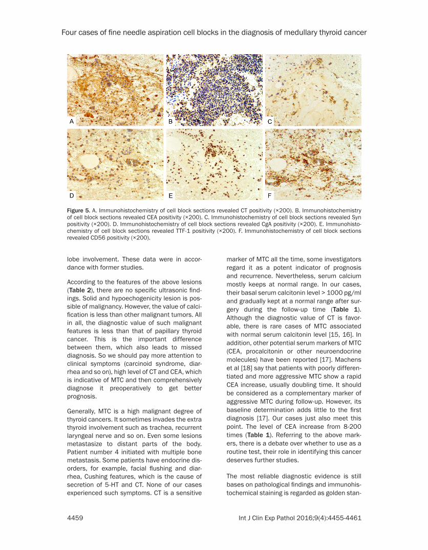
Figure 5. A. Immunohistochemistry of cell block sections revealed CT positivity (×200). B. Immunohistochemistry of cell block sections revealed CEA positivity (×200). C. Immunohistochemistry of cell block sections revealed Syn positivity (×200). D. Immunohistochemistry of cell block sections revealed CgA positivity (×200). E. Immunohisto-chemistry of cell block sections revealed TTF-1 positivity (×200). F. Immunohistochemistry of cell block sections revealed CD56 positivity (×200).

Four cases of fine needle aspiration cell blocks in the diagnosis of medullary thyroid cancer
4460 Int J Clin Exp Pathol 2016;9(4):4455-4461
dard to diagnose MTC. The tumor cell express-es some important markers, such as CT, Syn, CgA, and CEA. In our study, we found that CT, Syn, CgA, CEA, TTF-1, and CD56 are positive, yet TG is negative in our cell block specimens.
Nowadays, surgical excision is considered to be the first choice to treat MTC. Preoperative diag-nosis of MTC predominantly impacts on the prognosis. Although some new diagnostic idea has appeared, such as FNAB-CT and Gallium-68 dotatate PET/CT, cytological examination can-not be replaced because of very high specificity and sensitivity [6, 9]. Yet, there are still rare cases missed. In our study, we use a relatively simple and inexpensive cell block technology to identify special markers and histological morphology.
In conclusion, diagnosis of MTC is complex work. We should combine every method to comprehensively diagnose. Particularly, the clinical, imaging and serum indicators are com-plementary to the cytological procedure. This study was based on a relatively small sample size. However, it should be considered as a reli-able and useful adjunct to FNAB for establish-ing a definitive MTC diagnosis. More samples should be preserved for future studies.
Acknowledgements
This work was supported by grants from Capital University Clinical Cooperation Project, China (No.15JL10).
Disclosure of conflict of interest
None.
Address correspondence to: Dr. Linxue Qian, De- partment of Ultrasound, Beijing Friendship Hospital, Capital Medical University, 95 Yongan Road, Xicheng District, Beijing, China. Tel: +86-10-63138576; E-mail: [email protected]
References
[1] American Thyroid Association (ATA) Guidelines Taskforce on Thyroid Nodules and Diff- erentiated Thyroid Cancer, Cooper DS, Doherty GM, Haugen BR, Kloos RT, Lee SL, Mandel SJ, Mazzaferri EL, McIver B, Pacini F, Schlumberger M, Sherman SI, Steward DL, Tuttle RM. Revised American Thyroid Association management guidelines for patients with thyroid nodules
and differentiated thyroid cancer. Thyroid 2009; 19: 1167-214.
[2] Yoon JH, Moon HJ, Kim EK, Kwak JY. Inadequate cytology in thyroid nodules: should we repeat aspiration or follow-up? Ann Surg Oncol 2011; 18: 1282-9.
[3] Richards ML, Bohnenblust E, Sirinek K, Bingener J. Nondiagnostic thyroid fine-needle aspiration biopsies are no longer a dilemma. Am J Surg 2008; 196: 398-402.
[4] Yoon JH, Lee HS, Kim EK, Moon HJ, Kwak JY. A nomogram for predicting malignancy in thyroid nodules diagnosed as atypia of undetermined significance/follicular lesions of undetermined significance on fine needle aspiration. Surgery 2014; 155: 1006-13.
[5] Trimboli P, Treglia G, Guidobaldi L, Romanelli F, Nigri G, Valabrega S, Sadeghi R, Crescenzi A, Faquin WC, Bongiovanni M, Giovanella L. Detection rate of FNA cytology in medullary thyroid carcinoma: a meta-analysis. Clin Endocrinol (Oxf) 2015; 82: 280-5.
[6] Tran K, Khan S, Taghizadehasl M, Palazzo F, Frilling A, Todd JF, Al-Nahhas A. Gallium-68 Dotatate PET/CT is superior to other imaging modalities in the detection of medullary carci-noma of the thyroid in the presence of high serum calcitonin. Hell J Nucl Med 2015; 18: 19-24.
[7] Trimboli P, Rossi F, Baldelli R, Laurenti O, Nigri G, Ventura C, Appetecchia M, Attanasio D, Romanelli F, Guidobaldi L, Guarino M, Crescenzi A, Valabrega S. Measuring calcitonin in washout of the needle in patients undergo-ing fine needle aspiration with suspicious med-ullary thyroid cancer. Diagn Cytopathol 2012; 40: 394-8.
[8] Trimboli P, Cremonini N, Ceriani L, Saggiorato E, Guidobaldi L, Romanelli F, Ventura C, Laurenti O, Messuti I, Solaroli E, Madaio R, Bongiovanni M, Orlandi F, Crescenzi A, Valabrega S, Giovanella L. Calcitonin measure-ment in aspiration needle washout fluids has higher sensitivity than cytology in detecting medullary thyroid cancer: a retrospective mul-ticentre study. Clin Endocrinol (Oxf) 2014; 80: 135-40.
[9] de Crea C, Raffaelli M, Maccora D, Carrozza C, Canu G, Fadda G, Bellantone R, Lombardi CP. Calcitonin measurement in fine-needle aspi-rate washouts vs. cytologic examination for di-agnosis of primary or metastatic medullary thyroid carcinoma. Acta Otorhinolaryngol Ital 2014; 34: 399-405.
[10] Zhang S, Yu X, Zheng Y, Yang Y, Xie J, Zhou X. Value of fine needle aspiration cell blocks in the diagnosis and classification of lymphoma. Int J Clin Exp Pathol 2014; 7: 7717-25.

Four cases of fine needle aspiration cell blocks in the diagnosis of medullary thyroid cancer
4461 Int J Clin Exp Pathol 2016;9(4):4455-4461
[11] Us-Krasovec M, Auersperg M, Bergant D, Golouh R, Kloboves-Prevodnik V. Medullary carcinoma of the thyroid glad: diagnostic cytopathological characteristics. Pathologica 1998; 90: 5-13.
[12] Trimboli P, Nigri G, Romanelli F, Cicciarella Modica DD, Crescenzi A, Valabrega S, Giovanella L. Medullary thyroid nodules by measurement of calcitonin (Ct) in aspiration needle washout in patients with multinodular goiter and moderately elevated serum Ct. Exp Clin Endocrinol Diabetes 2012; 120: 234-7.
[13] American Thyroid Association Guidelines Task Force, Kloos RT, Eng C, Evans DB, Francis GL, Gagel RF, Gharib H, Moley JF, Pacini F, Ringel MD, Schlumberger M, Wells SA Jr. Medullary thyroid cancer: management guidelines of the American Thyroid Association. Thyroid 2009; 19: 565-612.
[14] Hofstra RM, Landsvater RM, Ceccherini I, Stulp RP, Stelwagen T, Luo Y, Pasini B, Höppener JW, van Amstel HK, Romeo G, et al. A mutation in the RET proto-oncogene associated with mul-tiple endocrine neoplasia type 2B and sporad-ic medullary thyroid carcinoma. Nature 1994; 367: 375-6.
[15] Dora JM, Canalli MH, Capp C, Puñales MK, Vieira JG, Maia AL. Normal perioperative se-rum calcitonin levels in patients with advanced medullary thyroid carcinoma: case report and review of the literature. Thyroid 2008; 18: 895-9.
[16] Giovanella L, Crippa S, Cariani L. Serum calci-tonin-negative medullary thyroid carcinoma: role of CgA and CEA as complementary mark-ers. Int J Biol Markers 2008; 23: 129-31.
[17] Trimboli P, Giovanella L, Crescenzi A, Romanelli F, Valabrega S, Spriano G, Cremonini N, Guglielmi R, Papini E. Medullary thyroid cancer diagnosis: an appraisal. Head Neck 2014; 36: 1216-23.
[18] Machens A, Ukkat J, Hauptmann S, Dralle H. Abnormal carcinoembryonic antigen levels and medullary thyroid cancer progression: a multivariate analysis. Arch Surg 2007; 142: 289-93; discussion 94.