case report in pediatrics and literature review
TRANSCRIPT
Page 1/12
Adenosquamous Carcinoma of Sigmoid Colon: FirstCase Report in Pediatrics and Literature ReviewShuaibing Lu
A�liated Tumor Hospital of Zhengzhou University: Henan Cancer HospitalFusheng Ge
A�liated Tumor Hospital of Zhengzhou University: Henan Cancer HospitalChen Liu
A�liated Tumor Hospital of Zhengzhou University: Henan Cancer HospitalLiangqun Peng
A�liated Tumor Hospital of Zhengzhou University: Henan Cancer HospitalFei Ma
A�liated Tumor Hospital of Zhengzhou University: Henan Cancer HospitalWei Yang
A�liated Tumor Hospital of Zhengzhou University: Henan Cancer HospitalZhandong Zhang
A�liated Tumor Hospital of Zhengzhou University: Henan Cancer HospitalSuxia Luo
A�liated Tumor Hospital of Zhengzhou University: Henan Cancer HospitalYawei Hua ( [email protected] )
A�liated Tumor Hospital of Zhengzhou University: Henan Cancer Hospital
Research Article
Keywords: sigmoid colon, adenosquamous carcinoma, pediatrics, liver metastasis, case report
Posted Date: October 25th, 2021
DOI: https://doi.org/10.21203/rs.3.rs-982723/v1
License: This work is licensed under a Creative Commons Attribution 4.0 International License. Read Full License
Page 2/12
AbstractBackground Colon adenosquamous carcinoma is an extremely rare cancer, especially in young people.No case of colon adenosquamous carcinoma in pediatric patient has been reported so far.
Case presentation Here we present the case of a 16-year-old Chinese girl diagnosed with adenosquamouscarcinoma of sigmoid colon. We performed laparoscopic sigmoidectomy with lymphadenectomy on thepatient. Unfortunately, she developed liver metastasis less than 1 month after surgery. Then weimplemented percutaneous radiofrequency ablation of liver mass and 4 cycles of adjuvant chemotherapyon her. However, liver metastasis recurred less than 4 months after radiofrequency ablation. The girleventurally died in 8 months after surgical resection of sigmoid colon. As far as we know, this girl is theyoungest patient with sigmoid colon adenosquamous carcinoma reported so far, and the �rst reportedcase of colon adenosquamous carcinoma in pediatrics.
Conclusion Only 8 months from detection to death, this case indicates the prognosis of colonadenosquamous carcinoma is poor, and adenosquamous carcinoma of colon may even have worseprognosis in pediatrics than in adults. In addition, cases with liver metastasis have a awfully unfavorableprognosis for colon adenosquamous carcinoma patients.
BackgroundAccording to the global cancer statistics for 2020 released by the International Agency for Research onCancer of the World Health Organization, the incidence of colorectal cancer ranked third among allmalignant tumors on a global scale(1). However, colorectal cancer is very rare among pediatric patients (<21 years old) and the incidence is only about 1 per million(2).
Adenosquamous carcinoma(ASC) refers to malignancy that contains both glandular and squamoushistologic components. ASC is most common in respiratory system (73.8%), but it is rare in digestivesystem(16.2%)(3). Studies have shown that ASC accounts for only 0.06%-0.09% of colorectal cancer(4,5). What’s more, this subtype of colorectal cancer is extremely rare in pediatric patients. There is only onecase of pediatric ASC of rectum has been reported(6). To the best of our knowledge, no case of colonASC in pediatric patient has been reported so far. The patient presented here is the youngest reportedcase of colon ASC, and the �rst reported case of colon ASC in pediatrics.
Since it is rare in clinical practice, the treatment for colonic ASC mainly refers to colonadenocarcinoma(ADC). Currently, surgical resection and adjuvant chemotherapy are the main treatmentmethods for this disease. But the results are generally unsatisfactory, especially for patients with distantmetastases. In this report, the girl diagnosed with ASC of sigmoid colon, and developed liver metastasisafter radical sigmoidectomy. Then she received 4 cycles of XELOX (consists of oxaliplatin andcapecitabine) chemotherapy after radiofrequency ablation of liver mass. But, it is unfortunate that livermetastasis recurred soon after radiofrequency ablation. The timeline of the patient’s diagnosis and

Page 3/12
treatment course is shown in table1. We present the following case in accordance with the CAREreporting checklist.
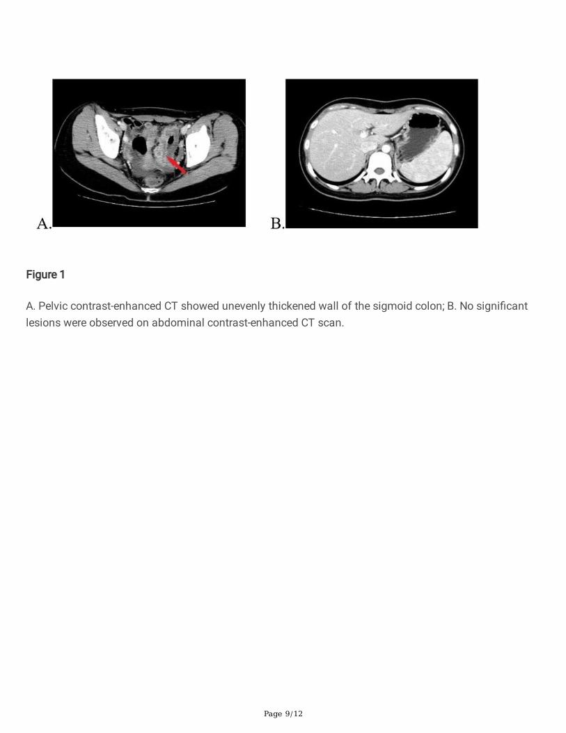
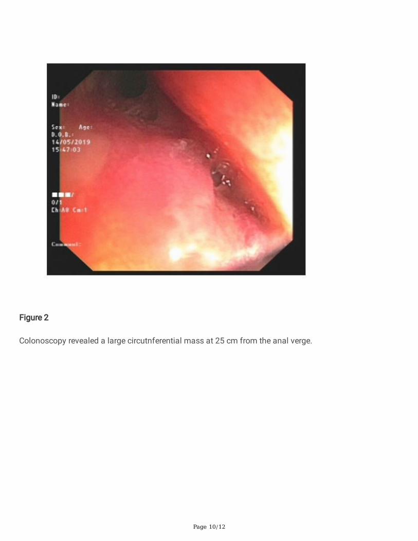
Case PresentationA 16-year-old Chinese girl was referred to our hospital with 1 month history of intermittent left lowerabdominal pain. Laboratory tests showed the following values: hemoglobin, 104g/L; CA19-9, 49.55U/mL;CEA, 0.82ng/ml; AFP, 1.58ng/ml; CA125, 30.12U/mL; CA724, 5.61U/ml. The stool examination for occultblood was positive. Pelvic contrast-enhanced CT demonstrated unevenly thickened wall of the sigmoidcolon(Figure1A). No signi�cant lesions were observed on abdominal contrast-enhanced CTscan(Figure1B). Colonoscopy revealed a large circutnferential mass at 25 cm from the analverge(Figure2). The mass narrowed the lumen so that the body of colonoscope could not pass through.Pathologic analysis of biopsy specimen indicated adenocarcinoma of sigmoid colon. The patient hadneither a signi�cant past medical history nor a family history of colorectal cancer.
During laparoscopic exploration, we found the tumor was located in the sigmoid colon (approximately7cmx6cmx5cm), invading the serosal membrane and closely adhering to part of the distal ileum. As aresult, we performed laparoscopic radical resection of sigmoid colon plus partial ileectomy on the girl.Pathological examination of the sigmoid colon specimen revealed the tumor was adenosquamouscarcinoma, including mucinous adenocarcinoma and poorly differentiated squamous cell carcinoma.Figure3 shows the hematoxylin-eosin staining of the sigmoid colon specimen.
Immunohistochemistry staining showed positive for CK5/6, P63, focal positive for CK20 CEA CDX-2,negative for P16 Syn CD56 and high Ki-67 expression(80%). Genetic testing showed wild type KRAS,microsatellite stable(MSS) and no mutation in BARF gene. The tumor invaded the adventitia, nerves andvessel. Three out of 39 lymph nodes contained metastatic tumor. According to the 7th edition of the UICCTNM classi�cation system, the �nal pathological diagnosis of the sigmoid colon mass was stage B(pT3N1bM0) adenosquamous carcinoma.
Less than 1 month after surgery, abdominal MRI showed a mass at the junction of liver S5 andS8(Figure4A), which was considered to be tumor metastasize to liver. After consultation with the girl andher parents, we implemented percutaneous radiofrequency ablation of liver mass and adjuvantchemotherapy on her. The regimen of chemotherapy was XELOX, which includes oxaliplatin(200mg perday) and capecitabine(1.5g BID d1-14). This girl tolerated XELOX chemotherapy regimen well. After 2cycles of adjuvant chemotherapy, re-examination of abdominal MRI showed stable condition(Figure4B),thus we continue to give the above chemotherapy regimen for 2 cycles.
After 4 cycles of adjuvant chemotherapy, abdominal MRI indicated recurrence of livermetastasis(Figure4C). The edge of the mass at the junction of liver S5 and S8 was enhanced andenlarged compared to the previous one. Nodules were found in the hilar area, which is considered to belymph node metastasis. Genetic testing revealed no mutations in B-raf and N-ras gene. Then we inviteddoctors from the department of hepatobiliary surgery and medical oncology for consultation. After
Page 4/12
discussion, reoperation and targeted therapy were recommended. The girl and her family membersrefused further treatment for �nancial reason and the girl eventurally died in 2 months later.
Discussion And ConclusionsASC is a malignant tumor that contains both glandular and squamous histologic components. Accordingto the relationship between glandular and squamous ingredients, ASC can be classi�ed into compositeand collision types. Composite type refers to the mixed distribution of adenocarcinoma and squamouscell carcinoma, while collision type refers to the adenocarcinoma composition and squamous cancercomposition has a clear boundary(7,8 9). There are four hypotheses about the histogenesis of squamouscell carcinoma in colonic ASC patients: 1) Ectopic squamous cells of the colonic mucosa directlytransform into squamous cell carcinoma; 2) basal cells transform into squamous cells; 3)Squamousmetaplasia of glandular cells;4 Squamous metaplasia of adenocarcinoma cells. At present, the fourthhypothesis is most accepted by scholars.
The common clinical manifestations of colon ASC patients are similar to that of colon ADC, includingabdominal pain, weight loss, altered bowel habits, bloody stool. Noteworthy, hypercalcemia isoccasionally encountered in patients with colon ASC(10 11 12). The cause of hypercalcemia in colonASC patients is mainly due to the release of parathyroid hormone by tumor, it means that hypercalcemiaappears as a paraneoplastic syndrome of ASC(10). Secondly, for patients with advanced ASC, bonemetastasis can also cause elevated serum calcium(11).
Colon ASC is rare in clinical practice and thus the understanding and treatment experience about thisdisease is very limited. Therefore, no consensus on effective treatment for colon ASC has been reached.At present, the treatment of colon ASC is mainly referred to colon ADC, that is surgery plus adjuvantchemotherapy. Regarding postoperative chemotherapy of colon ASC, FOLFOX (consists of �uorouracil,folinic acid and oxaliplatin) regimen is mostly used(13, 14, 15).
In recent years, postoperative chemotherapy combined with targeted therapy or immunotherapy has beenproposed. For patients with suitable genetic test results, postoperative chemotherapy combined withtargeted therapy or immunotherapy can be tried, although the effects of these treatments are not yetclear. A study showed that patients with BRAF V600E colorectal cancer have a poor clinical response toFOLFOX chemotherapy plus bevacizumab, leading to poor prognosis(16). In the future, more clinical trialsare needed to optimize the postoperative treatment regimen of ASC and determine the exact extent of itsclinical bene�ts.
Several studies have shown that the prognosis of colon ASC is worse than that of colon ADC(17, 18, 19,20). In 2020, Nasseri et al. conducted the largest retrospective cross-sectional examination of ASC from anational database for over 30 years. The mean age of patients in this study was approximately 64 years.The results showed the median overall survival for colon ASC patients was 13.9 months (95% CI: 10.98-16.83). For stage IV ASC and ADC of colon, the median overall survival was 14.1 months and 8.0months(P 0.0001), respectively(17). According to a study conducted by Frizelle et al, the prognosis of
Page 5/12
colorectal ASC is worse than that of ADC in stage III or IV, but similar to ADC in stage I or II. The 5-yearsurvival rate for patients with stage I-III colorectal ASC was about 65%, while the median survival rate forpatients with stage IV disease was 8.5 months(18).
The prognosis of ASC is worse than that of ADC, which may be due to: 1) Both adenocarcinoma andsquamous cell carcinoma can metastasise, but the later appears to spread more easily and moreaggressively than the former(21 22). 2) At the time of diagnosis, the incidence of distant metastasis(stage IV) of colon ASC was signi�cantly higher than that of ADC(5, 17). The most common metastasissites of colon squamous cancer are liver, peritoneum and lung in order(18).
In this case, no distant metastasis was found on preoperative examinations. After radical resection,pathological diagnosis of the sigmoid colon mass was stage B (pT3N1bM0) adenosquamouscarcinoma. When adjuvant chemotherapy is about to be performed 4 weeks after surgery, the imagingexamination revealed a liver mass, which was considered to be tumor metastasis. It can be seen thatcolonic ASC is awfully aggressive in adolescents. Then we performed percutaneous radiofrequencyablation of liver mass on this patient. XELOX chemotherapy was started after radiofrequency ablation,and the patient was well tolerated. However, re-examination showed recurrence of liver metastasis afterthe completion of 4 cycles of chemotherapy. Eventually, the patient and her family decided to give upfurther treatment and the girl died 8 months postoperatively.
In Choi’s report, a 43-year-old female underwented laparoscopic right colectomy. Pathological diagnosisof the colon specimen was stage IIIB (T3 N2a M0) adenosquamous carcinoma. The female patientreceived 6 cycles of FOLFOX4 (oxaliplatin, leucovorin, and �uorouracil) chemotherapy postoperative, andno tumor recurrence was found during the 10 months follow-up examinations since the completion ofchemotherapy(13). Compared with the case reported by Choi, the outcomes of the girl in our report isquiet frustrsting. It’s not di�clut to draw the conclusion that cases with liver metastasis have anunfavorable prognosis for colon ASC patients. Besides, We speculate that the effect of FOLFOXchemotherapy regimen is superior or non-inferior to XELOX regimen in patients with colonic ASC.
Our medical team specially held a seminar on this case. The lessons we have learned from this case areas follows: 1) Colonic ASC can also occur in pediatrics. Therefore, do not ignore the possibility of colonASC in adolescents with related symptoms. 2) For patients diagnosed with colon ASC, comprehensiveexamination must be performed to determine whether distant metastases have occurred. 3) After surgery,genetic testing of pathological tissue is required to determine if there is a suitable immunotherapy ortargeted therapy. 4) During the treatment of colon ASC, the frequency of imaging examinations should beappropriately increased in order to detect changes in patients' condition in time. Then adjust to the mostappropriate treatment in a timely manner.
Our team has two main expectations about future research on colon ASC. On one hand, lots of researchon the etiology and tissue origin of colon adenosquamous carcinoma are needed. Only in this way canwe understand this disease more clearly, and thus adopt more effective prevention and treatment

Page 6/12
methods. On the other hand, studies on postoperative adjuvant treatment of colon ASC patients indifferent age groups are also needed, so as to obtain a better prognosis as much as possible.
In conclusion, colon adenosquamous carcinoma is an exceedingly rare malignancy. To our knowledge,this is the �rst reported case of colon ASC in pediatrics. This case demonstrates the prognosis of thisdisease is poor, and colon ASC may even have worse prognosis in adolescents than in adults. For colonASC patients, cases with liver metastasis have an awfully unfavorable prognosis. In order to achievebetter outcomes, more research are needed to �nd out the histogenesis of ASC, and determine the bestpostoperative adjuvant treatment for colon ASC patients.
AbbreviationsASCAdenosquamous carcinomaADCAdenocarcinoma
DeclarationsEthics approval and consent to participate
All procedures performed in studies involving human participants were in accordance with the ethicalstandards of the ethics committee of our hospital (No. 2019156). Written informed consent was obtainedfrom the patient’s parent for publication of this case report and accompanying images. A copy of thewritten consent is available for review by the editor of this journal.
Consent for publication
The patient’s parents has given their consents for the case report to be published. The written informedconsent to publish this information was obtained from the patient’s parent. A copy of the written consentis available for review by the editor o�ce of this journal.
Availability of data and materials
All data during the study are included within the article.
Competing interests
The authors declare that they have no competing interests.
Funding
This work was supported by the National Cancer Climbing Fund (No.NCC201816B048); and Tackle KeyProblems in Medicine of Henan Province (No.LHGJ20200188).

Page 7/12
Authors' contributions
S Lu designed this report and write the manuscript. F Ge and C Liu helped to design this report and writethe manuscript. L Peng, F Ma, W Yang, assisted in writing and editing the manuscript. Z Zhang, S Luo, YHua reviewed and revised the manuscript. All authors contributed to the article and approved thesubmitted version.
Acknowledgements
Here we extend our deep condolences to the family. Meanwhile, we want to thank the girl’s parents fortheir permission to report this study.
Authors' information
1Department of General Surgery, the A�liated Tumor Hospital of Zhengzhou University, No.127Dongming Rd, Zhengzhou 450008, Henan, China; 2Department of Medical Oncology, the A�liated TumorHospital of Zhengzhou University, No.127 Dongming Rd, Zhengzhou 450008, Henan, China.
References1. Sung H, Ferlay J, Siegel LR, et al. Global Cancer Statistics 2020: GLOBOCAN Estimates of Incidence
and Mortality Worldwide for 36 Cancers in 185 Countries. CA: Cancer J Clin. 2021;0:1–41.
2. Poles GC, Clark DE, Mayo SW, et al. Colorectal carcinoma in pediatric patients: A comparison withadult tumors, treatment and outcomes from the National Cancer Database. J Pediatr Surg.2016;51(7):1061–6.
3. Lan Y, Huang K, Liu C, et al. A nation-wide cancer registry-based study of adenosquamous carcinomain Taiwan. PLoS ONE. 2015;10(10):e0139748.
4. Cagir B, Nagy MW, Topham A, et al. Adenosquamous carcinoma of the colon, rectum, and anus:epidemiology, distribution, and survival characteristics. Dis Colon Rectum. 1999;42:258–63.
5. Masoomi H, Ziogas A, Lin BS, et al. Population-based evaluation of adenosquamous carcinoma ofthe colon and rectum. Dis Colon Rectum. 2012;55(5):509–14.
�. García N, Cubero JH, Orduña VA, et al. Adenosquamous Cell Carcinoma of the Rectum in a Girl: FirstCase Reported and Review of the Literature. J Pediatr Hematol Oncol. 2015;37(6):e364–7.
7. Di Prete M, Baciorri F, FranceschilliM, et al. Adenosquamous carcinoma of the right colon withdiffuse signet-ring mucinous component. Clin J Gastroenterol. 2020;13(4):555–9.
�. Levendoglu H, Cox CA, Nadimpalli V. Composite (adenocarcinoid) tumors of the gastrointestinaltract. Dig Dis Sci. 1990;35:519–25.
9. Fukui H, Takada M, Chiba T, et al. Concurrent occurrence of gastric adenocarcinoma and duodenalneuroendocrine cell carcinoma: a composite tumour or collision tumours? Gut. 2001;48:853–6.

Page 8/12
10. Chevinsky AH, Berelowitz M, Hoover HC. Adenosquamous carcinoma of the colon presenting withhypercalcemia. Cancer. 1987;01(5):1111–6. 60(.
11. Palvio DH, Sørensen FB, Kløve-Mogensen M. Stem cell carcinoma of the colon and rectum. Report oftwo cases and review of the literature. Dis Colon Rectum. 1985;28(6):440–5.
12. Thompson JT, Paschold EH, Levine EA. Paraneoplastic hypercalcemia in a patient withadenosquamous cancer of the colon. Am Surg. 2001;67(6):585–8.
13. Choi JW, Park HU. Adenosquamous Carcinoma of the Ascending Colon: A Case Report and Review ofthe Literature. Annals of coloproctology. 2013;29(2):83–6.
14. Toumi O, Hamida B, Njima M, et al. Adenosquamous carcinoma of the right colon: A case report andreview of the literature. International Journal of Surgery Case Reports International journal of surgerycase reports. 2018;50:119–21.
15. Kang DB, Oh JT, Jo HJ, et al. Primary adenosquamous carcinoma of the colon. J Korean Surg Soc.2011;80(Suppl 1):31-5.
1�. Ishida H, Yamaguchi T, Chiba K, et al. A case report of ascending colon adenosquamous carcinomawith BRAF V600E mutation. Int Cancer Conf J. 2017;6(3):93–7.
17. Nasseri Y, Cox B, Shen W, et al. Adenosquamous carcinoma: An aggressive histologic sub-type ofcolon cancer with poor prognosis. Am J Surg. 2021;03(3):649–53. 221(.
1�. Frizelle FA, Hobday KS, Batts KP, et al. Adenosquamous and squamous carcinoma of the colon andupper rectum: a clinical and histopathologic study. Dis Colon Rectum. 2001;44(3):341–6.
19. Nozoe T, Anai H. Adenosquamous carcinoma of the sigmoid colon: report of a case. Surg Today.2001;31:830–2.
20. Dong Y, Wang J, Ma H, et al. Primary adenosquamous carcinoma of the colon: report of �ve cases.Surg Today. 2009;39:619–23.
21. Kontozoglou TE, Moyana TN. Adenosquamous carcinoma of the colon--an immunocytochemicaland ultrastructural study. Report of two cases and review of the literature. Dis Colon Rectum.1989;32(8):716–21.
22. Cerezo L, Alvarez M, Edwards O, et al. Adenosquamous carcinoma of the colon. Dis Colon Rectum.1985;28:597–603.
Figures
Page 9/12
Figure 1
A. Pelvic contrast-enhanced CT showed unevenly thickened wall of the sigmoid colon; B. No signi�cantlesions were observed on abdominal contrast-enhanced CT scan.
Page 10/12
Figure 2
Colonoscopy revealed a large circutnferential mass at 25 cm from the anal verge.

Page 11/12
Figure 3
Hematoxylin-eosin staining of the sigmoid colon specimen.
Figure 4
A. Abdominal MRI showed a mass at the junction of liver S5 and S8; B. Abdominal MRI showed stablecondition after 2 cycles of adjuvant chemotherapy; C. Abdominal MRI indicated recurrence of liver
Page 12/12
metastasis after 4 cycles of adjuvant chemotherapy.
Supplementary Files
This is a list of supplementary �les associated with this preprint. Click to download.
Table1.jpg