case report langerhans cell histiocytosis (lch) occurring ... · case report langerhans cell...
TRANSCRIPT

Int J Clin Exp Pathol 2017;10(1):695-701www.ijcep.com /ISSN:1936-2625/IJCEP0041815
Case Report Langerhans cell histiocytosis (LCH) occurring in the skull: report of 3 cases and review of the literature
Jincheng Fang*, Zhiquan Jiang*
Department of Neurosurgery, The First Affiliated Hospital, Bengbu Medical College, Bengbu, Anhui, China. *Equal contributors and co-first authors.
Received October 13, 2016; Accepted November 23, 2016; Epub January 1, 2017; Published January 15, 2017
Abstract: Langerhans cell histiocytosis (LCH) is a far-between disease of unknown etiology and is characterized by a clonal proliferation of Langerhans cells from single organ to multisystem involvement. Here we report 3 cases which lesions occurring in the head of the skull: right temporal bone, right occipital bone and the left frontal tempo-ral bone. Microsurgical resection of lesions was performed, and the pathologic diagnosis was LCH. Because of it’s infrequency and possibly variable display, LCH should be considered and included in the differential diagnosis at the time when we encounter head of the skull placeholder.
Keywords: Langerhans cell histiocytosis, LCH, head of the skull, temporal bone, occipital bone, frontal temporal bone, and placeholder
Introduction
Langerhans cell histiocytosis (LCH) is a far-between condition characterized by neoplastic proliferation of Langerhans cells [1]. The etiol-ogy is still unclear, common hypotheses include uncontrolled and irregular multiplication of lym-phocytes and histiocytes after a gene mutation or infection, some fragmentary manifestations are not believed to be of a reactive pattern though but effectively represent real neo-plasms [2, 3]. Although it’s can be diagnosed in any age group, a majority of LCH affects chil-dren between 1 and 4 years of age with a mor-bidity 5-6 cases per million children [4, 5]. Head and neck involvement, primarily skull base, is noted in about 60% of the cases [6]. It’s involv-ing the head of the skull region is uncommon and can resemble malignant excrescences on imaging due to high cellularity. One of the most continual presenting imaging features is osteal involvement with lytic lesions according to their clinical and pathologic features. Although understanding the presence of sharp margins with beveled-edges can be helpful, tissue sam-pling is usually necessary for confirming the diagnosis [7]. But head of the skull lesions are
extremely rare. Almost no related articles report characteristic imaging of LCH occurring in head of the skull lesions up to now. We report 2 male teenager patients and a female adult with head of the skull Langerhans cell histiocytosis, with a literature review.
Case report
Case 1
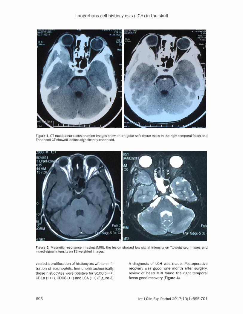
A 12-year-old male with pain in the right tempo-ral half for half month and aggravated with right eyelid swelling for a week. And there was no his-tory of trauma. Computed tomography (CT) mul-tiplanar reconstruction images revealed the irregular soft tissue mass in the right temporal bone and Enhanced CT showed lesions signifi-cantly enhanced (Figure 1). On magnetic reso-nance imaging (MRI), the lesion showed low signal intensity on T1-weighted images and mixed-signal intensity on T2-weighted images (Figure 2). There were no other lesions in the systemic survey. All laboratory results were normal.
Microsurgical resection of the lesion was per-formed, and histopathologic examination re-
Langerhans cell histiocytosis (LCH) in the skull
696 Int J Clin Exp Pathol 2017;10(1):695-701
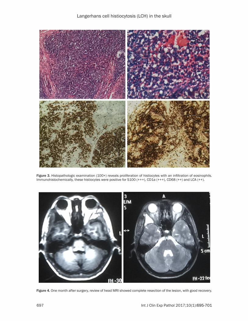
vealed a proliferation of histiocytes with an infil-tration of eosinophils. Immunohistochemically, these histiocytes were positive for S100 (+++), CD1a (+++), CD68 (++) and LCA (++) (Figure 3).
A diagnosis of LCH was made. Postoperative recovery was good, one month after surgery, review of head MRI found the right temporal fossa good recovery (Figure 4).
Figure 1. CT multiplanar reconstruction images show an irregular soft tissue mass in the right temporal fossa and Enhanced CT showed lesions significantly enhanced.
Figure 2. Magnetic resonance imaging (MRI), the lesion showed low signal intensity on T1-weighted images and mixed-signal intensity on T2-weighted images.
Langerhans cell histiocytosis (LCH) in the skull
697 Int J Clin Exp Pathol 2017;10(1):695-701
Figure 3. Histopathologic examination (100×) reveals proliferation of histiocytes with an infiltration of eosinophils. Immunohistochemically, these histiocytes were positive for S100 (+++), CD1a (+++), CD68 (++) and LCA (++).
Figure 4. One month after surgery, review of head MRI showed complete resection of the lesion, with good recovery.
Langerhans cell histiocytosis (LCH) in the skull
698 Int J Clin Exp Pathol 2017;10(1):695-701
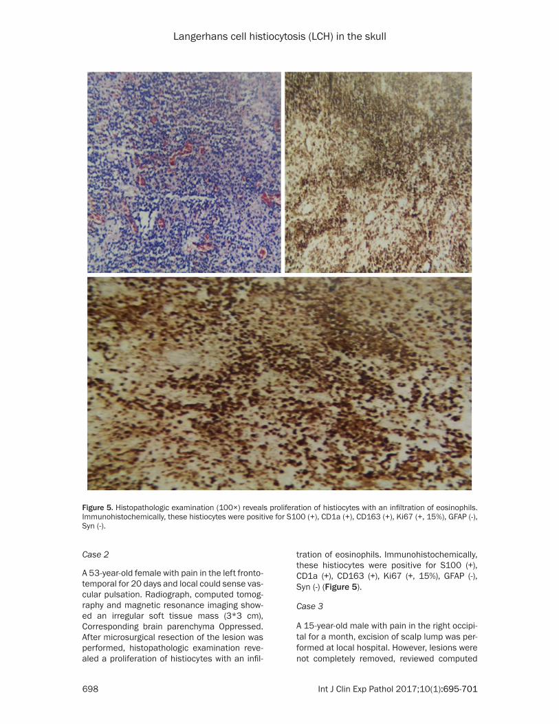
Figure 5. Histopathologic examination (100×) reveals proliferation of histiocytes with an infiltration of eosinophils. Immunohistochemically, these histiocytes were positive for S100 (+), CD1a (+), CD163 (+), Ki67 (+, 15%), GFAP (-), Syn (-).
Case 2
A 53-year-old female with pain in the left fronto-temporal for 20 days and local could sense vas-cular pulsation. Radiograph, computed tomog-raphy and magnetic resonance imaging show- ed an irregular soft tissue mass (3*3 cm), Corresponding brain parenchyma Oppressed. After microsurgical resection of the lesion was performed, histopathologic examination reve- aled a proliferation of histiocytes with an infil-
tration of eosinophils. Immunohistochemically, these histiocytes were positive for S100 (+), CD1a (+), CD163 (+), Ki67 (+, 15%), GFAP (-), Syn (-) (Figure 5).
Case 3
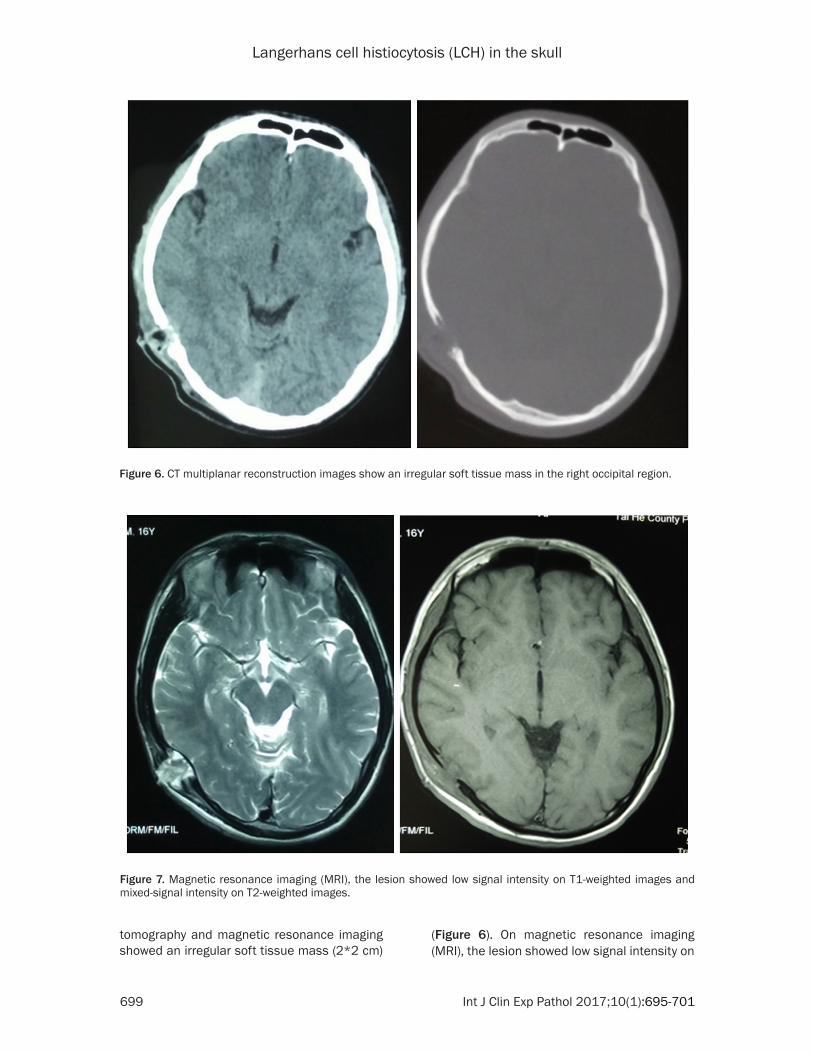
A 15-year-old male with pain in the right occipi-tal for a month, excision of scalp lump was per-formed at local hospital. However, lesions were not completely removed, reviewed computed
Langerhans cell histiocytosis (LCH) in the skull
699 Int J Clin Exp Pathol 2017;10(1):695-701
tomography and magnetic resonance imaging showed an irregular soft tissue mass (2*2 cm)
(Figure 6). On magnetic resonance imaging (MRI), the lesion showed low signal intensity on
Figure 6. CT multiplanar reconstruction images show an irregular soft tissue mass in the right occipital region.
Figure 7. Magnetic resonance imaging (MRI), the lesion showed low signal intensity on T1-weighted images and mixed-signal intensity on T2-weighted images.
Langerhans cell histiocytosis (LCH) in the skull
700 Int J Clin Exp Pathol 2017;10(1):695-701
T1-weighted images and mixed-signal intensity on T2-weighted images (Figure 7).
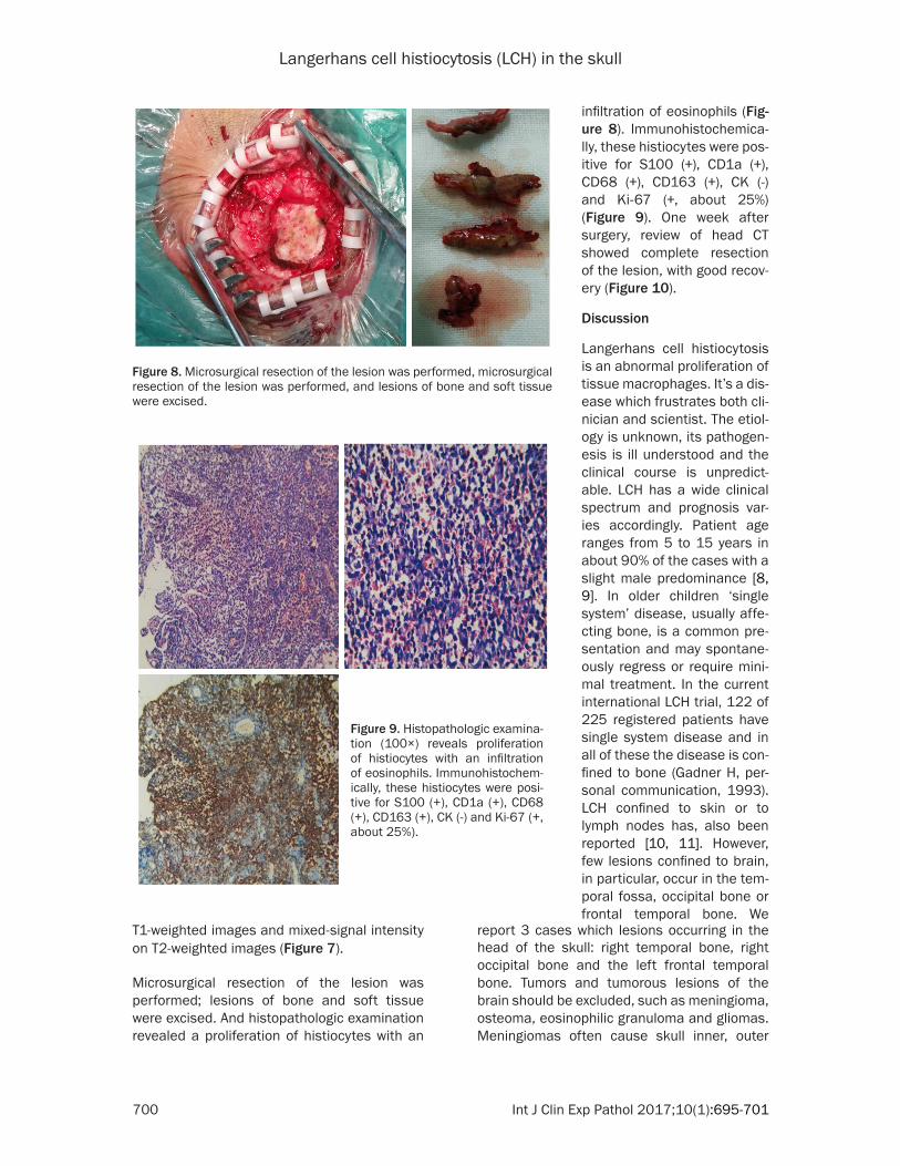
Microsurgical resection of the lesion was performed; lesions of bone and soft tissue were excised. And histopathologic examination revealed a proliferation of histiocytes with an
report 3 cases which lesions occurring in the head of the skull: right temporal bone, right occipital bone and the left frontal temporal bone. Tumors and tumorous lesions of the brain should be excluded, such as meningioma, osteoma, eosinophilic granuloma and gliomas. Meningiomas often cause skull inner, outer
Figure 8. Microsurgical resection of the lesion was performed, microsurgical resection of the lesion was performed, and lesions of bone and soft tissue were excised.
Figure 9. Histopathologic examina-tion (100×) reveals proliferation of histiocytes with an infiltration of eosinophils. Immunohistochem-ically, these histiocytes were posi-tive for S100 (+), CD1a (+), CD68 (+), CD163 (+), CK (-) and Ki-67 (+, about 25%).
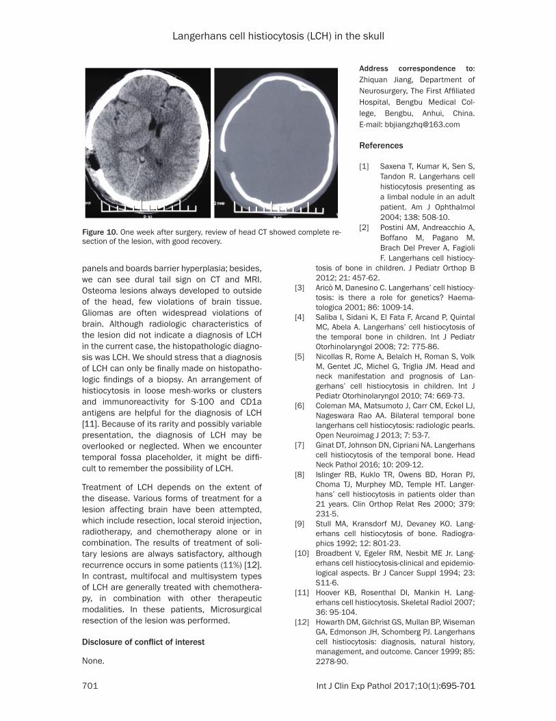
infiltration of eosinophils (Fig- ure 8). Immunohistochemica- lly, these histiocytes were pos-itive for S100 (+), CD1a (+), CD68 (+), CD163 (+), CK (-) and Ki-67 (+, about 25%) (Figure 9). One week after surgery, review of head CT showed complete resection of the lesion, with good recov-ery (Figure 10).
Discussion
Langerhans cell histiocytosis is an abnormal proliferation of tissue macrophages. It’s a dis-ease which frustrates both cli-nician and scientist. The etiol-ogy is unknown, its pathogen-esis is ill understood and the clinical course is unpredict-able. LCH has a wide clinical spectrum and prognosis var-ies accordingly. Patient age ranges from 5 to 15 years in about 90% of the cases with a slight male predominance [8, 9]. In older children ‘single system’ disease, usually affe- cting bone, is a common pre-sentation and may spontane-ously regress or require mini-mal treatment. In the current international LCH trial, 122 of 225 registered patients have single system disease and in all of these the disease is con-fined to bone (Gadner H, per-sonal communication, 1993). LCH confined to skin or to lymph nodes has, also been reported [10, 11]. However, few lesions confined to brain, in particular, occur in the tem-poral fossa, occipital bone or frontal temporal bone. We
Langerhans cell histiocytosis (LCH) in the skull
701 Int J Clin Exp Pathol 2017;10(1):695-701
panels and boards barrier hyperplasia; besides, we can see dural tail sign on CT and MRI. Osteoma lesions always developed to outside of the head, few violations of brain tissue. Gliomas are often widespread violations of brain. Although radiologic characteristics of the lesion did not indicate a diagnosis of LCH in the current case, the histopathologic diagno-sis was LCH. We should stress that a diagnosis of LCH can only be finally made on histopatho-logic findings of a biopsy. An arrangement of histiocytosis in loose mesh-works or clusters and immunoreactivity for S-100 and CD1a antigens are helpful for the diagnosis of LCH [11]. Because of its rarity and possibly variable presentation, the diagnosis of LCH may be overlooked or neglected. When we encounter temporal fossa placeholder, it might be diffi- cult to remember the possibility of LCH.
Treatment of LCH depends on the extent of the disease. Various forms of treatment for a lesion affecting brain have been attempted, which include resection, local steroid injection, radiotherapy, and chemotherapy alone or in combination. The results of treatment of soli-tary lesions are always satisfactory, although recurrence occurs in some patients (11%) [12]. In contrast, multifocal and multisystem types of LCH are generally treated with chemothera-py, in combination with other therapeutic modalities. In these patients, Microsurgical resection of the lesion was performed.
Disclosure of conflict of interest
None.
tosis of bone in children. J Pediatr Orthop B 2012; 21: 457-62.
[3] Aricò M, Danesino C. Langerhans’ cell histiocy-tosis: is there a role for genetics? Haema- tologica 2001; 86: 1009-14.
[4] Saliba I, Sidani K, El Fata F, Arcand P, Quintal MC, Abela A. Langerhans’ cell histiocytosis of the temporal bone in children. Int J Pediatr Otorhinolaryngol 2008; 72: 775-86.
[5] Nicollas R, Rome A, Belaïch H, Roman S, Volk M, Gentet JC, Michel G, Triglia JM. Head and neck manifestation and prognosis of Lan- gerhans’ cell histiocytosis in children. Int J Pediatr Otorhinolaryngol 2010; 74: 669-73.
[6] Coleman MA, Matsumoto J, Carr CM, Eckel LJ, Nageswara Rao AA. Bilateral temporal bone langerhans cell histiocytosis: radiologic pearls. Open Neuroimag J 2013; 7: 53-7.
[7] Ginat DT, Johnson DN, Cipriani NA. Langerhans cell histiocytosis of the temporal bone. Head Neck Pathol 2016; 10: 209-12.
[8] Islinger RB, Kuklo TR, Owens BD, Horan PJ, Choma TJ, Murphey MD, Temple HT. Langer- hans’ cell histiocytosis in patients older than 21 years. Clin Orthop Relat Res 2000; 379: 231-5.
[9] Stull MA, Kransdorf MJ, Devaney KO. Lang- erhans cell histiocytosis of bone. Radiogra- phics 1992; 12: 801-23.
[10] Broadbent V, Egeler RM, Nesbit ME Jr. Lang- erhans cell histiocytosis-clinical and epidemio-logical aspects. Br J Cancer Suppl 1994; 23: S11-6.
[11] Hoover KB, Rosenthal DI, Mankin H. Lang- erhans cell histiocytosis. Skeletal Radiol 2007; 36: 95-104.
[12] Howarth DM, Gilchrist GS, Mullan BP, Wiseman GA, Edmonson JH, Schomberg PJ. Langerhans cell histiocytosis: diagnosis, natural history, management, and outcome. Cancer 1999; 85: 2278-90.
Figure 10. One week after surgery, review of head CT showed complete re-section of the lesion, with good recovery.
Address correspondence to: Zhiquan Jiang, Department of Neurosurgery, The First Affiliated Hospital, Bengbu Medical Col- lege, Bengbu, Anhui, China. E-mail: [email protected]
References
[1] Saxena T, Kumar K, Sen S, Tandon R. Langerhans cell histiocytosis presenting as a limbal nodule in an adult patient. Am J Ophthalmol 2004; 138: 508-10.
[2] Postini AM, Andreacchio A, Boffano M, Pagano M, Brach Del Prever A, Fagioli F. Langerhans cell histiocy-