case report open access clinically suspected acute
TRANSCRIPT

JOURNAL OF MEDICALCASE REPORTS
Kasamatsu et al. Journal of Medical Case Reports 2013, 7:129http://www.jmedicalcasereports.com/content/7/1/129
CASE REPORT Open Access
Clinically suspected acute myopericarditis withcardiac tamponade associated with peripheralblood eosinophilia presenting in early pregnancy:a case reportYu Kasamatsu1*, Takashi Kida1, Mayumi Shigeru2, Toru Tagashira2, Naoki Murai2, Eiji Takai2 and Hideyuki Takaoka2
Abstract
Introduction: The clinical presentation of eosinophilic myocarditis may vary from asymptomatic to themanifestation of severe symptoms, including cardiac tamponade and arrhythmias. In pregnant patients with thiscondition, drugs must be used cautiously up to approximately the 4th month of pregnancy because drug useshould be limited during the period of fetal organogenesis.
Case presentation: A 30-year-old Asian woman at 14 weeks of pregnancy with progressive malaise was hospitalized.The electrocardiogram revealed ST elevation and low QRS voltage. Echocardiography revealed massive pericardialeffusion and myocardial swelling. A laboratory examination revealed an increase in her white blood cell count, with apredominance of neutrophils. Pericardial drainage was performed for relief of the cardiac tamponade. The pericardialeffusion revealed an abundance of eosinophils. Subsequently, the peripheral blood eosinophil count began to rise, andthe patient was clinically diagnosed with eosinophilic myopericarditis. The patient’s condition improved rapidlyfollowing the initiation of prednisolone treatment, and she finally delivered a full-term normal infant.
Conclusions: A patient with clinically suspected myopericarditis in the early stage of pregnancy who improved rapidlywith pericardial drainage and prednisolone therapy, and successfully delivered a normal full-term infant; the diagnosiswas made in the early stage of the disease, based on the detection of an abundance of eosinophils in the pericardialeffusion preceding the subsequent development of peripheral blood eosinophilia.
Keywords: Cardiac tamponade, Eosinophil, Myopericarditis, Prednisolone, Pregnancy
IntroductionEosinophilic myocarditis is characterized by myocarditis as-sociated with marked eosinophilic infiltration of the myo-cardium and peripheral blood eosinophilia. In addition, theclinical presentation of myocarditis may vary from asymp-tomatic to the manifestation of severe symptoms [1]. Somepatients develop cardiac tamponade due to the associatedpericarditis, whereas others may show rapid progression ofmyocardial necrosis, manifesting as fatal arrhythmias, car-diac arrest, or a clinical state necessitating mechanical as-sistance for the maintenance of circulation. The mortality
* Correspondence: [email protected] of Internal Medicine, Matsushita Memorial Hospital, 5-55,Sotojimacho, Moriguchi-shi, Osaka 570-8540, JapanFull list of author information is available at the end of the article
© 2013 Kasamatsu et al.; licensee BioMed CenCreative Commons Attribution License (http:/distribution, and reproduction in any medium
associated with the acute stage of this disease is reported tobe 7% [2].In pregnant patients with this condition, drugs must be
used cautiously up to approximately the 4th month of preg-nancy (the period of fetal organogenesis) because drug useshould be limited during this period [3]. We encountered apatient with acute clinically suspected myopericarditis whopresented, at 14 weeks of pregnancy, with cardiac tampon-ade and marked peripheral blood eosinophilia. Here wepresent this case.
Case presentationA 30-year-old Asian woman who was 14 weeks and 5 dayspregnant (gravida, 1; para, 0) presented to us with general-ized malaise and facial edema in June 2007. She had no
tral Ltd. This is an Open Access article distributed under the terms of the/creativecommons.org/licenses/by/2.0), which permits unrestricted use,, provided the original work is properly cited.
Kasamatsu et al. Journal of Medical Case Reports 2013, 7:129 Page 2 of 5http://www.jmedicalcasereports.com/content/7/1/129
significant past medical history or history of allergy. Shewas a non-smoker and gave no history of drug abuse oreating raw food. She visited a local medical clinic, where aroutine blood examination revealed leukocytosis. As themalaise worsened, the patient was brought by ambulanceto our hospital for further management. Analysis of hervaginal secretions and ultrasonography of the fetusconducted by an obstetrician/gynecologist revealed nomajor abnormalities. Pregnancy-induced hypertension wasruled out at that time. Therefore, the patient was referredto the Department of Cardiovascular Medicine of ourhospital.The findings on physical examination at admission
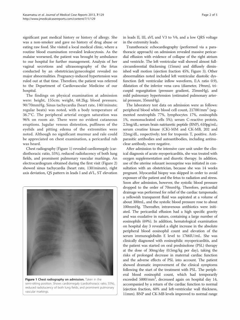
were: height, 155cm; weight, 68.2kg; blood pressure,90/70mmHg. Sinus tachycardia (heart rate, 140/minute;regular beats) was noted, with a body temperature of36.7°C. The peripheral arterial oxygen saturation was96% on room air. There were no evident cutaneouseruptions. Jugular venous distention, puffiness of theeyelids and pitting edema of the extremities werenoted. Although no significant murmur and rale couldbe appreciated on chest examination, a pericardial rubwas heard.Chest radiography (Figure 1) revealed cardiomegaly (car-
diothoracic ratio, 55%), reduced radiolucency of both lungfields, and prominent pulmonary vascular markings. Anelectrocardiogram obtained during the first visit (Figure 2)showed sinus tachycardia (heart rate, 130/minute), rightaxis deviation, QS pattern in leads I and aVL, ST elevation
Figure 1 Chest radiography on admission. Taken in thesemi-sitting position. Shows cardiomegaly (cardiothoracic ratio, 55%),reduced radiolucency of both lung fields, and prominent pulmonaryvascular markings.
in leads II, III, aVf, and V3 to V6, and a low QRS voltagein the extremity leads.Transthoracic echocardiography (performed via a para-
thoracic approach) on admission revealed massive pericar-dial effusion with evidence of collapse of the right atriumand ventricle. The left ventricular wall showed almost full-circumferential thickening (15mm) and diffusely dimin-ished wall motion (ejection fraction 45%, Figure 3). Otherabnormalities noted included left ventricular diastolic dys-function (left ventricular inflow waveform, E:A ratio 0.9),dilatation of the inferior vena cava (diameter, 19mm), tri-cuspid regurgitation (pressure gradient, 25mmHg), andmild pulmonary hypertension (estimated pulmonary arter-ial pressure, 35mmHg).The laboratory test data on admission were as follows:
peripheral blood white blood cell count, 21700/mm3 (seg-mented neutrophils 77%, lymphocytes 17%, eosinophils1%, mononucleated cells 5%); serum C-reactive protein,2.3mg/dL; serum brain natriuretic peptide (BNP), 610pg/mL;serum creatine kinase (CK)-MM and CK-MB, 202 and22mg/dL, respectively; test for troponin T, positive. Anti-parasitic antibodies and autoantibodies, including antinu-clear antibody, were negative.After admission to the intensive care unit under the clin-
ical diagnosis of acute myopericarditis, she was treated withoxygen supplementation and diuretic therapy. In addition,use of the uterine relaxant isoxsuprine was initiated in con-sultation with an obstetrician, because she was 14 weekspregnant. Myocardial biopsy was skipped in order to avoidexposure of the patient and the fetus to radiation and stress.Soon after admission, however, the systolic blood pressuredropped to the order of 70mmHg. Therefore, pericardialdrainage was performed for relief of the cardiac tamponade;a yellowish transparent fluid was aspirated at a volume ofabout 300mL, and the systolic blood pressure rose to about100mmHg. Thereafter, intravenous antibiotics were initi-ated. The pericardial effusion had a high specific gravityand was exudative in nature, containing a large number ofeosinophils (69%). In addition, hematological examinationon hospital day 3 revealed a slight increase in the absoluteperipheral blood eosinophil count and elevation of theserum immunoglobulin E level to 1766IU/mL. She wasclinically diagnosed with eosinophilic myopericarditis, andthe patient was started on oral prednisolone (PSL) therapyat the dose of 30mg/day (0.5mg/kg per day), taking therisks of prolonged decrease in maternal cardiac functionand the adverse effects of PSL into account. The patientshowed dramatic improvement of the clinical symptomsfollowing the start of the treatment with PSL. The periph-eral blood eosinophil count, which had temporarilyexceeded 5000/mm3, decreased again on hospital day 14,accompanied by a return of the cardiac function to normal(ejection fraction, 60% and left-ventricular wall thickness,11mm). BNP and CK-MB levels improved to normal range

Figure 2 Electrocardiogram taken at the first visit. Sinus tachycardia (heart rate, 140/minute), ST elevation in leads I, II, III, aVL, aVf, and V3 toV6, and low QRS voltage in the limb leads.
Figure 3 Echocardiogram on admission. Long axial view (left) and short axial view (right) obtained via a parathoracic approach. Almostfull-circumferential thickening of the left ventricular wall and massive pericardial effusion are noted.
Kasamatsu et al. Journal of Medical Case Reports 2013, 7:129 Page 3 of 5http://www.jmedicalcasereports.com/content/7/1/129

Kasamatsu et al. Journal of Medical Case Reports 2013, 7:129 Page 4 of 5http://www.jmedicalcasereports.com/content/7/1/129
on hospital day 20. The PSL dose was reduced to 20mg, andthe patient was discharged from the hospital in a stable clin-ical condition on hospital day 22 and followed up thereafteras an out-patient. Measurement of viral antibody titers(coxsackievirus, echovirus, and so on) in paired sera revealedno significant elevation of the titers of any of the antibodies.During the out-patient management, the PSL dose was
gradually reduced, without relapse of symptoms. On day63 after the diagnosis of the disease, oral PSL therapy wasdiscontinued. Although the patient had to be hospitalizedfor approximately 1 week because she temporarily sufferedfrom pregnancy-induced hypertension, the clinical coursewas favorable overall, and the patient delivered a healthybaby at the gestational age of 41 weeks.
DiscussionEosinophilic myopericarditis is believed to be caused bycytotoxic substances, such as eosinophil cationic proteinand major basic protein present in the granules of eosino-phils [4]. The diagnosis is established by myocardial biopsy.Eosinophilic myopericarditis may be divided etiologicallyinto three types: (1) parasitic infestation, (2) cardiomyop-athy due to chronic eosinophilia as a component of thehypereosinophilic syndrome or Churg–Strauss syndromeand (3) transient and temporary acute myopericarditis dueto hypersensitivity which is generally caused by exposure toirritants such as drugs and allergens [1,5,6]. In our patient,although we can rule out parasitic infestation and drug re-action, the role of pregnancy in the pathogenesis of this dis-ease could not be ruled out. According to our literaturesearch, we could not find a relationship between eosino-philia and pregnancy. However, the course of the diseasewas transient and favorable; thus, the disease was thoughtto be the acute hypersensitivity type. The clinical course ofacute eosinophilic myocarditis, with the exception of ful-minant type, is generally transient and its prognosis is good.However, a few cases had major basic protein expression inmyocardial biopsy specimens longitudinally after the onsetof the disease, therefore, we should cautiously follow upeven after alleviation of the acute-phase inflammation [2].Abundant accumulation of eosinophils in the target
organ was noted on admission, although no significantincrease in the peripheral blood eosinophil count wasnoted. One possible explanation for the accumulation ofeosinophils in the target organ that has been proposed isthat some stimuli induce migration of eosinophils to-wards the target organ, but the formation of eosinophilsin the bone marrow cannot compensate for this change,resulting in a transient paradoxic eosinopenia. Later, eo-sinophil formation in the bone marrow continues evenafter tissue infiltration by eosinophils has decreased,resulting in peripheral blood eosinophilia [7]. Althoughinhalation of the antigen might cause the accumulationof eosinophilic leukocytes via inteleukin-5 in lung
disease [8], the precise mechanism of accumulation ofeosinophils in the myocardium preceding the increase inthe peripheral blood eosinophil count remains unknown.The symptoms of eosinophilic myocarditis are akin tothose of viral myocarditis. In the early stages of eosino-philic myocarditis, peripheral blood eosinophilia is some-times absent [9]. Therefore, it is desirable to check theblood cell counts at intervals of 2 to 3 days when myocar-ditis is suspected.Steroids have been shown to be effective in the treat-
ment of eosinophilic myocarditis [1,5]. In regard to theuse of steroids during pregnancy, however, animal studieshave shown an increased death rate of the fetus followingadministration of high PSL doses and an increased risk forthe development of cleft lip following the use of PSL, evenat ordinary doses, during the first trimester of pregnancy[3,10]. Steroid therapy is certainly acceptable in patientswith severe disease, as in the case of our patient whopresented with cardiac tamponade, because maternal lowblood pressure caused by cardiac dysfunction andhypercytokinemia can adversely affect the continuation ofpregnancy and the mother’s prognosis. However, unex-pected outcomes may be encountered with the use of ste-roids and cardiac biopsy using X-ray during pregnancymay lead to unexpected outcomes, since adequate clinicaldata related to its use during pregnancy is still not avail-able [3]. A typical myopericarditis pattern on T2 and lategadolinium-enhanced sequences of cardiac magnetic res-onance imaging can be useful in the case of pregnancy[11]. Both patients and their family members tend to beanxious about the use of these drugs, especially duringpregnancy. Therefore, we should explain the risks andbenefits of this therapy to patients and their family mem-bers prior to the start of this treatment.In a previous report of a patient with eosinophilic myo-
carditis developing at 24 weeks of pregnancy, thickening ofthe ventricular septum was observed, with subsequent in-crease in the peripheral blood eosinophil count, whichclinched the diagnosis. The disease was mild in that case,and resolved in response to symptomatic therapy with ver-apamil, furosemide, and so on [12]. In our present patient,the disease was severe, and the patient presented with car-diac tamponade. Treatment was difficult because only alimited number of drugs could be used because the patientwas in the early stages of pregnancy. However, appropriatepericardial drainage and PSL therapy resulted in early alle-viation of the condition, which allowed continuation of thepregnancy and successful child delivery. This is a type ofcase as yet unrepresented the literature, which may there-fore prove clinically informative.
ConclusionsWe encountered a patient with myopericarditis in theearly stage of pregnancy who improved rapidly with

Kasamatsu et al. Journal of Medical Case Reports 2013, 7:129 Page 5 of 5http://www.jmedicalcasereports.com/content/7/1/129
pericardial drainage and PSL therapy, and successfully de-livered a normal full-term infant; the diagnosis was madein the early stage of the disease, based on the detection ofan abundance of eosinophils in the pericardial effusionpreceding the subsequent development of peripheralblood eosinophilia.
ConsentWritten informed consent was obtained from the patient(who also served as legal guardian for the baby) for publi-cation of this case report and any accompanying images.A copy of the written consent is available for review by theEditor-in-Chief of this journal.
Competing interestsNone of the authors have any conflict of interest to report.
Authors’ contributionsAll authors except for TK worked in the ward of Takatsuki General Hospitaland conferred on the treatment and patient’s condition. The data wereanalyzed and interpreted mainly by HT and TK. All authors read andapproved the final manuscript.
Author details1Department of Internal Medicine, Matsushita Memorial Hospital, 5-55,Sotojimacho, Moriguchi-shi, Osaka 570-8540, Japan. 2Department ofCardiovascular Medicine, Takatsuki General Hospital, 1-3-13, Kosobecho,Takatsuki-shi, Osaka 569-1192, Japan.
Received: 17 December 2012 Accepted: 11 April 2013Published: 13 May 2013
References1. Kawano S, Kato J, Kawano N, Yoshimura Y, Masuyama H, Fukunaga T, Sato
Y, Maruyama H, Mihara K, Ueda A, Toyoda K, Imamura T, Kitamura K: Clinicalfeatures and outcomes of eosinophilic myocarditis patients treated withprednisolone at a single institution over a 27-year period. Intern Med2011, 50(9):975–981.
2. Morimoto S, Kubo N, Hiramitsu S, Uemura A, Ohtsuki M, Kato S, Kato Y,Sugiura A, Miyagishima K, Mori N, Yoshida Y, Hishida H: Changes in theperipheral eosinophil count in patients with acute eosinophilicmyocarditis. Hear Vessel 2003, 18:193–196.
3. Parisi MA, Spong CY, Zajicek A, Guttmacher AE: We don't know what wedon't study: the case for research on medication effects in pregnancy.Am J Med Genet C Semin Med Genet 2011, 157(3):247–250.
4. Tai PC, Ackerman SJ, Spry CJF, Dunnette S, Olsen EGJ, Gleich GJ: Depositsof eosinophil granule proteins in cardiac tissues of patients witheosinophilic endomyocardial disease. Lancet 1987, 8534:643–647.
5. Al Ali AM, Straatman LP, Allard MF, Ignaszewski AP: Eosinophilicmyocarditis: case series and review of literature. Can J Cardiol 2006,22(14):1233–1237.
6. De Alava E, Panizo-Santos A, Fernandez-Gonzalez AL, Pardo-Mindan FJ:Eosinophilic myocarditis in patients waiting for heart transplantation.Cardiovasc Pathol 1995, 4:43–46.
7. Ubukata M, Natori S, Hasegawa A, Suzuki T, Murata K: A case ofhypereosinophilic myocarditis which showed degranulated eosinophilsby immunocytochemical technique. Nippon Naika Gakkai Zasshi 1989,78:1617–1618.
8. Luiz E, Endes C, Fernando L, Ferreira P: Pulmonary eosinophilia. J BrasPneumol 2009, 35(6):561–573.
9. Suk S, Jong Chun P, Jae Hun C, Kye Hun K, Youngkeun A, Myung Ho J,Jeong Gwan C: A case of acute eosinophilic myopericarditis presentingwith cardiogenic shock and normal peripheral eosinophil count. Korean JIntern Med 2006, 21:136–140.
10. Park-Wyllie L, Mazzotta P, Pastuszak A, Moretti ME, Beique L, Hunnisett L,Friesen MH, Jacobson S, Kasapinovic S, Chang D, Diav-Citrin O, Chitayat D,Nulman I, Einarson TR, Koren G: Birth defects after maternal exposure to
corticosteroids: prospective cohort study and meta-analysis ofepidemiological studies. Teratology 2000, 62(6):385–392.
11. Yelgec NS, Dymarkowski S, Ganame J, Bogaert J: Value of MRI in patientswith a clinical suspicion of acute myocarditis. Eur Radiol 2007,17(9):2211–2217.
12. Sagesaka T, Liang SG, Morioka H, Watanabe T, Kaibara M, Dohba N: Acutemyocarditis with eosinophilia presenting as asymmetric septalhypertrophy during pregnancy. J Obstet Gynecol Res 1997, 23(2):147–151.
doi:10.1186/1752-1947-7-129Cite this article as: Kasamatsu et al.: Clinically suspected acutemyopericarditis with cardiac tamponade associated with peripheralblood eosinophilia presenting in early pregnancy: a case report. Journalof Medical Case Reports 2013 7:129.
Submit your next manuscript to BioMed Centraland take full advantage of:
• Convenient online submission
• Thorough peer review
• No space constraints or color figure charges
• Immediate publication on acceptance
• Inclusion in PubMed, CAS, Scopus and Google Scholar
• Research which is freely available for redistribution
Submit your manuscript at www.biomedcentral.com/submit