case report open access scrotal extratesticular schwannoma ... · case presentation clinical...
TRANSCRIPT

Palleschi et al. BMC Urology 2014, 14:32http://www.biomedcentral.com/1471-2490/14/32
CASE REPORT Open Access
Scrotal extratesticular schwannoma: a case reportand review of the literatureGiovanni Palleschi1,2, Antonio Carbone1,2, Jessica Cacciotti3, Giorgia Manfredonia4, Natale Porta3, Andrea Fuschi1,Cosimo de Nunzio5, Vincenzo Petrozza3 and Antonio Luigi Pastore1,2*
Abstract
Background: Schwannomas are tumours arising from Schwann cells, which sheath the peripheral nerves. Here,we report a rare case of left intrascrotal, extratesticular schwannoma. Although rare, scrotal localisation ofschwannomas has been reported in male children, adult men, and elderly men. They are usually asymptomatic andare characterised by slow growth. Patients generally present with an intrascrotal mass that is not associated withpain or other clinical signs, and such cases are self-reported by most patients. Imaging modalities (such asultrasonography, computed tomography, and magnetic resonance imaging) can be used to determine tumour size,exact localisation, and extension. However, the imaging findings of schwannoma are non-specific. Therefore, onlycomplete surgical excision can result in diagnosis, based on histological and immunohistochemical analyses. If thetumour is not entirely removed, recurrences may develop, and, although malignant change is rare, this may occur,especially in patients with a long history of an untreated lesion. Thus, follow up examinations with clinical andimaging studies are recommended for scrotal schwannomas.
Case presentation: A 52-year-old man presented with a 3-year history of asymptomatic scrotal swelling. Physicalexamination revealed a palpable, painless, soft mass in the left hemiscrotum. After surgical removal of the mass, itshistological features indicated schwannoma.
Conclusions: Schwannoma should be considered in cases of masses that are intrascrotal but extratesticular.Ultrasonography provides the best method of confirming the paratesticular localisation of the tumour, beforesurgical removal allows histopathological investigation and definitive diagnosis. Surgery is the standard therapeuticapproach. To prevent recurrence, particular care should be taken to ensure complete excision. This case reportincludes a review of the literature on scrotal schwannomas.
Keywords: Scrotal schwannoma, Diagnosis, Histopathology
BackgroundSchwannomas are tumours arising from Schwann cells,which sheath the peripheral nerves. Schwannomas candevelop in any region of the human body, either sporad-ically or in association with neurofibromatosis [1]. Mostclinical findings associated with schwannoma are notspecific to the disease, because schwannomas growslowly and do not result in symptoms until they enlarge
* Correspondence: [email protected] University of Rome, Department of Medical and SurgicalBiotechnologies, Unit of Urology, ICOT, Via Franco Faggiana 1668, 04100Latina, Italy2Uroresearch Association, non profit association, Via Franco Faggiana 1668,04100 Latina, ItalyFull list of author information is available at the end of the article
© 2014 Palleschi et al.; licensee BioMed CentraCommons Attribution License (http://creativecreproduction in any medium, provided the orDedication waiver (http://creativecommons.orunless otherwise stated.
and compress the surrounding structures [2]. The exactincidence of schwannoma is not known [3]. Scrotal lo-calisation of schwannoma is rare, but has been the oc-casional subject of reports in the literature [4,5]. Werecently treated a case of intrascrotal, extratesticularschwannoma, which was found in an adult man whohad a 3-year history of an asymptomatic lesion in theleft hemiscrotum. Here, we describe the case with acomplete report, including intraoperative findings. Wealso provide a review of the literature, which was per-formed to collect and summarise the present state ofknowledge on this type of tumour.
l Ltd. This is an Open Access article distributed under the terms of the Creativeommons.org/licenses/by/2.0), which permits unrestricted use, distribution, andiginal work is properly credited. The Creative Commons Public Domaing/publicdomain/zero/1.0/) applies to the data made available in this article,

Palleschi et al. BMC Urology 2014, 14:32 Page 2 of 6http://www.biomedcentral.com/1471-2490/14/32
Case presentationClinical historyA 52-year-old man presented with a 3-year history ofasymptomatic scrotal swelling. He had a body massindex of 28 kg/m2 at the time of presentation. The pa-tient reported that his scrotum had significantly enlargedduring the last 6 months. Physical examination revealeda palpable, painless, soft mass in the left hemiscrotum,approximately the size of an egg. Further, an adhesionhad formed between the mass and the left testicle. Thelesion appeared not to be attached to the skin of thescrotum or the underlying tissues. The abdominal exam-ination findings were unremarkable, and rectal explor-ation showed enlargement of the prostate gland withoutany palpatory pathological findings. The patient did notreport any systemic symptoms. Accordingly, the initialdiagnostic hypothesis was suspicion of a left testiculartumour. Written informed consent was obtained fromthe patient for publication of this case report and any ac-companying images.
ImagingThe patient underwent scrotal ultrasonography (US),which revealed a hypoechoic solid mass. The mass was4 × 7 cm in size and had a nodular appearance. It wasfound in the mid-left hemiscrotum, behind the left tes-ticle, which appeared reduced in size (Figure 1). Leftvaricocele was observed, whereas hydrocele was absentbilaterally. Colour Doppler evaluation showed poor peri-lesional and intralesional hypervascularisation. Quantita-tive elastography revealed that the lesion had a lowelastic modulus relative to the left testicular parenchyma
Figure 1 Ultrasonography and colour Doppler examination of the schhypoechogenic, and had poor hypervascularisation.
and low intralesional vascularity (Figure 2). The patient’sright testicle was normal. To better examine the patient,contrast-enhanced computed tomography (CT) of theabdomen, scrotum, and chest was performed. The scanidentified a 7.1 × 3.9 cm mass, which was localised medi-ally in the left testicle, without extension into the tunicavaginalis. Lymphadenopathy was not present.
Laboratory findingsBlood tests showed normal levels of alpha-fetoprotein,beta-human chorionic gonadotropin, and carcinoem-bryonic antigen. Prostate specific antigen levels were alsonormal.
Therapeutic approachBased on this evidence, we decided to surgically removethe mass, and the patient agreed. A left inguinal 4-cmincision was made, the spermatic cord was clamped, andthe testicle was tractioned from the left inguinal ring.The mass presented with some adhesions to the left tes-ticle, but did not infiltrate the testicle (Figure 3). Surgicalremoval appeared to be simple and was performed usingmonopolar scissors in a few minutes. The spermaticcord was then released, and the left testicle was pushedback into the scrotum. The patient was discharged 48hours after surgery, without complications. After 12months of follow-up, physical examination and scrotalultrasound were negative.
Macroscopic findingsThe surgical specimen was immediately measured andfound to be 7.1 × 3.8 cm. It comprised a white mass,
wannoma. The lesion appeared inhomogeneous, was partially
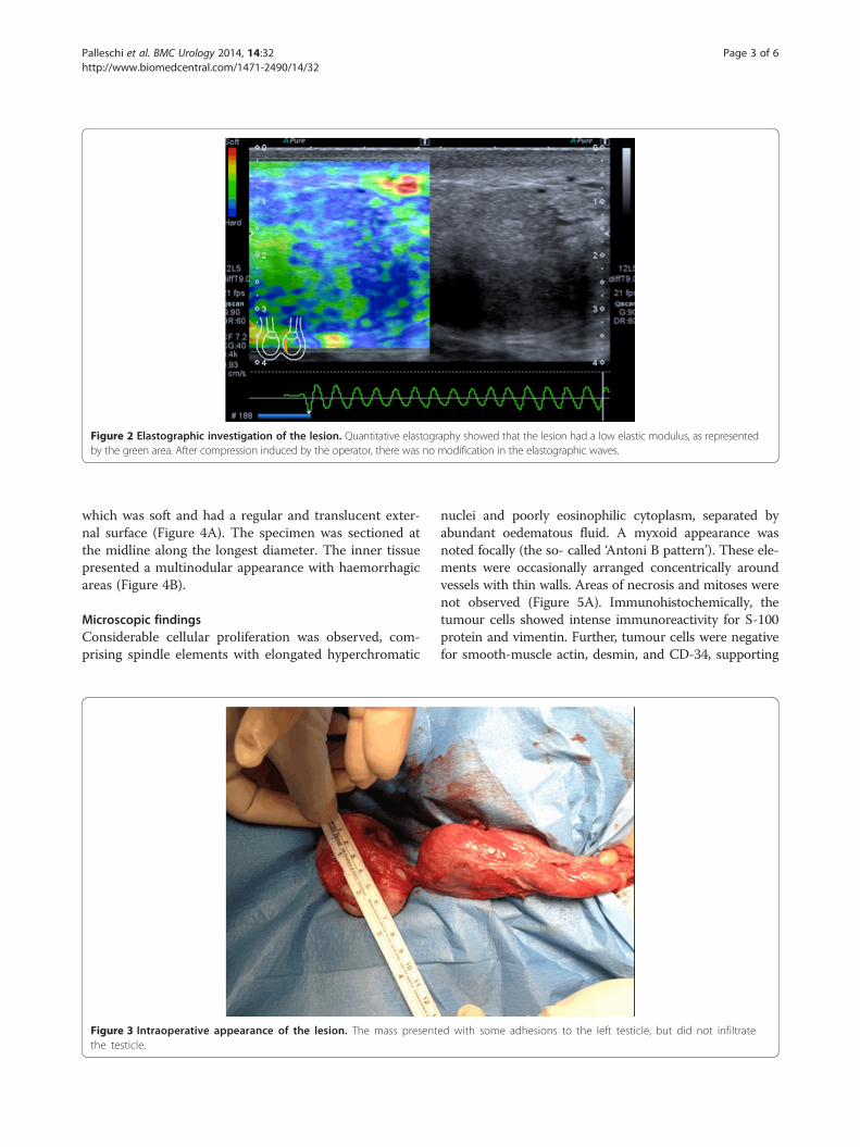
Figure 2 Elastographic investigation of the lesion. Quantitative elastography showed that the lesion had a low elastic modulus, as representedby the green area. After compression induced by the operator, there was no modification in the elastographic waves.
Palleschi et al. BMC Urology 2014, 14:32 Page 3 of 6http://www.biomedcentral.com/1471-2490/14/32
which was soft and had a regular and translucent exter-nal surface (Figure 4A). The specimen was sectioned atthe midline along the longest diameter. The inner tissuepresented a multinodular appearance with haemorrhagicareas (Figure 4B).
Microscopic findingsConsiderable cellular proliferation was observed, com-prising spindle elements with elongated hyperchromatic
Figure 3 Intraoperative appearance of the lesion. The mass presentthe testicle.
nuclei and poorly eosinophilic cytoplasm, separated byabundant oedematous fluid. A myxoid appearance wasnoted focally (the so- called ‘Antoni B pattern’). These ele-ments were occasionally arranged concentrically aroundvessels with thin walls. Areas of necrosis and mitoses werenot observed (Figure 5A). Immunohistochemically, thetumour cells showed intense immunoreactivity for S-100protein and vimentin. Further, tumour cells were negativefor smooth-muscle actin, desmin, and CD-34, supporting
ed with some adhesions to the left testicle, but did not infiltrate

Figure 4 Macroscopic examination and sectioning of the lesion. (A) Macroscopic examination after surgical removal showed a whitemass that was soft and had a regular, translucent external surface. (B) After sectioning, the specimen was multinodular in appearance,with haemorrhagic areas.
Palleschi et al. BMC Urology 2014, 14:32 Page 4 of 6http://www.biomedcentral.com/1471-2490/14/32
the neural differentiation of the tissue (Figure 5B and C).These histological features were suggestive of schwannoma.
Literature review and comparison with previous cases(Table 1)Scrotal masses require a precise diagnosis to prevent
therapeutic errors. Among various types of intrascrotalextratesticular lesions, schwannomas have been describedpreviously and have always represented a diagnostic chal-lenge for clinicians. During differential diagnosis of thesetumours, clinicians must also consider benign and malig-nant neoplasms of supporting structures of the scrotum:leiomyoma, leiomyosarcoma, and adenosarcoma [3].
Figure 5 Microscopic findings. (A) Proliferation was characterised by spineosinophilic cytoplasm, separated by abundant oedematous fluid. Thesvessels with thin walls (magnification × 4). (B) The cells show intense improtein (magnification × 20).
The clinical history of the disease was similar in eachof the previous reports that we examined [3-6]. Each ofthe subjects presented with painless scrotal swelling,which had been present for between a few months [3]and 3 years, as in our case. The ages of patients variedconsiderably across the reports, although older ages (>60years) were most common. Physical examination usuallyrevealed a soft mass without adhesion to the scrotal skinor the surrounding tissues. Yet, examining the patientdoes not entirely clarify whether the lesion originatedfrom the testicle or another location. In previously re-ported cases, therefore, US, magnetic resonance imaging(MRI), and CT have variously been used to arrive at more
dle elements with elongated hyperchromatic nuclei and poorlye elements were occasionally arranged concentrically aroundmunoreactivity for vimentin (magnification × 20) and (C) S-100
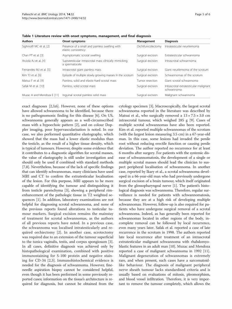
Table 1 Literature review with onset symptoms, management, and final diagnosis
Authors Onset symptoms Management Diagnosis
Sighinolfi MC et al. [2] Presence of a small and painless swelling withelastic consistency
Orchifunicolectomy Intratesticular neurilemoma
Chan PT et al. [3] Asymptomatic scrotal swelling Surgical excision Extratesticular schwannoma
Arciola AJ et al. [4] Supratesticular intrascrotal mass clinically mimickinga spermatocele
Surgical excision Intrascrotal schwannoma
Fernandez MJ et al. [5] Intrascrotal giant painless mass Surgical excision Giant neurilemoma of the scrotum
Kim YJ et al. [6] Episode of multiple slowly growing masses in the scrotum Surgical excision Schwannomas of the scrotum
Matsui F et al. [9] Painless, solid and elastic-hard scrotal mass Tumor resection Giant scrotal schwannoma
Safak M et al. [10] Painless, solid scrotal mass Surgical excision Intrascrotal extratesticular malignantschwannoma
Muzac A and Mendoza E [11] Inguinal scrotal painless solid mass Surgical excision Malignant schwannoma
Palleschi et al. BMC Urology 2014, 14:32 Page 5 of 6http://www.biomedcentral.com/1471-2490/14/32
exact diagnoses [2,3,6]. However, none of these optionshave allowed schwannoma to be identified, because thereis no pathognomonic finding for this disease [6]. On US,schwannoma generally appears as a well-circumscribedmass with a hypoechoic pattern [2], and on colour Dop-pler imaging, poor hypervascularisation is noted. In ourcase, we also performed quantitative elastography, whichshowed that the mass had a lower elastic modulus thanthe testicle, as the result of a higher tissue density, whichis typical of tumours. However, despite some evidence thatit contributes to a diagnostic algorithm for scrotal masses,the value of elastography is still under investigation andshould only be used if combined with standard methods[7,8]. Nevertheless, because of the lack of specific findingsthat can identify schwannomas, many clinicians have usedMRI and CT to confirm the extratesticular localisationof the lesion. For this purpose, MRI appears to be morecapable of identifying the tumour and distinguishing itfrom testicle parenchyma [3], showing a peripheral rim-enhancement of the pathologic tissue in T2 weighted se-quences [1]. In addition, laboratory examinations are nothelpful for diagnosing scrotal schwannoma, and none ofthe previous reports found alterations to testicular tu-mour markers. Surgical excision remains the mainstayof treatment for scrotal schwannomas, as the authorsof all previous reports have noted. In a previous case,the schwannoma was localised intratesticularly and re-quired orchiectomy [2]. In another case, scrotectomywas required due to an extension of the tumour superficialto the tunica vaginalis, testis, and corpus spongiosum [3].In all cases, definitive diagnosis was achieved only byhistopathological examination, combined with positiveimmunostaining for S-100 protein and negative stain-ing for CD-34 [2,3]. Immunohistochemical evidence isneeded for the diagnosis of schwannoma. However, fine-needle aspiration biopsy cannot be considered helpful,even though it has been performed in some previously re-ported cases; information on the tissue’s architecture is re-quired for diagnosis, but cannot be obtained from the
cytology specimen [3]. Macroscopically, the largest scrotalschwannoma reported in the literature was described byMatsui et al., who surgically removed a 13 × 7.5 × 3.0 cmintrascrotal tumour, which weighed 285 g [9]. Cases ofmultiple scrotal schwannomas have also been reported.Kim et al. reported multiple schwannomas of the scrotum(with the largest lesion measuring 3.5 cm) in a 67-year-oldman. In this case, some lesions had invaded the penileroot without reducing erectile function or causing peniledeviation. The author reported no recurrence for at least6 months after surgery. For patients with the systemic dis-ease of schwannomatosis, the development of a single ormultiple scrotal masses should lead the clinician to sus-pect peripheral localisation of schwannoma. In anothercase, reported by Ikary et al., a scrotal schwannoma devel-oped in a 66-year-old man who had previously undergonesurgical excision of a brain tumour, which itself originatedfrom the glossopharyngeal nerve [1]. The patient’s histo-logical diagnosis was schwannoma. Therefore, regular sur-veillance is needed for patients with schwannomatosis,because they are at a high risk of developing multipleschwannomas. However, follow-up is also required for pa-tients who have undergone surgical removal of a scrotalschwannoma. Indeed, as has generally been reported forschwannomas located in other regions of the body, in-complete removal can be followed by local recurrence,even many years later. Safak et al. reported a case of laterecurrence in the scrotum in 1998. The authors reportedlate local recurrence after treatment of an intrascrotalextratesticular malignant schwannoma with rhabdomyo-blastic features in an adult man [10]. Muzac and Mendozareported a case of malignant schwannoma in 1992 [11].Malignant degeneration of schwannomas is extremelyrare, and when present, such cases have a sarcomatoid-like behaviour. The diagnosis of malignant peripheralnerve sheath tumour lacks standardised criteria and isusually based on evaluations of mitosis, pleomorphism,and blood vessel infiltration. Therefore, it is very impor-tant to remove the tumour completely, which allows the

Palleschi et al. BMC Urology 2014, 14:32 Page 6 of 6http://www.biomedcentral.com/1471-2490/14/32
most precise histopathological investigation. From a histo-pathological perspective, the microscopic appearance ofschwannoma is distinctive, with 2 recognisable patterns,which are not always present together: Antoni A areasand Antoni B areas. Antoni A areas are composed of com-pacted spindle cells that are arranged in palisades or in anorganoid arrangement (Verocay bodies). Antoni B areasconsist of tumour cells suspended in a myxomatous mat-rix that may appear microcystic. Several histological var-iants have been described previously: cellular, glandular,epithelioid, and ancient, which involves bizarre hyperchro-matic nuclei without mitosis [3]. However, each of thesevariants is benign, and the ancient type is rare, especiallyin the scrotum (most cases occur in the head and neckregion).
ConclusionOur experience with the reported case and our review ofthe literature strongly suggest that schwannoma shouldbe considered in cases involving masses that are intras-crotal and extratesticular. US (followed by MRI) is thebest modality with which to confirm the paratesticularlocalisation of the tumour, before surgical removal al-lows histopathological investigation and definitive diag-nosis. Surgery is the standard therapeutic approach, whichallows the testicle to be spared in most cases. To preventrecurrence, particular care should be taken to ensurecomplete excision, especially in cases that involve large ormultiple masses. Eventhough it is hard to establish a spe-cific follow-up plan considering the limited number ofcases reported in literature, basing on all the experiencesreported in literature, in case of benign lesions completelyremoved it might be suggested a clinical post-operativeevaluation at 6 and 12 months. Differently, in case ofhistopathological suspicious or clear evidence of malig-nancy, a MRI after 6 months from surgery should beproposed.Early diagnosis can help limit the extent of surgical ex-
cision that is required. Further, early diagnosis can pre-vent the onset of malignant degeneration, which is rare,but has been reported in the literature.
ConsentPatient has given his consent for the case report to bepublished. A copy of the written consent is available, atanytime, for review by the Editor of this journal.
AbbreviationsCT: Computed tomography; MRI: Magnetic resonance imaging;US: Ultrasound.
Competing interestsNo financial support has been received for this study, and none of theauthors have any conflicts of interest pertinent to the content of themanuscript.
Authors’ contributionsAC and VP ideated the study. GP, NP, JC, GM, ALP acquired the data. GP andNP drafted the manuscript. AC, GP, ALP, VP critically revised the manuscript.All authors read and approved the final manuscript.
AcknowledgementsNone source of funding supported the study.
Author details1Sapienza University of Rome, Department of Medical and SurgicalBiotechnologies, Unit of Urology, ICOT, Via Franco Faggiana 1668, 04100Latina, Italy. 2Uroresearch Association, non profit association, Via FrancoFaggiana 1668, 04100 Latina, Italy. 3Department of Medical and SurgicalBiotechnologies, Histopathology Unit, ICOT Latina, via Faggiana 1668, Latina,Italy. 4ICOT Hospital, CADI Centre, via Faggiana 1668, Latina, Italy. 5SapienzaUniversity of Rome, Department of Urology, Sant’Andrea Hospital, Rome,Italy.
Received: 27 November 2013 Accepted: 16 April 2014Published: 28 April 2014
References1. Ikari R, Keisei Okamoto K, Tetsuya Yoshida T, Johnin K, Okabe H, Okada Y:
A rare case of multiple Schwannomas presenting with scrotal mass: aprobable case of Schwannomatosis. Int J Urol 2010, 17:734–736.
2. Sighinolfi MC, Mofferdin A, De Stefani S, Celia A, Micali S, Saredi G, Rossi G,Bianchi G: Benign intratesticular schwannoma: a rare finding. Asian JAndrol 2006, 8(1):101–103.
3. Chan PT, Tripathi S, Low SE, Robinson LQ: Case report – ancientschwannoma of the scrotum. BMC Urol 2007, 7(I):1–4.
4. Arciola AJ, Golden S, Zapinsky J, Fracchia JA: Primary intrascrotalnontesticular schwannoma. Urology 1985, 26:304–306.
5. Fernandez MJ, Martino A, Khan H, Considine TJ, Burden J: Giantneurilemoma: unusual scrotal mass. Urology 1987, 30:74–76.
6. Kim YJ, Kim SD, Huh JS: Intrascrotal and extratesticular multipleschwannoma. World J Mens Health 2013, 31(2):179–181.
7. Cantisani V, Olive M, Di Segni M, Di Leo N, Grazdhani H, D’Ettorre G,Ceccarelli G, Fioravanti C, Ricci P: Contrast-enhanced ultrasonographic(CEUS) and elastosonographic features of a case of testicular Leydigtumor. Ultraschall Med 2012, 33(5):407–409.
8. Aigner F, De Zordo T, Pallwein-Prettner L, Junker D, Schäfer G, Pichler R,Leonhartsberger N, Pinggera G, Dogra VS, Frauscher F: Real-timesonoelastography for the evaluation of testicular lesions. Radiology 2012,263(2):584–589.
9. Matsui F, Kobori Y, Takashima H, Amano T, Takemae K: A case ofintrascrotal schwannoma. Hinyokika Kiyo 2002, 48(12):749–751.
10. Safak M, Baltaci S, Ozer G, Turkolmez K, Uluoglu O: Long-term outcome ofa patient with intrascrotal extratesticular malignant schwannoma.Urol Int 1998, 60(3):202–204.
11. Muzac A, Mendoza E: Malignant schwannoma presenting as aninguinoscrotal mass. Eur Urol 1992, 21(4):340–342.
doi:10.1186/1471-2490-14-32Cite this article as: Palleschi et al.: Scrotal extratesticular schwannoma: acase report and review of the literature. BMC Urology 2014 14:32.
Submit your next manuscript to BioMed Centraland take full advantage of:
• Convenient online submission
• Thorough peer review
• No space constraints or color figure charges
• Immediate publication on acceptance
• Inclusion in PubMed, CAS, Scopus and Google Scholar
• Research which is freely available for redistribution
Submit your manuscript at www.biomedcentral.com/submit