case report pulmonary squamous cell carcinoma … j clin exp pathol 2016;9(7):7668-7673...
TRANSCRIPT
Int J Clin Exp Pathol 2016;9(7):7668-7673www.ijcep.com /ISSN:1936-2625/IJCEP0029043
Case Report Pulmonary squamous cell carcinoma with lepidic growth pattern: new insights into lung cancer classification
Alessandro Del Gobbo1*, Andrea Vingiani1*, Emilia Masci2, Giuseppe Faillace2, Salvatore Barbaro2, Eleonora Bonaparte1, Stefano Ferrero1,3
1Division of Pathology, Fondazione IRCCS Ca’ Granda, Ospedale Maggiore Policlinico, via F. Sforza, 35, 20122 Mi-lan, Italy; 2Division of General Surgery, Ospedale Bassini ICP, via Gorki 50, 20092 Cinisello Balsamo (Milan), Italy; 3Department of Biomedical, Surgical and Dental Sciences, University of Milan Medical School, Milan, Italy, via F. Sforza, 35, 20122 Milan, Italy. *Equal contributors.
Received March 24, 2016; Accepted May 28, 2016; Epub July 1, 2016; Published July 15, 2016
Abstract: Lung squamous cell carcinoma (SCC) has always been considered a monomorphic entity, different from lung adenocarcinoma which is known to be a very heterogeneous tumor from morphological and molecular point of view. Just two histological subtypes of SCC are recognised, the basaloid and lymphoepithelioma-like histotypes, as in other sites different from the lung. Recently, different studies tried to expand the classification of SCC by adding different subtypes based on morphological characteristics (such as keratinization or clear cell features) or different growth patterns (papillary or basaloid). We report a case of squamous cell carcinoma with a previously unreported, distinctive and predominant “lepidic” growth pattern, with its immunophenotypical and molecular characterization.
Keywords: Lung pathology, squamous cell carcinoma, lepidic growth pattern
Introduction
Squamous cell carcinoma (SCC) accounts for 25-30% of all non-small cell lung cancers, often linked to a history of smoking [1]. During the past century, SCC was the most common sub-type of NSCLC, but starting from ’70 s a de- crease of SCC prevalence relative to adenocar-cinomas of the lung was noted and, today, ade-nocarcinoma of the lung is the most common subtype of NSCLC [2]. Histologically, SCC is a malignant epithelial tumour composed of cells with large and abundant cytoplasm and irregu-lar, hyperchromatic nuclei with small nucleoli showing some degrees of keratinisation, pearls formation and intercellular bridges [3].
Histological variants of SCC are not fully evalu-ated and just two subtypes are recognised, the basaloid and lymphoepithelioma-like subtypes, as in other sites different from the lung [2].
According to the previous literature, only a minority of cases arises in small peripheral air-ways [4], although some recent studies suggest a proportional increase of peripheral type carci-noma. Funai et al [5] subdivided peripheral
type squamous cell carcinoma according to the growth pattern into three types: the alveolar space-filling type, in which tumoral cell nests grow filling the alveolar space separated by thin preexisting septa; the expanding type, showing solid growth, destroying or compressing the surrounding parenchyma, and the combined type.
The molecular landscape of SCCs appears to be characterized by different driver mutations respect to lung adenocarcinomas (ADCs). On- cogenic activation of PIK3CA, AKT1 and DDR2 genes occur respectively in 9%, 7.1% and 3.8% of SCC, while EGFR, KRAS, MEK1, ERBB2 and ALK activating mutations are more frequently involved in ADCs [6, 7].
We report a case of squamous cell carcinoma with a previously unreported, distinctive pre-dominant “lepidic” growth pattern.
Materials and methods
A 73-year-old male patient with an history of HCV-related hepatopathy, hypertension, subre-nal abdominal aortic aneurysm, chronic ob-
Lepidic pulmonary squamous cell carcinoma
7669 Int J Clin Exp Pathol 2016;9(7):7668-7673
structive pulmonary disease (COPD) due to tabagism and left radical nefrectomy for renal cell carcinoma came to our attention for dizzi-ness and collapse.
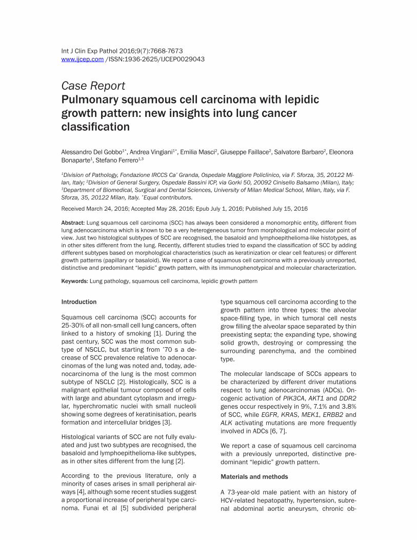
A routine chest X-ray revealed a mass on the right lung, and high-resolution computed-to- mography (CT) scan of the chest and the abdo-men confirmed a nodule measuring 2.5 cm in the middle right pulmonary lobe, another nod-ule measuring 1.5 cm with speculated, irregu-lar and infiltrative margins in the upper left pul-monary lobe and the presence of right hilar and mediastinal lymphadenopathy (Figure 1).
The PET/CT scan indicated an intense 18F-FDG activity in the middle right lobe (Standard Uptake Value = 5) and in the upper left lobe (SUV = 3, 4).
No other abnormal 18F-FDG uptake and no other localization of disease were identified at the clinical and imaging staging. The blood tumor markers were all within the normal rang-es. Conventional bronchoscopy with flexible endoscope did not show pathological findings and bronchoalveolar lavage (BAL) was not diagnostic.
Left pulmonary nodule was not candidated for CT-guided percutaneous biopsy because of dif-ficult lesion accessibility without a safe window for needle entry. In consideration of the clinical and imaging findings, the patient was candi-date for a surgical lung biopsy of the left pulmo-nary solid nodule. Video-assisted thoracic sur-gery (VATS) with wedge resection of the upper left lobe was performed. The surgical thoraco-scopic procedure was performed with the use
of three ports: the pulmonary wedge resection of the left lung apex was made by the use of an endo-GIA linear stapling device removing the nodule completely. No additional lymph node sampling was performed considering the pres-ence of controlateral pulmonary disease. A PET/CT scan performed forty days after surgery detected no more metabolic activity in the mid-dle right pulmonary lobe suggesting the resolu-tion of a flogistic disease; therefore non addi-tional surgery procedure and no adjuvant th- erapy for lung cancer was indicated by onco- logists.
Formalin-fixed, paraffin-embedded surgical specimens were stained with haematoxylin and eosin, and immunohistochemical stainings with antibodies directed against cytokeratin 7 (clone SP52), cytokeratin 20 (clone Ks20.8), cytokera-tin 5-6 (clone D5/16 B4), p40 (clone BC28), TTF-1 (clone SP141) and Napsin A (clone MRQ40) were performed.
Immunohistochemistry was performed using the automatic system BenchMark XT (Ventana Medical Systems, Inc. Tucson, Arizona, USA). Reactions were revealed using the UltraViewTM Universal DAB, a biotin-free, multimer-based detection system, according to the manufac-ture’s instruction.
Tumour genotyping by MassARRAY
In order to characterize the molecular profile of the tumor, a panel of mutations, insertions and deletions of the genes most frequently involved in NSCL genetic alterations was investigated. The Catalogue of somatic mutations in cancer (COSMIC) database was consulted to select the hotspot regions to analyze. Tumor DNA was iso-lated from formalin fixed paraffin embedded (FFPE) tissue sections, using the Biostic FFPE tissue DNA isolation Kit (Mo Bio Laboratories, Carlsbad, USA), following the manufacturer’s instructions. The MALDI-TOF (Matrix Assisted Laser Desorption Ionization Time-of-Flight) analysis was performed on 30 ng of DNA. MassARRAY Analyzer 4 was employed (Agena Bioscience, Hamburg, Germany), according to the iPLEX Pro application guide, using Complete iPLEX Pro Genotyping Reagent Set and SpectroCHIP II Arrays and Clean Resin Kit (Agena Bioscience, Hamburg, Germany). Mass- ARRAY Typer 4.0 software was used for data analysis.
Figure 1. High-resolution computed-tomography (CT) scan of the chest and the abdomen showing a nod-ule nodule measuring 1.5 cm in the upper left pul-monary lobe.

Lepidic pulmonary squamous cell carcinoma
7670 Int J Clin Exp Pathol 2016;9(7):7668-7673
Results
Histopathological findings
Macroscopically, the nodule measured 1.3 cm and, on cut sections, it had a grayish appear-ance, with ill-defined margins and a sub-pleural
localization. All the nodule was sampled and paraffin-embedded for histological examina- tion.
Microscopical examination showed a moder-ately differentiated (G2) squamous cell carci-noma, exhibiting focal but clearly visible kerati-
Figure 2. Morphological features of the nodule. Low-power magnification showed a central, solid scar with periph-eral lepidic growth pattern (A. 0.5x). At a higher magnification, lepidic growth pattern can be better appreciated, with squamous cell neoplastic population growing over the pre-existing alveolar septa, without destruction of the parenchyma (B, C. 5x; D-G. 10x). Cytological details of the neoplastic population morphologically indicate squamous differentiation (H. 20x).
Lepidic pulmonary squamous cell carcinoma
7671 Int J Clin Exp Pathol 2016;9(7):7668-7673
nization, characterized by an extensive lepidic growth: together with a central scar-like compo-nent, with an infiltrative growth pattern in the centre of the lesion, some areas showed malig-nant squamous cells growing along preexisting alveolar walls, preserving parenchymal archi-tecture, frequently in multiple layers, displacing pneumocytes toward the central portions of alveolar lumen (Figure 2).
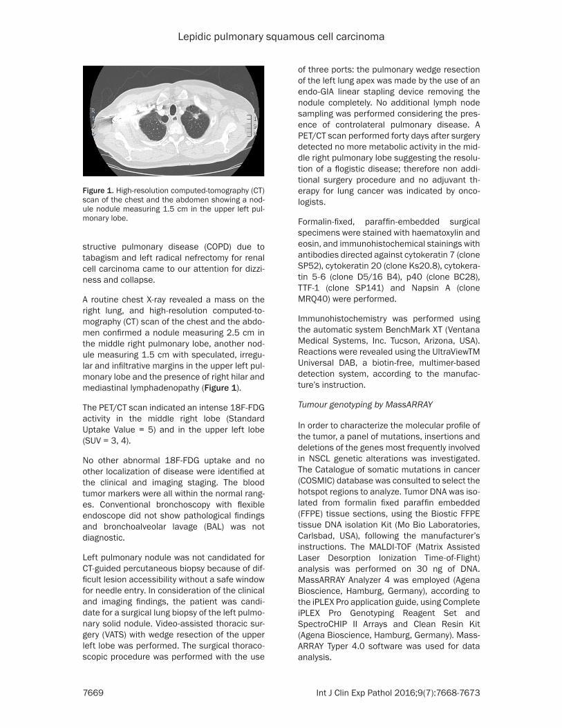
Strong immunoreactivity for p40 (Figure 3A), p63 (Figure 3B) and cytokeratins 5-6 (Figure 3C) confirmed the squamous nature of the lesion, while TTF-1 (Figure 3D) and Napsin A (Figure 3E) highlighted the intact frame of alve-olar pneumocytes, with no staining in the neo-plastic elements. Other immunohistochemical showed negativity in tumoral cells for cytokera-tin 7 and 20.
Molecular findings
We genotyped the tumor investigating the most frequent driver mutations of NSCLC.
We did not find any mutations in PIK3CA, AKT1 and DDR2, which are typical of SCCs, either in EGFR, KRAS, MEK1, ERBB2 and ALK, generally found in ADCs.
Discussion
If primary lung adenocarcinoma is known to be a very heterogeneous tumor in many aspects with different morphological subtypes and molecular mutations, on the contrary squa-mous cell carcinoma has always been consid-ered a “monomorphic”, solid entity [3].
Funai et al [5] tried to investigate different sub-types also in SCC, identifying in peripheral tumors three different growth pattern, the alve-olar space-filling type, the expanding type and the combined type.
The alveolar space-filling type of SCC is charac-terized by filling of the alveolar space without destroying alveolar septa and it has the most favorable clinical prognosis. The expanding
Figure 3. Immunohisto-chemical results. Stain-ings for p40 (A), p63 (B) and cytokeratins 5-6 (C) highlighted the neoplastic population, while TTF1 (D) and Nap-sin A (E) stained normal pneumocytes.

Lepidic pulmonary squamous cell carcinoma
7672 Int J Clin Exp Pathol 2016;9(7):7668-7673
type corresponds to the classic tumor nodule with infiltrative margins and with destruction of the pre-existing parenchyma and the combined type is a mixture of these two entitites.
Alveolar space-filling type can be considered a squamous evolution, a more advanced phase, of lepidic growth type, which is peculiar of ade-nocarcinoma: in lepidic-growth adenocarcino-ma, the neoplasm arises from cells lining the alveoli and spreads along their walls, but in alveolar space-filling type squamous cell carci-onoma, the neoplastic elements are not limited to cover septa, but they fill entirely the alveolar spaces.
In addition, a recent work by Kadota et al [8] indicates for SCCs five different subtypes: kera-tinizing, non-keratinizing, clear cell (which are not properly a growth pattern at all), basaloid and papillary type, but this is simpler and far from the complex classification of adenocar- cinoma.
In our case, in the majority of the tumor nodule we did not have a proper alveolar filling but just only a lepidic growth along alveolar walls and we can speculate that the presence of this pat-tern can be explained with a squamous meta-plastic change of the alveolar epithelium that has subsequently undergone cancerization and invasion.
Moreover, immunohistochemical of the lesion confirmed the squamous nature of the neo- plasia.
Despite its relatively monomorphic presenta-tion, molecular alterations in squamous cell lung cancers have not been comprehensively studied, and no targeted therapies have been developed.
Significantly altered pathways includes NFE2L2 and KEAP1 in 34%, squamous differentiation genes in 44%, PI3K pathway genes in 47%, and CDKN2A and RB1 in 72% of tumours [9].
In our opinion, the discovery of new subtypes within squamous cell carcinomas can have sig-nificant clinical impact, since research of molecular mutations, in mixed forms, should be guided by the morphology and could lead to new therapeutic strategies in these patients. This is well known in adenocarcinoma, where tumor heterogeneity influences evaluation of prognostic biomarkers [10].
We investigated the molecular profile of the tumor in order to identify the typical driver mutation of SCCs, but no alterations were found.
To our knowledge this is the first reported, mor-phologically and molecularly documented case of lung squamous cell carcinoma with this pe- culiar histological presentation, and its detec-tion confirms the recent studies indicating that squamous cell carcinoma is an entity which may arise with different subtypes and histologi-cal growth patterns.
To highlight this tumor heterogeneity, a further measure should be to do a high number of sam-ples of the nodules. In our case, the relatively small size of the nodule has allowed a sample of the tumor in its entirety.
Disclosure of conflict of interest
None.
Address correspondence to: Dr. Alessandro Del Gobbo, Division of Pathology, Fondazione IRCCS Ca’ Granda, Ospedale Maggiore Policlinico, via F. Sforza, 35, 20122 Milan, Italy. Tel: +39-025-503-8494; Fax: +39-025-503-2860; E-mail: [email protected]
References
[1] Drilon A, Rekhtman N, Ladanyi M, Paik P. Squa-mous-cell carcinomas of the lung: emerging biology, controversies, and the promise of tar-geted therapy. Lancet Oncol 2012; 13: e418-426.
[2] Dodds L, Davis S, Polissar L. A population-based study of lung cancer incidence trends by histologic type, 1974-81. J Natl Cancer Inst 1986; 76: 21-29.
[3] Travis WD. Pathology of lung cancer. Clin Chest Med 2011; 32: 669-692.
[4] Sakurai H, Asamura H, Watanabe S, Suzuki K, Tsuchiya R. Clinicopathologic features of pe-ripheral squamous cell carcinoma of the lung. Ann Thorac Surg 2004; 78: 222-227.
[5] Funai K, Yokose T, Ishii G, Araki K, Yoshida J, Nishimura M, Nagai K, Nishiwaki Y, Ochiai A.Clinicopathologic characteristics of peripheral squamous cell carcinoma of the lung. Am J Surg Pathol 2003; 27: 978-984.
[6] Liu SV, Subramaniam D, Cyriac GC, Abdul-Khalek FJ, Giaccone G. Emerging protein ki-nase inhibitors for non-small cell lung cancer. Expert Opin Emerg Drugs 2014; 19: 51-65.
[7] Mantripragada K, Khurshid H. Targeting Ge-nomic Alterations in Squamous Cell Lung Can-cer. Front Oncol 2013; 3: 195.
Lepidic pulmonary squamous cell carcinoma
7673 Int J Clin Exp Pathol 2016;9(7):7668-7673
[8] Kadota K, Nitadori J, Woo KM, Sima CS, Finley DJ, Rusch VW, Adusumilli PS, Travis WD. Com-prehensive pathological analyses in lung squa-mous cell carcinoma: single cell invasion, nu-clear diameter, and tumor budding are independent prognostic factors for worse out-comes. J Thorac Oncol 2014; 9: 1126-1139.
[9] Cancer Genome Atlas Research Network. Com-prehensive genomic characterization of squa-mous cell lung cancers. Nature 2012; 489: 519-525.
[10] Del Gobbo A, Pellegrinelli A, Gaudioso G, Cas-tellani M, Zito Marino F, Franco R, Palleschi A, Nosotti M, Bosari S, Vaira V, Ferrero S. Analysis of NSCLC tumour heterogeneity, proliferative and 18F-FDG PET indices reveals Ki67 prog-nostic role in adenocarcinomas. Histopatholo-gy 2016; 68: 746-51.