case study 30 - alphabioendpoint.azureedge.netalphabioendpoint.azureedge.net/...case_study_30... ·...
TRANSCRIPT
Dr. Lucian Toma Ciocan, Dmd, Os, Msd, PhdDr. Ion Nicolescu, Dmd, Dds, Oms, PhdMTD Arnon Kiperman
Bone stability around implants in full mouth fixed reconstruction:7 years follow up
Case study 30
2
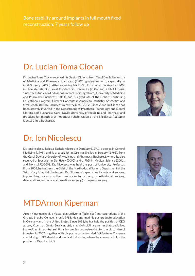
Dr. Lucian Toma CiocanDr. Lucian Toma Ciocan received his Dental Diploma from Carol Davila Universityof Medicine and Pharmacy, Bucharest (2002), graduating with a specialty in Oral Surgery (2005). After receiving his DMD, Dr. Ciocan received an MSc in Biomaterials, Bucharest Polytechnic University (2004) and a PhD (Thesis: “Interface Studies on Endoseous Implant Biointegration”), University of Medicine and Pharmacy, Bucharest (2011), and is a graduate of the Linhart Continuing Educational Program: Current Concepts in American Dentistry-Aesthetics and Oral Rehabilitation, Faculty of Dentistry, NYU (2012). Since 2002, Dr. Ciocan has been actively involved in the Department of Prosthetic Technology and Dental Materials of Bucharest, Carol Davila University of Medicine and Pharmacy and practices full mouth prosthodontics rehabilitation at the Nicolescu-Agatstein Dental Clinic, Bucharest.
Bone stability around implants in full mouth fixed reconstruction: 7 years follow up
BONE STABILITY AROUND IMPLANTS IN FULL MOUTH FIXED RECONSTRUCTION: 7-YEAR FOLLOW-UP
Case Study
Authors
Dr. LUCIAN TOMA CIOCAN, DMD, OS, MsD, PhD Dr. ION NICOLESCU, DMD, DDS, OMS, PhD MTD ARNON KIPERMAN
Dr. Lucian Toma Ciocan
Dr. Lucian Toma Ciocan received his Dental Diploma from Carol Davila University of Medicine and Pharmacy, Bucharest (2002), graduating with a specialty in Oral Surgery (2005). After receiving his DMD, Dr. Ciocan received an MSc in Biomaterials, Bucharest Polytechnic University (2004) and a PhD (Thesis: “Interface Studies on Endoseous Implant Biointegration”), University of Medicine and Pharmacy, Bucharest (2011), and is a graduate of the Linhart Continuing Educational Program: Current Concepts in American Dentistry-Aesthetics and Oral Rehabilitation, Faculty of Dentistry, NYU (2012). Since 2002, Dr. Ciocan has been actively involved in the Department of Prosthetic Technology and Dental Materials of Bucharest, Carol Davila University of Medicine and Pharmacy and practices full mouth prosthodontics rehabilitation at the Nicolescu-Agatstein Dental Clinic, Bucharest.
Dr. Ion NicolescuDr. Ion Nicolescu holds a Bachelor degree in Dentistry (1991), a degree in General Medicine (1999), and is a specialist in Oro-maxillo-facial Surgery (1995), from the Carol Davila University of Medicine and Pharmacy, Bucharest, where he also received a Specialist in Dentistry (2000) and a PhD in Medical Science (2001), and from 1992-2008, Dr. Nicolescu was held the post of University Professor. From 2008, he has been the Chief of the Maxillo-facial Surgery Department at the Saint Mary Hospital, Bucharest. Dr. Nicolescu’s specialties include oral surgery, implantology, reconstructive dento-alveolar surgery, maxillo-facial surgery, deformations and facial malformations surgery (orthognatic surgery).
Dr. Ion Nicolescu
Dr. Ion Nicolescu holds a Bachelor degree in Dentistry (1991), a degree in General Medicine (1999), and is a specialist in Oro-maxillo-facial Surgery (1995), from the Carol Davila University of Medicine and Pharmacy, Bucharest, where he also received a Specialist in Dentistry (2000) and a PhD in Medical Science (2001), and from 1992-2008, Dr. Nicolescu was held the post of University Professor. From 2008, he has been the Chief of the Maxillo-facial Surgery Department at the Saint Mary Hospital, Bucharest. Dr. Nicolescu’s specialties include oral surgery, implantology, reconstructive dento-alveolar surgery, maxillo-facial surgery, deformations and facial malformations surgery (orthognatic surgery).
Arnon Kiperman Arnon Kiperman holds a Master degree (Dental Technician) and is a graduate of the Ort Yad Shapira College (Israel), 1985. He continued his postgraduate education in Germany and in the United States. Since 1993, he has held the position of CEO at Levy Kiperman Dental Services, Ltd., a multi-disciplinary center that specializes in providing integrated solutions in complex reconstruction for the global dental industry. In 2007, together with his partners, he founded MS Systems Company specializing in 3D dental and medical industries, where he currently holds the position of Director, R&D.
MTDArnon KipermanArnon Kiperman holds a Master degree (Dental Technician) and is a graduate of the Ort Yad Shapira College (Israel), 1985. He continued his postgraduate education in Germany and in the United States. Since 1993, he has held the position of CEO at Levy Kiperman Dental Services, Ltd., a multi-disciplinary center that specializes in providing integrated solutions in complex reconstruction for the global dental industry. In 2007, together with his partners, he founded MS Systems Company specializing in 3D dental and medical industries, where he currently holds the position of Director, R&D.
Dr. Ion Nicolescu
Dr. Ion Nicolescu holds a Bachelor degree in Dentistry (1991), a degree in General Medicine (1999), and is a specialist in Oro-maxillo-facial Surgery (1995), from the Carol Davila University of Medicine and Pharmacy, Bucharest, where he also received a Specialist in Dentistry (2000) and a PhD in Medical Science (2001), and from 1992-2008, Dr. Nicolescu was held the post of University Professor. From 2008, he has been the Chief of the Maxillo-facial Surgery Department at the Saint Mary Hospital, Bucharest. Dr. Nicolescu’s specialties include oral surgery, implantology, reconstructive dento-alveolar surgery, maxillo-facial surgery, deformations and facial malformations surgery (orthognatic surgery).
Arnon Kiperman Arnon Kiperman holds a Master degree (Dental Technician) and is a graduate of the Ort Yad Shapira College (Israel), 1985. He continued his postgraduate education in Germany and in the United States. Since 1993, he has held the position of CEO at Levy Kiperman Dental Services, Ltd., a multi-disciplinary center that specializes in providing integrated solutions in complex reconstruction for the global dental industry. In 2007, together with his partners, he founded MS Systems Company specializing in 3D dental and medical industries, where he currently holds the position of Director, R&D.
3
AbstractThis case study presents a 7-year follow-up bone stability deploying 17 ATID Alpha-Bio Tec implants inserted in a 44 year old patient, full mouth rehabilitation with porcelain fused to metal restorations. The long term stability of the clinical outcome was evaluated on clinical check-ups, periodontal charting and panoramic x-rays.
IntroductionImplant placement in aggressive periodontitis has been a questionable issue of debate. Although there are still some clinicians who consider periodontal disease an absolute contraindication for dental implants, randomized clinical studies show that there is no statistically significant difference in implant survival rates in patients with implants replacing teeth lost due to chronic periodontitis (90.5%). This is compared to patients with implants replacing teeth lost due to reasons other than periodontitis (96.5%) during a 10-year maintenance period and in particular, those who give up smoking and improved their oral hygiene routine [1].
Case Description
Clinical Examination
A 44 year old female patient presented in our clinic in March 2006, with the major complaint of gradually increasing teeth mobility, bleeding when brushing and recently spontaneous loss of tooth 1.2. (U.S. system #7). She denied any alteration of the systemic conditions, being at that time a heavy smoker (more than 20 cigarettes p/day). Anamnesis revealed nocturne bruxism parafunctional activity.
• Facial examinationshowed a rounded symmetric face (Fig. 1) with the face midline perpendicular to the bipupilary line, light alteration (1-2mm) of the VDO (Vertical Dimension of Occlusion). Class 1 (low) lip line. Mouth opening 40mm, normal TMJ movement and well developed masticatory muscles.
• Oral examination(Fig. 2) revealed a labio-genial mucosa of normal aspect, as well as the one of tongue, palate and floor of the mouth, normal tongue mobility, normal saliva secretion in both quality and quantity, presence of scanty calculus with moderate to severe gingival inflammation and poor oral hygiene and Kenedy class III edentulous maxillae and mandible restored by four fixed partial PFM dentures with poor fit: grade 1 mobility for the one from the 4th quadrant teeth/dentures and grade 2 mobility for the ones from quadrants 1st, 2nd and 3rd. The remaining anterior non-covered teeth presented attrition grade 2. BOP and deep probing depth of all remaining teeth (Figs. 4-5).
• Panoramic x-ray examinationOrthopantomography (Fig. 3) shows vertical bone loss in relation to all teeth present on the both arches (1.8., 1.7., 1.3., 1.1., 2.1., 2.2., 2.3., 2.8., 3.7., 3.5., 3.4., 3.3., 3.2., 3.1., 4.1., 4.3., 4.4., 4.5., 4.7.).
4
DiagnosisSevere deep generalized periodontitis without indication of conservatory treatment of the remaining teeth.
Restoration TypeCement implant supported PFM fixed full dentures.
MaterialsATID Alpha-Bio Tec implants, Xenograft and Collagen Membrane.
Treatment Plan and GoalsIn this case, the treatment options were presented to the patient using Dental Master software [2]: full upper and lower denture, maxillary and mandibulary implants supported overdentures and maxillary and mandibulary porcelain fused to metal fixed implants supported full bridges (Fig. 6). Patient selected the full PFM bridges.
• Step I24-05-2006: During the first treatment visit, we proceeded with the extractions (Fig. 7), the curettage of alveolar pockets and the appliance of immediate full upper and lower dentures (Figs. 7-10).
• Step II20-07-2006 (2 months following Step I): Early placement with soft tissue healing implant insertion according to SAC classification in Implant Dentistry [3] in the position of 1.7. (ATI 4.2/10mm); 1.6. (ATI 4.2/11,5mm); 1.4. (ATI 3.75/11,5mm); 1.3. (ATI 3.75/11,5mm); 1.2. (ATI 3.75/10mm); 2.2. (ATI 3.75/10mm); 2.4. (ATI 3.75/11.5mm); 2.6. (ATI 3.75/11.5mm); 2.7. (ATI 4.2/10mm); 3.7. (ATI 4.2/11.5mm); 3.6. (ATI 3.75/11.5mm); 3.4. (ATI 3.75/16mm), 3.2. (ATI 3.75/16mm); 4.2. (ATI 3.75/16mm); 4.4. (ATI 3.75/16mm); 4.6. (ATI 3.75/11.5mm); 4.7. (ATI 4.2/11.5mm), bilaterally closed sinus lifting at the levels of 1.7. and 2.7., GBR with Xenograft and Collagen Membrane at the level of implants 3.7. and 4.7. were realized by Dr. Ion Nicolescu. Dentures soft tissue relining using GC Tissue Conditioner with Nistatin in concentration below 1.000.000U [4] (Figs. 11-14).
• Step II26-09-2006 (2 months following Step II): The removable dentures were anchored to the 1.4., 2.4., 4.4. and 3.4. implants using ball attachments to reduce overpressure on the facial bone over the implants and to ensure patient comfort for the remaining 5 months by uncovering the upper denture's palatal area (Figs. 15-20).
Bone stability around implants in full mouth fixed reconstruction: 7 years follow up
5
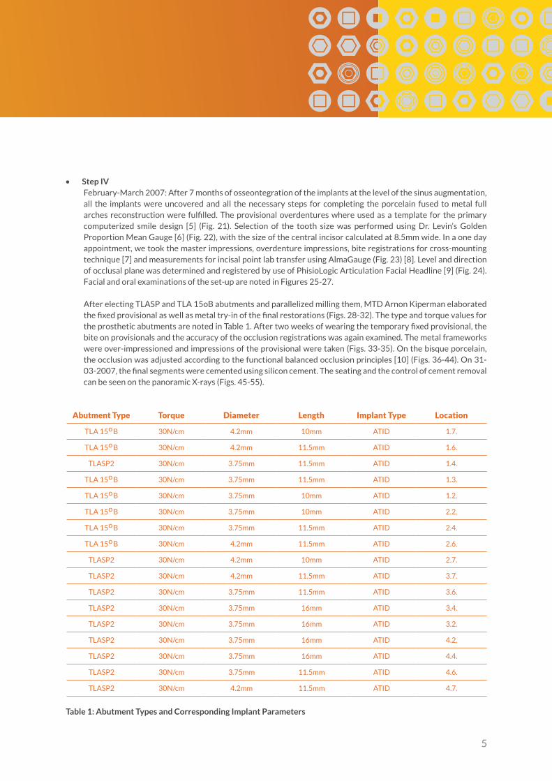
• Step IVFebruary-March 2007: After 7 months of osseontegration of the implants at the level of the sinus augmentation, all the implants were uncovered and all the necessary steps for completing the porcelain fused to metal full arches reconstruction were fulfilled. The provisional overdentures where used as a template for the primary computerized smile design [5] (Fig. 21). Selection of the tooth size was performed using Dr. Levin’s Golden Proportion Mean Gauge [6] (Fig. 22), with the size of the central incisor calculated at 8.5mm wide. In a one day appointment, we took the master impressions, overdenture impressions, bite registrations for cross-mounting technique [7] and measurements for incisal point lab transfer using AlmaGauge (Fig. 23) [8]. Level and direction of occlusal plane was determined and registered by use of PhisioLogic Articulation Facial Headline [9] (Fig. 24). Facial and oral examinations of the set-up are noted in Figures 25-27.
After electing TLASP and TLA 15oB abutments and parallelized milling them, MTD Arnon Kiperman elaborated the fixed provisional as well as metal try-in of the final restorations (Figs. 28-32). The type and torque values for the prosthetic abutments are noted in Table 1. After two weeks of wearing the temporary fixed provisional, the bite on provisionals and the accuracy of the occlusion registrations was again examined. The metal frameworks were over-impressioned and impressions of the provisional were taken (Figs. 33-35). On the bisque porcelain, the occlusion was adjusted according to the functional balanced occlusion principles [10] (Figs. 36-44). On 31-03-2007, the final segments were cemented using silicon cement. The seating and the control of cement removal can be seen on the panoramic X-rays (Figs. 45-55).
Abutment Type Torque Diameter Length Implant Type Location
TLA 15o B 30N/cm 4.2mm 10mm ATID 1.7.
TLA 15o B 30N/cm 4.2mm 11.5mm ATID 1.6.
TLASP2 30N/cm 3.75mm 11.5mm ATID 1.4.
TLA 15o B 30N/cm 3.75mm 11.5mm ATID 1.3.
TLA 15o B 30N/cm 3.75mm 10mm ATID 1.2.
TLA 15o B 30N/cm 3.75mm 10mm ATID 2.2.
TLA 15o B 30N/cm 3.75mm 11.5mm ATID 2.4.
TLA 15o B 30N/cm 4.2mm 11.5mm ATID 2.6.
TLASP2 30N/cm 4.2mm 10mm ATID 2.7.
TLASP2 30N/cm 4.2mm 11.5mm ATID 3.7.
TLASP2 30N/cm 3.75mm 11.5mm ATID 3.6.
TLASP2 30N/cm 3.75mm 16mm ATID 3.4.
TLASP2 30N/cm 3.75mm 16mm ATID 3.2.
TLASP2 30N/cm 3.75mm 16mm ATID 4.2.
TLASP2 30N/cm 3.75mm 16mm ATID 4.4.
TLASP2 30N/cm 3.75mm 11.5mm ATID 4.6.
TLASP2 30N/cm 4.2mm 11.5mm ATID 4.7.
Table 1: Abutment Types and Corresponding Implant Parameters
6
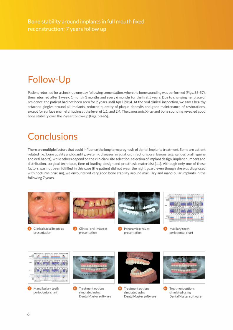
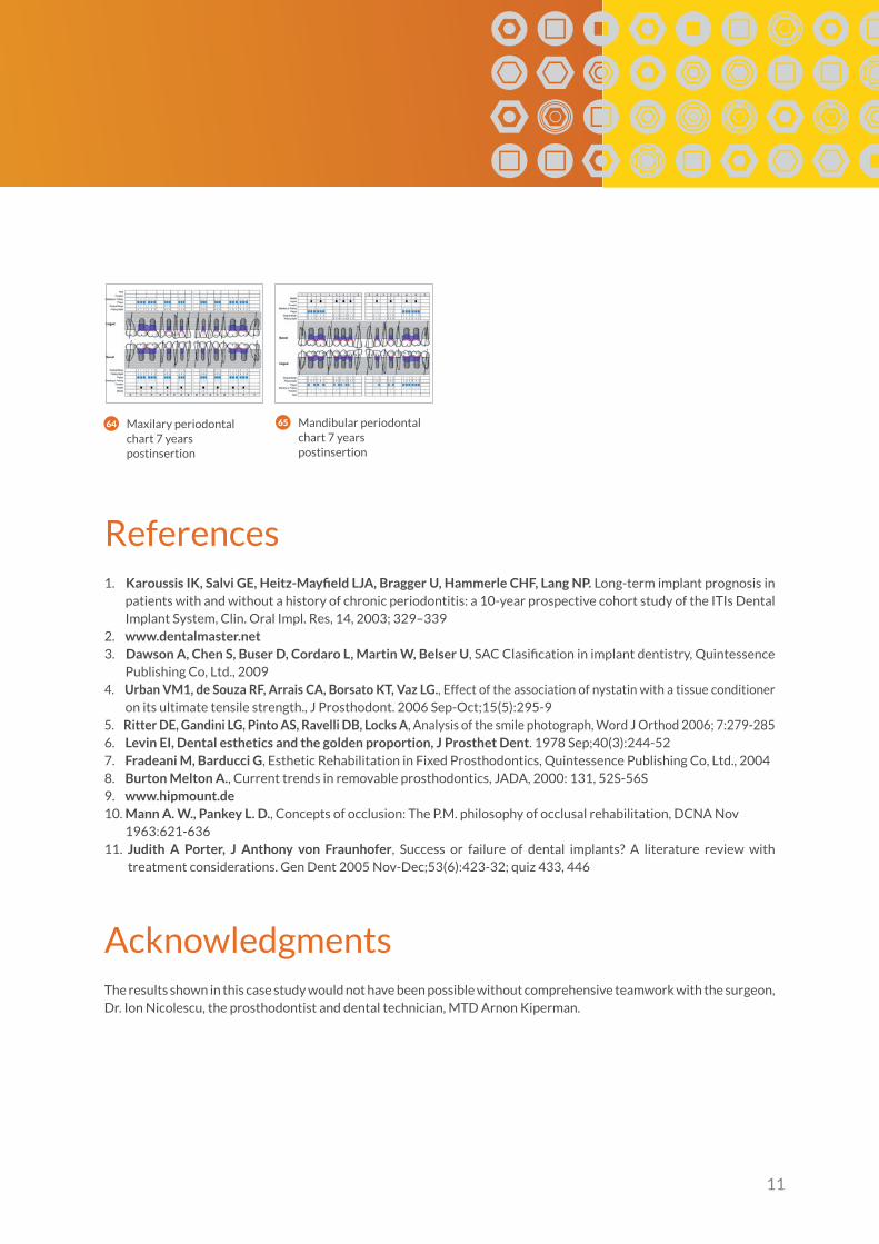
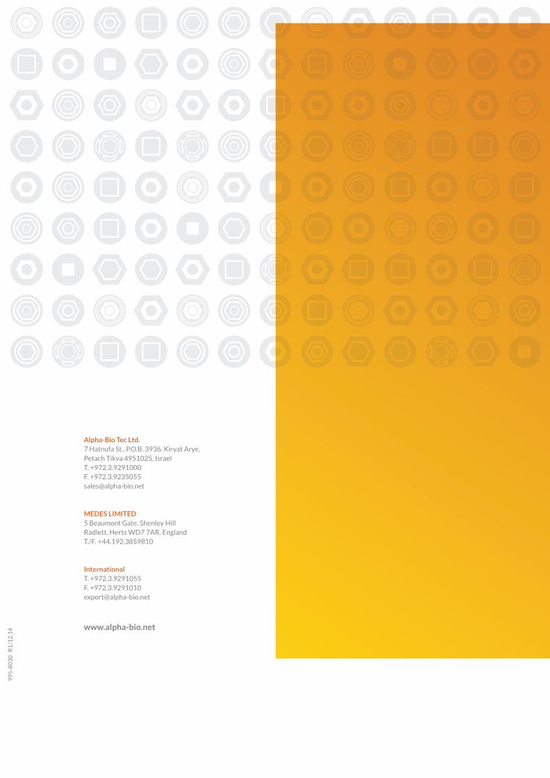
Follow-UpPatient returned for a check-up one day following cementation, when the bone sounding was performed (Figs. 56-57), then returned after 1 week, 1 month, 3 months and every 6 months for the first 5 years. Due to changing her place of residence, the patient had not been seen for 2 years until April 2014. At the oral clinical inspection, we saw a healthy attached gingiva around all implants, reduced quantity of plaque deposits and good maintenance of restorations, except for surface enamel chipping at the level of 1.1. and 2.4. The panoramic X-ray and bone sounding revealed good bone stability over the 7-year follow-up (Figs. 58-65).
ConclusionsThere are multiple factors that could influence the long term prognosis of dental implants treatment. Some are patient related (i.e., bone quality and quantity, systemic diseases, irradiation, infections, oral lesions, age, gender, oral hygiene and oral habits), while others depend on the clinician (site selection, selection of implant design, implant numbers and distribution, surgical technique, time of loading, design and prosthesis materials) [11]. Although only one of these factors was not been fulfilled in this case (the patient did not wear the night guard even though she was diagnosed with nocturne bruxism), we encountered very good bone stability around maxillary and mandibular implants in the following 7 years.
1 3 42Clinical facial image at presentation
Panoramic x-ray atpresentation
Maxilary teeth periodontal chart
Clinical oral image at presentation
5 6a 6b 6cMandibulary teeth periodontal chart
Treatment options simulated using DentalMaster software
Treatment options simulated using DentalMaster software
Treatment options simulated using DentalMaster software
Bone stability around implants in full mouth fixed reconstruction: 7 years follow up

7
8 10 119Oral image of postextractionalveolar ridges
Oral image of immediateoverdentures soft relined “in situ”
Oral image of maxillary ridge after implants insertions
Panoramic x-ray after extraction
76d 6e 6fTreatment options simulated using DentalMaster software
Treatment options simulated using DentalMaster software
Immediate full denturespreextractional prepared
Treatment options simulated using DentalMaster software
12 13 14 15Oral image of mandibularridge after implants insertion
Re-relined dentures afterimplant insertion
Oral image of maxilary ridge 2 month after implant insertion. Mounted TB4 straight ball attachment at the level of 1.4. and 2.4.,3,75/11,5 mm ATID implants
Panoramic x-ray after implant insertion (Surg. Ion Nicolescu)
16 17 18 19Oral image of mandible ridge 2 month after implant insertion. Mounted TB3 straight ball attachment at the level of 3.4. and 4.4., 3,75/16mm implants
Clinical oral image 2 monthafter implants insertion–removable upper denture relined, directanchorated on ball attachments and palatalreductioned
Clinical oral image 2 month after implants insertion – removablelower denture relined and direct anchorated on ball attachments
Clinical oral image 2 month after implants insertion – good softtissue healing arround healing abutments
8
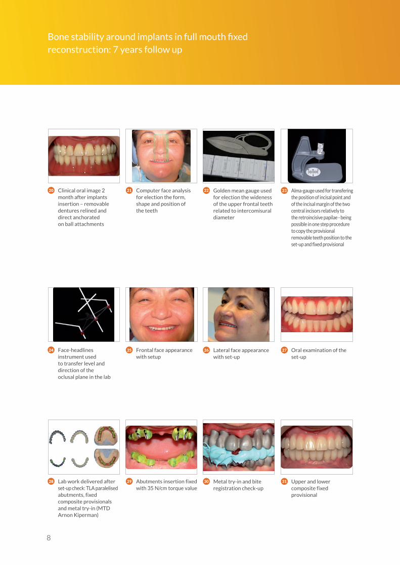
28 29 30 31Lab work delivered afterset-up check: TLA paralelisedabutments, fixed composite provisionals and metal try-in (MTD Arnon Kiperman)
Metal try-in and biteregistration check-up
Upper and lower composite fixed provisional
Abutments insertion fixed with 35 N/cm torque value
262524 27Face-headlines instrument usedto transfer level and direction of theoclusal plane in the lab
Lateral face appearance with set-up
Oral examination of the set-up
Frontal face appearance with setup
Bone stability around implants in full mouth fixed reconstruction: 7 years follow up
2320 21 22Clinical oral image 2 month after implants insertion – removabledentures relined and direct anchoratedon ball attachments
Golden mean gauge used for election the wideness of the upper frontal teeth related to intercomisuraldiameter
Computer face analysis for election the form, shape and position ofthe teeth
Alma-gauge used for transfering the position of incisal point and of the incisal margin of the two central incisors relatively to the retroincisive papilae - being possible in one step procedure to copy the provisional removable teeth position to the set-up and fixed provisional
9
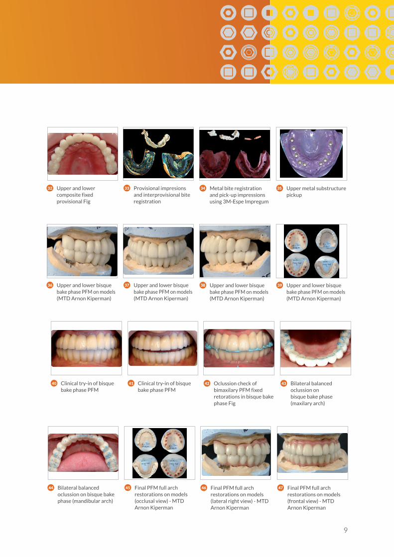
32 33 34 35Upper and lower composite fixed provisional Fig
Metal bite registration and pick-up impressions using 3M-Espe Impregum
Upper metal substructure pickup
Provisional impresions and interprovisional bite registration
3936 37 38Upper and lower bisque bake phase PFM on models(MTD Arnon Kiperman)
Upper and lower bisque bake phase PFM on models(MTD Arnon Kiperman)
Upper and lower bisque bake phase PFM on models(MTD Arnon Kiperman)
Upper and lower bisque bake phase PFM on models(MTD Arnon Kiperman)
44 45 46 47Bilateral balanced oclussion on bisque bake phase (mandibular arch)
Final PFM full archrestorations on models (lateral right view) - MTD Arnon Kiperman
Final PFM full arch restorations on models (frontal view) - MTD Arnon Kiperman
Final PFM full arch restorations on models (occlusal view) - MTDArnon Kiperman
424140 43Clinical try-in of bisque bake phase PFM
Oclussion check of bimaxilary PFM fixed retorations in bisque bake phase Fig
Bilateral balanced oclussion onbisque bake phase (maxilary arch)
Clinical try-in of bisque bake phase PFM

10
60 62 63PFM full arches restorations in disclusion 7yrs after
Panoramic x-ray 7 yrs after
Facial appearence 7 years after
61 PFM mandibulary arch restoration (oclusal view) 7 yrs after
48 49 50 51Final PFM full archrestorations on models (lateral left view) - MTD Arnon Kiperman
PFM full arches restorations cemented on implants in oclusion(frontal view)
PFM maxilary arch restoration (oclusal view)
Oral image of abutments before cementations Fig
585756 59Maxilary periodontalpostinsertion
Frontal view of PFM full arches restorations in oclusion 7 yrs after
PFM maxilary arch restoration (oclusal view) 7 yrs after
Mandibular periodontalpostinsertion
5552 53 54PFM full arches restorations cemented on implants in disclusion
Panoramic x-ray at the day of insertion
Facial appearence in the day of delivery
PFM mandibulary archrestoration (oclusal view)
Bone stability around implants in full mouth fixed reconstruction: 7 years follow up
11
64 Maxilary periodontal chart 7 years postinsertion
65 Mandibular periodontal chart 7 years postinsertion
References1. Karoussis IK, Salvi GE, Heitz-Mayfield LJA, Bragger U, Hammerle CHF, Lang NP. Long-term implant prognosis in
patients with and without a history of chronic periodontitis: a 10-year prospective cohort study of the ITIs Dental Implant System, Clin. Oral Impl. Res, 14, 2003; 329–339
2. www.dentalmaster.net3. Dawson A, Chen S, Buser D, Cordaro L, Martin W, Belser U, SAC Clasification in implant dentistry, Quintessence
Publishing Co, Ltd., 20094. Urban VM1, de Souza RF, Arrais CA, Borsato KT, Vaz LG., Effect of the association of nystatin with a tissue conditioner
on its ultimate tensile strength., J Prosthodont. 2006 Sep-Oct;15(5):295-95. Ritter DE, Gandini LG, Pinto AS, Ravelli DB, Locks A, Analysis of the smile photograph, Word J Orthod 2006; 7:279-2856. Levin EI, Dental esthetics and the golden proportion, J Prosthet Dent. 1978 Sep;40(3):244-527. Fradeani M, Barducci G, Esthetic Rehabilitation in Fixed Prosthodontics, Quintessence Publishing Co, Ltd., 20048. Burton Melton A., Current trends in removable prosthodontics, JADA, 2000: 131, 52S-56S9. www.hipmount.de10. Mann A. W., Pankey L. D., Concepts of occlusion: The P.M. philosophy of occlusal rehabilitation, DCNA Nov
1963:621-63611. Judith A Porter, J Anthony von Fraunhofer, Success or failure of dental implants? A literature review with
treatment considerations. Gen Dent 2005 Nov-Dec;53(6):423-32; quiz 433, 446
AcknowledgmentsThe results shown in this case study would not have been possible without comprehensive teamwork with the surgeon, Dr. Ion Nicolescu, the prosthodontist and dental technician, MTD Arnon Kiperman.
99
5-8
03
0
R1
/12
.14
Alpha-Bio Tec Ltd.7 Hatnufa St., P.O.B. 3936. Kiryat Arye,Petach Tikva 4951025, IsraelT. +972.3.9291000F. [email protected]
MEDES LIMITED5 Beaumont Gate, Shenley HillRadlett, Herts WD7 7AR. EnglandT./F. +44.192.3859810
InternationalT. +972.3.9291055 F. [email protected]
www.alpha-bio.net