case three-dimensional images of extra-routine grafts in
TRANSCRIPT

333Ann Thorac Cardiovasc Surg Vol. 14, No. 5 (2008)
CaseReport
Three-Dimensional Images of Extra-RoutineGrafts in CABG by Multidetector
Computed Tomography
Akihiro Nabuchi, MD, PhD,1 Atsushi Kurata, MD,1 Hiroshi Okuyama, MD,1
Takeshi Kondo, MD, PhD,2 Yasuji Muto, MD,1 and Yuki Endo, MD 1
From 1Department of Cardiovascular Surgery, Yamato Seiwa Hospital, Yamato; and 2Department of Cardiology, Takase Clinic, Takasaki, Japan
Received September 8, 2008; accepted for publication September 22, 2008Address reprint requests to Akihiro Nabuchi, MD, PhD: Department of Cardiovascular Surgery, Yamato Seiwa Hospital, 9–8–2 Minami-rinkan, Yamato, Kanagawa 242–0006, Japan.
The multidetector computed tomography (MDCT) scan is now widely used especially to find lesions of the coronary artery stenosis. In this report the images of a postoperative study of coronary artery bypass grafting (CABG) by MDCT are introduced to reveal their feasibility and reliability. Shown is one of the patients whose saphenous vein graft (SVG) was connected from the descending aorta to the left anterior descending artery (LAD) via the obtuse marginal branch (OM). This is because the left internal thoracic artery (ITA) was not available and the ascending aorta could not be used for highly calcified degenera-tion. That kind of graft in CABG should be recognized as “extra routine,” and its use will cause some difficulties to arise in postoperative elucidation for graft function. The images by MDCT reported here showed an excellent view of the route and lumen of the graft, sug-gesting the feasibility and usefulness of MDCT in CABG postoperative study. (Ann Thorac Cardiovasc Surg 2008; 14: 333–335)
Key words: coronary artery bypass grafting, postoperative study, multidetector computed tomography, volume-rendering three-dimensional image
graft.” The arising angles of the grafts at the proximal anastomosis are also important to predict their longevity as a graft conduit. It is hard to estimate patency and these features in the postoperative course, even by angiogram catheter.
Multidetector computed tomography (MDCT) scan is now widely used especially to find lesions of the coronary artery stenosis3–5) or plaque,6–8) but few findings have been reported in postoperative studies for CABG grafts emphasizing its feasibility to visually show satisfactorily the volume-rendering three-dimensional (3-D) image.9,10)
We have reported on one patient with an extra-routine graft and present volume-rendering 3-D images taken by MDCT scan.
Case
An 82-year-old gentleman presented with a severely calcified left coronary orifice. The diagnosis called for performing CABG on both the left coronary artery and
Background
In coronary artery bypass grafting (CABG) surgery, a cardiac surgeon is sometimes faced with patients for whom a routine graft origin is not available, especially in the redo of a CABG case.1)
We have experienced several patients in whom the internal thoracic artery (ITA) was not available and the descending aorta was used as a proximal end of the saphenous vein graft (SVG).2) These routes are quite unusual because their proximal ends are inside the thoracic cavity and are easily altered by compression of the lung, which can be recognized as an “extra-routine

334
Nabuchi et al.
Ann Thorac Cardiovasc Surg Vol. 14, No. 5 (2008)
one of the left circumflex branches. The left ITA was unavailable because of occlusion of the left subclavian artery resulting from atherosclerotic degeneration. The wall of the ascending aorta had highly calcified degen-eration, and it was decided not to touch it. During general anesthesia, a left thoracotomy was conducted and the SVG harvested. A stabilizer was used to per-form end-to-side anastomosis on the left anterior descending artery (LAD) and side-to-side anastomosis on the obtuse marginal branch (OM) with the SVG in
Fig. 1. Intraoperative view just after the SVG anastomosis proce-dure under the stabilizer has been completed.
The proximal end has not been done.
Fig. 2. The 3-D view three years and three months after CABG is shown as a still picture of the volume-rendering re-construction method.
The 360° multiangular views are avail-able in the monitor of the equipment. Anastomotic sites of the SVG from the descending aorta are well visualized and appear to bulge slightly, suggesting con-sistent flow into the LAD and OM (CT scanner: AquilionTM 64, Toshiba Medical Systems Corp.).
the usual manner (Fig. 1). The proximal end of the SVG was anastomosed to the descending aorta with a side-biter clamp beneath the hilus of the left lung. The postoperative course was uneventful, and the patient was discharged on postoperative day 14.
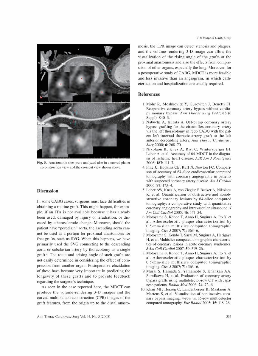
Three years and three months after the surgery, a postoperative study was done by MDCT (Fig. 2). The features throughout the route of the SVG have been shown clearly in volume-rendering 3-D images, as well in other reconstructed images (Fig. 3).
3-D Image of CABG Graft
Ann Thorac Cardiovasc Surg Vol. 14, No. 5 (2008) 335
Discussion
In some CABG cases, surgeons must face difficulties in obtaining a routine graft. This might happen, for exam-ple, if an ITA is not available because it has already been used, damaged by injury or irradiation, or dis-eased by atherosclerotic change. Moreover, should the patient have “porcelain” aorta, the ascending aorta can-not be used as a portion for proximal anastomosis for free grafts, such as SVG. When this happens, we have primarily used the SVG connecting to the descending aorta or subclavian artery by thoracotomy as a single graft.2) The route and arising angle of such grafts are not easily determined in considering the effect of com-pression from another organ. Postoperative elucidation of these have become very important in predicting the longevity of these grafts and to provide feedback regarding the surgeon’s technique.
As seen in the case reported here, the MDCT can produce the volume-rendering 3-D images and the curved multiplanar reconstruction (CPR) images of the graft features, from the origin up to the distal anasto-
mosis, the CPR image can detect stenosis and plaques, and the volume-rendering 3-D image can allow the visualization of the rising angle of the grafts at the proximal anastomosis and also the effects from compre-ssion of other organs, especially the lung. Moreover, for a postoperative study of CABG, MDCT is more feasible and less invasive than an angiogram, in which cath-eterization and hospitalization are usually required.
References
1. Mohr R, Moshkovitz Y, Gurevitch J, Benetti FJ. Reoperative coronary artery bypass without cardio-pulmonary bypass. Ann Thorac Surg 1997; 63 (6 Suppl): S40–3.
2. Nabuchi A, Kurata A. Off-pump coronary artery bypass grafting for the circumflex coronary artery via the left thoracotomy in redo CABG with the pat-ent left internal thoracic artery graft to the left anterior descending artery. Ann Thorac Cardiovasc Surg 2000; 6: 268–70.
3. Nikolaou K, Knez A, Rist C, Wintersperger BJ, Leber A, et al. Accuracy of 64-MDCT in the diagno-sis of ischemic heart disease. AJR Am J Roentgenol 2006; 187: 111–7.
4. Fine JJ, Hopkins CB, Ruff N, Newton FC. Compari-son of accuracy of 64-slice cardiovascular computed tomography with coronary angiography in patients with suspected coronary artery disease. Am J Cardiol 2006; 97: 173–4.
5. Leber AW, Knez A, von Ziegler F, Becker A, Nikolaou K, et al. Quantification of obstructive and nonob-structive coronary lesions by 64-slice computed tomography: a comparative study with quantitative coronary angiography and intravascular ultrasound. J Am Coll Cardiol 2005; 46: 147–54.
6. Motoyama S, Kondo T, Anno H, Sugiura A, Ito Y, et al. Atherosclerotic plaque characterization by 0.5-mm-slice multislice computed tomographic imaging. Circ J 2007; 71: 363–6.
7. Motoyama S, Kondo T, Sarai M, Sugiura A, Harigaya H, et al. Multislice computed tomographic characteris-tics of coronary lesions in acute coronary syndromes. J Am Coll Cardiol 2007; 50: 319–26.
8. Motoyama S, Kondo T, Anno H, Sugiura A, Ito Y, et al. Atherosclerotic plaque characterization by 0.5-mm-slice multislice computed tomographic imaging. Circ J 2007; 71: 363–6.
9. Murai S, Hamada S, Yamamoto S, Khankan AA, Sumikawa H, et al. Evaluation of coronary artery bypass grafts using multidetector-row CT with Japa-nese patients. Radiat Med 2006; 24: 72–6.
10. Khan MF, Herzog C, Landenberger K, Maataoui A, Martens S, et al. Visualisation of non-invasive coro-nary bypass imaging: 4-row vs. 16-row multidetector computed tomography. Eur Radiol 2005; 15: 118–26.
Fig. 3. Anastomotic sites were analyzed also in a curved planer reconstruction view and the crosscut view shown above.