caseation of human tuberculosis granulomas correlates with...
TRANSCRIPT

Research ArticleLipid metabolism in human tuberculous granulomas
258
Caseation of human tuberculosis granulomascorrelates with elevated host lipidmetabolism
Mi-Jeong Kim1, Helen C. Wainwright2, Michael Locketz2, Linda-Gail Bekker3, Gabriele B. Walther4y,Corneli Dittrich2, Annalie Visser2, Wei Wang5, Fong-Fu Hsu6, Ursula Wiehart1z, Liana Tsenova7z,Gilla Kaplan7, David G. Russell1*
Keywords: foamy macrophage;
granuloma; Mycobacterium;
tuberculosis
DOI 10.1002/emmm.201000079
Received January 16, 2010
Revised May 03, 2010
Accepted May 28, 2010
(1) Department of Microbiology and Immunology, C
Medicine, Cornell University, Ithaca, NY, USA.
(2) Division of Anatomical Pathology, Faculty of Health
of Cape Town, Cape Town, South Africa.
(3) Department of Medicine, The Desmond Tutu HIV
Infectious Disease and Molecular Medicine, Unive
Cape Town, South Africa.
(4) Chris Barnard Division of Cardio-Thoracic Surg
Hospital, University of Cape Town, Cape Town, Sou
(5) Department of Biomedical Sciences, College of
Cornell University, Ithaca, NY, USA.
� 2010 EMBO Molecular Medicine
The progression of human tuberculosis (TB) to active disease and transmission
involves the development of a caseous granuloma that cavitates and releases
infectious Mycobacterium tuberculosis bacilli. In the current study, we exploited
genome-wide microarray analysis to determine that genes for lipid sequestration
and metabolism were highly expressed in caseous TB granulomas. Immunohis-
tological analysis of these granulomas confirmed the disproportionate abun-
dance of the proteins involved in lipid metabolism in cells surrounding the
caseum; namely, adipophilin, acyl-CoA synthetase long-chain family member
1 and saposin C. Biochemical analysis of the lipid species within the caseum
identified cholesterol, cholesteryl esters, triacylglycerols and lactosylceramide,
which implicated low-density lipoprotein-derived lipids as the most likely
source. M. tuberculosis infection in vitro induced lipid droplet formation in
murine and human macrophages. Furthermore, the M. tuberculosis cell wall
lipid, trehalose dimycolate, induced a strong granulomatous response in mice,
which was accompanied by foam cell formation. These results provide molecular
and biochemical evidence that the development of the human TB granuloma
to caseation correlates with pathogen-mediated dysregulation of host lipid
metabolism.
INTRODUCTION infections is a cause for considerable concern, and TB has emerged
Despite advances in modern medicine, tuberculosis (TB) still
remains one of the most deadly infectious diseases with about
8 million infected individuals progressing to active disease and
about 2 million people dying of TB annually (Dye et al, 2008). The
synergy between HIV and Mycobacterium tuberculosis (Mtb)
ollege of Veterinary
Sciences, University
Centre, Institute of
rsity of Cape Town,
ery, Groote Schuur
th Africa.
Veterinary Medicine,
as the diagnostic infection for the progression to acquired
immunity deficiency syndrome (AIDS) in sub-Saharan Africa.
Shortly after Mtb infection, the tissue site becomes organized
into a granuloma, which comprises of a core of infected macro-
phages surrounded by foamy and epithelioid macrophages,
monocytes andmultinucleated giant cells (MGCs) (Russell, 2007).
(6) Department of Internal Medicine, Washington University School of
Medicine, St. Louis, MO, USA.
(7) Laboratory of Mycobacterial Immunity and Pathogenesis, Public Health
Research Institute Center at UMDNJ, Newark, NJ, USA.
*Corresponding author: Tel: þ1 607 253 4272; Fax: þ1 607 253 4058;
E-mail: [email protected] Present address: Department for General, Abdominal, Vascular and Thoracic
Surgery, Klinikum Bogenhausen, Teaching Hospital of the TU Munchen,
Munich, Germany.zMedical Biosciences Department, University of the Western Cape Bellville,
South Africa.
EMBO Mol Med 2, 258–274 www.embomolmed.org
Research ArticleMi-Jeong Kim et al.
The periphery of the granuloma is marked by fibroblasts that lay
down a fibrous capsule around the macrophage-rich centre. Once
the capsule has formed, lymphocytes are scarce within the
granuloma centre and are most abundant at the periphery of
granuloma. In this state, the granuloma represents a stable
structure that maintains a fairly constant bacterial load but does
not cause any overt signs of disease in the infected individual.
Progression of the latent TB towards active disease correlates with
the increased accumulation of caseum in granulomas, which is
thought to result from the death of macrophages in the granuloma
centre (Dannenberg, 1994; Dannenberg & Sugimoto, 1976; Russell
et al, 2009). The granuloma becomes increasingly necrotic until
the centre liquefies and ruptures into the lung airway. At this late
stage, many of the Mtb bacilli are extracellular and free bacilli are
released into the airways to be exhaled into the atmosphere, thus
facilitating spread of the infection.
Progression of disease is determined locally at the level of the
granuloma; however, the factors that lead to granuloma
development remain to be determined. Progression to active
disease is almost universally regarded as a ‘failure’ of the host
immune system to control the infection. However, this is a ‘host-
centered’ view that minimizes Mtb’s ability to manipulate the
host response locally and to induce the damage that is required
for the pathogen to complete its life cycle. Recent data on
granuloma model systems have revealed how Mtb is extremely
adept at inducing responses that lead to extensive tissue
remodelling and pathology (Actor et al, 2001; Geisel et al, 2005;
Karakousis et al, 2004; Puissegur et al, 2004; Rhoades et al,
2005). These data indicate that Mtb actively modulates the
localized tissue response throughout the course of infection.
Despite the critical importance of understanding the changes in
tissue metabolism within TB granuloma, few studies have
looked beyond immune mechanisms to probe the underlying
pathology required to generate active disease.
In the current study, we exploited laser capture microdissec-
tion (LCM) and genome-wide microarray analysis to investigate
human TB granulomas. Amongst several physiological changes
revealed by transcriptional profiling, we noted that host genes
for lipid sequestration and metabolism, namely, adipophilin
(ADFP), acyl Co-A synthase long chain family member 1
(ACSL1) and prosaposin (PSAP), were upregulated in caseous
human pulmonary TB granulomas. Immunohistological analy-
sis of human TB granulomas confirmed the disproportionate
abundance of ADFP, ACSL1 and the PSAP product, saposin C
(SapC), in cells surrounding the caseum. Biochemical analysis
of the major lipid species within the caseum revealed an
abundance of cholesterol ester (CE), cholesterol (CHO), and
triacylglycerol (TAG) and also a high level of lactosylceramide
(LacCer), which is known to influence both CHO sequestration
and cell death (Chatterjee et al, 1997; Garner et al, 2002). Mtb
bacilli induced similar lipid sequestration and foam cell forma-
tion in murine and human macrophages in vitro. Significantly,
using a murine granuloma model we developed previously
(Rhoades et al, 2005), we observed that the Mtb cell wall lipid
trehalose dimycolate (TDM) injected with Matrigel into the
subcutis of mice induced a strong granulomatous response and
extensive foam cell formation.
www.embomolmed.org EMBO Mol Med 2, 258–274
The data presented here provide compelling evidence that
the development of the lipid-rich caseum in the human TB
granuloma correlates with a re-alignment in host lipid meta-
bolism within the granuloma. The induction of a similar
response by the bacterial lipid TDM implies that this is a
pathogen-driven response, which results in a metabolic shift
that induces the pathology necessary for the pathogen to
complete its life cycle.
RESULTS
Transcriptional profiling of caseous human pulmonary TB
granulomas
Previous studies of the human response to Mtb infection at the
molecular level have examined the gene expression profiles of
bronchoalveolar lavage fluid (BALF) samples, peripheral blood
mononuclear cells (PBMCs), human cell lines or human TB
lesions by using quantitative real-timeRT-PCR and/ormicroarray
for a subset of genes (Grassi et al, 2006; Kim et al, 2006; Ragno et
al, 2001; Ulrichs & Kaufmann, 2006; Volpe et al, 2006; Zhu et al,
2003). Although these studies have provided valuable informa-
tion, we feel that characterization of global gene expression
within human pulmonary TB granulomas is required to further
our appreciation of the disease process. In the current study, lung
tissues were surgically excised from TB patients with extensive
lung cavitation and tissue degeneration, and LCM was employed
to dissect out granulomas, excluding uninvolved lung tissue. We
specifically selected caseous granulomas for this study because
accumulation of caseum is a histologically defined process that is
correlative of granuloma liquefaction and cavitation, which leads
ultimately to transmission (Fig S1) (Dannenberg, 1994; Dannen-
berg & Sugimoto, 1976). Total ribonucleic acids (RNAs) were
extracted from LCM samples, and messenger RNAs (mRNAs)
were amplified, biotin-labelled and hybridized on to genome-
wide Human X3P array that is specifically designed based on 200
base pairs towards 30 end of mRNA.
We analyzed the array data in two different ways. First,
selection of an appropriate control is problematic because the
TB granuloma is strikingly different from uninfected lung pare-
nchyma and contains a markedly different set of cell lineages;
therefore the comparison between infected and uninfected
tissue is of debatable value. In addition, there is no justification
to biopsy healthy lung tissue, so we are limited to uninvolved
lung tissue from TB patients. Nonetheless, we did generate an
array profile from ‘normal’, uninvolved lung parenchyma to
include as a control and used this to produce a relative list of
transcripts from caseous granulomas compared to the normal
lung tissue sample (GEO Accession# GSE20050, Table SII).
Second, we generated a hierarchical list of absolute transcript
abundance from caseous granulomas (GEO Accession#
GSE20050, Table SI), in case the comparative list generated
misleading results. Fortunately, both the comparative and hier-
archical array lists showed very similar rankings of transcript
abundance.
Preliminary examination of granuloma-associated abundant
transcripts identified gene subsets involved in a number of
� 2010 EMBO Molecular Medicine 259

Research ArticleLipid metabolism in human tuberculous granulomas
260
destructive tissue pathologies such as invasive cancers and
metabolic diseases, suggesting overlap in pathological mechan-
isms (Bild et al, 2006; Ma et al, 2009; Tuomisto & Yla-Herttuala,
2005). However, we focused on lipid metabolism as previous
reports have documented the abundance of lipids in Mtb-
infected host cells in mice and human (D’Avila et al, 2006;
Hunter et al, 2007; Kondo & Kanai, 1976; Kondo & Murohashi,
1971; Kondo et al, 1970; Peyron et al, 2008; Russell et al, 2009).
We noted that many of the genes encoding enzymes involved
in lipid catabolism and synthesis were markedly upregulated
in the transcriptome of the TB granuloma (Table 1) and
generated an Ingenuity� metabolism plot that was centered
around this extensive array of lipid-processing enzymes (Fig 1).
The upregulated genes involved in lipid metabolism are
interconnected and many of them are unified around the pro-
inflammatory cytokine tumour necrosis factor-a (TNF-a),
indicating that this response is consistent with a sustained
pro-inflammatory insult.
Table 1. Genes encoding enzymes involved in lipid metabolism
Gene Description
ABHD5 Abhydrolase domain containing 5
ACLY ATP citrate lyase
ACSL1 Acyl-CoA synthetase long chain fatty acid family member 1
ACSL3 Acyl-CoA synthetase long chain fatty acid family member 3
ACSL4 Acyl-CoA synthetase long chain fatty acid family member 4
ACSL5 Acyl-CoA synthetase long chain fatty acid family member 5
ADFP Adipose differentiation-related protein
CRAT Carnitine O-acetyltransferase
CYP1B1 Cytochrome P450, family 1, subfamily B, polypeptide 1
CYP27A1 Cytochrome P450, family 27, subfamily A, polypeptide 1
DEGS1 Degenerative spermatocyte homologue 1, lipid desaturase
DHCR7 7-Dehydrocholesterol reductase
EBP Emopamil binding protein
ELOVL5 Elovl family member 5, elongation of long chain fatty acids
FADS1 Fatty acid desaturase 1
FDPS Farnesyl diphosphate synthase
GBA Glucosidase, beta, acid
GLA Galactosidase, alpha
GLB1 Galactosidase, beta 1
GPD2 Glycerol-3-phosphate dehydrogenase 2
HADHA Hydroxyacyl-Coenzyme A dehydrogenase/3-ketoacyl-Coenzyme A
thiolase/enoyl-Coenzyme A hydratase, alpha subunit
HMGCR 3-Hydroxy-3methylglutaryl-Coenzyme A reductase
IDI1 Isopentenyl-diphosphate delta isomerase 1
LIPA Lipase A, lysosomal acid, cholesterol esterase
LSS Lanosterol synthase
PLSCR1 Phospholipid scramblase 1
PSAP Prosaposin (precursor of sapC)
SCD Stearoyl-CoA desaturase
SC5DL Sterol-C5-desaturase
SOAT1 Sterol O-acyltransferase 1
SPHK2 Sphingosine kinase 2
TPI1 Triosephosphate isomerase 1
aGene expression profiles from three independent caseous human TB granulom
generated.bGene expression profiles from three independent caseous human TB granulomas
upregulation of selected genes were statistically significant (P<0.05).
� 2010 EMBO Molecular Medicine
Localization of proteins associated with lipid synthesis and
sequestration in human TB granulomas
We decided to characterize in greater depth the proteins
encoded by three highly abundant transcripts relevant to three
different aspects of the lipid sequestration and metabolism
themes revealed in Fig 1. The selected genes encode ADFP,
ACSL1 and PSAP, the precursor of SapC (Table 1). These
three proteins play different, but key roles in the intracellular
processing and sequestration of lipids. ADFP is known to be
required for lipid droplet synthesis and remains strongly
associated with these droplets following their formation
(Robenek et al 2005). Upregulation of ADFP expression
increases sequestration of CE, increases synthesis of long-chain
fatty acid (LCFA) and TAG, and inhibits catalysis of lipid
through the b-oxidation pathway (Bickel et al, 2009; Chang &
Chan, 2007; Larigauderie et al, 2004). ACSL1 can also lead to the
lipid droplet formation through the de novo synthesis of LCFAs
that are incorporated into TAG (Parkes et al, 2006; Soupene &
Hierarchical signal
intensityaFold changeb Function
9.6 34.3 Triacylglycerol metabolism
12.4 22.7 Acetyl-CoA synthesis
12.7 39.4 Fatty acyl-CoA ester synthesis
11.1 71.2 Fatty acyl-CoA ester synthesis
10.5 261.5 Fatty acyl-CoA ester synthesis
11.7 75.5 Fatty acyl-CoA ester synthesis
13.1 19.1 Lipid accumulation
10.6 14.8 Acyl-CoA/CoA regulation
13.6 36.5 Sterol synthesis
12.6 48.0 Sterol synthesis
11.8 180.2 Unsaturated fatty acid synthesis
10.0 8.8 Cholesterol synthesis
10.4 18.3 Cholesterol synthesis
12.4 36.8 Fatty acid synthesis
12.2 45.0 Fatty acid synthesis
11.2 8.6 Cholesterol synthesis
11.6 32.0 Sphingolipid metabolism
13.0 152.5 Sphingolipid metabolism
11.4 19.9 Sphingolipid metabolism
10.2 60.9 Glycerophospholipid metabolism
11.0 16.7 Fatty acid metabolism
8.9 38.4 Cholesterol synthesis
10.9 7.3 Cholesterol synthesis
12.6 209.6 Sterol metabolism
11.0 8.1 Cholesterol synthesis
11.8 137.1 Regulation of lipid accumulation
14.6 30.0 Sphingolipid metabolism
13.1 76.8 Fatty acid synthesis
10.1 18.7 Cholesterol synthesis
10.3 47.2 Cholesterol esterification
11.6 32.1 Sphingolipid metabolisum
12.4 10.9 Fatty acid synthesis
as were averaged, and the hierarchy of the absolute expression levels was
were averaged, and then compared to that of normal lung tissue sample. The
EMBO Mol Med 2, 258–274 www.embomolmed.org

Research ArticleMi-Jeong Kim et al.
Figure 1. The network of genes involved in lipid metabolism. Genes in blue were upregulated in caseous human pulmonary TB granulomas (Table 2), and their
relationship was generated by using Ingenuity Pathway Analysis program. ADFP, ACSL1 and PSAP (SapC) are highlighted in yellow. (B) The genes in pink were either
not upregulated or did not generate statistically significant values (P<0.05).
Kuypers, 2008). Finally, SapC is required for the turnover of
sphingolipids, an essential activity to maintain the balance of
lipid species in cellular membranes (Harzer et al, 2001; Kolter &
Sandhoff, 2005), which is critical to cells experiencing lipid
overload.
To investigate the localization of these proteins within the
human TB granulomas, we performed immunohistological
analysis on lung tissues excised from TB patients. Tissues from
32 independent Mtb-infected samples (each tissue sample had
multiple granulomas) were categorized into four stages: nascent
(n¼ 28), caseous (n¼ 16), fibrocaseous (n¼ 66) and resolved
granulomas (n¼ 9) (Fig 2). The expression profile of the lipid
droplet-associated protein, ADFP, varied markedly through the
different stages of granuloma. Nascent granulomas showed
weak or minimal staining (Fig 3A), whereas caseous and
fibrocaseous granulomas exhibited the highest expression of
ADFP (Fig 3 B and C). Interestingly, the actual caseum of several
www.embomolmed.org EMBO Mol Med 2, 258–274
granulomas revealed strong staining for ADFP, suggesting that
the lipids of the caseum were likely derived from the lipid
droplets sequestered within foam cells upon the subsequent
death of those cells (Fig 3D). Finally, resolved or calcified
granulomas lacked detectable ADFP label (Fig 3E). Type II
pneumocytes produce surfactant proteins to reduce surface
tension, and the surfactant proteins are cleared or recycled by
type II pneumocytes and alveolar macrophages. As a result,
those cells possess lipid droplets, and ADFPwas readily detected
in the normal lung parenchyma (Fig 3F).
The expression pattern of ACSL1 was found to be very similar
to that of ADFP. Nascent granulomas had almost no staining,
while MGCs occasionally showed very weak staining (Fig 4A).
In contrast, the cellular regions subtending the capsules of
caseous and fibrocaseous granulomas were robustly stained
with the ACSL1 antibody (Fig 4B and C). Similarly to ADFP,
resolved granulomas showed no ACSL1 expression, with the
� 2010 EMBO Molecular Medicine 261

Research ArticleLipid metabolism in human tuberculous granulomas
Figure 2. Definition of the Stages of Human TB lung Granulomas. Human TB lung granulomas were categorized into stages as illustrated by the following
examples.
A, B. nascent granuloma (A) (�40) and nascent granuloma (B) (�100).
C, D. caseous granuloma (C) (�100) and caseous granuloma (D) (�40).
E, F. fibrocaseous granulomas (�40), and
G, H. resolved granulomas (�40).
Figure 3. ADFP expression in human TB granulomas. Immunofluorescence signals were obtained for each granuloma, and a representative image (right) and
the corresponding region from an H&E stained slide (left, boxed) are shown. Nuclei are seen in blue and antigens in red.
A. Nascent granulomas exhibit weak ADFP expression.
B. Caseous granulomas stain strongly for ADFP.
C. Fibrocaseous granulomas also label strongly for ADFP expression.
D. The caseous center also has intense ADFP expression, together with nuclear debris.
E. Resolved granulomas exhibit low levels of ADFP labeling.
F. Control normal lung parenchyma shows ADFP expression in pneumocytes and alveolar macrophages (the left image is a merged image with bright field). Scale
bar is 50mm.
262 � 2010 EMBO Molecular Medicine EMBO Mol Med 2, 258–274 www.embomolmed.org

Research ArticleMi-Jeong Kim et al.
Figure 4. ACSL1 expression in human TB granulomas. Immunofluorescence signals were obtained for each granuloma, and a representative image (right) and
the corresponding area from an H&E stained slide (left, boxed) are shown. Nuclei are seen in blue and antigens in red.
A, D. ACSL1 expression is weak in nascent granulomas (A) and in resolved granulomas (D).
B, C. ACSL1 is strongly expressed in caseous (B) and fibrocaseous granulomas (C).
E. Normal lung parenchyma shows ACSL1 expression (the left is a merged image with bright field). Scale bar is 50mm.
exception of a few cells (Fig 4D). ACSL1 is important for normal
cell functions; thereby it was detectable in normal lung
parenchyma (Fig 4E). SapC expression differed slightly from
ADFP and ACSL1 in that it was strongly expressed in macro-
Figure 5. SapC expression in human TB granulomas. Immunofluorescence signal
corresponding area from an H&E stained slide (left, boxed) are shown. Nuclei ar
A, B, C. SapC is expressed in nascent granuloma (A), caseous granulomas (B), an
D. Resolved granulomas show weak expression. Scale bar is 50mm.
www.embomolmed.org EMBO Mol Med 2, 258–274
phages and MGCs even in nascent granulomas (Fig 5A). In
caseous and fibrocaseous granulomas, SapC was expressed
along the capsule that contained many macrophages and MGCs
(Fig 5B and C). Similarly to ADFP and ACSL1, SapC expression
s were obtained for each granuloma, and a representative image (right) and the
e seen in blue and antigens in red.
d fibrocaseous granulomas (C).
� 2010 EMBO Molecular Medicine 263

Research ArticleLipid metabolism in human tuberculous granulomas
264
was very weak or not detected in resolved granulomas (Fig 5D).
There was no detectable level of SapC expression in normal lung
parenchyma (data not shown).
The enhanced expression of these proteins, most notably
ADFP, in the macrophage-rich centre of the granuloma is
indicative of lipid sequestration and foam cell formation. We
examined cryosections of human pulmonary TB tissues by
staining neutral lipids with Oil Red O and observed lipid-laden
foam cells in both respiratory bronchioles and caseous
granulomas (Fig S2A–D).
The immunohistological analysis of human TB granulomas
enables us to form a more dynamic interpretation of granuloma
biology. Both ADFP and ACSL1 are indicative of lipid ac-
cumulation through either sequestration or de novo synthesis.
The paucity of ADFP and ACSL1 expression in nascent granu-
lomas, coupledwith their increased expression upon acquisition
of the lipid-laden caseum in caseous and fibrocaseous granu-
lomas, suggests a functional linkage.
Analysis of caseum: neutral lipids and lactosylceramide
The lipid accumulation in Mtb-infected tissues has been re-
ported in studies dating back to early last century (Caldwell,
Figure 6. Thin-layer chromatographic analysis of lipids from the caseum and
A. Dual solvent separation of granuloma lipids by TLC. Total lipids were extracted
(lane 7–9, 120mg), and then equivalent amounts were run in parallel with total l
2, 50mg), phosphatidylcholine (lane 3, 50 mg), CHOL (lane 10, 20mg), CE (lan
abundance of CHOL, CE, and TAG.
B. Single solvent separation of lipid species with similar migration to the sphingo
from the caseum (lane 2, 120mg) were run together with sphingomyelin stan
Caseous and fibrocaseous granuloma samples were derived from seven indep
� 2010 EMBO Molecular Medicine
1919; Kondo & Murohashi, 1971; Kondo et al, 1970; Pagel &
Pagel, 1925). Moreover, our data indicate that it is a re-
alignment in host metabolism that is the most likely explanation
for lipid accumulation. To test this hypothesis, we sought to
determine the identity of the major lipid species present in the
caseum from human pulmonary TB granulomas.
We extracted total lipids from the caseum of caseous or
fibrocaseous human pulmonary TB granulomas and from
uninvolved lung parenchyma and analyzed them by thin-layer
chromatography (TLC). There were noticeable differences in
lipid profiles; the caseum had markedly increased amounts of
lipids that co-migrated with the CE, CHO and TAG standards,
when compared to lipids isolated from normal lung tissues
(Fig 6A). The identities of CE, CHO and TAG were confirmed by
mass spectrometric analysis. Electron impact gas chromato-
graphy/mass spectrometry (EI-MS) and total ion current (TIC)
chromatogram generated a peak at 12.23min and an EI mass
spectrum that confirmed the presence of CHO (Fig S3A and B).
The tandemmass spectrum contained the major [MþNa]þ ions
atm/z 671.5 andm/z 673.8, corresponding to CE (Fig S3C). The
dominating [MþNH4]þ ions were at m/z 876.8 corresponding
to TAG possessing a total of C52 with the presence of two total
normal lung tissues.
from normal lung tissues (lane 4–6, 120mg) and caseous TB lung granulomas
ipids fromMtb CDC1551 (lane 1, 400mg), and standard lipids, cardiolipin (lane
e 11, 25mg), TAG (lane 12, 20mg). The lipids from the TB caseum exhibit an
lipids, illustrating the enrichment of LacCer in the caseum extract. Total lipids
dard (lane 1, 5mg) and neutral glycosphingolipids standard (lane 3, 30mg).
endent tissue samples, and normal lung tissue samples.
EMBO Mol Med 2, 258–274 www.embomolmed.org
Research ArticleMi-Jeong Kim et al.
unsaturated bonds situated on the fatty acyl chains (52:2)
(Fig S3D).
In addition to these neutral lipids, we observed lipids near the
origin that migrated similarly to glycosphingolipids (Fig 6B).
Among glycosphingolipids, we detected a unique double band
that was identified as LacCer, and ESI-MS analysis revealed
[MþNa]þ ions at m/z 884.6 and m/z 994.8, corresponding
to d18:1/16:0-LacCer and d18:1/24.1-LacCer, respectively
(Fig S3E).
These data support the hypothesis that there is a functional
correlation between the major lipid species in the caseum and
the upregulated transcripts and proteins (Fig 1). Foamy macro-
phages acquire much of their lipids from low-density lipopro-
teins (LDLs), which contains CHO, TAG and glycosphingolipids.
The CHO becomes esterified to CE during incorporation into
lipid droplets, which are formed through the activity of ADFP.
Therefore, the presence of CE implicates passage through a
macrophage prior to delivery to the caseum.
Mtb-infected macrophages exhibit upregulation of lipid
synthesis and sequestration pathways
It has been reported previously that Mtb infection induces lipid
droplet formation in vitro (Kondo & Kanai, 1976; Peyron et al,
2008). To determine if this process was associated with
increased expression of ADFP, ACSL1 and PSAP (SapC), we
infected murine bone marrow-derived macrophages (BMMF)
and human PBMC-derived macrophages with Mtb. Quantitative
real-time RT-PCR (Table 2) analysis revealed increased expres-
sion ofADFP,ACSL1 and PSAP (SapC) in infected-macrophages.
Mtb-infected cells demonstrated lipid droplet formation con-
comitant with the expression of ADFP, ACSL1 and SapC, re-
vealed by confocal microscopy (Fig 7A–F). The upregulation of
these genes was modest in the human PBMC-derived macro-
phages, however we observed lipid droplet formation even in
uninfected human cells (Fig 7G) suggesting that they were
already pre-disposed to accumulate lipids.
TDM, a strong inducer of foam cells in mice
Recently it was reported that mycolic acids from the cell wall of
Mtb were strong inducers of foamy macrophage formation
in vitro (Korf et al, 2005; Peyron et al, 2008). We had shown
previously that Mtb cell wall lipids are released in copious
Table 2. Expressions of ADFP, ACSL1 and PSAP (SapC) are induced by Mtb
infection
Gene Fold induction
Murine BMMF Human PBMM
ADFP 2.95 1.22
ACSL1 32.67 1.86
SapC 2.28 1.37
Murine and human macrophages were infected with Mtb H37Rv and mRNA
levels of ADFP, ACSL1 and PSAP (SapC) were determined by quantitative real-
time RT-PCR. Three technical replicates were included for all conditions and
genes. The fold change was calculated using the formula, 2�DDCt , and the
average of replicates is shown here.
www.embomolmed.org EMBO Mol Med 2, 258–274
amounts and traffic out of infected macrophages (Beatty et al,
2000, 2001). We had also demonstrated that amongst these
lipids, TDM was the most potent inducer of a granulomatous
response and would be an ideal candidate for triggering the
pathological response at the infection site (Geisel et al, 2005;
Hunter et al, 2006a,b).
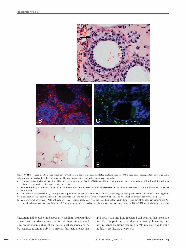
To determine whether or not this granulomatous response
was accompanied by lipid sequestration, we used the granu-
loma model we had developed previously (Geisel et al, 2005;
Rhoades et al, 2005). Polystyrene beads coated with TDM from
Mtb were suspended in Matrigel and inoculated into mice
subcutaneously. Murine cells invade thematrix and remodel the
site under the influence of the bacterial lipid. Histology of the
tissue site revealed an abundance of Adfp-expressing foam
cells clustering around the inoculated beads (Fig 8A and B).
Cryosections of TDM-induced granuloma also showed an
abundance of lipid droplets in foam cells (Fig 8C). Control
lipid phosphatidylglycerol (PG)-coated beads recruited very few
cells and did not induce foam cell formation (Fig 8D and E).
Acsl1 was also detectable in the cells recruited to the TDM-
induced granuloma; however, we did not observe detectable
levels of SapC expression (data not shown). These data indicate,
not surprisingly, that the Mtb lipid TDM is capable of inducing
some, but not all, of the features found in the human TB
granuloma.
DISCUSSION
The human TB granuloma is a complex structure that has
evolved to enable both the host and the pathogen to survive and
replicate. However, unlike many chronic infectious agents, Mtb
actually promotes the development of a robust, systemic
immune response, which it subverts at the localized tissue-
level to facilitate its prolonged survival, and, ultimately facilitate
its transmission (Russell, 2007). The TB granuloma plays a
pivotal role in TB; yet, our knowledge of what determines
containment versus active disease is superficial.
In the present study, we sought to elucidate tissue changes
that accompany granuloma progression through LCM and
microarray analysis of caseous granulomas from human TB
patients. Because of the caveats associated with the use of
uninfected lung tissue as a control, we generated both
hierarchical and comparative lists of transcript abundance;
the overall transcriptional profiles from both methods were
strikingly similar. One of the interesting themes to emerge from
the microarray data was the apparent re-alignment of lipid
metabolism. We chose to focus on the proteins ADFP, ACSL1
and SapC as three key representatives of the lipid-modulating
pathways that are impacted by the proinflammatory cytokine
TNF-a (Fig 1). ADFP is a member of PAT (perilipin, ADRP
and tail-interacting protein of 47 kDa) domain proteins and is
present on the surface and the core of intracellular lipid droplets
formed in foam cells. Its mRNA level is increased by excess
LCFAs, fasting, modified LDL, peroxisome proliferator-activated
receptor (PPAR) agonists or transcription factors that play a role
in lipid homeostasis (Dalen et al, 2006; Edvardsson et al, 2006;
� 2010 EMBO Molecular Medicine 265

Research ArticleLipid metabolism in human tuberculous granulomas
266
Gao & Serrero, 1999; Larigauderie et al, 2004; Wang et al, 1999;
Wei et al, 2005). In the current study, ADFP was detected in the
cells subtending the caseum in caseous and fibrocaseous human
pulmonary TB granulomas. ADFP was also observed in newly
caseating granulomas, mingled with nuclear debris, implying
that the protein was associated with lipid droplets that were
released by cells undergoing necrotic or apoptotic death
(Fig 3D). ACSL1 and SapC were also strongly expressed in
caseous and fibrocaseous human pulmonary TB granulomas.
ACSL1mediates the formation of fatty acyl-CoA esters from fatty
acids (preferably chain length of 12–20 carbons), which is a key
step for lipid biosynthesis and fatty acid degradation, and is also
found associated with lipid droplets (Soupene & Kuypers, 2008).
Figure 7. ADFP, ACSL1, and SapC in Mtb-infected murine and human macropha
24hr and examined by confocal microscopy.
A, B. Expression of Adfp in (A) control cells versus (B) Mtb-infected cells.
C, D. Expression of Acsl1 in (C) control and (D) Mtb-infected macrophages.
E, F. Expression of SapC in (E) control and (F) Mtb-infectedmacrophages. Blue, nuc
H37Rv for 4 days, and the antigens were detected by confocal microscope
G, H. ADFP is expressed and is associated tightly with lipid droplets (G) control
uninfected and infected human PBMC-derived macrophages.
I, J. ACSL1 is also detected in association with lipid droplets, (I) control and (J)
K, L. SapC is expressed and localizes throughout the cytoplasm (K) control and (L
� 2010 EMBO Molecular Medicine
SapC aids the degradation of glycosphingolipids into ceramide,
and sphingosine in acidic compartments, which is critical in
maintaining the turn-over of cellular components (Hannun &
Obeid, 1995; Kolter & Sandhoff, 2005; Qi et al, 1994; Schuette
et al, 2001). In addition to glycosphingolipid metabolism, SapC
is also known to transfer mycobacterial lipid antigen from
intralysosomal membrane to CD1b, thereby activating the
antigen specific T cells (Winau et al, 2004). Our hypothesis that
the TB granuloma undergoes a shift in lipid metabolism and
accumulates host-derived lipids was supported further by the
analysis of the lipid constituents of the caseous material isolated
from human pulmonary TB granulomas. Compared to normal
lung tissues, the caseum samples were enriched for CE, CHO,
ges.Murine bone marrow-derived macrophages infected with Mtb H37Rv for
lei, Green: antigens. Human PBMC-derivedmacrophages were infected byMtb
.
and (H) Mtb-infected macrophages. Lipid droplets were observed in both
Mtb-infected macrophages.
) Mtb-infected macrophages. Blue: nuclei, red: antigens, green: lipid droplets.
EMBO Mol Med 2, 258–274 www.embomolmed.org

Research ArticleMi-Jeong Kim et al.
TAG and LacCer (Fig 6A and B). This profile of lipid enrichment
is most consistent with the lipids coming from the lipid droplets
formed in macrophages that have acquired their lipids from
LDL.
The TB literature is rich with reports of lipid accumulation
in Mtb infections such as lipid-laden foam cells in human TB
lung tissues (Hunter et al, 2006b, 2007; Pagel & Pagel, 1925),
the accumulation of CE in Mtb infected murine lungs during
persistent infection (Kondo & Murohashi, 1971; Kondo et al,
1970) and the development of lipid droplets in murine or human
macrophages infected with virulent Mtb or BCG (D’Avila et al,
2006, 2008; Kondo & Kanai, 1976; Peyron et al, 2008). Lipid
droplets are attracting increased attention as dynamic organelles
that participate in numerous biological functions linked to
metabolic syndromes and diseases including atherosclerosis,
diabetes, obesity, heart attack, sepsis, Chagas’ disease and AIDS
(Bozza et al, 2009; Desruisseaux et al, 2007; Ducharme & Bickel,
2008; Murphy, 2001). The data presented in this study provide a
new hypothesis for the alterations in tissue physiology that
accompany the progression of the human TB granuloma to
caseation.
Cholesterol has long been linked to TB; however, data on
role(s) of the sterol in disease progression or control are
contradictory. The administration of CHO-rich diets to TB
patients reduced the number of bacilli in the sputum (Deniz
et al, 2007; Kozarevic et al, 1981; Perez-Guzman et al, 2005;
Taylor & Bamgboye, 1979). However, hypercholesterolemic
mice through deletion of ApoE showed increased Mtb growth
and exacerbated lung pathology (Martens et al, 2008). This is
consistent with the observations of Pandey (Pandey & Sassetti,
2008) that CHO supports the growth of persistent Mtb, and the
findings in this current study that the local tissue response to
Mtb infection is tightly associated with lipid accumulation.
Garton et al reported that Mtb bacilli isolated from sputum
samples had TAG-rich intracellular lipophilic inclusions (ILIs)
(Garton et al, 2002, 2008). Prokaryotes have the ability to
accumulate lipids (Alvarez & Steinbuchel, 2002; Waltermann
et al, 2005), and Mtb is known to synthesize and accumulate
TAG under stress, e.g. starvation and hypoxia (Daniel et al,
2004; Deb et al, 2009; Sirakova et al, 2006), and encode enzymes
to utilize the TAG (Deb et al, 2006; Mishra et al, 2008).
Therefore, the accumulated CHO and TAG in caseum of human
granulomasmay represent a convenient carbon source for either
intracellular or extracellular bacilli on the cavitary surface of TB
granulomas. LacCer is an extremely interesting glycosphingo-
lipid to be found in abundance in the caseum with respect to
its biological properties in glycosphingolipid metabolism. It is
normally present in trace amounts in mammalian cells because
it is an intermediate in the glycosphingolipids metabolism
(Kolter & Sandhoff, 2005, 2006). Its accumulation in cells could
be the result of either increased ceramide synthase activity or the
increased catabolism of complex ceramides (Chatterjee &
Pandey, 2008). The upregulation of SapCwithin the granulomas
favors the explanation that the tissue may be experiencing
LacCer excess and is trying to degrade the lipid through
promoting the activities of b-galactosidase and b-glucosidase,
the expression of which were upregulated 20-fold and 32-fold,
www.embomolmed.org EMBO Mol Med 2, 258–274
respectively, in the TB granuloma (Table 1). In addition to the
endogenous generation of LacCer, treatment of cells with LacCer
is known to reduce CHO efflux and enhances foam cell
formation (Glaros et al, 2005). Further studies need to be done
to fully verify the origin(s) of CE, CHO, TAG and LacCer that
accumulate in human TB granuloma caseum.
Lesions of Mycobacterium leprae-infected lepromatous
leprosy patients show significant accumulation of oxidized
phospholipids (Cruz et al, 2008). Intriguingly, several of the
genes reported as upregulated in our study overlap with those
observed in lepromatous leprosy lesions, including PSAP and
ADFP (Cruz et al, 2008). Another study also demonstrated the
accumulation of neutral lipids including CE and the localization
of ADFP protein in leprosy lesions (Mattos et al, 2010). These
data indicate that there are many parallels to the metabolic
reprogramming induced by bothMtb andM. leprae. The point at
which the two infection sites diverge could hinge on the
formation of the fibrous capsule in the TB granuloma, which
may focus and constrain the inflammatory response leading to
the accumulation of the sequestered lipids as caseum. This
pathology is unique to Mtb infection and precedes the lique-
faction and cavitation of the lesion. In mice, this late-stage
pathology was absent from experimental Mtb infections, which
has led many researchers to be concerned about the validity of
the mouse as an experimental model. Recently, however,
Hunter and colleagues reported caseum formation in reactiva-
tion disease in antibiotic-treated mice (Hunter et al, 2007).
Significantly, these murine caseous lesions exhibited a highly
developed fibrous capsule comparable to that observed in
fibrocaseous human granulomas.
We also have shown thatMtb infection ofmacrophages in vitro
induces foam cell formation, and that inoculation of beads coated
with the bacterial cell wall lipid TDM into mice caused the
clustering of Adfp-expressing foam cells around the particles.
These data argue strongly that lipid sequestration is a pathogen-
induced response. We have demonstrated previously that
mycobacterial cell wall lipid-containing vesicles, which contain
TDM, are released from infected macrophages and spread the
influence of the bacterium to neighbouring cells (Beatty et al,
2000; Rhoades et al, 2003; Russell et al, 2002). TDM activates
macrophages by signalling through TLR2, which forms a
functional complex with the scavenger receptor MARCO to bind
the mycobacterial lipid (Bowdish et al, 2009). This amplification
of the response from infected cells to uninfected bystander cells
could explain the extreme tissue reaction observed in human TB
granulomas when the bacterial density is known to be relatively
low (data not shown) (Ulrichs et al, 2005).
The current study provides a new framework for appreciating
the physiological responses to Mtb infection that accompany the
progression of TB granulomas to caseation and cavitation, and
ultimately disease transmission. We hypothesize that bacterial
components, such as TDM, stimulate the host’s innate immune
response to enhance synthesis and/or sequestration of host
lipids in the form of lipid droplets. The foamy macrophages
accumulate in the macrophage-rich centre of the granuloma,
where ultimately they die through either apoptosis or necrosis.
This leads to the buildup of lipids as caseum, and, finally to
� 2010 EMBO Molecular Medicine 267
Research Article
Figure 8. TDM-coated beads induce foam cell formation in mice in an experimental granuloma model. TDM-coated beads resuspended in Matrigel were
subcutaneously injected to wild-type mice and the granulomas were excised at day6 post-inoculation.
A. Histological examination demonstrated the extensive recruitment of cells to TDM-coated beads, many of which had the appearance of lipid droplet-filled foam
cells (A representative cell is marked with an arrow).
B. Immunohistology on the consecutive section of the same tissue block revealed a strong expression of lipid droplet-associated protein, Adfp (nuclei in blue and
Adfp in red).
C. Lipid droplets were detected by staining neutral lipids with Nile Red on cryosections from TDM-induced granuloma (nuclei in blue and neutral lipids in green).
D. In contrast, control lipid PG-coated beads demonstrated considerably reduced recruitment of cells and no induction of foam cell formation (H&E).
E. Moreover, probing with anti-Adfp antibody on the consecutive section cut from the same tissue block as (D) did not label any of the cells surrounding the PG-
coated beads (nuclei in blue and Adfp in red). The experiments were repeated three times, and threemice were used for PG- or TDM-Matrigel mixture injection.
268
cavitation and release of infectious Mtb bacilli (Fig 9). Our data
argue that the development of novel therapeutics should
encompass manipulation of the host’s local response and not
be restricted to antimicrobials. Targeting fatty acid metabolism,
� 2010 EMBO Molecular Medicine
lipid deposition and lipid-mediated cell death in host cells are
unlikely to impact on bacterial growth directly, however, they
may influence the tissue response to Mtb infection and thereby
modulate TB disease progression.
EMBO Mol Med 2, 258–274 www.embomolmed.org

Research ArticleMi-Jeong Kim et al.
Figure 9. A model illustrating the linkages between Mtb-infection, foam cell formation and accumulation of caseum in the human TB granuloma.
A. Intracellular Mtb bacilli synthesize and release cell wall components inside their host cells. We have demonstrated previously that these lipids accumulate in
the internal vesicles in multi-vesicular bodies, which are exocytosed from the cell in vesicular form.
B. Because of the release of these vesicles, both infected and uninfected macrophages are exposed to cell wall mycolates and induced to form foamy
macrophages, as illustrated in Fig 7. The foamy macrophages have been shown to support the maintenance and growth of persistent bacteria.
C. We now propose that these cells die via an inflammatory, necrotic process and release their lipid droplets into the extracellular milieu within the granuloma. As
a result of the fibrotic capsule, the human granuloma is an enclosed, isolated structure with minimal vasculature. The enclosed nature of the human
granuloma leads to accumulation of necrotic debris as caseum. In this model, this process is an integral part of the pathology that leads to active disease and
transmission.
MATERIALS AND METHODS
Human subjects
Human subjects were included in this study according to protocols
approved by Institutional Review Boards at University of Cape Town,
South Africa, the Public Health Research Institute, Newark, NJ and
Cornell University, Ithaca, NY. Informed consent was obtained from all
of patients.
Animals
C57BL/6J mice were obtained from the Jackson Laboratory and housed
under specific-pathogen-free conditions. All experiments were approved
by Cornell University Institutional Animal Care and Use Committee.
Tissue specimens
Human TB lung tissues were surgically excised from TB patients who
had extensive lung cavitation and tissue degeneration, that showed
little response to the antibiotics. Snap-frozen tissues were transported
from Groote Schuur Hospital of Cape Town to our biosafety level-3
(BL3) laboratory. Frozen samples were embedded in OCT compound
(Tissue-Tek1), cut into 10mm-thick sections at 258C on a Cryocut
1800 cryostat (Leica Microsystems) and attached onto PET-membrane
frame slides (Leica Microsystems) for LCM or Superfrost Plus Micro
slides (Erie Scientific) for histological examination.
www.embomolmed.org EMBO Mol Med 2, 258–274
Histology of human tissues
Human TB lung tissues embedded in paraffin were cut at 3mm
thickness and stained with hematoxylin & eosin (H&E) for histological
examination. Cryosections of human TB lung tissues were fixed in 4%
paraformaldehyde in PBS for 30min and stained with H&E, and then
paired to PET-membrane slides prepared for LCM. For the detection of
neutral lipids, fixed cryosections were stained with Oil Red O (Alfa
Aesar) and counterstained with hematoxylin.
Laser capture microdissection
Tissue sections mounted onto PET-membrane slides were fixed in
graded ethanol (70, 75, 96 and 100%) containing 0.5% sodium azide
for 30 s per each step. Slides were air dried briefly, and areas of interest
were dissected by using Leica AS LMD system (Leica Microsystems).
Microarray
Total RNA was isolated from LCM-derived materials using PicoPureTM
RNA Isolation Kit (Arcturus) and treated with DNase using TURBO DNA-
freeTM (Ambion). Purity, quality and quantity of RNA were assessed by
the Agilent NanoChip Bioanalyzer assay, spectrophotometer and reverse-
transcription PCR (RT-PCR) of b-actin (234bp), glyceraldehyde-3-
phosphate dehydrogenase (GAPDH, 189bp), 18S rRNA (236bp) and
ubiquitin (198bp). To obtain sufficient quantities of RNA for microarray,
two-rounds of linear amplification of RNA were conducted using
RiboAmp HSTM Amplification Kit (Arcturus) and BioArray High YieldTM
� 2010 EMBO Molecular Medicine 269

Research ArticleLipid metabolism in human tuberculous granulomas
270
RNA Transcript Labeling Kit (T7) (Enzo Life Sciences, Inc.). Biotinylated,
amplified antisense RNA was fragmented and hybridized onto
GeneChip1 Human X3P Array (Affymetrix), and the chip was scanned
by GeneArray 300 scanner (Affymetrix).We performed three independent
arrays on one LCM-derived granuloma sample, which confirmed the
reliability of the technique. Thereafter, two other granulomas were
further analyzed by microarray for biological replicates.
Microarray data analysis
For the generation of a hierarchical list of absolute gene expressions in
caseous human TB lung granulomas, raw CEL files were analyzed at the
probe level using R and Bioconductor. Only perfect match (PM) probes
were included in the estimation for expression level of probe sets. Raw
PM intensities from each array were subjected to local background
correction using MAS5 method, log2-transformed and median-centered.
Informative PM probes from each probe set were selected as having the
highest mean across all arrays. In this study, the mean signal of top 5
informative PM probes of each probe set was used to represent its
expression level. For a list of genes differentially expressed between
caseous human pulmonary TB granulomas and normal lung parench-
yma, raw CHP files were summarized by MAS5 method and scaled to
median of all samples, and the baseline was transformed to the median
of all samples, by using GeneSpring GX 10.0.2 program (Agilent
Technologies). The networks were generated through the use of
Ingenuity Pathway Analysis (Ingenuity1 Systems, www.ingenuity.com).
Statistical analysis
The statistical significance of differences in gene expressions between
normal human lung tissues and caseous human TB lung granulomas was
calculated using an unpaired t-test with Benjamini–Hochberg correction.
Results with a P-value less than 0.05 were considered significant.
Culture and in vitro infection of macrophages
Bone marrow cells were isolated from femurs and tibias of 6- to 8-
week-old female C57BL/6 mice and cultured in Dulbecco’s Modified
Eagle Medium (DMEM, Gibco) supplemented with 20% L-929 cell-
conditioned medium, 10% heat-inactivated foetal calf serum (Hy-
clone), 2mM L-glutamine (Gibco), 1mM sodium pyruvate (Gibco) and
100U/ml penicillin-100mg/ml streptomycin (Gibco) at 378C, 5% CO2
and 90% humidity for 5 days. BMMF were incubated with Mtb
H37Rv at a multiplicity of infection 20:1 (bacteria per macrophage)
for 2 h, washed with PBS and cultured DMEM medium without
antibiotics.
Monocytes were isolated from human blood (Zen-Bio Inc.) by
gradient separation. Briefly, blood was mixed with an equal volume of
PBS, and 25ml of this mixture was laid over 12ml of Histopaque1-
10771 (Sigma, density¼1.0771 g/ml). The leukocyte-containing layer
was harvested, washed and cultured in DMEM medium as in BMMF
culture, but without L-929 cell-conditioned medium. Differentiated
human PBMC-derived macrophages were incubated with Mtb H37Rv
at a MOI 10:1 (bacteria per MF) for 2 h, washed with PBS and
cultured in DMEM medium without antibiotics.
Quantitative real-time RT-PCR
Infected and uninfected macrophages were lysed with TRIZOL1
Reagent (Invitrogen) and homogenized by passing cell lysate through
� 2010 EMBO Molecular Medicine
a pipette. The homogenized samples were mixed with 0.2 volume of
chloroform per 1 volume of TRIZOL1 Reagent, and phases were
separated by centrifugation. The RNA-containing aqueous phase was
column-purified using RNEasy mini kit (Qiagen). RNA samples were
treated with DNase using TURBO DNA-freeTM (Ambion). The cDNA was
synthesized using iScriptTM cDNA Synthesis Kit (BIO-RAD) and used for
real-time PCR using iTaqTM SYBR1 Green Supermix with ROX (BIO-RAD)
in 7500 Fast Real-Time PCR System (Applied Biosystems). Each PCR was
done in triplicate, and PCR conditions are as follows: initial activation at
958C for 2min and 40 cycles of denaturation at 958C for 30 s and
annealing and extension at 538C for 45 s. Forward and reverse primers
designed by using Primer3 are shown in Table SIII (Rozen & Skaletsky,
2000). The calculated threshold cycle (Ct) value for each gene was
normalized to Ct of house-keeping gene 18S rRNA, and then the fold
induction of gene of interest between control MF and Mtb-infected
MF was calculated and presented by using the formula, 2�DDCt .
Confocal microscopy of macrophages
Cells were fixed in 4% paraformaldehyde in PBS for 30min, washed and
blocked with 5% bovine serum albumin (Sigma), 1.5% glycine (VWR
International) and 0.1% saponin in PBS at room temperature for 1 h.
Cells were incubated with guinea pig polyclonal antibody against
murine Adfp or mouse monoclonal antibody against human ADFP
(1:100, RDI division of Fitzgerald Industries Intl.), rabbit polyclonal
antibody against human ACSL1 (1:200, GenWay Biotech) or rabbit
polyclonal antibody against human SapC (1:50, Santa Cruz Biotechnol-
ogy) at 378C for 1h. After washing with PBS, cells were incubated with
Texas Red1-conjugated antibodies against guinea pig, mouse or rabbit
(1:250, Jackson ImmunoResearch) and BODIPY1 493/503 (1mg/ml,
Invitrogen) for lipid droplets, at room temperature for 1 h. After wash
with PBS, the nuclei were stained with 5mg/ml of bisbenzimide (Sigma)
prior to mounting the cells in ProLong Gold Antifade Reagent (Molecular
Probes1). Cells without primary antibodies were included as a control.
Cells were examined by Leica TCS SP5 confocal laser scanning
microscope (Leica Microsystems) using a 63� oil immersion lens.
Immunofluorescence microscopy of human TB lung tissues
Paraffin-embedded sections of 3mm thickness were heat-fixed,
deparaffinized and rehydrated. Epitopes were heat-retrieved in a
pressure cooker with 1mM EDTA buffer pH 8.0, and tissue sections
were blocked with 0.2% gelatin in PBS with 0.05% Tween-20 (PBS-T)
at room temperature for 1 h. Primary antibodies, mouse monoclonal
antibody against human ADFP (1:100, RDI division of Fitzgerald
Industrial Intl.), rabbit polyclonal antibody against human ACSL1
(1:150, GenWay Biotech) or rabbit polyclonal antibody against human
SapC (1:50, Santa Cruz Biotechnology) were applied to tissue sections
at 378C for 1 h. After washing h BS-T, sections were incubated with 1%
Sudan Black B (Sigma) in 7% ethanol at room temperature for 10min.
Sections were washed with PBS-T and incubated with andCy3TM-goat
antibody against mouse or rabbit (1:250, Jackson ImmunoResearch) at
room temperature for 45min. After PBS-T wash, nuclei were stained
with 1mg/ml of DAPI (Invitrogen) and the tissue sections were
mounted in ProLong Gold Antifade Reagent, prior to examination.
Images were captured by Zeiss Axioskop 40 epifluorescence micro-
scope with AxioVision 4.3 software (Carl Zeiss MicroImaging). Further
image analysis was done by AxioVision LE software (Carl Zeiss
MicroImaging).
EMBO Mol Med 2, 258–274 www.embomolmed.org
Research ArticleMi-Jeong Kim et al.
The paper explained
PROBLEM:
Tuberculosis has a serious impact on global health with a
disproportionate impact on low-income countries. It is estimated
that one-third of the planet is infected with its causative agent,
M. tuberculosis, yet only 5–15% of these individuals will go on to
develop disease. The balance between containment of the
infection and the progression to active disease is determined at
the site of infection, the granuloma. Despite the significance of
this issue, little is known about the factors that regulate this
process.
RESULTS:
We use human whole genome array analysis of granulomas from
TB patients to demonstrate that progression of the infection in
the granuloma is accompanied by dysregulation of host lipid
metabolism. Furthermore, M. tuberculosis induces lipid seques-
tration and the formation of foamy macrophages (loaded with
lipid droplets) both in vitro and in vivo. The accumulation of the
caseum appears to result from the death of these foamy
macrophages. The expansion of the necrotic, caseous centre of
the granuloma is significant both for the progression of the
disease and as a potential carbon source for bacterial growth.
IMPACT:
These findings enhance our understanding of the factors, both
host- and pathogen derived, that impact progression of the
infection from containment to active disease.
Lipid analysis
Frozen human TB lung tissues or normal lung tissues were thawed, and
caseum or normal tissue segments were isolated and transferred into
glass vials (Wheaton Science) and then mixed with chloroform:metha-
nol (2:1, v/v). After sonication at 508C for 1h, the extracts were filtered
through 0.2mm-pore size PTFE membrane (GD/X1 syringe filter,
Whatman) and dried down using nitrogen. TLC plates (aluminium-
backed Silica gel 60 plates, EMD) were pre-run in chloroform:methanol
(90:10, v/v) to remove contaminants from silica gel followed by heat-
activation at 1108C for 30min. Total lipids were developed either in a
dual solvent system of chloroform:methanol:water (65:25:4, v/v/v) and
petroleum ether:ethyl ether:acetic acid (70:30:2, v/v/v), or in a single
solvent system of chloroform:methanol:water (65:25:4, v/v/v). Plates
were sprayed with 50% sulfuric acid in ethanol and charred. The lipid
standards used were cholesteryl oleate, TAG, cardiolipin and phospha-
tidylcholine (Sigma), CHO (MP Biomedicals), sphingomyelin (Avanti
Polar Lipids) and neutral glycosphingolipid qualmix (Matreya). Caseous
and fibrocaseous granuloma samples were derived from seven
independent tissue samples, and normal lung tissues were from the
unaffected lung area of seven independent tissue samples.
Mass-spectrometry
Structural analysis of lipids including SM, LacCer, CE and TAG were
carried out by a Finnigan TSQ-7000 triple stage quadrupole mass
spectrometer (Thermo Electron Corporation, CA) equipped with a
Finnigan ESI source and controlled by Finnigan ICIS software operated
on a DEC alpha workstation. Lipid extract dissolved in methanol/
chloroform (2:1, v/v) were continuously infused into the ESI source
with a Harvard syringe pump at a flow rate of 1ml/min. The skimmer
was at ground potential, and the electrospray needle was at 4.5 kV.
The heated capillary temperature was 2508C. The collisionally
activated dissociation (CAD) tandem mass spectra were obtained
under collision energy from 30 to 45 eV, and argon (2.3mTorr) was
used as target gas. The product ion spectra were acquired in the profile
mode at the scan rate of one scan per 3 s.
www.embomolmed.org EMBO Mol Med 2, 258–274
Structural characterization of CH was conducted by Agilent 5973
MSD GC/MS system operated by ChemStation software. Lipid extract
in methanol/chloroform (2:1, v/v) was injected into a Hewett-Packard
6890 GC in a splitless mode and analyzed with a 15m DB-1701
column (0.25mm i.d, 0.25mm film thickness; J & W Scientific). The
initial temperature of GC was set at 1008C for 1min, increased to
2208C at a rate of 508C/min, then to 2908C at a rate of 108C/min and
maintained at 2908C for another 10min. The temperatures of the
injector, transfer line of the GC column and of the ion-source were set
at 280, 280 and 2308C, respectively. The full scan mass spectra (50–
500Da) were acquired at a rate of 1 scan/0.25 s.
In vivo murine granuloma model
Mycobacterial lipid-bearing beads were prepared as described
previously, with slight modifications (Geisel et al, 2005; Rhoades et
al, 2005). Briefly, 9.25�103 of 80mm polystyrene microspheres (Duke
Scientific) were coated with 15mg of Mtb-derived TDM (Sigma) or
control lipid PG (Sigma) by alternative cycles of vortexing and
sonication. The TDM- or PG-coated beads were resuspended in
300ml of ice-cold growth-factor reduced Matrigel (BD). The mixture
was injected sub-dermally into the skin fold at the scruff of the neck.
Granulomas were excised at 6 days post-inoculation. Three or four
mice were used for the injection of TDM- or PG-Matrigel mixture. The
experiment was repeated more than three times.
Histology and immunohistology of murine granulomas
Granulomas were fixed in 4% paraformaldehyde in PBS at 48C for
over 24 h and transferred to 70% ethanol. Sections were stained with
H&E for histological examination. For immunostaining, paraffin-
embedded sections were heat-fixed, deparaffinized and rehydrated.
Epitopes were heat-retrieved in 1mM EDTA buffer pH 8.0, and tissue
sections were blocked with 5% BSA in PBS at room temperature for
1 h. Guinea pig polyclonal antibody against murine Adfp (1:200, RDI
division of Fitzgerald Industrial Intl.) or rabbit polyclonal antibody
against human ACSL1 (1:150, GenWay Biotech) was applied to
� 2010 EMBO Molecular Medicine 271

Research ArticleLipid metabolism in human tuberculous granulomas
272
sections at 378C for 1 h. After washing with PBS three times, sections
were incubated with 1% Sudan Black B (Sigma) in 70% ethanol at
room temperature for 10min. Sections were washed with PBS three
times and incubated with Texas Red1-donkey antibody against
guinea pig IgG (Hþ L) (1:250, Jackson ImmunoResearch) or Texas
Red1-goat antibody against rabbit IgG (Hþ L) (1:250, Jackson
ImmunoResearch) at room temperature for 45min. After three
washes with PBS, nuclei were stained with 5mg/ml of bisbenzimide
(Sigma) in PBS at room temperature for 5min. After thorough wash
with PBS, sections were mounted in ProLong Gold Antifade Reagent
(Molecular Probes1). Sections without primary antibody were
included as a negative control. Images were captured by Zeiss Axio
Imager with AxioVision LE software (Carl Zeiss MicroImaging).
Cryosections were cut at 10mm thickness and fixed in 4%
paraformaldehyde in PBS for 20min. The sections were stained with
Nile Red (1mg/ml, Sigma) for neutral lipids and with bisbenzimide for
nuclei. Mounted sections were visualized by Zeiss Axio Imager with
AxioVision LE software (Carl Zeiss MicroImaging).
Author ContributionsM-JK, GK and DGR planned the experiments. M-JK, GK and
DGR devised the concept. M-JK, F-FH and UW performed the
experiments. GBW performed the surgery. HCW, ML, L-GB,
GBW, CD, AV and LT generated and processed samples and
performed pathology on the tissues. M-JK and WW performed
microarray data analysis. M-JK, GK and DGR wrote and edited
the manuscript.
AcknowledgementsThis work was supported by funds from the National Institutes
of Health HL055936 to DGR and AI054338 to GK.
Supporting information is available at EMBO Molecular
Medicine online.
The authors declare that they have no conflict of interest.
For more information
GEO Database; Human TB granuloma array data:
http://www.ncbi.nlm.nih.gov/geo/query/acc.cgi?token=nnslpmyesqeiqb-
m&acc=GSE20050
OMIM Online Mendelian Inheritance in Man:
Adipophilin (PLIN2)
http://www.ncbi.nlm.nih.gov/omim/103195
ACSL1
http://www.ncbi.nlm.nih.gov/omim/152425
PSAP
http://www.ncbi.nlm.nih.gov/omim/176801
Author Websites:
Linda-Gail Bekker and the Desmond Tutu HIV Center
http://www.iidmm.uct.ac.za/bekkerwood/index.htm
Gilla Kaplan
http://www.phri.org/research/res_pikaplan.asp
David Russell
http://www.vet.cornell.edu/microbiology/faculty/Russell/
� 2010 EMBO Molecular Medicine
ReferencesActor JK, Olsen M, Hunter RL, Jr., Geng YJ (2001) Dysregulated response to
mycobacterial cord factor trehalose-6,60-dimycolate in CD1D�/� mice.
J Interferon Cytokine Res 21: 1089-1096
Alvarez HM, Steinbuchel A (2002) Triacylglycerols in prokaryotic
microorganisms. Appl Microbiol Biotechnol 60: 367-376
Beatty WL, Rhoades ER, Ullrich HJ, Chatterjee D, Heuser JE, Russell DG (2000)
Trafficking and release of mycobacterial lipids from infected macrophages.
Traffic 1: 235-247
Beatty WL, Ullrich HJ, Russell DG (2001) Mycobacterial surface moieties are
released from infected macrophages by a constitutive exocytic event. Eur J
Cell Biol 80: 31-40
Bickel PE, Tansey JT, Welte MA (2009) PAT proteins, an ancient family of lipid
droplet proteins that regulate cellular lipid stores. Biochim Biophys Acta
1791: 419-440
Bild AH, Yao G, Chang JT, Wang Q, Potti A, Chasse D, Joshi MB, Harpole D,
Lancaster JM, Berchuck A, et al (2006) Oncogenic pathway signatures in
human cancers as a guide to targeted therapies. Nature 439: 353-357
Bowdish DM, Sakamoto K, Kim MJ, Kroos M, Mukhopadhyay S, Leifer CA,
Tryggvason K, Gordon S, Russell DG (2009) MARCO, TLR2, and CD14 are
required for macrophage cytokine responses to mycobacterial trehalose
dimycolate and Mycobacterium tuberculosis. PLoS Pathog 5: e1000474
Bozza PT, Magalhaes KG, Weller PF (2009) Leukocyte lipid bodies—biogenesis
and functions in inflammation. Biochim Biophys Acta 1791: 540-551
Caldwell G (1919) Chemical changes in tuberculous tissues. J Infect Dis 24: 81-
113
Chang BH, Chan L (2007) Regulation of triglyceride metabolism. III. Emerging
role of lipid droplet protein ADFP in health and disease. Am J Physiol
Gastrointest Liver Physiol 292: G1465-G1468
Chatterjee S, Pandey A (2008) The Yin and Yang of lactosylceramide
metabolism: implications in cell function. Biochim Biophys Acta 1780: 370-
382
Chatterjee SB, Dey S, Shi WY, Thomas K, Hutchins GM (1997) Accumulation of
glycosphingolipids in human atherosclerotic plaque and unaffected aorta
tissues. Glycobiology 7: 57-65
Cruz D, Watson AD, Miller CS, Montoya D, Ochoa MT, Sieling PA, Gutierrez MA,
Navab M, Reddy ST, Witztum JL, et al (2008) Host-derived oxidized
phospholipids and HDL regulate innate immunity in human leprosy. J Clin
Invest 118: 2917-2928
Dalen KT, Ulven SM, Arntsen BM, Solaas K, Nebb HI (2006) PPARalpha
activators and fasting induce the expression of adipose differentiation-
related protein in liver. J Lipid Res 47: 931-943
Daniel J, Deb C, Dubey VS, Sirakova TD, Abomoelak B, Morbidoni HR,
Kolattukudy PE (2004) Induction of a novel class of diacylglycerol
acyltransferases and triacylglycerol accumulation in Mycobacterium
tuberculosis as it goes into a dormancy-like state in culture. J Bacteriol 186:
5017-5030
Dannenberg AM Jr (1994) Roles of cytotoxic delayed-type hypersensitivity and
macrophage-activating cell-mediated immunity in the pathogenesis of
tuberculosis. Immunobiology 191: 461-473
Dannenberg AM, Jr, Sugimoto M (1976) Liquefaction of caseous foci in
tuberculosis. Am Rev Respir Dis 113: 257-259
D’Avila H, Melo RC, Parreira GG, Werneck-Barroso E, Castro-Faria-Neto HC,
Bozza PT (2006) Mycobacterium bovis bacillus Calmette-Guerin induces
TLR2-mediated formation of lipid bodies: intracellular domains for
eicosanoid synthesis in vivo. J Immunol 176: 3087-3097
D’Avila H, Roque NR, Cardoso RM, Castro-Faria-Neto HC, Melo RC, Bozza PT
(2008) Neutrophils recruited to the site of Mycobacterium bovis BCG
infection undergo apoptosis and modulate lipid body biogenesis and
prostaglandin E production by macrophages. Cell Microbiol 10: 2589-
2604
Deb C, Daniel J, Sirakova TD, Abomoelak B, Dubey VS, Kolattukudy PE (2006) A
novel lipase belonging to the hormone-sensitive lipase family induced
under starvation to utilize stored triacylglycerol in Mycobacterium
tuberculosis. J Biol Chem 281: 3866-3875
EMBO Mol Med 2, 258–274 www.embomolmed.org

Research ArticleMi-Jeong Kim et al.
Deb C, Lee CM, Dubey VS, Daniel J, Abomoelak B, Sirakova TD, Pawar S, Rogers
L, Kolattukudy PE (2009) A novel in vitro multiple-stress dormancy model
for Mycobacterium tuberculosis generates a lipid-loaded, drug-tolerant,
dormant pathogen. PLoS ONE 4: e6077
Deniz O, Gumus S, Yaman H, Ciftci F, Ors F, Cakir E, Tozkoparan E, Bilgic H, Ekiz
K (2007) Serum total cholesterol, HDL-C and LDL-C concentrations
significantly correlate with the radiological extent of disease and the degree
of smear positivity in patients with pulmonary tuberculosis. Clin Biochem
40: 162-166
Desruisseaux MS, Nagajyothi, Trujillo ME, Tanowitz HB, Scherer PE (2007)
Adipocyte, adipose tissue, and infectious disease. Infect Immun 75: 1066-
1078
Ducharme NA, Bickel PE (2008) Lipid droplets in lipogenesis and lipolysis.
Endocrinology 149: 942-949
Dye C, Bassili A, Bierrenbach AL, Broekmans JF, Chadha VK, Glaziou P, Gopi PG,
Hosseini M, Kim SJ, Manissero D, et al (2008) Measuring tuberculosis
burden, trends, and the impact of control programmes. Lancet Infect Dis 8:
233-243
Edvardsson U, Ljungberg A, Linden D, William-Olsson L, Peilot-Sjogren H,
Ahnmark A, Oscarsson J (2006) PPARalpha activation increases triglyceride
mass and adipose differentiation-related protein in hepatocytes. J Lipid Res
47: 329-340
Gao J, Serrero G (1999) Adipose differentiation related protein (ADRP)
expressed in transfected COS-7 cells selectively stimulates long chain fatty
acid uptake. J Biol Chem 274: 16825-16830
Garner B, Mellor HR, Butters TD, Dwek RA, Platt FM (2002) Modulation of THP-
1 macrophage and cholesterol-loaded foam cell apolipoprotein E levels by
glycosphingolipids. Biochem Biophys Res Commun 290: 1361-1367
Garton NJ, Christensen H, Minnikin DE, Adegbola RA, Barer MR (2002)
Intracellular lipophilic inclusions of mycobacteria in vitro and in sputum.
Microbiology 148: 2951-2958
Garton NJ, Waddell SJ, Sherratt AL, Lee SM, Smith RJ, Senner C, Hinds J,
Rajakumar K, Adegbola RA, Besra GS, et al (2008) Cytological and transcript
analyses reveal fat and lazy persister-like bacilli in tuberculous sputum.
PLoS Med 5: e75
Geisel RE, Sakamoto K, Russell DG, Rhoades ER (2005) In vivo activity of
released cell wall lipids of Mycobacterium bovis bacillus Calmette-Guerin is
due principally to trehalose mycolates. J Immunol 174: 5007-5015
Glaros EN, Kim WS, Quinn CM, Wong J, Gelissen I, Jessup W, Garner B (2005)
Glycosphingolipid accumulation inhibits cholesterol efflux via the ABCA1/
apolipoprotein A-I pathway: 1-phenyl-2-decanoylamino-3-morpholino-1-
propanol is a novel cholesterol efflux accelerator. J Biol Chem 280: 24515-
24523
Grassi M, Bocchino M, Marruchella A, Volpe E, Saltini C, Colizzi V, Mariani F
(2006) Transcriptional profile of the immune response in the lungs of
patients with active tuberculosis. Clin Immunol 121: 100-107
Hannun YA, Obeid LM (1995) Ceramide: an intracellular signal for apoptosis.
Trends Biochem Sci 20: 73-77
Harzer K, Hiraiwa M, Paton BC (2001) Saposins (sap) A and C activate the
degradation of galactosylsphingosine. FEBS Lett 508: 107-110
Hunter RL, Olsen M, Jagannath C, Actor JK (2006a) Trehalose 6,60-dimycolate
and lipid in the pathogenesis of caseating granulomas of tuberculosis in
mice. Am J Pathol 168: 1249-1261
Hunter RL, Olsen MR, Jagannath C, Actor JK (2006b) Multiple roles of cord
factor in the pathogenesis of primary, secondary, and cavitary tuberculosis,
including a revised description of the pathology of secondary disease. Ann
Clin Lab Sci 36: 371-386
Hunter RL, Jagannath C, Actor JK (2007) Pathology of postprimary tuberculosis
in humans and mice: contradiction of long-held beliefs. Tuberculosis
(Edinb) 87: 267-278
Karakousis PC, Yoshimatsu T, Lamichhane G, Woolwine SC, Nuermberger EL,
Grosset J, Bishai WR (2004) Dormancy phenotype displayed by extracellular
Mycobacterium tuberculosis within artificial granulomas in mice. J Exp Med
200: 647-657
www.embomolmed.org EMBO Mol Med 2, 258–274
Kim DK, Park GM, Hwang YI, Kim HJ, Han SK, Shim YS, Yim JJ (2006) Microarray
analysis of gene expression associated with extrapulmonary dissemination
of tuberculosis. Respirology 11: 557-565
Kolter T, Sandhoff K (2005) Principles of lysosomal membrane digestion:
stimulation of sphingolipid degradation by sphingolipid activator proteins
and anionic lysosomal lipids. Annu Rev Cell Dev Biol 21: 81-103
Kolter T, Sandhoff K (2006) Sphingolipid metabolism diseases. Biochim
Biophys Acta 1758: 2057-2079
Kondo E, Kanai K (1976) Accumulation of cholesterol esters in macrophages
incubated with mycobacteria in vitro. Jpn J Med Sci Biol 29: 123-
137
Kondo E, Murohashi T (1971) Esterification of tissue cholesterol with fatty
acids in the lungs of tuberculous mice. Jpn J Med Sci Biol 24: 345-
356
Kondo E, Kanai K, Nishimura K, Tsumita T (1970) Analysis of host-originated
lipids associated with ‘‘in vivo grown tubercle bacilli’’. Jpn J Med Sci Biol 23:
315-326
Korf J, Stoltz A, Verschoor J, De Baetselier P, Grooten J (2005) The
Mycobacterium tuberculosis cell wall component mycolic acid elicits
pathogen-associated host innate immune responses. Eur J Immunol 35:
890-900
Kozarevic D, McGee D, Vojvodic N, Gordon T, Racic Z, ZukelW, Dawber T (1981)
Serum cholesterol and mortality: the Yugoslavia Cardiovascular Disease
Study. Am J Epidemiol 114: 21-28
Larigauderie G, Furman C, Jaye M, Lasselin C, Copin C, Fruchart JC, Castro G,
Rouis M (2004) Adipophilin enhances lipid accumulation and prevents lipid
efflux from THP-1 macrophages: potential role in atherogenesis.
Arterioscler Thromb Vasc Biol 24: 504-510
Ma XJ, Dahiya S, Richardson E, Erlander M, Sgroi DC (2009) Gene expression
profiling of the tumor microenvironment during breast cancer progression.
Breast Cancer Res 11: R7
Martens GW, Arikan MC, Lee J, Ren F, Vallerskog T, Kornfeld H (2008)
Hypercholesterolemia impairs immunity to tuberculosis. Infect Immun 76:
3464-3472
Mattos KA, D’Avila H, Rodrigues LS, Oliveira VG, Sarno EN, Atella GC, Pereira
GM, Bozza PT, Pessolani MC (2010) Lipid droplet formation in leprosy: toll-
like receptor-regulated organelles involved in eicosanoid formation and
Mycobacterium leprae pathogenesis. J Leukoc Biol 87: 371-384
Mishra KC, de Chastellier C, Narayana Y, Bifani P, Brown AK, Besra GS, Katoch
VM, Joshi B, Balaji KN, Kremer L (2008) Functional role of the PE domain and
immunogenicity of the Mycobacterium tuberculosis triacylglycerol
hydrolase LipY. Infect Immun 76: 127-140
Murphy DJ (2001) The biogenesis and functions of lipid bodies in animals,
plants and microorganisms. Prog Lipid Res 40: 325-438
Pagel W, Pagel M (1925) Zur Histochemie der Lungentuberkulose, mit
besonderer Berucksichtigung der Fettsubstanzen und Lipoide. Virchows
Archiv 256: 629-640
Pandey AK, Sassetti CM (2008) Mycobacterial persistence requires the
utilization of host cholesterol. Proc Natl Acad Sci USA 105: 4376-4380
Parkes HA, Preston E, Wilks D, Ballesteros M, Carpenter L, Wood L, Kraegen EW,
Furler SM, Cooney GJ (2006) Overexpression of acyl-CoA synthetase-1
increases lipid deposition in hepatic (HepG2) cells and rodent liver in vivo.
Am J Physiol Endocrinol Metab 291: E737-744
Perez-Guzman C, Vargas MH, Quinonez F, Bazavilvazo N, Aguilar A (2005) A
cholesterol-rich diet accelerates bacteriologic sterilization in pulmonary
tuberculosis. Chest 127: 643-651
Peyron P, Vaubourgeix J, Poquet Y, Levillain F, Botanch C, Bardou F, Daffe M,
Emile JF, Marchou B, Cardona PJ, et al (2008) Foamy macrophages from
tuberculous patients’ granulomas constitute a nutrient-rich reservoir forM.
tuberculosis persistence. PLoS Pathog 4: e1000204
Puissegur MP, Botanch C, Duteyrat JL, Delsol G, Caratero C, Altare F (2004) An
in vitro dual model of mycobacterial granulomas to investigate the
molecular interactions between mycobacteria and human host cells. Cell
Microbiol 6: 423-433
� 2010 EMBO Molecular Medicine 273
Research ArticleLipid metabolism in human tuberculous granulomas
274
Qi X, Leonova T, Grabowski GA (1994) Functional human saposins expressed in
Escherichia coli. Evidence for binding and activation properties of saposins C
with acid beta-glucosidase. J Biol Chem 269: 16746-16753
Ragno S, RomanoM, Howell S, Pappin DJ, Jenner PJ, ColstonMJ (2001) Changes
in gene expression in macrophages infected with Mycobacterium
tuberculosis: a combined transcriptomic and proteomic approach.
Immunology 104: 99-108
Rhoades E, Hsu F, Torrelles JB, Turk J, Chatterjee D, Russell DG (2003)
Identification and macrophage-activating activity of glycolipids released
from intracellular Mycobacterium bovis BCG. Mol Microbiol 48: 875-
888
Rhoades ER, Geisel RE, Butcher BA, McDonough S, Russell DG (2005) Cell wall
lipids from Mycobacterium bovis BCG are inflammatory when inoculated
within a gel matrix: characterization of a new model of the granulomatous
response to mycobacterial components. Tuberculosis (Edinb) 85: 159-
176
Robenek H, Robenek MJ, Buers I, Lorkowski S, Hofnagel O, Troyer D, Severs NJ
(2005) Lipid droplets gain PAT family proteins by interaction with
specialized plasma membrane domains. J Biol Chem 280: 26330-26338
Rozen S, Skaletsky H (2000) Primer3 on the WWW for general users and for
biologist programmers. Methods Mol Biol 132: 365-386
Russell DG (2007) Who puts the tubercle in tuberculosis? Nat Rev Microbiol 5:
39-47
Russell DG, Mwandumba HC, Rhoades EE (2002) Mycobacterium and the coat
of many lipids. J Cell Biol 158: 421-426
Russell DG, Cardona PJ, Kim MJ, Allain S, Altare F (2009) Foamy macrophages
and the progression of the human tuberculosis granuloma. Nat Immunol
10: 943-948
Schuette CG, Pierstorff B, Huettler S, Sandhoff K (2001) Sphingolipid activator
proteins: proteins with complex functions in lipid degradation and skin
biogenesis. Glycobiology 11: 81R-90R
Sirakova TD, Dubey VS, Deb C, Daniel J, Korotkova TA, Abomoelak B,
Kolattukudy PE (2006) Identification of a diacylglycerol acyltransferase
gene involved in accumulation of triacylglycerol in Mycobacterium
tuberculosis under stress. Microbiology 152: 2717-2725
� 2010 EMBO Molecular Medicine
Soupene E, Kuypers FA (2008) Mammalian long-chain acyl-CoA synthetases.
Exp Biol Med (Maywood) 233: 507-521
Taylor GO, Bamgboye AE (1979) Serum cholesterol and diseases in Nigerians.
Am J Clin Nutr 32: 2540-2545
Tuomisto TT, Yla-Herttuala S (2005) What have we learnt about microarray
analyses of atherogenesis? Curr Opin Lipidol 16: 201-205
Ulrichs T, Kaufmann SH (2006) New insights into the function of granulomas
in human tuberculosis. J Pathol 208: 261-269
Ulrichs T, Lefmann M, Reich M, Morawietz L, Roth A, Brinkmann V, Kosmiadi
GA, Seiler P, Aichele P, Hahn H, et al (2005) Modified immunohistological
staining allows detection of Ziehl-Neelsen-negative Mycobacterium
tuberculosis organisms and their precise localization in human tissue.
J Pathol 205: 633-640
Volpe E, Cappelli G, Grassi M, Martino A, Serafino A, Colizzi V, Sanarico N,
Mariani F (2006) Gene expression profiling of human macrophages at late
time of infection with Mycobacterium tuberculosis. Immunology 118: 449-
460
Waltermann M, Hinz A, Robenek H, Troyer D, Reichelt R, Malkus U, Galla HJ,
Kalscheuer R, Stoveken T, von Landenberg P, et al (2005) Mechanism of
lipid-body formation in prokaryotes: how bacteria fatten up.Mol Microbiol
55: 750-763
Wang X, Reape TJ, Li X, Rayner K, Webb CL, Burnand KG, Lysko PG (1999)
Induced expression of adipophilin mRNA in human macrophages
stimulated with oxidized low-density lipoprotein and in atherosclerotic
lesions. FEBS Lett 462: 145-150
Wei P, Taniguchi S, Sakai Y, Imamura M, Inoguchi T, Nawata H, Oda S,
Nakabeppu Y, Nishimura J, Ikuyama S (2005) Expression of adipose
differentiation-related protein (ADRP) is conjointly regulated by PU.1 and
AP-1 in macrophages. J Biochem 138: 399-412
Winau F, Schwierzeck V, Hurwitz R, Remmel N, Sieling PA, Modlin RL, Porcelli
SA, Brinkmann V, Sugita M, Sandhoff K, et al (2004) Saposin C is required for
lipid presentation by human CD1b. Nat Immunol 5: 169-174
Zhu G, Xiao H, Mohan VP, Tanaka K, Tyagi S, Tsen F, Salgame P, Chan J (2003)
Gene expression in the tuberculous granuloma: analysis by laser capture
microdissection and real-time PCR. Cell Microbiol 5: 445-453
EMBO Mol Med 2, 258–274 www.embomolmed.org