cathepsin b-mediated activation of trypsinogen in ... · inflammatory pathways whereby components...
TRANSCRIPT
Gastroenterology 2018;154:704–718
BASICAND
TRANSLATIONALPANCREAS
Cathepsin B-Mediated Activation of Trypsinogen in EndocytosingMacrophages Increases Severity of Pancreatitis in Mice
Matthias Sendler,1 Frank-Ulrich Weiss,1 Janine Golchert,2 Georg Homuth,2Cindy van den Brandt,1 Ujjwal M. Mahajan,6 Lars-Ivo Partecke,3 Paula Döring,4 Ilya Gukovsky,5
Anna S. Gukovskaya,5 Preshit R. Wagh,1 Markus M. Lerch,1,§ and Julia Mayerle1,6,§
1Department of Medicine A, University Medicine Greifswald, Greifswald, Germany; 2Interfaculty Institute for Genetics andFunctional Genomics, University Medicine Greifswald, Greifswald, Germany; 3Department of Surgery, University MedicineGreifswald, Greifswald, Germany; 4Institute of Pathology, University Medicine Greifswald, Greifswald, Germany; 5VA GreaterLos Angeles Healthcare System; David Geffen School of Medicine, University of California at Los Angeles, California; and6Medizinische Klinik und Poliklinik II, Universitätsklinikum der Ludwig-Maximilians-Universität, Klinikum Grosshadern,Munich, Germany
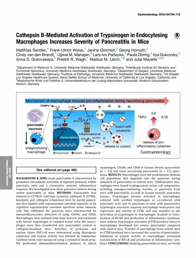
phagocytosingmacrophage
MyD88
TLR IL1βR
pro-IL1β
IL1β
caspase 1
ASC NLRP3
trypsinogen trypsin cathepsin B
DAMPs
phagocytosis
fusion with lysosome
release to cytosol
inflammasome
release of pro-inflammatory cytokines
TNFαIL6
IL1β
transcriptionNFκB
DAMPsMechanism of macrophage induced pro-inflammation during necrotising pancreatitis
pancreatic necrosis
NFκB activation
See editorial on page 482.
BACKGROUND & AIMS: Acute pancreatitis is characterized bypremature intracellular activation of digestive proteases withinpancreatic acini and a consecutive systemic inflammatoryresponse. We investigated how these processes interact duringsevere pancreatitis in mice. METHODS: Pancreatitis wasinduced in C57Bl/6 wild-type (control), cathepsin B (CTSB)-knockout, and cathepsin L-knockout mice by partial pancre-atic duct ligation with supramaximal caerulein injection, or byrepetitive supramaximal caerulein injections alone. Immunecells that infiltrated the pancreas were characterized byimmunofluorescence detection of Ly6g, CD206, and CD68.Macrophages were isolated from bone marrow and incubatedwith bovine trypsinogen or isolated acinar cells; the macro-phages were then transferred into pancreatitis control orcathepsin-knockout mice. Activities of proteases andnuclear factor (NF)-kB were determined using fluorogenicsubstrates and trypsin activity was blocked by nafamostat.Cytokine levels were measured using a cytometric bead array.We performed immunohistochemical analyses to detect
trypsinogen, CD206, and CD68 in human chronic pancreatitis(n ¼ 13) and acute necrotizing pancreatitis (n ¼ 15) speci-mens. RESULTS: Macrophages were the predominant immunecell population that migrated into the pancreas duringinduction of pancreatitis in control mice. CD68-positive mac-rophages were found to phagocytose acinar cell components,including zymogen-containing vesicles, in pancreata frommice with pancreatitis, as well as human necrotic pancreatictissues. Trypsinogen became activated in macrophagescultured with purified trypsinogen or co-cultured withpancreatic acini and in pancreata of mice with pancreatitis;trypsinogen activation required macrophage endocytosis andexpression and activity of CTSB, and was sensitive to pH.Activation of trypsinogen in macrophages resulted in trans-location of NF-kB and production of inflammatory cytokines;mice without trypsinogen activation (CTSB-knockout mice) inmacrophages developed less severe pancreatitis comparedwith control mice. Transfer of macrophage from control miceto CTSB-knockout mice increased the severity of pancreatitis.Inhibition of trypsin activity in macrophages preventedtranslocation of NF-kB and production of inflammatory cyto-kines. CONCLUSIONS: Studying pancreatitis in mice, we found

EDITOR’S NOTES
BACKGROUND AND CONTEXT
Necrotizing acute pancreatitis induces an overwhelmingsystemic inflammatory response (SIRS), which ismediated by infiltrating phagocytosing macrophages.Persistent SIRS results in multiorgan failure.
NEW FINDINGS
Pancreatic protease activation, a hallmark of acutepancreatitis, is not restricted to acinar cells butphagocytosing macrophages are able to intracellularlyactivate trypsinogen to trypsin in a cathepsin Bdependent manner. Active trypsin inside macrophagesacts as DAMP and fuels systemic inflammation.
LIMITATIONS
Macrophage mediated protease activation is limited to asevere model of acute necrotising pancreatitis.
IMPACT
Intrapancreatic protease activation within macrophages isdirectly linked to NFkB activation and the systemicinflammatory response determining severity of thedisease. This allows therapeutic targeting of two causalsmechanism in pancreatitis.
February 2018 Protease-driven Macrophage Activation 705
CREA
S
activation of digestive proteases to occur not only in acinarcells but also in macrophages that infiltrate pancreatic tissue.Activation of the proteases in macrophage occurs duringendocytosis of zymogen-containing vesicles, and depends onpH and CTSB. This process involves macrophage activation viaNF-kB-translocation, and contributes to systemic inflamma-tion and severity of pancreatitis.
Keywords: Pancreatic Inflammation; Mechanisms; ImmuneResponse; Mouse Model.
cute pancreatitis is common and of increasing1
§ Authors share co-senior authorship.
Abbreviations used in this paper: ATP, adenosine triphosphate; BMDM,bone marrow-derived macrophages; CCK, cholecystokinin; CTSB,cathepsin B; CTSL, cathepsin L; DAMP, damage-associated molecularpattern; IL, interleukin; LPS, lipopolysaccharide; MCP-1, monocytechemoattractant protein-1; NKkBp65, nuclear factor kB p65; NLRP3,nucleotide-binding oligomerization domain-like receptor family pyrindomain-containing 3; TNFa, tumor necrosis factor a; WT, wild-type.
Most current article
© 2018 by the AGA Institute. Published by Elsevier Inc. This is an openaccess article under the CC BY-NC-ND license (http://creativecommons.
org/licenses/by-nc-nd/4.0/).0016-5085
https://doi.org/10.1053/j.gastro.2017.10.018
BASICAN
DTR
ANSLAT
IONA
LPA
N
Aincidence in Western countries. The majority ofcases suffer from a mild form of the disease but approxi-mately 20% of patients develop severe pancreatitis, asso-ciated with pancreatic necrosis, systemic inflammation, andorgan failure.2 An overwhelming systemic immune reaction,the so-called systemic inflammatory response syndrome,accounts for persistent multiorgan failure and seems to beresponsible for the majority of systemic complications andmortality.3 The triggering events of this immune reactionand the pathophysiological mechanism that determineseverity of the disease are still poorly understood.4Self-digestion by its own proteases is considered to be atrigger of pancreatitis. Under pathologic conditions, thedigestive serine protease trypsinogen is converted toactive trypsin by the lysosomal hydrolase cathepsin B(CTSB) within the acinar cells.5–7 Premature intracellularprotease activation is then followed by acinar cell death,which is accompanied by a pro-inflammatory response andleads to a prominent translocation of leukocytes. Especiallycells of the innate immune system migrate into the injuredorgan.8 These cells are the first cells that reach the
pancreas.9,10 This immune reaction boosts local damageand results in an increase in severity of disease.10,11 Theimportance of the protease/anti-protease balance for thedevelopment of pancreatitis is supported by the observa-tion that germline mutations that increase the suscepti-bility toward developing pancreatitis mostly affect genes ofthe protease/anti-protease system, such as cationic-trypsinogen (PRSS1), chymotrypsin-C (CTRC) or thetrypsin-inhibitor SPINK1.12–16 How the intracellular acti-vation of digestive proteases is linked to local and systemicinflammatory reaction is still being debated. On the onehand, acinar cells under experimental conditions activatenuclear factor kB,17 a key transcription factor for the pro-inflammatory response, but still do so in animals in whichT7-trypsinogen, a major mouse-isoform of trypsinogen, hasbeen deleted.18 On the other hand, acinar cell that undergoautodigestion and necrosis release damage-associatedmolecular patterns (DAMPs) such as free DNA, histones,or free adenosine triphosphate (ATP), which are recog-nized by immune cell receptors inducing a pro-inflammatory reaction and activation of the inflamma-some pathway.19 The main task of infiltrating immune cellsin the pancreas is thought to be defensive and includes theremoval of cellular debris and necrosis. Most of theclearance is achieved through the action of macrophagesthat not only phagocytose cellular debris,20 but whoseinvolvement in the subsequent immune reaction is pro-foundly influenced by what cellular components they areexposed to and which ones they ingest.21 For example, theirclearance of apoptotic cellular waste involves largely non-inflammatory pathways whereby components of necroticcells can trigger a pro-inflammatory response.21 In the presentstudy we found that, in areas of pancreatic necrosis, macro-phages ingest zymogen-containing vesicles from damagedacinar cells that they convert these zymogens to active pro-teases in a CTSB-dependent manner and that the intra-macrophage activation of trypsinogen is an important driverof local and systemic inflammation and disease severity.
Material and MethodsIsolation of Pancreatic Acini
Acini were isolated from mouse pancreas by collagenasedigestion under sterile conditions, as previously reported.6 Fordetails see Supplementary Materials and Methods.
706 Sendler et al Gastroenterology Vol. 154, No. 3
BASICAND
TRANSLATIONALPANCREAS
Isolation of Bone Marrow-derived MacrophagesBone marrow-derived macrophages (BMDM) were isolated
from the femur and tibia of mice. A detailed protocol is pro-vided in the Supplementary Materials.
Biochemical AssaysSerum amylase and different protease activities were
measured as previously reported using substrates R110-Ile-Pro-Arg for trypsin, Suc-Ala-Ala-Pro-Phe-AMC for chymo-trypsin, R110-Phe-Arg for CTSL, and AMC-Arg2 for CTSB.
22 Fordetails on in vivo imaging of proteases see SupplementaryMaterials.
Induction of Pancreatitis in MiceAll animal experiments were performed after prior
approval by the Institutional Animal Care committee. C57Bl/6mice were obtained from Charles River (Sulzfeld, Germany),CTSB-/-, cathepsin L (CTSL)-/-, and nucleotide-binding oligo-merization domain-like receptor family pyrin domain-containing 3 (NLRP3)-/- mice were maintained in our animalfacility; all mice strains are bred with a C57Bl/6 background.
Histology, Immunohistochemistry, andImmunofluorescence
Paraffin sections were used for H&E staining and Masson-Goldner-trichrome staining as previously reported.23 Fordetails see Supplementary Materials.
Human Pancreatic SamplesHuman chronic pancreatitis tissue was collected in the
context of the ChroPac trial (ISRCTN38973832; http://www.isrctn.com/ISRCTN38973832).24 Necrotic tissue was har-vested during endoscopic necrosectomy under the ethicscommittee approval for the ProZyt trial.
Statistical AnalysisAll data are expressed as means ± standard error of the
mean from at least 5 animals or experiments. Statistical anal-ysis was performed using SigmaPlot and SigmaStat. Unpaired2-tailed Student’s t-test or Rank-Sum test were used. Differ-ences were considered significant for P < .05.
ResultsMigration of Macrophages in Acute Pancreatitis
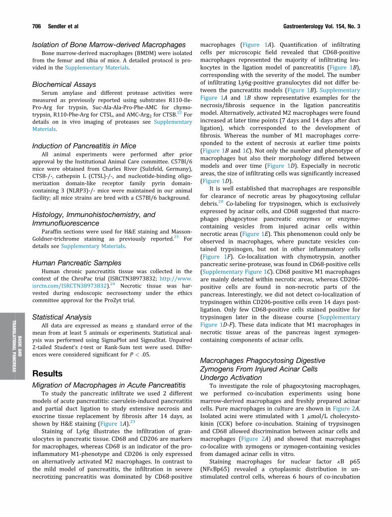
To study the pancreatic infiltrate we used 2 differentmodels of acute pancreatitis: caerulein-induced pancreatitisand partial duct ligation to study extensive necrosis andexocrine tissue replacement by fibrosis after 14 days, asshown by H&E staining (Figure 1A).23
Staining of Ly6g illustrates the infiltration of gran-ulocytes in pancreatic tissue. CD68 and CD206 are markersfor macrophages, whereas CD68 is an indicator of the pro-inflammatory M1-phenotype and CD206 is only expressedon alternatively activated M2 macrophages. In contrast tothe mild model of pancreatitis, the infiltration in severenecrotizing pancreatitis was dominated by CD68-positive
macrophages (Figure 1A). Quantification of infiltratingcells per microscopic field revealed that CD68-positivemacrophages represented the majority of infiltrating leu-kocytes in the ligation model of pancreatitis (Figure 1B),corresponding with the severity of the model. The numberof infiltrating Ly6g-positive granulocytes did not differ be-tween the pancreatitis models (Figure 1B). SupplementaryFigure 1A and 1B show representative examples for thenecrosis/fibrosis sequence in the ligation pancreatitismodel. Alternatively, activated M2 macrophages were foundincreased at later time points (7 days and 14 days after ductligation), which corresponded to the development offibrosis. Whereas the number of M1 macrophages corre-sponded to the extent of necrosis at earlier time points(Figure 1B and 1C). Not only the number and phenotype ofmacrophages but also their morphology differed betweenmodels and over time (Figure 1D). Especially in necroticareas, the size of infiltrating cells was significantly increased(Figure 1D).
It is well established that macrophages are responsiblefor clearance of necrotic areas by phagocytosing cellulardebris.20 Co-labeling for trypsinogen, which is exclusivelyexpressed by acinar cells, and CD68 suggested that macro-phages phagocytose pancreatic enzymes or enzyme-containing vesicles from injured acinar cells withinnecrotic areas (Figure 1E). This phenomenon could only beobserved in macrophages, where punctate vesicles con-tained trypsinogen, but not in other inflammatory cells(Figure 1F). Co-localization with chymotrypsin, anotherpancreatic serine-protease, was found in CD68-positive cells(Supplementary Figure 1C). CD68 positive M1 macrophagesare mainly detected within necrotic areas, whereas CD206-positive cells are found in non-necrotic parts of thepancreas. Interestingly, we did not detect co-localization oftrypsinogen within CD206-positive cells even 14 days post-ligation. Only few CD68-positive cells stained positive fortrypsinogen later in the disease course (SupplementaryFigure 1D-F). These data indicate that M1 macrophages innecrotic tissue areas of the pancreas ingest zymogen-containing components of acinar cells.
Macrophages Phagocytosing DigestiveZymogens From Injured Acinar CellsUndergo Activation
To investigate the role of phagocytosing macrophages,we performed co-incubation experiments using bonemarrow-derived macrophages and freshly prepared acinarcells. Pure macrophages in culture are shown in Figure 2A.Isolated acini were stimulated with 1 mmol/L cholecysto-kinin (CCK) before co-incubation. Staining of trypsinogenand CD68 allowed discrimination between acinar cells andmacrophages (Figure 2A) and showed that macrophagesco-localize with zymogens or zymogen-containing vesiclesfrom damaged acinar cells in vitro.
Staining macrophages for nuclear factor kB p65(NFkBp65) revealed a cytoplasmic distribution in un-stimulated control cells, whereas 6 hours of co-incubation

Figure 1. Severe acute pancreatitis was induced by duct ligation and supramaximal caerulein stimulation. Infiltrating immunecells of the innate immune system were stained in pancreatic tissue (A) and quantified by cell counting (B). Time course andamount of neutrophil infiltration (Ly6g) and M1 macrophage (CD68) infiltration in the severe model of acute pancreatitis wascompared with the mild form of the disease. H&E staining of the pancreas illustrating the severity of disease. The inflammatoryinfiltrate correlates with the amount of necrotic areas and the decrease of unaffected healthy exocrine tissue (C). The numberof M2 macrophages (CD206) rises with increasing fibrosis (C). Three days after duct ligation the shape and volume of mac-rophages is significantly altered and increased in CD68-positive macrophages (D). Large CD68-positive macrophages displayco-localization with pancreatic trypsinogen (E), indicating phagocytosis of dying acini or cellular waste. Neutrophils did notshow any co-localization with trypsinogen (F). Asterisks (*) indicate significant differences with P < .05 (n ¼ 5–8).
February 2018 Protease-driven Macrophage Activation 707
BASICAN
DTR
ANSLAT
IONA
LPA
NCRE
AS
with CCK-stimulated acini induced a translocation ofNFkBp65 into the nucleus of CD68-positive macrophages(Figure 2B). In the same setting, co-incubation of macro-phages with CCK-stimulated acini resulted in a significantincrease of cytokine secretion, here shown for the pro-inflammatory cytokines interleukin-6 (IL6) and tumor ne-crosis factor a (TNFa), as well as the chemokine monocytechemoattractant protein-1 (MCP-1) (Figure 2C). Lipopoly-saccharide (LPS)-stimulated macrophages served as con-trols. Acini were also able to secrete cytokines, but themajority of cytokine release originated from macrophages(Figure 2C). When morphology of macrophages wasanalyzed for cell size (Figure 2D), co-incubation with aciniin vitro resulted in a significant change in macrophagemorphology, resembling the results of tissue sections(Figure 1D). Macrophages developed a spreading phenotypeand changed their morphology in forward side scatter on
fluorescence-activated cell sorter analysis (Figure 2E).Macrophages are activated upon co-incubation with acini orisolated zymogen granules (Figure 2F), which are engulfedby macrophages (Supplementary Figure 2A-C). These dataindicate that co-incubation of macrophages with acinar cellsresults in activation of macrophages and a differentiation toa pro-inflammatory M1 phenotype.
Macrophages Phagocytose Zymogen-containingVesicles and Activate Trypsinogen Intracellularlyin a CTSB-dependent Manner
The detection of trypsin activity within phagocytosingmacrophages raised the question whether activated trypsinwas engulfed or whether macrophages activate trypsinogenintracellularly. We therefore isolated macrophages frombone marrow of C57Bl/6 mice and co-incubated them with

Figure 2. BMDM were co-incubated with CCK-stimulated acini. Staining of CD68 marked macrophages, whereas trypsinogenwas used as a marker for acini (A). Phagocytosis of acinar cell compartments is demonstrated by intra-macrophage tryp-sinogen staining (arrow). Six hours after co-incubation the transcription factor NFkBp65 showed nuclear translocation inmacrophages compared with untreated controls (B). Cytokine secretion of macrophages was significantly increased after 6hours of co-incubation, as seen by secretion IL6, TNFa, and MCP-1. Acini alone showed a significantly lower secretion ofthese cytokines (C). Treatment of macrophages with LPS served as control. Furthermore, macrophage morphology changedduring co-incubation (D-E). CD69, an early activation marker belonging to the C-type lectin domain family, is increased onmacrophages co-incubated with acini or isolated zymogen granules (F). Asterisks (*) indicate significant differences withP < .05 (n ¼ 6); # indicate a significant difference to all other conditions.
708 Sendler et al Gastroenterology Vol. 154, No. 3
BASICAND
TRANSLATIONALPANCREAS
bovine trypsinogen in a concentration of 10 mg/mL (seeSupplementary Figure 2D). Labelling for trypsinogen andCD68 indicated that trypsinogen is internalized by macro-phages (Figure 3A) and then converted to active trypsinintracellularly (Figure 3B). Intracellular trypsin activity inmacrophages could be blocked completely by 50 mmol/Lnafamostate, a potent digestive serine protease inhibitor, bybafilomycin-A1, an inhibitor of V-ATPases that neutralizesthe usually acidic intravesicular pH, and by CA074me, a cellpermeant CTSB inhibitor. Both substances led to a pro-nounced reduction in CTSB and trypsin activity in macro-phages (Figure 3B). Furthermore, in vivo imaging of isolatedmacrophages co-incubated with trypsinogen revealed activetrypsin inside macrophages (Figure 3C). Final evidence thattrypsinogen undergoes activation in a CTSB-dependentmanner within phagocytosing macrophages came fromexperiments using CTSB- and CTSL-deleted mousemacrophages incubated with bovine trypsinogen. Trypsinactivity was significantly reduced in macrophages fromCTSB-/- mice while it was increased in macrophages fromCTSL-/- mice (Figure 3D), which is in accordance with thecounteracting role of CTSL to CTSB.25 Immunofluorescencestaining for CTSB and trypsinogen in macrophages incu-bated with bovine trypsinogen revealed a punctate
localization of both enzymes in a vesicular compartment ofmacrophages (Figure 3E). These experiments show thatmacrophages activate phagocytosed trypsinogenintracellularly.
Co-incubation of BMDM with freshly prepared and CCK-stimulated acini (rather than trypsinogen) again resulted inintracellular localization of trypsinogen as well as amylasewithin macrophages. In addition, a co-localization of CTSBwith trypsinogen was observed (Figure 4A). Intracellularlocalization of protease activity showed a membrane-confined punctate localization for active trypsin and activechymotrypsin (Figure 4B). Furthermore, fluorescent-labeledzymogen granules showed trypsin activity visualized byR110-Ile-Pro-Arg cleavage (Supplementary Figure 2A).Western blotting of macrophage lysates confirmed theintracellular presence of trypsinogen and amylase(Figure 4C). Activity of trypsin in macrophages after co-incubation with acini was significantly increased. Evenwhen macrophages were co-incubated with acini isolatedfrom CTSB-/- mice, we observed a significant increase intrypsin activity (Figure 4D), indicating that macrophageCTSB, rather than acinar cell CTSB, drives trypsin activation.When we used macrophages isolated from CTSB-/- mice wefound no increase in trypsin activity after co-incubation

Figure 3. BMDM were co-incubated with bovine trypsinogen over a time period of 6 hours. Immunofluorescence staining oftrypsinogen revealed an intracellular membrane confined punctiform localization within CD68-positive macrophages after 6hours (A). Activity measurements in cell lysates of macrophages proved a significant increase of trypsin activity in macro-phages co-incubated with trypsinogen. Nafamostate, CA074me, or bafilomycin-A1 and lysosomal acidification were able tocompletely block trypsin activity (B), but only CA074me and bafilomycin-A1 caused a significant decrease in CTSB activity (B).Visual detection of active trypsin by the fluorogenic substrate R110-CBZ-Ile-Pro-Arg demonstrated intracellular located trypsinactivity within macrophages (C). Comparison of WT macrophages with CTSB or CTSL-deficient macrophages showedsignificantly decreased trypsin activity in CTSB-deleted macrophages and increased activity in CTSL-deleted cells (D).Immunofluorescence staining indicates co-localization of phagocytosed trypsinogen and macrophage CTSB (E). Asterisks (#)indicate significant differences with P < .05 (n ¼ 4–6).
February 2018 Protease-driven Macrophage Activation 709
BASICAN
DTR
ANSLAT
IONA
LPA
NCRE
AS
with acini from wild-type (WT) or CTSB-/- mice. Bothnafamostate and CA074me blocked trypsin activity after co-incubation (Figure 4E), indicating that intracellular trypsinactivation in macrophages is entirely dependent on thepresence and activity of CTSB. Amylase activity wasmeasured in macrophages as a marker of phagocytosis ofacinar cell components. Uptake was not impaired by nafa-mostate pretreatment, whereas pretreatment of macro-phages with CA074me resulted in an increase inintracellular amylase activity (Figure 4E), indicating thatinhibition of CTSB leads to impaired degradation of amylase.To investigate whether phagocytosis is the mechanism ofuptake for pancreatic zymogens, we used cytochalasin-B toblock phagocytosis by inhibition of the network formation ofactin filaments.26 Cytochalasin-B greatly reduced trypsin,chymotrypsin, and amylase activity in macrophages exposedto either bovine trypsinogen or acinar cells (Figure 4F). The
activity of CTSB was not affected by cytochalasin-B treat-ment (Figure 4F).
Blockade of Digestive Protease Activation WithinMacrophages Reduces Pro-inflammatorySignaling and Cytokine Secretion
Macrophages engulf cellular debris from injured aciniand incorporate pancreatic zymogens and zymogen-containing vesicles. Phagocytosed zymogens, especiallytrypsinogen, undergo activation in a CTSB-dependentmanner within macrophages. We therefore investigatedthe effect of this intracellular protease activation onmacrophages.
Translocation of NFkBp65 into the nucleus indicatespro-inflammatory differentiation of macrophages (M1).Co-incubation of BMDM with CCK-stimulated acini resulted

710 Sendler et al Gastroenterology Vol. 154, No. 3
BASICAND
TRANSLATIONALPANCREAS

February 2018 Protease-driven Macrophage Activation 711
in a nuclear redistribution of NFkBp65. This translocationwas blocked by nafamostate (Figure 5A), indicating thattrypsin activity is involved in this process. Untreated mac-rophages served as negative controls while LPS treatmentserved as positive controls (Figure 5A). Both nafamostateand bafilomycin-A1 abolished intracellular trypsin activitywithout affecting phagocytosis, as shown by an unimpairedamylase uptake (Figure 5B). Cytokine release of macro-phages co-incubated with acini was determined by cyto-metric bead array. Treatment with nafamostate significantlyreduced secretion of pro-inflammatory cytokines such asIL6, MCP-1, and TNFa. Treatment with bafilomycin-A1 had asimilar effect, with the exception of TNFa (Figure 5C). Theeffect of bafilomycin-A1 on TNFa secretion in macrophageshas been described previously.27 Comparing the cytokinerelease of macrophages from WT mice with CTSB-/- cells,we found significantly reduced IL6, TNFa, and MCP-1secretion after co-incubation with acini (Figure 5D). Thisis not a generalized defect because CTSB-/- macrophagesresponded to LPS adequately and in the same manner asWT macrophages (Supplementary Figure 2E). Tran-scriptome analysis of macrophages was performed 6 hoursafter co-incubation with acini. Transcriptional alteration ofNFkB-related genes was affected by pre-treatment withnafamostate, blocking intracellular trypsin activity andproving a trypsin-mediated effect on NFkB-signaling. Inparticular, Nfkb2 and Rela were significantly decreased(Student’s t-test of fold change) (Figure 5E and 5F).
These data suggest that the pro-inflammatory phenotypeof macrophages in pancreatitis is dependent on CTSB-mediated trypsinogen activation within phagocytosingmacrophages.
BASICAN
DTR
ANSLAT
IONA
LPA
NCRE
AS
Protease Activation Within MacrophagesActs as DAMP and Induces NLRP3Inflammasome Activation
Pathway analysis of macrophage transcriptome data byIngenuity Pathway Analysis shows a significant down-regulation of the toll-like receptor pathways, as well asthe NFkB pathway, after co-incubation with acini andtrypsin inhibition by nafamostate (Figure 6A). MyD88 is theupstream regulator (P ¼ 2.0E-6) that is affected most.Detailed analysis of the NFkB pathway suggests a criticalrole of IL1b for the activation of NFkB (Supplementary
=Figure 4. BMDM were co-incubated with freshly prepared acini swith acini led to intracellular localization of trypsinogen and panmore, co-localization of trypsinogen and CTSB was observed ((green fluorescent ± DAPI [40,6-diamidino-2-phenylindole]) or cpunctiform localization of active pancreatic enzymes within mapositive for trypsinogen when co-incubated with acini or trypsinoband for pancreatic amylase (C). Macrophages from WT animincubated with acini, even with acini of CTSB-/- mice, in contno increase in trypsin activity if co-incubated with WT or CTSB-dcompletely abolished trypsin activity. Similarly, inhibition of CTSmacrophages but the uptake of acinar cell proteins, as demonstror CA074me (E). Treatment of macrophages with cytochalastrypsinogen, resulting in decreased trypsin activity and decreasactivity was not affected by cytochalasin-B (F). Asterisks (*) ind
Figure 3). Secretion and maturation of IL1b depends oninflammasome activation and caspase-1.19,28 Immunohisto-chemistry of caspase-1 shows expression within infiltratinginflammatory cells but not in pancreatic acini 3 days afterduct ligation (Figure 6B). Secretion of mature IL1b frommacrophages in response to acini proves in vitro activationof the inflammasome; in contrast, CCK-stimulated acini didnot secret IL1b. Blockade of macrophage cathepsins byCa074me or E64d abolished IL1b secretion (Figure 6C).Genetic deletion of NLRP3, an essential part of the inflam-masome complex, did not affect macrophage phagocytosisand intracellular protease activation upon co-incubationwith WT acini (Figure 6D). NFkB translocation into thenucleus (Figure 6E), as well as cytokine secretion of IL6,TNFa, and MCP-1, was reduced in the same way as aftertreatment with nafamostate (Figure 6F).
Protease activation within macrophages results in IL1bmaturation and secretion, which enhances the pro-inflammatory macrophage phenotype by acting on theIL1b receptor expressed on macrophages in an autocrineloop (Supplementary Figure 2C).
Adoptive Transfer of WT Macrophages RestoresTrypsinogen Activation During SevereNecrotizing Pancreatitis in CTSB-/- Mice
As previously reported, significant numbers of macro-phages migrate into the pancreas during the early phase ofpancreatitis, and these infiltrating cells express considerableconcentrations of CTSB (Supplementary Figure 4A). In fact,CTSB is the second most abundant protein in macrophages(data not shown). This would make it plausible that mac-rophages also contribute to intrapancreatic trypsinogenactivation in vivo. We therefore induced severe pancreatitisin WT- and cathepsin B-deleted animals. Two days afterduct ligation we performed an adoptive transfer of BDMBgenerated from WT or CTSB-/- mice (Figure 7A) andsacrificed animals 24 hours after adoptive transfer of mac-rophages. Staining for CTSB indicated the presence of CTSBin all cells of WT animals; however, in WT animals receivingCTSB-/- macrophage transfer, we detected CTSB-negativecells in the pancreas. In contrast, in CTSB-/- mice nosignal for CTSB was detected and only CTSB-/- mice thathad received WT macrophages displayed some CTSB- pos-itive infiltrating cells in the pancreas (Figure 7B). We
timulated with 1 mmol/L CCK. Co-incubation of macrophagescreatic amylase in CD68-positive macrophages (A). Further-A). Live-cell imaging with fluorochrome substrates for trypsinhymotrypsin (blue fluorescent) demonstrated an intracellularcrophages (B). Western blotting of macrophage lysates staingen, but only macrophages co-incubated with acini showed aals revealed significantly increased trypsin activity when co-rast to macrophages of CTSB-deleted mice, which showedeleted acini (D). Inhibition of serine proteases by nafamostateB activity by CA074me reduced intracellular trypsin activity inated by amylase activity, was not reduced by the nafamostatein-B reduced the uptake of acinar cell proteins as well ased amylase or chymotrypsin activity in macrophages. CTSBicate significant differences with P < .05 (n ¼ 4–6).

Figure 5. BMDM were co-incubated with CCK-stimulated acini for 6 hours. Immunofluorescence staining of NFkBp65 showedtranslocation to the nucleus of LPS stimulated macrophages compared with untreated controls. Co-incubation ofmacrophages and acini resulted in nuclear translocation of NFkBp65, while treatment with nafamostate prohibited nucleartranslocation (A). Treatment with nafamostate as well as bafilomycin-A1 inhibited intracellular trypsin activity in macrophages,but did not affect phagocytosis, as demonstrated by amylase activity (B). After 6 hours of co-incubation, the cytokine release inthe supernatant was measured (C). Blockade of protease activation with nafamostate or bafilomycin-A1 significantly reducedIL6-, MCP-1, and TNFa release. The same effect was observed in CTSB-deleted macrophages compared with WT cells (D).Transcriptome analysis showed increased expression of NFkB-related genes in macrophages after co-incubation with acini,which is reduced upon nafamostate treatment for Nfkb2 and Rela (E-F). Asterisks (*) indicate significant differences withP < .05 (n ¼ 4–6); # indicate a significant difference of untreated cells compared with treatment.
712 Sendler et al Gastroenterology Vol. 154, No. 3
BASICAND
TRANSLATIONALPANCREAS
observed a prominent infiltration of F4/80-positive macro-phages in all animals, independently of the adoptive trans-fer. H&E histology showed pancreatic damage and necrosisin all animals, but to a significantly lesser degree in CTSB-deficient animals (Figure 7B).
CTSB activity in pancreatic homogenates was completelysuppressed in CTSB-/- animals and only in animals that hadreceived WT macrophages did we observed an increase inCTSB activity during pancreatitis (Figure 7C). Untreatedanimals without duct ligation pancreatitis served as control.In pancreatic homogenates from CTSB-/- mice, we detectedonly traces of active trypsin and even after induction ofpancreatitis we did not detect a significant increase intrypsin activity when CTSB-/- macrophages were adop-tively transferred. Only in animals that had received WTmacrophages we found a significant increase in trypsinactivity in pancreatic tissue homogenates (Figure 7C). Inline with these results, in CTSB-/- animals adoptive transferof WT macrophages significantly increased trypsinogen
activation in WT animals compared with mice that hadreceived CTSB-/- macrophages. These data confirm ourfindings from the in vitro studies that CTSB-containingmacrophages contribute to intrapancreatic trypsinogenactivation in the course of severe acute pancreatitis in vivo.The time course of trypsinogen activation in the ductligation model showed elevated trypsin activity over 14days, and we observed trypsinogen phagocytosing macro-phages at all time points (Supplementary Figure 5).Macrophage-derived protease activation depended on thepresence of necrosis. A much greater effect on diseaseseverity and systemic inflammation was observed inCTSB-/- animals 3 days after duct ligation (SupplementaryFigure 4B and 4C). The differences between CTSB-/- andCTSBþ/þ animals were more pronounced in the severemodel of pancreatitis compared with the mild caerulein-induced disease model.5 In a genetic model of chronicpancreatitis in Lamp2-deficient mice,29 we observedenlarged CD68-positive macrophages within the pancreas in

Figure 6. Pathway analysis of transcriptome data was performed by using Ingenuity Pathway Analysis software. Majorpathways that were induced or repressed in macrophages co-incubated with acini ± nafamostate reveal down-regulation ofthe toll-like receptor pathway, as well as the NFkB-pathway (A). The major inducer of the NFkB pathway via MyD88 was IL1b.54
IL-1bmaturation depends on caspase-1, which is exclusively expressed in infiltrating inflammatory cells and not in acini duringpancreatitis (B). In vitro assay of BMDM revealed secretion of IL1b into the medium 6 hours after co-incubation with acini, whiletreatment with 50 mmol/L CA074me or 20 mmol/L E64d abolished IL1b release. Acini by themselves were not able to releasemature IL1b (C). BMDM from NLRP3-/- mice phagocytose in the same way as macrophages from WT mice, as shown bytrypsin and amylase content (D). NFkB translocation into the nucleus was reduced in NLRP3-deleted macrophages afterco-incubation with acini (E), resulting in a decreased secretion IL6, TNFa, and MCP-1 (F).
February 2018 Protease-driven Macrophage Activation 713
BASICAN
DTR
ANSLAT
IONA
LPA
NCRE
AS
conjunction with intracellular trypsinogen and CTSBco-localization (Supplementary Figure 4D).
It is well established that inflammatory response de-termines the severity of pancreatic damage, and infiltratingmonocytes and neutrophils can directly induce acinar celldamage.10,11 Adoptive transfer of WT macrophages in CTSB-deleted animals resulted not only in an increase in serumamylase but also in serum MCP-1 in animals that hadreceived WT macrophages (Figure 7D). Systemic inflam-mation thus depends on intra-macrophage trypsinogenactivation, which may be the reason why CTSB-/- micedisplay less systemic inflammation.
Macrophages in Human Pancreatic TissueParaffin sections of human pancreatic tissue from
chronic pancreatitis patients, as well as necrotic tissue fromendoscopic necrosectomies, were stained for CD68. Macro-phage infiltration was observed in human chronic pancre-atitis tissue with regional differences. Tissue samples from
necrosectomy specimens contained only a low number ofvital cells, but macrophage infiltration was also observedhere (Figure 7E). Co-localization of trypsinogen and CD68 inhuman chronic pancreatitis samples proved that macro-phages phagocytose trypsinogen in humans in the same wayas in mouse models of pancreatitis (Figure 7F). In additionto CD68þ macrophages, CD206þ M2 macrophages werealso detected in pancreatic tissue, but we never observedco-localization of trypsinogen in CD206þ cells(Supplementary Figure 6). Our data suggest that phagocy-tosing macrophages can actively contribute to the regulationof the immune response in the course of pancreatitis in miceand men.
DiscussionMacrophages are cells of the innate immune system that
migrate into the pancreas within hours after the onset ofpancreatitis and are involved in local injury via the releaseof cytokines such as TNFa.10,30,31 Pancreatitis is primarily a

Figure 7. Adoptive-transfer of BMDM from WT mice into CTSB-deleted animals and vice versa was performed (A). Staining ofCTSB showed infiltrating CTSB-positive cells in the pancreas of CTSB-deleted mice after adoptive transfer of WT macro-phages, while adoptive transfer of CTSB-deleted macrophages in CTSB-deleted mice showed no signal (B). F4/80 stainingillustrates macrophage infiltration in all animals to the same extent and H&E staining of the pancreas showed pancreaticdamage and necrosis in all groups with a higher degree of severity in WT mice (B). Measurement of CTSB activity revealed asignificantly higher activity in CTSB-deleted animals treated with WT macrophages, while WT mice displayed the highestactivity (C). The adoptive transfer of WT macrophages restored trypsin activity in CTSB-deleted animals, and the additionaltransfer of WT macrophages in CTSBþ/þ mice resulted in an increase in trypsinogen activation compared with the transferof CTSB-/- macrophages (C). In addition to trypsin activity, serum amylase levels were also increased in CTSB-/- animalswith CTSBþ/þ macrophages, as well as serum levels of MCP-1 (D). Asterisks (*) indicate significant differences with P < .05(n ¼ 8–12). Human chronic pancreatitis and necrosectomy specimens were stained for CD68-positive macrophages. Chronicpancreatitis tissue showed in healthy parts a lower number of macrophages and an excessive infiltration of CD68-positive cellsin parts of the section where acute damage was observed (E). A human necrosectomy specimen showed only detached areaswith CD68-positive cells; for most parts of the tissue only ghosts of cells were detected (E). The highly infiltrated parts ofchronic pancreatitis tissue were stained for trypsin and CD68. Here, we detected co-localization of trypsin in CD68-positivemacrophages similar to that in mice (F).
714 Sendler et al Gastroenterology Vol. 154, No. 3
BASICAND
TRANSLATIONALPANCREAS
sterile inflammation and activation of immune cells in theearly disease course is largely independent of infectiouspathogens. The activation of immune cells and recruitmentof leukocytes is thought to be mediated by the release ofpro-inflammatory mediators and chemokines directly fromacinar cells17,30 as well as via DAMPs (eg, free DNA, his-tones, or free ATP) activating toll-like-receptors and theinflammasome complex on immune cells.19,28 This DAMP-related activation of immune cells links inflammationdirectly to pancreatic damage. In this context, acinar damageand local inflammation form a causal loop activating each
other in a chain reaction. Intracellular protease activation isknown to be a hallmark of pancreatitis32 but is only indi-rectly linked to the inflammatory response. From T7trypsinogen-deleted mice we have learned that NFkB acti-vation can occur independently of trypsinogen activation.18
So far, most investigations have either focused on acini or oninflammatory cells, while the present project addresses thecross-talk between intracellular protease activation andinflammation.
Macrophages are characterized by a high plasticity. Dif-ferentiation of macrophages changes their role in the

February 2018 Protease-driven Macrophage Activation 715
BASICAN
DTR
ANSLAT
IONA
LPA
NCRE
AS
pathways of inflammation. Alternatively activated macro-phages (M2) have mainly an anti-inflammatory profilepromoting wound healing or fibrosis,33,34 whereas classi-cally activated macrophages (M1) are characterized by aprominent pro-inflammatory phenotype and play a criticalrole for host defenses against pathogens.33 In pancreatitis,the role of macrophages appears to change during thedifferent disease stages. In the chronic phase of the diseasemainly alternatively activated M2 macrophages are detectedin the pancreas and involved in fibrosis formation viastimulation of pancreatic stellate cells.23,34 In acutepancreatitis, macrophages infiltrate into the pancreas,polarize into the pro-inflammatory M1 phenotype, and driveadditional tissue damage.10,23 Our results show a positivecorrelation between the extent of pancreatic damage andthe number of infiltrating macrophages, whereas for neu-trophils no such correlation existed. Because the major taskof neutrophils is bacterial clearance by respiratory burst inpancreatitis,35 a primarily sterile inflammation, the infiltra-tion of neutrophils is more or less an unspecific response,although their ability to form neutrophil extracellular trapswithin small pancreatic ducts appears to contribute to dis-ease progression.36 In contrast to neutrophils, monocytesand macrophages can fulfill multiple functions depending ontheir polarization.33 One of these functions is the eliminationof apoptotic cells, necrotic tissue, and cellular debris.20
Removal of apoptotic or necrotic cells is a crucial functionof macrophages and necessary for maintaining tissuehomeostasis and regeneration.37 We could show that CD68-positive macrophages are attracted to necrotic areas in thepancreas where they phagocytose cellular debris. Cells thatundergo apoptosis are rapidly removed without inducing apro-inflammatory response. Necrotic cell death, the majorform of cell death during severe forms of pancreatitis,38
leads to a pro-inflammatory response of macrophages andcan contribute to disease severity.21,37 We observed astrong pro-inflammatory reaction of macrophages exposedto CCK-stimulated acini, leading to nuclear translocation ofNFkBp65 and the release of high concentrations ofpro-inflammatory cytokines. Thus, macrophages trigger apro-inflammatory reaction during acute pancreatitis. Thepro-inflammatory phenotype (M1) depends on necroticacinar cell death, the lack of DAMP signaling leads to analternative macrophage activation (M2 phenotype) that isassociated with fibrosis induction3.4 The balancebetween M1 and M2 macrophages correlates to thenecrosis-fibrosis sequence, which underlines the crucial roleof macrophages during acute and chronicpancreatitis.23,34,39
The pancreas synthesizes and secretes a multitude ofdigestive enzymes and, in contrast to other organs, themacrophages of the pancreas could phagocytose largeamounts of zymogens when they ingest injured acini ortheir components. During phagosome maturation, fusionwith lysosomes is a critical event for the degradation ofphagocytosed content.40 In pancreatitis, CTSB (the majoractivator of trypsinogen5,6) and pancreatic digestiveproteases undergo co-localization within the same
compartment, not only in acinar cells, but apparently also inmacrophages. Here we show that trypsinogen undergoesactivation in macrophages. This protease activation followsthe same pathway as in acinar cells and depends on thepresence of CTSB and an acidic pH in the vesicularcompartment. Moreover, in mouse models of acutepancreatitis, we could restore protease activation in CTSB-deleted mice by adoptive transfer of CTSB-expressingmacrophages and demonstrated their migration into theinflamed pancreas. This means that pancreatic proteaseactivation is not restricted to acinar cells. Protease activa-tion is not only induced in acinar cells by transmigratingleukocytes,10 it also arises within phagosomes of macro-phages. It is well established that the rise in intrapancreatictrypsin activity during pancreatitis follows a biphasicpattern.5 Intracellular protease activation in acinar cells inthe early phase of the disease begins in a zymogen granule-enriched subcellular fraction,6,32 whereas the second peakof trypsin activity (after hours, rather than minutes)localizes to a lighter, non-zymogen fraction.6 While it istechnically impossible to distinguish the cellular source oftrypsin-containing subcellular vesicles, our results make itlikely that the second peak of protease activation duringpancreatitis originates from macrophages, rather than othertypes of leukocytes5 or acinar cells. Prior studies on mac-rophages during pancreatitis demonstrated a decreasedprotease activation in mice after macrophage depletion withclodronate-containing liposomes.10 Severity of disease de-pends on macrophage-derived cytokine secretion, such asTNFa,31 which is responsible for intra-acinar proteaseactivation and necrosis.10 Our data show that the second,macrophage-driven peak of intrapancreatic trypsin activityhas an effect on disease severity. While previous studieshave suggested that the initial, intra-acinar cell trypsinactivation does not affect the local or systemic inflammatoryresponse,18 our results indicate that trypsinogen activationwithin macrophages greatly increases inflammation. Thenuclear translocation of NFkBp65, as well as the pro-inflammatory release of cytokines from macrophages ortheir differentiation to the M1-phenotype, could be abol-ished by trypsin inhibition (nafamostate), pH neutralization(bafylomycin-A1), or by deletion or inhibition of CTSB. Theregulation of immune responses via serine proteases is notunheard of. During cystic fibrosis, cleavage of the phos-phatidylserine receptor by polymorphonuclear leukocyte(PMN) elastase results in impaired macrophage-mediatedtissue clearance and ongoing inflammation.41 In vitro ex-periments using isolated macrophages demonstrated apro-inflammatory effect of serine proteases that could beprevented by protease inhibitors.42 Activation of the phos-phatidylserine receptor was found to be involved in theanti-inflammatory response of macrophages43,44 and therecognition of apoptotic cells.43,45 Another crucial mecha-nism for macrophage activation is the formation of theinflammasome complex that leads to secretion of IL1b. It iswell known that phagosomal rupture and the release ofmultiple cathepsins into the cytosol induces formation of theinflammasome complex.46 On the other hand, active trypsin

716 Sendler et al Gastroenterology Vol. 154, No. 3
BASICAND
TRANSLATIONALPANCREAS
destabilizes cathepsin-containing compartments like phag-olysosomes.47 Taken together, active trypsin acts as adestabilizing agent on lysosomes that induces inflamma-some activation and IL1b release. Macrophages express theIL1b receptor and react in an autocrine pro-inflammatoryloop on IL1b stimulation with NFkB translocation to thenucleus.48 Thus, intra-macrophage protease activation canresult in an autocrine loop in macrophage activation thatadditionally enhances the pro-inflammatory response(Supplementary Figure 7). Even if, according to animal data,blockage of the IL1b pathway could be a therapeutic optionfor the treatment of severe acute pancreatitis in man, similarto TNFa blockage, we would like to propose a word ofcaution: It is well known that treating animals with a IL1receptor agonist (IL1ra) reduces systemic inflammation,and serum levels of IL6 and TNFa, as well as pancreaticdamage.49 Furthermore, genetic deletion of IL1b50 or com-ponents of the inflammasome complex reduce diseaseseverity and systemic inflammation.19,28,51 However, allthose experimental treatments were given prophylacticallyand we do not have data on the effect of IL1b inhibition infully established systemic inflammatory response syn-drome. Thus, pancreatic inflammation appears to be aself-sustaining mechanism driven by acinar cells and mac-rophages. Also, in human chronic pancreatitis samples ornecrosectomy specimens, macrophage infiltration andingestion of trypsinogen seem to be a relevantcharacteristic.
Macrophages appear to play a crucial role in regulatingthe immune response and are the dominant immune cellpopulation that migrates into the pancreas. In addition,some genetic models of chronic pancreatitis, such as Lamp2-deficient mice or IL1b-transgenic mice, support an impor-tant role of macrophages in the disease development.29,52
Activation and recruitment of macrophages was previouslythought to be mediated by cytokine release of acinar cells.53
Recent data further suggested a critical role of DAMPs,which activate the inflammasome/caspase-1 complex.19,28
According to our study, a third mechanism of macrophageactivation must be added – that induced by intra-macrophage trypsinogen activation. This mechanism isunique to the pancreas, depends on the phagocytosis ofcellular components of injured acini in necrotic tissue, de-pends on the activity of CTSB in macrophages (rather thanin acinar cells), and lends itself to therapeutic interventions.Most importantly, targeting intra-macrophage trypsinogenactivation would address disease severity at a time pointwhen pancreatitis and tissue necrosis are fully established,rather than trying to treat an early event that most often hasalready passed when the patient is admitted to the emer-gency room. This option would clearly be attractive fortherapeutic studies.
Supplementary MaterialNote: To access the supplementary material accompanyingthis article, visit the online version of Gastroenterology atwww.gastrojournal.org, and at https://doi.org/10.1053/j.gastro.2017.10.018.
References
1. Yadav D, Lowenfels AB. The epidemiology of pancrea-titis and pancreatic cancer. Gastroenterology 2013;144:1252–1261.
2. Hernández CA, Lerch MM. Sphincter stenosis and gall-stone migration through the biliary tract. Lancet 1993;341:1371–1373.
3. Working Group IAP/APA Acute Pancreatitis Guidelines.IAP/APA evidence-based guidelines for the managementof acute pancreatitis. Pancreatology 2013;13(Suppl 2):e1–e15.
4. Mayerle J, Dummer A, Sendler M, et al. Differential rolesof inflammatory cells in pancreatitis. J GastroenterolHepatol 2012;27(Suppl 2):47–51.
5. Halangk W, Lerch MM, Brandt-Nedelev B, et al. Role ofcathepsin B in intracellular trypsinogen activation and theonset of acute pancreatitis. J Clin Invest 2000;106:773–781.
6. Sendler M, Maertin S, John D, et al. Cathepsin B activityinitiates apoptosis via digestive protease activation inpancreatic acinar cells and experimental pancreatitis.J Biol Chem 2016;291:14717–14731.
7. Hirano T, Saluja A, Ramarao P, et al. Apical secretion oflysosomal enzymes in rabbit pancreas occurs via asecretagogue regulated pathway and is increased afterpancreatic duct obstruction. J Clin Invest 1991;87:865–869.
8. Schnekenburger J, Schick V, Krüger B, et al. The cal-cium binding protein S100A9 is essential for pancreaticleukocyte infiltration and induces disruption of cell-cellcontacts. J Cell Physiol 2008;216:558–567.
9. Sandoval D, Gukovskaya A, Reavey P, et al. The role ofneutrophils and platelet-activating factor in mediatingexperimental pancreatitis. Gastroenterology 1996;111:1081–1091.
10. Sendler M, Dummer A, Weiss FU, et al. Tumour necrosisfactor a secretion induces protease activation and acinarcell necrosis in acute experimental pancreatitis in mice.Gut 2013;62:430–439.
11. Gukovskaya AS, Vaquero E, Zaninovic V, et al. Neutro-phils and NADPH oxidase mediate intrapancreatictrypsin activation in murine experimental acute pancre-atitis. Gastroenterology 2002;122:974–984.
12. Whitcomb DC, Gorry MC, Preston RA, et al. Hereditarypancreatitis is caused by a mutation in the cationictrypsinogen gene. Nat Genet 1996;14:141–145.
13. Witt H, Luck W, Hennies HC, et al. Mutations in the geneencoding the serine protease inhibitor, Kazal type 1 areassociated with chronic pancreatitis. Nat Genet 2000;25:213–216.
14. Rosendahl J, Witt H, Szmola R, et al. Chymotrypsin C(CTRC) variants that diminish activity or secretion areassociated with chronic pancreatitis. Nat Genet 2008;40:78–82.
15. Keim V, Bauer N, Teich N, et al. Clinical characterizationof patients with hereditary pancreatitis and mutations inthe cationic trypsinogen gene. Am J Med 2001;111:622–626.
16. Whitcomb DC, LaRusch J, Krasinskas AM, et al. Com-mon genetic variants in the CLDN2 and PRSS1-PRSS2

February 2018 Protease-driven Macrophage Activation 717
BASICAN
DTR
ANSLAT
IONA
LPA
NCRE
AS
loci alter risk for alcohol-related and sporadic pancrea-titis. Nat Genet 2012;44:1349–1354.
17. Gukovsky I, Gukovskaya AS, Blinman TA, et al. EarlyNF-kappaB activation is associated with hormone-induced pancreatitis. Am J Physiol 1998;275:G1402–G1414.
18. Dawra R, Sah RP, Dudeja V, et al. Intra-acinar trypsin-ogen activation mediates early stages of pancreaticinjury but not inflammation in mice with acute pancrea-titis. Gastroenterology 2011;141:2210–2217.e2.
19. Hoque R, Sohail M, Malik A, et al. TLR9 and the NLRP3inflammasome link acinar cell death with inflammationin acute pancreatitis. Gastroenterology 2011;141:358–369.
20. Poon IKH, Hulett MD, Parish CR. Molecular mechanismsof late apoptotic/necrotic cell clearance. Cell Death Differ2010;17:381–397.
21. Lawrence T, Willoughby DA, Gilroy DW. Anti-inflammatorylipid mediators and insights into the resolution of inflam-mation. Nat Rev Immunol 2002;2:787–795.
22. Krüger B, Lerch MM, Tessenow W. Direct detection ofpremature protease activation in living pancreatic acinarcells. Lab Investig J Tech Methods Pathol 1998;78:763–764.
23. Sendler M, Beyer G, Mahajan UM, et al. Complementcomponent 5 mediates development of fibrosis, viaactivation of stellate cells, in 2 mouse models of chronicpancreatitis. Gastroenterology 2015;149:765–776.e10.
24. Diener MK, Hüttner FJ, Kieser M, et al. Partial pan-creatoduodenectomy versus duodenum-preservingpancreatic head resection in chronic pancreatitis: themulticentre, randomised, controlled, double-blind Chro-Pac trial. Lancet 2017;390:1027–1037.
25. Wartmann T, Mayerle J, Kähne T, et al. Cathepsin Linactivates human trypsinogen, whereas cathepsinL-deletion reduces the severity of pancreatitis in mice.Gastroenterology 2010;138:726–737.
26. Jungermann J, Lerch MM, Weidenbach H, et al. Disas-sembly of rat pancreatic acinar cell cytoskeleton duringsupramaximal secretagogue stimulation. Am J Physiol1995;268:G328–G338.
27. Bidani A, Heming TA. Effects of bafilomycin A1 onfunctional capabilities of LPS-activated alveolar macro-phages. J Leukoc Biol 1995;57:275–281.
28. Hoque R, Mehal WZ. Inflammasomes in pancreaticphysiology and disease. Am J Physiol Gastrointest LiverPhysiol 2015;308:G643–G651.
29. Mareninova OA, Sendler M, Malla SR, et al. Lysosomeassociated membrane proteins maintain pancreaticacinar cell homeostasis: LAMP-2 deficient mice developpancreatitis. Cell Mol Gastroenterol Hepatol 2015;1:678–694.
30. Gukovskaya AS, Gukovsky I, Zaninovic V, et al.Pancreatic acinar cells produce, release, and respond totumor necrosis factor-alpha. Role in regulating cell deathand pancreatitis. J Clin Invest 1997;100:1853–1862.
31. Perides G, Weiss ER, Michael ES, et al. TNF-alpha-dependent regulation of acute pancreatitis severity byLy-6C(hi) monocytes in mice. J Biol Chem 2011;286:13327–13335.
32. Hofbauer B, Saluja AK, Lerch MM, et al. Intra-acinarcell activation of trypsinogen during caerulein-inducedpancreatitis in rats. Am J Physiol 1998;275:G352–G362.
33. Gordon S, Taylor PR. Monocyte and macrophage het-erogeneity. Nat Rev Immunol 2005;5:953–964.
34. Xue J, Sharma V, Hsieh MH, et al. Alternatively activatedmacrophages promote pancreatic fibrosis in chronicpancreatitis. Nat Commun 2015;6:7158.
35. El-Benna J, Hurtado-Nedelec M, Marzaioli V, et al.Priming of the neutrophil respiratory burst: role in hostdefense and inflammation. Immunol Rev 2016;273:180–193.
36. Leppkes M, Maueröder C, Hirth S, et al. Externalizeddecondensed neutrophil chromatin occludes pancreaticducts and drives pancreatitis. Nat Commun2016;7:10973.
37. Cocco RE, Ucker DS. Distinct modes of macrophagerecognition for apoptotic and necrotic cells are notspecified exclusively by phosphatidylserine exposure.Mol Biol Cell 2001;12:919–930.
38. Louhimo JM, Steer ML, Perides G. Necroptosis is animportant severity determinant and potential therapeutictarget in experimental severe pancreatitis. Cell MolGastroenterol Hepatol 2016;2:519–535.
39. Gea-Sorlí S, Closa D. Role of macrophages in the pro-gression of acute pancreatitis. World J GastrointestPharmacol Ther 2010;1:107–111.
40. Levin R, Grinstein S, Canton J. The life cycle of phag-osomes: formation, maturation, and resolution. ImmunolRev 2016;273:156–179.
41. Vandivier RW, Fadok VA, Hoffmann PR, et al. Elastase-mediated phosphatidylserine receptor cleavage impairsapoptotic cell clearance in cystic fibrosis and bronchi-ectasis. J Clin Invest 2002;109:661–670.
42. Fadok VA, Bratton DL, Guthrie L, et al. Differential effectsof apoptotic versus lysed cells on macrophage produc-tion of cytokines: role of proteases. J Immunol 2001;166:6847–6854.
43. Huynh M-LN, Fadok VA, Henson PM. Phosphatidylser-ine-dependent ingestion of apoptotic cells promotesTGF-beta1 secretion and the resolution of inflammation.J Clin Invest 2002;109:41–50.
44. Hoffmann PR, Kench JA, Vondracek A, et al. Interactionbetween phosphatidylserine and the phosphatidylserinereceptor inhibits immune responses in vivo. J Immunol2005;174:1393–1404.
45. Fadok VA, Bratton DL, Rose DM, et al. A receptor forphosphatidylserine-specific clearance of apoptotic cells.Nature 2000;405:85–90.
46. Orlowski GM, Colbert JD, Sharma S, et al. Multiplecathepsins promote pro-IL-1b synthesis and NLRP3-mediated IL-1b activation. J Immunol 2015;195:1685–1697.
47. Talukdar R, Sareen A, Zhu H, et al. Release of cathepsinB in cytosol causes cell death in acute pancreatitis.Gastroenterology 2016;151:747–758.e5.
48. Jung YJ, Isaacs JS, Lee S, et al. IL-1beta-mediatedup-regulation of HIF-1alpha via an NFkappaB/COX-2pathway identifies HIF-1 as a critical link between inflam-mation and oncogenesis. FASEB J 2003;17:2115–2117.

718 Sendler et al Gastroenterology Vol. 154, No. 3
BASICAND
TRANSLATIONALPANCREAS
49. Norman J, Franz M, Messina J, et al. Interleukin-1receptor antagonist decreases severity of experimentalacute pancreatitis. Surgery 1995;117:648–655.
50. Denham W, Yang J, Fink G, et al. Gene targeting dem-onstrates additive detrimental effects of interleukin 1 andtumor necrosis factor during pancreatitis. Gastroenter-ology 1997;113:1741–1746.
51. Hoque R, Farooq A, Ghani A, et al. Lactate reduces liverand pancreatic injury in Toll-like receptor- andinflammasome-mediated inflammation via GPR81-mediated suppression of innate immunity. Gastroenter-ology 2014;146:1763–1774.
52. Marrache F, Tu SP, Bhagat G, et al. Overexpression ofinterleukin-1beta in the murine pancreas results inchronic pancreatitis. Gastroenterology 2008;135:1277–1287.
53. Neuhöfer P, Liang S, Einwächter H, et al. Deletion of IkBaactivates RelA to reduce acute pancreatitis in micethrough up-regulation of Spi2A. Gastroenterology 2013;144:192–201.
54. Li C, Zienkiewicz J, Hawiger J. Interactive sites in theMyD88 Toll/interleukin (IL) 1 receptor domain responsiblefor coupling to the IL1beta signaling pathway. J BiolChem 2005;280:26152–26159.
Author names in bold designate shared co-first authorship.
Received April 24, 2017. Accepted October 17, 2017.
Reprint requestsAddress requests for reprints to: Julia Mayerle, MD, Medizinische Klinik undPoliklinik II, Klinikum der LMU München-Grosshadern, Anstalt desöffentlichen Rechts, Marchioninistr. 15, D-81377 München, Germany. e-mail:[email protected]; fax: þ49 (0) 89 4400-78887.
Conflict of interestThe authors disclose no conflicts.
FundingThe Deutsche Forschungsgemeinschaft (DFG MA 4115/1-2/3, DFG SE 2702/2-1,GRK 1947;A3), the Federal Ministry of Education and Research (BMBF GANI-MED 03IS2061A and BMBF 0314107, 01ZZ9603, 01ZZ0103, 01ZZ0403,03ZIK012), and the European Union (EU-FP-7: EPC-TM), V-630-S-150-2012/132/133 and ESF/14-BM-A55-0045/16.

Supplementary Materials and Methods
Acini PreparationBriefly, cells were maintained and stimulated in Dul-
becco’s modified Eagle medium containing 10 mmol/L 4-(2-hydroxyethyl)-1-piperazine ethansulfonic acid (HEPES), 2%of bovine serum albumin (BSA) and 1% Penstrep. Stimula-tion of acinar cells was performed with 0.001 mmol/L CCKover 30 minutes; afterwards, cells were centrifuged for 30seconds at 500 rpm and resuspended in fresh media towash out residual CCK.
Reagents and AntibodiesMacrophage colony stimulating factor (MCSF) was pur-
chased from Miltenyi Biotec. (Auburn, CA). Collagenase ofClostridium histolyticum (EC.3.4.24.3) from Serva (lotno. 14007, Heidelberg, Germany) was used for acinar cellisolation. LPS from Escherichia coli O26:B6 was obtainedfrom Sigma (St. Louis, MO). Purified enzymes of pancreaticporcine amylase and bovine trypsinogen and caerulein,cholecystokinine (CCK), CA074me, E-64d, and cytochalasin-Bfrom Drechslera dematioideum were obtained from Sigma(St. Louis, MO). Bafilomycin-A1 was obtained from InvivoGen(San Diego, CA).
Protease activity was measured using the following fluo-rogenic substrates: trypsin R110-(CBZ-Ile-Pro-Arg)2 fromInvitrogen (Carlsbad, CA), cathepsin B AMC-Arg2, cathepsin LR110-(CBZ-Phe-Arg) from Invitrogen, and chymotrypsin Suc-Ala-Ala-Pro-Phe-AMC from Bachem (Bubendorf, Switzerland).
The following antibodies were used: anti-Ly6g(ab25377) from Abcam (Cambridge, UK), anti-CD68(ABIN181836) from antibodies-online, GmbH (Aachen,Germany), anti-CD206 (OASA05048) from Aviva SystemsBiology (San Diego, CA), anti-NF-kB p65 (#8242) from CellSignaling Technology (Danvers, MA), anti-cathepsin B(AF965) from R&D Systems (Minneapolis, MN), anti-amylase (sc-46657) from Santa Cruz Biotechnology (Dal-las, TX), anti-glyceraldehyde-3-phosphate dehydrogenase(clone 6C5) from Meridian (Memphis, TN), anti-chymotrypsin (sc-80750) from Santa Cruz Biotechnology,anti-F4/80 (MCA497R) from AbD Serotec (Raleigh, NC),anti-human CD68 (clone PG-M1) from Dako (Glostrup,Denmark), anti-trypsin (AB1823) from Chemicon Interna-tional (Temecula, CA), anti-caspase-1(#2225) from CellSignaling Technology, anti-human CD206 (MAB25341) fromR&D Systems, and anti-syncollin (ab178415) from Abcam.
Induction of Pancreatitis in MiceA mild form of pancreatitis was induced in C57Bl/6 mice
by hourly intraperitoneal injections of caerulein (50 mg/kgbody weight) up to 8 hours.
A second model of severe acute pancreatitis was inducedin C57Bl/6 and CTSB-/-mice by partial duct ligation of thepancreatic duct with a single additional supramaximalstimulation with caerulein 2 days after ligation, as
previously reported.1 Animals were sacrificed 3, 7, and 14days after partial duct ligation. Adoptive transfer experi-ments were performed 2 days after duct ligation by injec-tion of 2 million cells intravenously; 24 hours after theadoptive transfer animals were sacrificed. Cells for adoptivetransfer were isolated from bone marrow as describedabove and maintained in 10-cm dishes for 5–7 days.
Tissue HandlingPancreas was snap frozen in liquid nitrogen and stored
at -80�C for enzyme measurement. For histology, tissue wasfixed in 4.5% formalin for paraffin embedding or embeddedin TissueTec (OCT, Sakura, Los Angeles, CA) for cryo sec-tions. Collected blood samples were centrifuged and serumwas stored at -80�C.
Isolation of BMDMFemur and tibia of C57Bl/6, CTSB-/-, andCTSL-/-micewere
prepared under sterile conditions. Bone marrow was flushedout of the bones with sterile phosphate-buffered saline (PBS)and passed through a cell strainer (70 mmol/L). Cells werewashed with sterile PBS, counted, and maintained in 6-wellplates or chamber slides for immunofluorescence staining in aconcentration of 2.5 million cells/well with RPMI medium (1%Penstrep and 5% fetal calf serum [FCS]). Six hours after isola-tion from bone marrow medium, non-attaching cells wereremoved and cells resuspended in fresh medium containing 20mg/ml macrophage colony-stimulating factor (M-CSF). Cellswere used 5–7 days after isolation for experiments.
Before stimulation, cells were washed with sterile PBSand medium was replaced by fresh medium. Cells were co-incubated with freshly prepared acinar cells as describedbefore, or with 10 mg/ml bovine trypsinogen over 6 hours,untreated cells served as controls. For different experi-mental settings, 50 mmol/L nafamostate, 100 nmol/L bafi-lomycin A1, 50 mmol/L CA074me, or 10 mg/ml cytochalasinB were added. Stimulation with 1 mg/ml LPS served aspositive control for cytokine secretion or NFkBp65 trans-location into the nucleus.
Supernatant was harvested, centrifuged at 10,000 rpm,and frozen at -80�C for cytokine measurement. Cells werewashed 3 times with PBS to remove residual acinar cells,cellular waste or remaining bovine trypsinogen and subse-quently scraped from the plate in the presence of 100 mlPBS. Cell suspension was lysed by ultrasound sonificationand stored at -80�C for measurement of protease activity.Samples for Western blotting were lysed in buffer contain-ing 25 mmol/L HEPES, 75 mmol/L NaCl, 0.5% Triton X-100,5% glycerin, 1 mmol/L EDTA in the presence of 1 mmol/LPMSF (phenylmethylsulfonyl fluoride), 5 mmol/L Na4P2O7,10 mmol/L NaF (sodium fluoride), and 1 mg/ml aprotinin.
In Vivo Imaging of Proteases inMacrophages
Serum amylase and different protease activitieswere measured as previously reported using substrates
February 2018 Protease-driven Macrophage Activation 718.e1

R110-Ile-Pro-Arg for trypsin, Suc-Ala-Ala-Pro-Phe-AMCfor chymotrypsin, R110-Phe-Arg for cathepsin L, andAMC-Arg2for cathepsin B. In vivo imaging of activeproteases within macrophages was performed in m-dishfrom Ibidi (Martinsried, Germany). Cells were maintained inm-dishes for 5–7 days in the presence of 20 mg/ml MCSF.Co-incubation with freshly prepared acinar cells or bovinetrypsinogen was performed over 6 hours; afterwards,cells were washed 3 times with PBS before they wereloaded with fluorogen substrates, R110-Ile-Pro-Arg, and/orSuc-Ala-Ala-Pro-Phe-AMC in a concentration of 10 mmol/Lfor 30 minutes. Cells were washed again carefully andmaintained in PBS for microscopy.
FACS Analysis of BMDMCells were washed by centrifugation, re-suspended in
PBS, and strained through a 40-mm strainer. Cells were re-centrifuged and re-suspended in PBS and stained withZombie-NIR (1:100, BioLegend, San Diego, CA) as a markerfor necrotic cells in the dark for 30 minutes at room tem-perature. After that, cells werewashed by centrifugationwithFACS buffer (1% FCS in PBS) and then incubated with anti-bodies for 30 minutes at 4�C. BD Horizon V450-conjugatedrat anti-mouse/human CD11b (1:100, BD Biosciences, SanJose, CA) and PE-conjugated rat anti-mouse CD69 (1:100,BioLegend) were used. Cells were washed by centrifugationat þ4�C and re-suspended in 200 ml FACS buffer to be readyfor FACS analysis (Becton Dickinson, Franklin Lakes, NJ). Thepercentage of activated cells was analyzed by FlowJo(https://www.flowjo.com/). CD11bwas used as amarker formacrophages, whereas CD69 is an activation marker that isonly expressed on activated immune cells.
Isolation of Zymogen FractionSubcellular fractionation of healthy whole pancreas from
wild type (WT) mice was performed by sucrose gradientcentrifugation in a buffer containing 240 mmol/L sucrose,5mmol/LMOPS (3-(N-morpholino)propanesulfonic acid), and1mmol/LMgSO4, as previously described.
2 Zymogen-enrichedfraction was stained with membrane bound red fluorochromeVybrant CM-Dil cell-labeling solution from Invitrogen.
Cytokine Measurement in SupernatantThe cytokines IL6 and TNFa and the chemokine MCP-1
in medium were measured by Cytometric Bead Array(CBA) mouse inflammation Kit from Becton Dickinson andCompany (BD Bioscience). IL1b in supernatant was deter-mined by Quantikine ELISA from R&D Systems.
Histology, Immunohistochemistry, andImmunofluorescence
For Masson Goldner staining, a kit from Merck (Darm-stadt, Germany) was used. Immunohistochemical staining ofF4/80 and cathepsin B as well as CD68 in human chronicpancreatitis and necrosis samples was performed onparaffin sections. For antigen retrieval target, retrieval
solution from Dako was used. Antibodies were used in adilution of 1:100 in 20% fetal calf serum (FCS) from PANBiotech (Aidenbach, Germany).
Immunofluorescence stainings were performed fromcryo-embedded tissue as previously reported.3 Anti-CD68was used as marker for M1 macrophages, anti-CD206 forM2 macrophages, and anti-Ly6g for neutrophils. Quantifica-tion of immunologic infiltrate was performed by cell count/field of view. Aminimumof 5 pictures/animalwas quantified.
Immunofluorescence stainings of BMDM was performedin chamber slides. Cells were maintained as previouslydescribed in the presence of 20 mg/ml MCSF. Co-incubationswith bovine trypsinogen or freshly prepared acinar cellswereperformed under sterile conditions. Cells were washedcarefully after co-incubation with PBS before they were fixedin ice-cold acetone for 30 minutes; 20% FCS was used forblocking as well as for antibody dilution. Incubation withantibodies was performed over night at 4�C; antibodies wereused in a dilution of 1:100. Secondary antibody incubationwas performed for 1 hour at room temperature. Nuclei werestained by DAPI (1:10,000), slides were mounted with DACOmounting medium for fluorescence slides.
Area quantification of macrophages was performed bythe software CellSens Dimensions 1.7.1 from Olympuscooperation (Tokyo, Japan). Quantification of secretory tis-sue and fibrosis in Masson-Goldner staining was performedby color deconvolution technique using the software ImageJ(Supplement 1).
Microarray-based TranscriptomeAnalysis
Microarray-based transcriptome analysis was performedas previously described.4 Shortly, total RNA was extractedfrom cells using TRIzol reagent, followed by further columnpurification and quality control. Individual RNA samples(n ¼ 3) were subjected to transcriptome analysis usingAffymetrix GeneChip Mouse Gene 2.0 ST Arrays and Gen-eChip WT PLUS Reagent Kit (Thermo Fisher Scientific Inc.,Waltham, MA) according to the manufacturer’s instructions.Microarray data analysis was performed using the RosettaResolver software system (Rosetta Bio Software, Seattle,WA). Significantly different mRNA levels were defined usingthe following criteria: one-way ANOVA with Benjamini andHochberg FDR (P � .05), signal correction statistics (RatioBuilder software) P � .05), and an expression value ratiobetween the different conditions � 1.5-fold.
In-silico pathway and functional analysis of differentiallyexpressed genes was carried out using the commercialsystems biology-oriented package Ingenuity Pathway Anal-ysis (Ingenuity Systems, Inc. Redwood City, CA).
Western BlotProtein concentrations of samples were determined by
Bradford assay; 20 mg of total protein was loaded on poly-acrylamide gels and transferred onto nitrocellulose forimmunoblotting. Anti-amylase, anti-trypsin, and anti-GAPDHantibodies were used in a dilution of 1:1000.
718.e2 Sendler et al Gastroenterology Vol. 154, No. 3

References1. Sendler M, Beyer G, Mahajan UM, et al. Complement
component 5 mediates development of fibrosis, viaactivation of stellate cells, in 2 mouse models of chronicpancreatitis. Gastroenterology 2015;149:765–776.e10.
2. Sendler M, Maertin S, John D, et al. Cathepsin B activityinitiates apoptosis via digestive protease activation inpancreatic acinar cells and experimental pancreatitis.J Biol Chem 2016;291:14717–14731.
3. Lerch MM, Lutz MP, Weidenbach H, et al. Dissociationand reassembly of adherens junctions during experi-mental acute pancreatitis. Gastroenterology 1997;113:1355–1366.
4. Lietzow J, Golchert J, Homuth G, et al. 3,5-T2 altersmurine genes relevant for xenobiotic, steroid, andthyroid hormone metabolism. J Mol Endocrinol 2016;56:311–323.
Author names in bold designate shared co-first authorship.
February 2018 Protease-driven Macrophage Activation 718.e3

Supplementary Figure 1. H&E and Masson-Goldner staining of the pancreas from partial duct ligated C57Bl/6 animals. H&Estainings illustrate necrosis development 3 and 7 days after operation during the severe acute phase of the disease. Masson-Goldner staining revealed development of fibrosis over time, with a maximum at later time points such as day 14 (A). Thesestainings reflected the necrosis fibrosis sequence in this model. Quantification of fibrosis or healthy exocrine tissue wasperformed by color deconvolution with ImageJ (B), whereas red areas were regarded as exocrine tissue and green areas asfibrotic tissue. Quantification was calculated in percentage of image taken. Staining of CD68-positive macrophages (Cy3) andtrypsinogen (FITC) in pancreas showed intact acini and CD68-positive macrophages without co-localization in necrosis-freeareas, but a distinct intracellular localization of trypsinogen in macrophages in necrotic areas (C). Here large amounts oftrypsinogen were not associated with DAPI staining and represent extracellular zymogens within necrosis. Co-staining oftrypsinogen (FITC) with CD206 (Cy3), a marker for M2 macrophages, in contrast to CD68-positive M1 macrophages (D),showed no co-localization (D). Another pancreatic enzyme, chymotrypsin (Cy3), was also co-localized within CD68-positivemacrophages (FITC) (E). Fourteen days after duct ligation we observed an increase of CD206-positive M2 macrophages.These cells do not co-localize with pancreatic trypsinogen (F), but also during this late phase of disease some necrotic areascould be detected where CD68-positive macrophages showed distinct intracellular granules positive for trypsinogen (F).
718.e4 Sendler et al Gastroenterology Vol. 154, No. 3

Supplementary Figure 2. Co-incubation of BMDM with isolated and fluorescent-labeled zymogen granules revealed anuptake of zymogens by macrophages within 1 hour (A). Loading cells with fluorescent substrate for active trypsin (R110 IPA)demonstrated trypsin activity in phagocytosed zymogen granules. Fluorescent staining of CD68 and trypsinogen showedintracellular localization of trypsinogen in macrophages after co-incubation with isolated zymogen granules (B). Western blotanalysis of BMDM confirmed uptake of trypsinogen if macrophages were co-incubated with bovine trypsinogen, zymogengranules, or acini, but not in control cells or cells that were co-incubated with porcine amylase (C). Amylase could only bedetected in cells co-incubated with zymogen granules, acini, or purified protein. Syncollin, a zymogen marker, could only bedetected in cells co-incubated with zymogen granules or acini. GAPDH served as loading control. The IL1b receptor wasexpressed independently of co-incubation on macrophages (C). Schematic illustration of experimental set-up for the co-incubation experiments of isolated macrophages (D). BMDM were fed with (I) freshly prepared acinar cell that were stimu-lated with 0.001 mmol/L CCK before co-incubation, or (II) with 10 mg/ml of bovine trypsinogen. Macrophages phagocytoseboth dying acinar cells as well as bovine trypsinogen. To investigate the mechanism of macrophage-mediated intracellularprotease activation we use different compounds: (1) Cytochalasin B inhibits actin cytoskeleton reorganization and thus pre-vents phagocytosis; (2) Bafilomycin A1 inhibits V-ATPases and therefore prevents acidification of the lysosomal and phag-osomal compartment, which leads to reduced cathepsin B activity; (3) CA074me is a cell-permeable inhibitor of cathepsin B,the major activator of trypsinogen; (4) Nafamostate is a serine protease inhibitor that directly inhibits active trypsin. Stimulationof BMDM from WT and CTSB-/- mice with LPS did not show differences in cytokine secretion (E).
February 2018 Protease-driven Macrophage Activation 718.e5

Supplementary Figure 3. Transcritome analysis of macrophages co-incubated with acini compared with unstimulated con-trols suggested a pronounced induction of the NFkB pathway. One pathway to induce NFkB translocation into the nucleus isthe IL1b signaling. IPA analysis suggested a critical role for IL1b-mediating NFkB activation and translocation into the nucleus.
718.e6 Sendler et al Gastroenterology Vol. 154, No. 3

Supplementary Figure 4.Macrophages are the dominant infiltrating immune cells during severe necrotizing pancreatitis inmice, demonstrated by F4/80 staining of the pancreas 3 days after duct ligation (A). Staining of cathepsin B illustrates thatthese infiltrating immune cells carry large amounts of cathepsin B (A). Comparison of caerulein-induced pancreatitis with ductligation pancreatitis in CTSB-/- animals showed slightly decreased pancreatic damage in the model of caerulein pancreatitisand a significant reduction of severity in a severe model of necrotizing pancreatitis (B-C). Serum amylase and lipase weresignificantly reduced upon duct ligation pancreatitis in CTSB-/- mice. In contrast to caerulein-induced pancreatitis, MPO levelsin the lung were decreased in CTSB-/- 3 days after duct ligation (C).This illustrates the crucial role of CTSB mediating thesystemic immune response in a severe model of pancreatitis. LAMP2-deficient mice develop chronic pancreatitis sponta-neously. In this genetic model of chronic pancreatitis, CD68-positive phagocytosing macrophages co-localized with tryp-sinogen within the pancreas (D). Staining of CTSB shows CTSB-positive infiltrating cells within the pancreas. Enzymemeasurement of CTSB and CTSL demonstrated a significant increase of lysosomal enzymes within the pancreas andtrypsinogen activation follows this pattern. LAMP2-deficient mice also showed an increased systemic immune response,illustrated by increased levels of lung MPO. This form of chronic pancreatitis is associated with a strong infiltration ofCD68-positive M1 macrophages.
February 2018 Protease-driven Macrophage Activation 718.e7

Supplementary Figure 5. Trypsin activity was measured over the time course of 14 days after induction of partial duct ligationpancreatitis in pancreatic tissue homogenate. Trypsinogen activation was significantly elevated at all time points (3, 7, and 14days) compared with untreated control mice (0 days). In addition to the trypsin activity, we observed a co-localization oftrypsinogen within CD68-positive macrophages at all time points after induction of pancreatitis.
718.e8 Sendler et al Gastroenterology Vol. 154, No. 3

Supplementary Figure 6. Staining of alternatively activated macrophages (M2) by CD206 in human chronic pancreatitisresection specimen and trypsinogen staining excluded co-localization of these proteins. Phagocytosis of zymogens resulted inthe differentiation to na M1 phenotype, and thus the surface marker CD206 that is only expressed on M2 macrophages waslost. However, not all M1 macrophages phagocytosed zymogens but expressed CD68, suggesting alternative pathways ofactivation to be in place (B).
February 2018 Protease-driven Macrophage Activation 718.e9

Supplementary Figure 7. NFkB activation in phagocytosing macrophages depended in part on intracellular protease acti-vation and an autocrine loop via IL1b receptor signaling. Zymogens that were engulfed by macrophages undergo lysosomaldegradation and during this process trypsinogen was co-localized with macrophage-derived cathepsin B in a phagolysosomalcompartment. Cathepsin B-mediated activation of trypsinogen led to phagolysosomal leakiness, which resulted in thecytosolic redistribution of cathepsins. Inflammasome activation was induced by cytosolic cathepsin and resulted in thematuration of IL1b through the proteolytic processing of pro-IL1b by caspase-1. IL1b is a crucial activator of the NFkBpathway, as suggested by transcriptome analysis (Supplementary Figure 3).
718.e10 Sendler et al Gastroenterology Vol. 154, No. 3