catheter-related candidemia caused by candida lipolytica ... · features were examined on...
TRANSCRIPT

JOURNAL OF CLINICAL MICROBIOLOGY, Apr. 2002, p. 1381–1386 Vol. 40, No. 40095-1137/02/$04.00�0 DOI: 10.1128/JCM.40.4.1381–1386.2002Copyright © 2002, American Society for Microbiology. All Rights Reserved.
Catheter-Related Candidemia Caused by Candida lipolytica in aPatient Receiving Allogeneic Bone Marrow TransplantationDomenico D’Antonio,1 Ferdinando Romano,2* Eugenio Pontieri,3 Giuseppe Fioritoni,1
Claudia Caracciolo,4 Stefano Bianchini,3 Paola Olioso,1 Tommaso Staniscia,2 Roberta Sferra,4Stefania Boccia,2 Antonella Vetuschi,4 Giovanni Federico,5 Eugenio Gaudio,4 and Giuseppe Carruba3
Servizio di Microbiologia Clinica del Dipartimento di Ematologia ed Oncologia, Ospedale “Spirito Santo,” e Cattedra diEmatologia,1 and Direzione Sanitaria, ASL Pescara,5 Pescara, Dipartimento di Medicina e Scienze dell’Invecchiamento, Sezione diEpidemiologia e Sanità Pubblica, Università “G. d’Annunzio,” Chieti,2 and Cattedra di Virologia e Micologia Medica3 and Cattedra
di Anatomia Umana e Clinica,4 Dipartimento di Medicina Sperimentale, Università degli Studi dell’Aquila, L’Aquila, Italy
Received 14 May 2001/Returned for modification 28 November 2001/Accepted 1 February 2002
Candida lipolytica was recovered from the blood and the central venous catheter in a patient receivingallogeneic bone marrow transplantation. Two C. lipolytica strains from different geographical areas and theATCC 9773 strain of C. lipolytica were used as controls. C. lipolytica was identified by standard methods. MICsindicated antifungal susceptibilities to amphotericin B, fluconazole, and itraconazole for all strains. In vitrotesting and scanning electron microscopy showed that C. lipolytica was capable of producing large amounts ofviscid slime material in glucose-containing solution, likely responsible for the ability of the yeast to adhere tocatheter surfaces. Restriction fragment length polymorphisms revealed an identical profile for all clinicalisolates, unrelated to those observed for the control strains. This finding suggested the absence of microevo-lutionary changes in the population of the infecting strain, despite the length of the sepsis and the potentialselective pressure of amphotericin B, which had been administered to the patient for about 20 days. Thegenomic differences that emerged between the isolates and the control strains were indicative of a certaindegree of genetic diversity between C. lipolytica isolates from different geographical areas.
Invasive fungal infections have emerged as a frequent causeof morbidity and mortality in patients with hematological ma-lignancies, especially in patients who are severely immunocom-promised, such as those who undergo bone marrow transplan-tation (BMT) (6, 15). The risk for these infections is quite highduring the first 100 days posttransplant. This period corre-sponds to profound neutropenia of the preengraftment stageand early immune reconstitution postengraftment. Moreover,fungal infections are frequently seen in patients with graftfailure or significant delays in immune reconstitution, such asin recipients with severe graft-versus-host disease (GvHD) (5).Candida and Aspergillus species are the most frequently iso-lated fungal agents from BMT patients. For many years Can-dida albicans was the principal yeast-like fungus isolated fromthese infections. More recently, however, other species such asCandida tropicalis, Candida parapsilosis, Candida guillermondii,Candida krusei, Candida glabrata, and Candida inconspicuahave emerged as pathogens in BMT patients. These yeasts areoften associated with resistance to antifungal azoles and withhigher mortality (5, 7, 25, 27).
Candida lipolytica has not been a frequent agent of oppor-tunistic infections (9, 24). It is ubiquitous, having been isolatedfrom refrigerated meat products, petroleum products, agricul-tural processing plants, and soil (10). C. lipolytica has also beenisolated from the mouth, pulmonary tree, and intestines (26).
Documented infections caused by C. lipolytica have been de-scribed for two patients (alcohol abusers) with candidemia, forthree patients with traumatic ocular infection, for one patientwith chronic sinusitis, and for one BMT patient with dissemi-nated infection (18, 19, 26, 28). Moreover, we document theproduction of a biofilm (slime), which may have contributed tocolonization of the catheter and subsequent candidemia.
MATERIALS AND METHODS
Patient report. An 18-year-old female with acute lymphoblastic leukemia instage II complete remission underwent allogeneic BMT from an HLA-identicalbrother on 26 September 1996. Prior to intensive chemotherapy a central venouscatheter (CVC) was positioned. The pretransplant conditioning (myeloablative)regimen consisted of cyclophosphamide, VP16 (antineoplastic and immunosup-pressive agents), and fractionated total body irradiation. Her prophylactic treat-ment consisted of trimethoprim-sulfamethoxazole, oral amphotericin B (AMB),and acyclovir. Prophylaxis of GvHD involved administration of cyclosporine as ofday 1 prior to BMT. Engraftment was achieved on day 8 post-BMT. On day 12post-BMT, an acute grade II GvHD of the skin and alimentary tract developed,requiring the use of high-dose methylprednisolone. On day 46 post-BMT, feverunresponsive to broad-spectrum antimicrobials developed. On day 48 post-BMT,a yeast identified as C. lipolytica was recovered from blood cultures and persistedfor 7 days. Intravenous AMB (1 mg/kg of body weight/day) was administered,and for 10 days fever disappeared and blood cultures were negative. On day 65,during AMB treatment, fever reappeared and yeast-like organisms identified asC. lipolytica were isolated from consecutive blood cultures. Despite the absenceof erythema, edema, or tenderness at the CVC entry site, and thrombophlebitis,a new central line catheter was inserted and the old CVC was removed andcultured. Cultures of the CVC tip revealed yeast-like organisms identified as C.lipolytica. After CVC removal, the body temperature of the patient becamenormal and blood cultures were negative. The clinical course and the timing ofsamples are summarized in Fig. 1. Two weeks later, the patient had rapidlyprogressive dyspnea and subsequently developed fatal cytomegalovirus intersti-tial pneumonia that was confirmed with an autopsy. Moreover, the autopsyexamination and culturing of autopsy specimens of lungs, pericardium, liver,spleen, and kidneys did not reveal evidence of invasive yeast.
* Corresponding author. Mailing address: Dipartimento di Medi-cina e Scienze dell’Invecchiamento, Sezione di Epidemiologia e SanitàPubblica, Università “G. d’Annunzio,” Via dei Vestini, 5, 66013 Chieti,Italy. Phone: 0871-3554003. Fax: 0871-3554001. E-mail: [email protected].
1381
on February 15, 2019 by guest
http://jcm.asm
.org/D
ownloaded from

Specimen collection and processing. Blood samples were inoculated into aer-obic and anaerobic bottles (Vital Biomerieux). The bottles were incubated at37°C and read twice daily with an automatic detector (Biomerieux Italia, Rome,Italy). As bottles became positive, aliquots were removed for Gram staining.Based on the Gram stain results, the samples were subcultured onto appropriatemedia. Specimens positive for yeast-like cells were streaked for isolation onSabouraud glucose agar (SGA) plates (Difco Laboratories, Detroit, Mich.), andplates were incubated at 27 and 37°C for 72 h. After removal, the catheter tip wasalso cultured (12).
Strain identification. Yeasts grown on SGA plates were identified according totheir morphological characteristics and biochemical profiles. The morphologicalfeatures were examined on cornmeal-Tween 80 agar slide cultures (UnipathS.p.A., Garbagnate, Milan, Italy). Biochemical tests were performed by usingID32C strips with an ATB reader (API System; BioMerieux). Moreover, thefollowing characteristics of the isolates were studied: urease activity with acommercial medium (Liofilchem s.r.l., Roseto degli Abruzzi, Teramo, Italy) andgrowth in vitamin-free medium (Difco). The in vitro antifungal susceptibility testwas performed according to the National Committee for Clinical LaboratoryStandards guidelines. The reference method was the broth macrodilutionmethod. The medium used to prepare the 10� drug dilutions and inoculumsuspensions was RPMI 1640 medium buffered to pH 7.0 with 0.165 Morpho-linepropanesulfonic acid (MOPS) buffer. The MIC of AMB was the lowestconcentration at which there was 100% inhibition of growth; that of azoles wasdefined as the lowest concentration at which there was at least an 80% inhibitionof growth compared with that for the growth control tube (16).
Isolates. Ten strains of C. lipolytica were sequentially isolated from the patient.Nine strains were isolated from blood, and one was isolated from the CVC tip.Three C. lipolytica strains were used as controls: a human isolate (C211) fromM. G. Rinaldi, San Antonio, Tex.; an environmental isolate (C202) from E.Manzo, Ancona, Italy; and the strain ATCC 9773, listed as Yarrowia lipolytica.
Slime production test. Slime production was assayed according to the methodproposed by Branchini et al. (2). Briefly, a suspension of 105 cells from Sab-ouraud agar slants was grown in 5 ml of a liquid medium containing 1% yeastextract (Difco) and glucose at 2 or 8% (wt/vol) (Carlo Erba, Milan, Italy) in
15-ml Falcon 2095 polystyrene conic tubes and incubated at 37°C for 24 h at 150rpm using an orbital incubator. After removal of liquid medium, each Falcontube was examined visually for the presence of an adherent slime layer on theinternal wall. Slime production was scored as follows: 0 (no production); 1 (weakproduction), presence of slime at the bottom of the Falcon tube (cone); 2(moderate production), presence of slime both at the bottom and on the internalwall of the Falcon tube; and 3 (high production), presence of slime at the bottom,on the internal wall, and at the top of the Falcon tube (ring). Each isolate wastested at least three times and read independently by two different observers.
Ultrastructural analysis (SEM). Isolates were grown in 2 and 8% glucose andstudied by scanning electron microscopy (SEM). Samples from different sectionsof the culture tube (ring, tube, and cone) were examined. For each isolate 105
cells were inoculated in 5 ml of a liquid medium composed of 0.67% (wt/vol)yeast nitrogen base (Difco) and 0.9% (wt/vol) glucose (Carlo Erba), correspond-ing to a 50 mM concentration, in a 15-ml Falcon 2095 polystyrene tube at 37°Cfor 24 h at 150 rpm. The liquid medium was removed, tubes were rinsed inter-nally with 5 ml of phosphate-buffered saline containing 1 mM MnCl2 and CaCl2(pH 7.2), and 2.5 mg of lectin from Canavalia ensiformis (concanavalin A)(Pharmacia) per ml was added for 1 h at room temperature (13, 16). After thesolution was discarded, the tubes were rinsed two times with the same buffer;cells adhering to the Falcon tube were fixed in 3% (vol/vol) glutaraldehydesolution in 0.1 M cacodylate buffer, pH 7.4, containing 0.2% ruthenium red for24 h at room temperature. After three washes in the same solution, the cells werepostfixed in 1% osmium tetroxide and 1% thiocarbohydrazide in distilled waterfor 2 to 4 h at room temperature with gentle agitation (1, 13). The cells were thendehydrated in graded ethanol; 0.5-cm2 sections of tubes were mounted on stubsby means of silver dag glue, covered with gold by means of an SCD040 Balzersputterer, and observed by SEM (Philips 505).
DNA typing and restriction endonuclease analysis. Independently, severalsingle colonies of each C. lipolytica isolate were grown to the stationary phase inYPD medium (1% yeast extract, 2% Bacto Peptone, 2% [wt/vol] dextrose)(Difco) at 30°C in a horizontal shaker incubator, and total cellular DNA wasextracted (3, 14). The following restriction endonucleases were used: EcoRI,HindIII, HinfI, and HpaII (Boehringer Mannheim GmbH). Approximately 10 �g
FIG. 1. Clinical pattern of C. lipolytica catheter-related candidemia. The arrows indicate the day of occurrence of the event in the frame. AMK,amikacin; CAZ, ceftazidime; VAN, vancomycin.
1382 D’ANTONIO ET AL. J. CLIN. MICROBIOL.
on February 15, 2019 by guest
http://jcm.asm
.org/D
ownloaded from

of total cellular DNA was incubated for 4 to 6 h with 40 to 60 enzyme units,electrophoresed in a 1% agarose gel (Bio-Rad) at 30 V overnight in 1� TBEbuffer (49 mM Tris-HCl, 49 mM boric acid, 1.5 mM EDTA, pH 8.2), stained inethidium bromide, destained in distilled water, and photographed under UVlight with Polaroid T57-type film. Bands of interest were sized by using GELSsoftware version 3.1 (Bio-Rad), and lambda phage DNA cut with HindIII endo-nuclease was used as the molecular size marker.
RESULTS
Morphological and physiological analysis. The morpholog-ical and physiological data gathered on blood and CVC isolatescorrelated well with those of C. lipolytica. On SGA, the yeastsformed distinctive cerebriform, convoluted firm white colonies.Microscopic examination after 3 days at 25°C showed ellipsoi-dal yeasts, either single, paired, or single in small clusters.Cultures on cornmeal agar showed features compatible with C.lipolytica such as pseudohyphae and true hyphae. The septa oftrue hyphae presented a single central pore. The API ID32Csystem yielded a 2300011011 code with excellent identificationof the genus C. lipolytica, and all three control strains yieldedan identical profile, assimilating glucose, glycerol, and erythri-tol and being urease positive. The in vitro antifungal suscepti-bility results for the clinical isolates and the three controlstrains are reported in Table 1. All C. lipolytica strains weresusceptible to AMB, fluconazole, and itraconazole. No sub-stantial differences in MICs were detected between clinicalisolates and control strains.
Slime production. Table 1 reports the slime score assignedto each strain after growth in 2 or 8% glucose. All isolates andcontrol strains showed moderate to high slime production, andno substantial differences were observed in slime productionbetween 2 and 8% glucose concentrations.
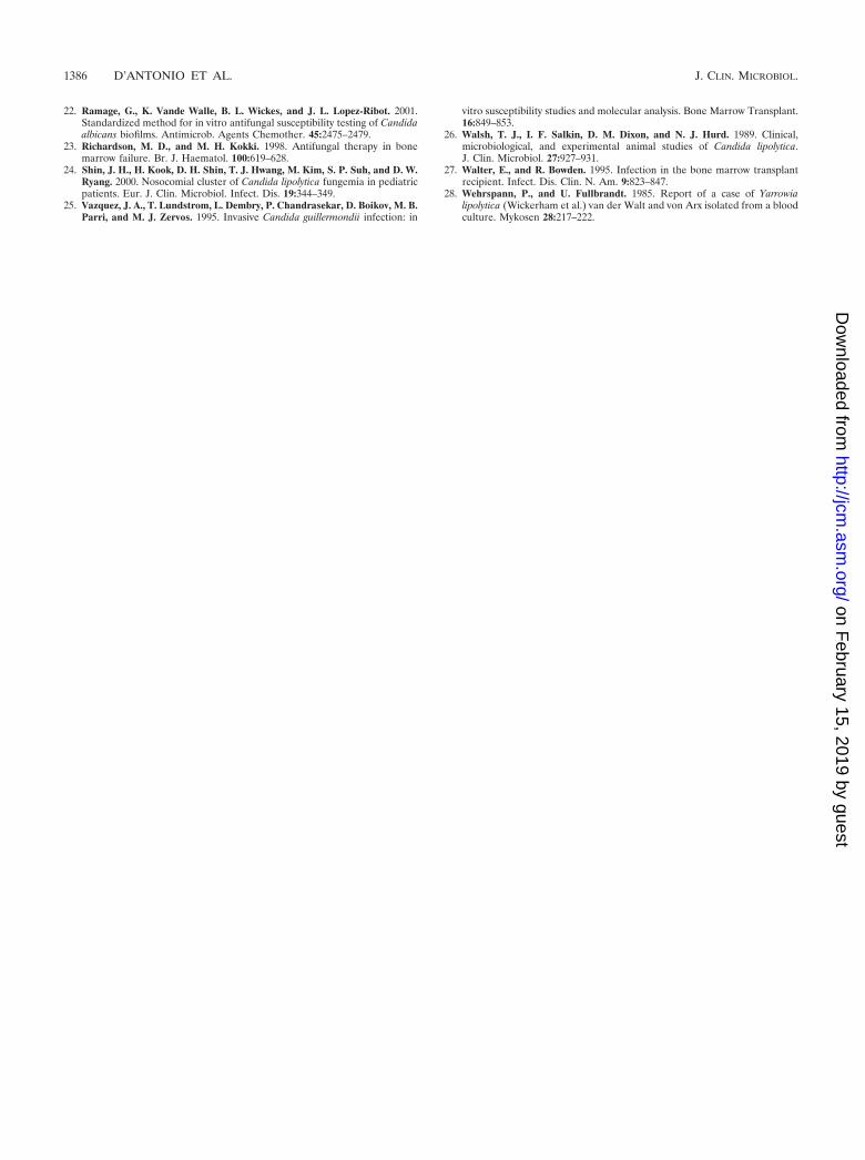
Ultrastructural analysis. Slime production was confirmedthrough SEM (Fig. 2), which showed amorphous material(slime) unevenly distributed on the cell surface. The slimeseemed to entangle the yeast cells and to contribute both tointrayeast cohesion and to their adhesion to the plastic. SEMalso showed that the concentration of yeast cells was quitestable across the different sections of the tube for the strains
grown with 2% glucose. When the strains were grown with 8%glucose, a consistent decrease in yeast cell concentration wasobserved moving from the ring of the culture tube to the cone,and there were no differences in production between our iso-lates and the control strains.
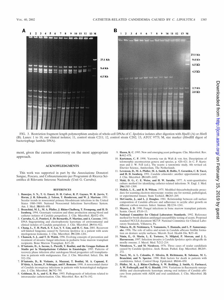
Genomic typing. All isolates from the patient had identicalrestriction profiles with HpaII and HinfI endonucleases, whiledifferences emerged with control strains (Fig. 3A). After re-striction with HpaII, the control strains presented the sameprofile but were clearly different from our isolates, in that the7,485-kb band was absent. Conversely, HinfI digestion pro-duced substantial differences among the control strains. TheC211 strain presented the same profile as did the clinical iso-lates, the C202 strain showed a unique pattern characterized bythe absence of the 2,050-kb band, and the ATCC strain showeda supplementary band of 23,650 kb, compared with the otherstrains (Fig. 3B).
DISCUSSION
Over the past 25 years the incidence of invasive candidiasisin patients with hematological malignancies has increased dra-matically; as well, the variety of AMB-resistant species isolatedduring infections in these patients has also changed. The pro-portion of C. albicans strains has decreased, and isolations ofnon-C. albicans spp. have increased (23; W. G. Merz, Abstr.Invasive Mycoses Crit. Trends Clin. Challenge Focus Itracon-azole Congr., 1993). In a recent report, non-C. albicans speciesaccounted for 63.4% of the candidemias studied (20). Ofnon-C. albicans spp. isolated from opportunistic infections, C.lipolytica has infrequently been identified as a cause. Most ofthese isolations have been due to line infections (18), as wasthe case in this study. Positive cultures of the CVC tip from thispatient in the absence of other potential sources of infectionwere very suggestive of CVC-associated candidemia. Further-more, BMT-related procedures, persistent use of broad-spec-trum antimicrobial agents, and GvHD predisposed this patientto infusion-related infections. Finally, after CVC removal, sub-
TABLE 1. Biofilm production scores and antifungal susceptibilities (16) of all isolates and control strains
Strain No. of days fromonset of fever Source
Slime productiona MIC (�g/ml) of drug:
2% glucose 8% glucose AMB Fluconazole Itraconazole
Clinical1 2 Blood 2 2 0.39 2.5 0.52 4 Blood 3 2 0.78 1.25 0.53 4 Blood 2 2 0.39 1.25 0.54 9 Blood 2 2 0.39 1.25 0.55 9 Blood 3 3 0.78 2.5 0.56 9 Blood 3 2 0.78 2.5 0.57 19 Blood 2 2 0.78 1.25 0.58 23 Blood 2 2 0.39 2.5 0.59 25 Blood 2 2 0.78 2.5 0.510 25 CVC 2 2 0.39 2.5 0.5
Control11 (C211) 2 2 0.19 2.5 0.512 (C202) 2 2 0.19 1.25 0.513 (ATCC 9773) 2 3 0.19 1.25 0.5
a Score: 1, low; 2, moderate; 3, high.
VOL. 40, 2002 CATHETER-RELATED CANDIDEMIA CAUSED BY C. LIPOLYTICA 1383
on February 15, 2019 by guest
http://jcm.asm
.org/D
ownloaded from

sequent blood cultures were persistently negative, and the ep-isode of candidemia appeared to be finished.
In all cases of infections reported in the literature C. lipoly-tica has been shown to be weakly virulent. This is in agreementwith the absence of mortality and the very low frequency ofvisceral lesions observed in mice inoculated with C. lipolytica(26). The weak virulence of this organism is further supportedby our findings. In this case, despite the long persistence ofcandidemia, there was no evidence of deep visceral infection atautopsy examination or through positive autopsy cultures.
The consistent slime production that we observed for thisisolate might explain the capability of this organism to adhereto and colonize the plastic CVC. Slime production might alsoserve to explain the reappearance of a persistent fever indica-tive of a blood-borne infection during treatment with AMB,despite the susceptibility of our C. lipolytica isolates to thisdrug. Similar to the inability of antimicrobial agents to pene-trate bacterial biofilms (8, 17, 22), extracellular slime of C.lipolytica might have impaired the efficacy of antifungal agentsin penetrating the thick biofilm on the catheter surface (11).
The appropriate management of catheter-related C. lipoly-tica fungemia has yet to be defined. A recent paper suggeststhat a stand-back approach without catheter removal or anti-fungal therapy may be acceptable (4). Conversely, according toour findings and in line with those reported by others (24, 26),management of catheter-related C. lipolytica fungemia shouldinclude a course of systemic antifungal therapy and removal ofthe potentially infected vascular catheter.
It has been reported elsewhere that colonizing and infecting
Candida populations may undergo microevolution, particularlyin immunocompromised individuals such as BMT patients(21). This genetic variation is due primarily to the reorganiza-tion of genomic sequences. Therefore, genomic typing wasperformed to compare strains of C. lipolytica isolated from thepatient during the two distinct febrile episodes. Restrictionfragment length polymorphism profiles proved to be identicalover time for all our isolates. This finding suggests the absenceof microevolutionary changes in the population of the infectingstrain, despite the length of the sepsis and the potential selec-tive pressure of AMB, which had been administered to thepatient for about 20 days. The lack of differences between ourisolates did not depend on a low discriminatory power of thetyping method. In fact, coupling the results of restriction anal-yses with HpaII and HinfI we were able both to differentiateour isolates from the control strains and to determine genotypedifferences among the control strains. These differences be-tween our isolates and the control strains are also suggestive ofa certain degree of genetic diversity between C. lipolytica iso-lates from widely divergent geographical areas. This variabilityseems to be present also in strains isolated in different placeswithin the same country, as indicated by the diverse genomicprofiles observed for our isolates compared to one of the twocontrol strains coming from another Italian city.
In conclusion, the increasing evidence of catheter-related C.lipolytica infections suggests that C. lipolytica should be in-cluded in any list of emerging yeast pathogens and also posesthe problem of defining the best strategy of patient manage-
FIG. 2. (A) Scanning electron microphotograph of blood isolate. The microphotograph shows microcolonial aggregate intimately associatedwith an amorphous material which seems to envelope single cells and join them (original magnification, �950). (B) Enlargement of the imageoutlined in panel A (original magnification, �2,150). The arrowhead indicates the amorphous material (slime).
1384 D’ANTONIO ET AL. J. CLIN. MICROBIOL.
on February 15, 2019 by guest
http://jcm.asm
.org/D
ownloaded from
ment, given the current controversy on the most appropriateapproach.
ACKNOWLEDGMENTS
This work was supported in part by the Associazione DonatoriSangue, Pescara, and Cofinanziamento per Programmi di Ricerca Sci-entifica di Rilevante Interesse Nazionale (Unit G. Carruba).
REFERENCES
1. Banerjee, S. N., T. G. Emori, D. H. Culver, R. P. Gaynes, W. R. Jarvis, T.Horan, J. R. Edwards, J. Tolson, T. Henderson, and W. J. Martone. 1991.Secular trends in nosocomial primary bloodstream infections in the UnitedStates 1980–1989. National Nosocomial Infections Surveillance System.Am. J. Med. 3B:86S–89S.
2. Branchini, M. L., M. A. Pfaller, J. Rhine-Chalberg, T. Frempong, and H. D.Isenberg. 1994. Genotypic variation and slime production among blood andcatheter isolates of Candida parapsilosis. J. Clin. Microbiol. 32:452–456.
3. Carruba, G., E. Pontieri, F. De Bernardis, P. Martino, and A. Cassone. 1991.DNA fingerprinting and electrophoretic karyotype of environmental andclinical isolates of Candida parapsilosis. J. Clin. Microbiol. 29:916–922.
4. Chang, L., T. H. Park, E. Y. Lee, Y. T. Lim, and H. C. Son. 2001. Recurrentself-limited fungemia caused by Yarrowia lipolytica in a patient with acutemyelogenous leukemia. J. Clin. Microbiol. 39:1200–1201.
5. Chanock, S. J., and T. D. Walsh. 1996. Evolving concepts of prevention andtreatment of invasive fungal infections in pediatric bone marrow transplantrecipients. Bone Marrow Transplant. 3:15–20.
6. D’Antonio, D., A. Iacone, L. Pierelli, T. Bonfini, and the Gruppo Italiano diStudio per la Manipolazione Cellulare in Ematologia. 1995. Patterns ofrecovery phase infection after autologous blood progenitor cell transplanta-tion in patients with malignancies. Eur. J. Clin. Microbiol. Infect. Dis. 14:552–556.
7. D’Antonio, D., B. Violante, A. Mazzoni, T. Bonfini, M. A. Capuani, F.D’Aloia, A. Iacone, F. Schioppa, and F. Romano. 1998. A nosocomial clusterof Candida inconspicua infections in patients with hematological malignan-cies. J. Clin. Microbiol. 36:792–795.
8. Goldman, D. A., and G. B. Pier. 1993. Pathogenesis of infections related tointravascular catheterization. Clin. Microbiol. Rev. 6:176–192.
9. Hazen, K. C. 1995. New and emerging yeast pathogens. Clin. Microbiol. Rev.8:462–478.
10. Kurtzman, C. P. 1998. Yarrowia van de Walt & von Arx. Descriptions oftelomorphic ascomycetous genera and species, p. 420–421. In C. P. Kurtz-man and J. W. Fell (ed.), The yeasts: a taxonomic study, 4th revised ed.Elsevier Science, Amsterdam, The Netherlands.
11. Levenson, D., M. A. Pfaller, M. A. Smith, R. Hollis, T. Gerarden, C. B. Tucci,and H. D. Isenberg. 1991. Candida zelanoides: another opportunistic yeast.J. Clin. Microbiol. 29:1689–1692.
12. Maki, D. G., C. E. Weise, and H. W. Sarafin. 1977. A semi-quantitativeculture method for identifying catheter-related infections. N. Engl. J. Med.296:1305–1309.
13. Malick, L. E., and R. B. Wilson. 1975. Modified thiocarbohydrazide proce-dure for scanning electron microscopy: routine use for normal, pathological,or experimental tissues. Stain Technol. 50:265–269.
14. McCourtie, J., and L. J. Douglas. 1981. Relationship between cell surfacecomposition of Candida albicans and adherence to acrylic after growth ondifferent carbon sources. Infect. Immun. 32:1234–1241.
15. Meyers, J. D. 1990. Fungal infections in bone marrow transplant patients.Semin. Oncol. 17:10–13.
16. National Committee for Clinical Laboratory Standards. 1992. Referencemethod for broth dilution antifungal susceptibility testing of yeasts. Proposedstandard NCCLS document M27-P. National Committee for Clinical Labo-ratory Standards, Villanova, Pa.
17. Nikava, H., H. Nishimura, T. Yamamoto, T. Hamada, and L. P. Samaranay-ake. 1996. The role of saliva and serum in Candida albicans biofilm forma-tion on denture acrylic surfaces. Microb. Ecol. Health Dis. 9:35–48.
18. Ninin, E., O. Morin, L. E. Tortorec, N. Milpied, P. Moreau, and J. L.Harousseau. 1997. Infection invasive à Candida lipolytica après allogreffe demoelle osseuse. J. Mycol. Med. 7:212–214.
19. Nitzulescu, V., and M. Niculescu. 1976. Three cases of ocular candidiasiscaused by Candida lipolytica. Arch. Roum. Pathol. Exp. Microbiol. 35:269–272.
20. Nucci, M., A. L. Colombo, F. Silveira, R. Richtmann, R. Salomao, M. L.Branchini, and N. Spector. 1998. Risk factors for death in patients withcandidemia. Infect. Control Hosp. Epidemiol. 19:846–850.
21. Pfaller, M. A., J. Rhine-Chalberg, S. W. Redding, J. Smith, G. Farinacci,A. W. Fothergill, and M. G. Rinaldi. 1994. Variations in fluconazole suscep-tibility and electrophoretic karyotype among oral isolates of Candida albi-cans from patients with AIDS and oral candidiasis. J. Clin. Microbiol. 32:59–64.
FIG. 3. Restriction fragment length polymorphism analysis of whole-cell DNAs of C. lipolytica isolates after digestion with HpaII (A) or HinfI(B). Lanes: 1 to 10, our clinical isolates; 11, control strain C211; 12, control strain C202; 13, ATCC 9773; M, size marker (HindIII digest ofbacteriophage lambda DNA).
VOL. 40, 2002 CATHETER-RELATED CANDIDEMIA CAUSED BY C. LIPOLYTICA 1385
on February 15, 2019 by guest
http://jcm.asm
.org/D
ownloaded from
22. Ramage, G., K. Vande Walle, B. L. Wickes, and J. L. Lopez-Ribot. 2001.Standardized method for in vitro antifungal susceptibility testing of Candidaalbicans biofilms. Antimicrob. Agents Chemother. 45:2475–2479.
23. Richardson, M. D., and M. H. Kokki. 1998. Antifungal therapy in bonemarrow failure. Br. J. Haematol. 100:619–628.
24. Shin, J. H., H. Kook, D. H. Shin, T. J. Hwang, M. Kim, S. P. Suh, and D. W.Ryang. 2000. Nosocomial cluster of Candida lipolytica fungemia in pediatricpatients. Eur. J. Clin. Microbiol. Infect. Dis. 19:344–349.
25. Vazquez, J. A., T. Lundstrom, L. Dembry, P. Chandrasekar, D. Boikov, M. B.Parri, and M. J. Zervos. 1995. Invasive Candida guillermondii infection: in
vitro susceptibility studies and molecular analysis. Bone Marrow Transplant.16:849–853.
26. Walsh, T. J., I. F. Salkin, D. M. Dixon, and N. J. Hurd. 1989. Clinical,microbiological, and experimental animal studies of Candida lipolytica.J. Clin. Microbiol. 27:927–931.
27. Walter, E., and R. Bowden. 1995. Infection in the bone marrow transplantrecipient. Infect. Dis. Clin. N. Am. 9:823–847.
28. Wehrspann, P., and U. Fullbrandt. 1985. Report of a case of Yarrowialipolytica (Wickerham et al.) van der Walt and von Arx isolated from a bloodculture. Mykosen 28:217–222.
1386 D’ANTONIO ET AL. J. CLIN. MICROBIOL.
on February 15, 2019 by guest
http://jcm.asm
.org/D
ownloaded from