ccrn review: neurological system - aacn edema and increased icp: seizures, htn, cushing’s
TRANSCRIPT

CCRN/PCCN
Review:
Neurological System
Chris Szabo, PhD, RN,CNRN,CCRN
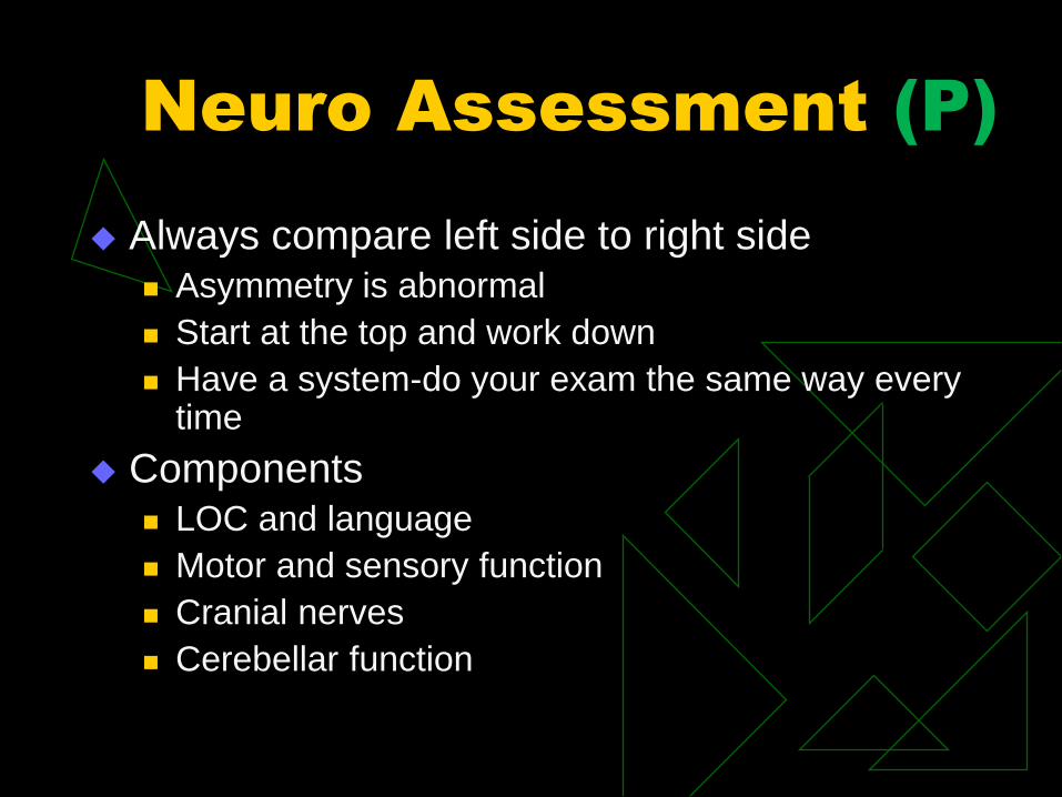
Neuro Assessment (P)
Always compare left side to right side Asymmetry is abnormal
Start at the top and work down
Have a system-do your exam the same way every time
Components LOC and language
Motor and sensory function
Cranial nerves
Cerebellar function

Anatomy Review (P)
Bones Fontanelles close 18-24 mos
Meninges Dura, arachnoid, pia
Ventricles CSF, choroid plexus,
arachnoid villi
Basal ganglia Fine motor control

A Little Anatomy…
Frontal Lobe
Primary motor cortex
Judgment, reasoning,
intellect, personality,
abstract thinking
Long term memory
Broca’s area
Parietal Lobe
Interpretation and
discrimination of
sensory input
Define shape, size,
texture, consistency
Touch, pressure,
position
Awareness of body
parts, spatial orientation

A Little Anatomy…
Occipital Lobe
Interpretation and
discrimination of visual
input
Primary visual cortex
receives direct signals
from macula
Secondary visual cortex
interprets vision and
meaning of written words
Temporal Lobe
Hearing and
discrimination of
auditory input
Primary auditory area
detects loudness, tones
Secondary auditory
area interprets meaning
of spoken word and
music
Wernicke’ area

Anatomy Review
Cerebellum Coordinates voluntary muscle control, equilibrium
Chiari malformation
Cerebral Circulation Arteries and areas supplied
Venous system
Blood-brain Barrier Restricts certain molecules and cells from passing
Spine and Spinal Cord

Cerebral Arterial
Circulation
Anterior cerebral
Anterior communicating
Internal carotids
Middle cerebral
Posterior cerebral
Posterior communicating
Basilar

Arterial Supply

Anatomy Review
Thalamus Initial processing of sensory input
Hypothalamus Temperature, appetite, thirst, emotional
expression, sleep-wake cycles
Pituitary Gland (Hypophysis) Hormones
Brainstem Midbrain, pons, medulla

Level of
Consciousness
Most sensitive indicator of problems
Studying the brain without studying consciousness would be like studying the stomach without studying digestion… John R. Searle, Philosopher

Definitions of LOC
Full consciousness Awake, alert and oriented
Confused Disorientation to one or more spheres,
↓ attention span and memory, difficulty following commands
Lethargic May or may not be fully oriented, follows
commands but mental and motor activities slow
Obtunded Arouse to tactile stimuli, responds with 1-2 words,
may not follow commands

Definitions of LOC
Stuporus
Shows little spontaneous movement, moans
Responds purposefully to noxious stimuli
Comatose
Total absence of awareness
No response to verbal stimuli
May be purposeful to totally unresponsive
GCS <8

Aphasia
Broca’s Aphasia Motor, expressive
Cannot convert thoughts to words
Automatic speech may be preserved
Wernicke’s Aphasia Sensory, receptive
Speech lacks content and meaning
Cannot understand written and/or spoken words
Global Aphasia Motor and sensory
Expressive and receptive
Dysarthria Loss of articulation, phonation due to motor deficits
or loss of breath control

Glasgow Coma
Scale
Points Adult/Child Infant/Preverbal
Eye Opening 4 Spontaneous Spontaneous
3 To speech To speech
2 To pain To pain
1 None None
Verbal Response 5 Oriented Coos, babbles
4 Confused Irritable cry
3 Inappropriate words Cries to pain
2 Sounds Moans to pain
1 None None
Motor Response 6 Obeys Normal, spontaneous
5 Localizes pain Withdraws to touch
4 Withdraws to pain Withdraws to pain
3 Abnormal flexion Abnormal flexion
2 Abnormal extension Abnormal extension
1 None None

Components of the
Neurological Assessment
Verbal response Orientation
Memory
Fund of Knowledge
Motor Response Obey
Localize
Withdraw
Abnormal Flexion
Extension
Eye Opening
Spontaneous
To speech
To pain
None

Pupillary Assessment
Size
Shape
Reaction to light
Direct and
consensual

EOM and OCR

Decorticate Posturing:
Abnormal Flexion

Decerebrate Posturing:
Extension

Pathological
Reflexes
Babinski
Present or absent
Presence in adults is
abnormal
Occulocephalic Reflex
Absent (negative) if
eyes don’t move

Cranial Nerves
CN 1 Smell
CN 2 See
CN 3,4,6 Move eyes, 3 constricts, lid up
CN 5 Chew and feel front of face
CN 7 Moves face, tastes, salivates, cries, lid down
CN 8 Hears, regulates balance
CN 9 Taste, salivate,swallow, monitors carotid body and sinus
CN 10 Tastes, swallow,lift palate, communicates with viscera
CN 11 Turns head and lifts shoulders
CN 12 Moves tongue

Anatomy Review
Peripheral Nervous System Dorsal root is sensory, ventral is motor
Cranial Nerves Names and functions
Autonomic Nervous System Sympathetic and Parasympathetic
Reflexes Babinski’s

Intracranial
Contents and ICP
Blood 10%
Brain 80%
CSF 10%
Normal ICP 0-15mmHg
Constantly changing phenomenon
Intracranial hypertension ICP >20mmHg

Increased Intracranial
Pressure
Munro-Kellie Hypothesis
Contents of cranial vault (brain, blood,
CSF) are in a state of dynamic equilibrium
Increase in one components requires a
reciprocal decrease in one or both of the
others
Normal ICP 0-15mmHg
CPP=MAP-ICP
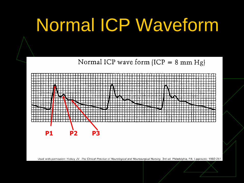
Normal ICP Waveform
P1 P2 P3
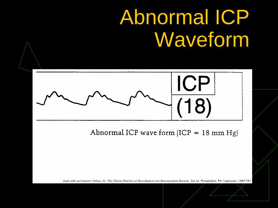
Abnormal ICP Waveform

A Waves

B Waves

C Waves
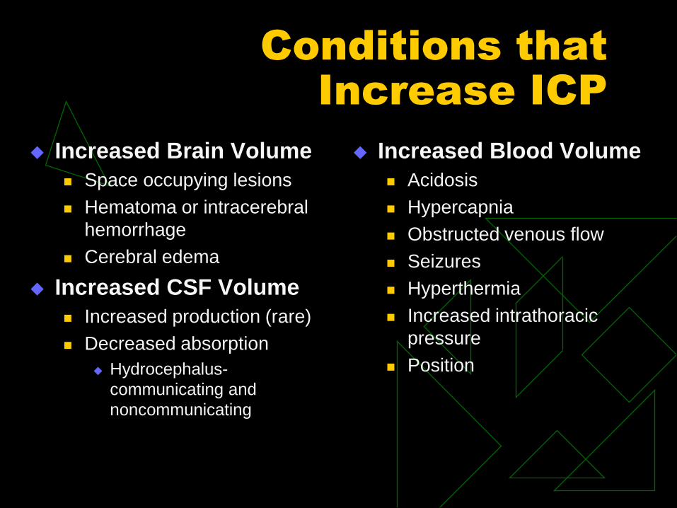
Conditions that
Increase ICP
Increased Brain Volume
Space occupying lesions
Hematoma or intracerebral
hemorrhage
Cerebral edema
Increased CSF Volume
Increased production (rare)
Decreased absorption
Hydrocephalus-
communicating and
noncommunicating
Increased Blood Volume
Acidosis
Hypercapnia
Obstructed venous flow
Seizures
Hyperthermia
Increased intrathoracic
pressure
Position

Clinical Signs of
Increased ICP
Early Deterioration in LOC
Deterioration in motor function-contralateral
Pupillary dysfunction-ipsilateral
Possible headache
Late Signs Coma
Pupils fixed and dilated-bilateral
Profound change in motor exam-bilateral
Cushing’s Triad- systolic pressure, widening pulse pressure, bradycardia
Alterations in respirations and temperature

Medical Interventions to
Control ICP
Analgesia and sedation
Hyperventilation for
rapid reduction
Oxygenation and BP
control
Maintain CPP: CSF
drainage
Mannitol
Hypertonic saline
Neuromuscular
blockade
Fluid management
Temperature control-
hypothermia
Seizure prophylaxis
High-dose barbiturate
coma
Surgery

Nursing Management
Positioning
HOB elevation-reverse Trendelenburg
No lateral neck flexion or rotation
Suctioning
Hyperventilate and limit to 10 seconds
Avoid clustering of activities
Control extraneous activities
Noise, visitation, light

Aneurysm (P)
Saccular outpouching
Occur at arterial bifurcations in Circle of
Willis
Incidence increases with age, may be
familial
More common in women
Classified by size or by size and shape

Aneurysm
Berry -most common, has a neck or stem
Fusiform -no stem
Traumatic -any aneurysm that results from trauma
Mycotic -septic emboli lead to formation
Charcot-Bouchard -microscopic, associated with hypertension, involves basal ganglion and brainstem
Dissecting -related to atherosclerosis, inflammation or trauma; separation of intimal and medial layers of artery

Aneurysm
Location
Carotid system: 85-95%
Anterior Communicating Artery: 30%
ACA: more common in men
Posterior Communicating Artery: 25%
Posterior circulation: 5-15%
20%-30% of patients who suffer an
aneurysm have more than one

Aneurysm:
Unruptured
Assymptomatic until aneurysm bleeds
Warning signs (prodromal)
Dilated pupil
Abnormal EOMs
Ptosis
Pain above or behind the eye
Localized headache
Nuccal rigidity
Photophobia

Aneurysm:
Unruptured
Management
Assess risk for rupture
No definitive recommendations
Risk factor reduction: Hypertension
Cigarette smoking
Use of oral contraceptives
Alcohol consumption
Surgery if technically possible

Aneurysm: Ruptured
Most frequent presentation is subarachnoid hemorrhage (SAH)
At time of rupture, blood forced into subarachnoid space
Patient experiences sudden explosive headache; c/o “worst headache of my life”
Decreased or immediate loss of consciousness
Mortality 25% and most die within first 24 hours
50% of patients who survive aneurysmal rupture left with severe disabilities

Aneurysm: Ruptured
Other signs and symptoms
Cranial nerve deficits-3,4,and 6
Meningeal irritation: nausea and vomiting, neck and
back pain, nuchal rigidity, blurred vision, photophobia,
mild temperature elevation
Stroke syndrome: paresis or plegia, aphasia, cognitive
deficits
Cerebral edema and increased ICP: seizures, HTN,
Cushing’s
Pituitary dysfunction: diabetes insipidus, hyponatremia
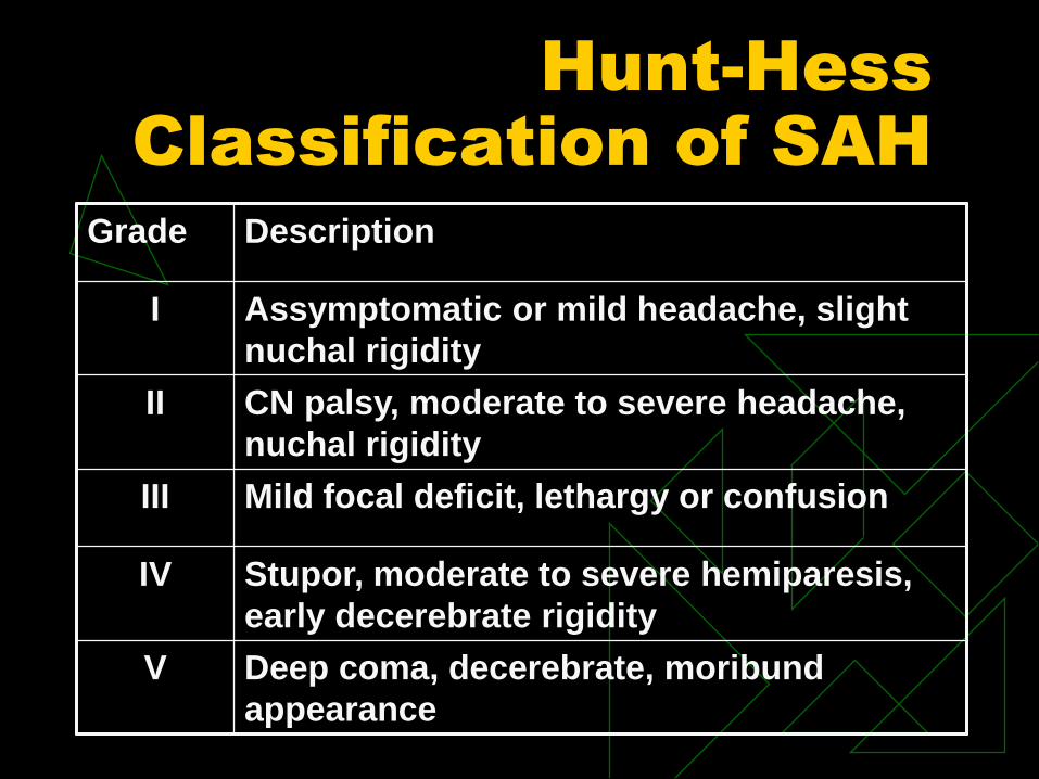
Hunt-Hess
Classification of SAH
Grade Description
I Assymptomatic or mild headache, slight
nuchal rigidity
II CN palsy, moderate to severe headache,
nuchal rigidity
III Mild focal deficit, lethargy or confusion
IV Stupor, moderate to severe hemiparesis,
early decerebrate rigidity
V Deep coma, decerebrate, moribund
appearance

Diagnosis of
Aneurysm
Neurological exam
CT without and with contrast
LP if CT negative
CTA
MRI/MRA
Not sensitive within first 24-48 hours, best after 4-7 days
Cerebral Angiography – “Gold Standard”
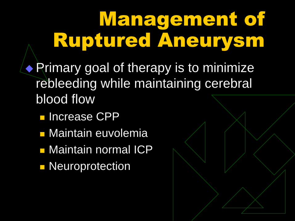
Management of
Ruptured Aneurysm
Primary goal of therapy is to minimize
rebleeding while maintaining cerebral
blood flow
Increase CPP
Maintain euvolemia
Maintain normal ICP
Neuroprotection

Management of
Ruptured Aneurysm
Fluid management
NS, albumin
BP control
Unsecured: SBP 120-150mmHg
Labetalol, Nipride, Nicardipine
Pulmonary artery catheter for Hunt-Hess ≥3,
SIADH or hemodynamically unstable
Intubation if comatose or need airway protection
ICP monitoring in patients who are developing
hydrocephalus and Hunt-Hess grade ≥3

Nursing
Responsibilities
VS and neuro exam every hour
Pulse oximetry
Bedrest with HOB elevation 30 degrees
Minimize external stimulation
Strict I&O
DVT prophylaxis-SCD,TED hose, etc..
Indwelling urinary catheter as needed

Aneurysm
Treatment
Surgical
Clipping
Wrapping
Endovascular
Coiling
Stent-assisted coiling
For wide-necked aneurysms and fusiform
aneurysms

Cerebral Vasospasm
Major cause of mortality and morbidity in
patients who survive initial rupture of
aneurysm
Causes reduced blood flow which may lead
to delayed ischemic deficit and infarction
Occurs in approximately 30% of patients
Clinically evident in 30% of these patients, 70%
are found only through transcranial doppler or
angio

Signs and Symptoms
of Vasospasm
Gradual neurological deterioration
Confusion
Decreased LOC
Paresis/plegia
Cranial nerve deficits
Aphasia

Treatment of Vasospasm:
Triple H Therapy
Hypervolemia: CVP 8-10mmHg, PCOP 14-18mmHg
NS, colloids
Hypertension
Unsecured:120-150 mmHg for the duration of spasm
Secured: 160-170 mmHg for the duration of spasm
Phenylephrine, Levophed, Dopamine, Dobutamine
Hemodilution
Target Hct ≤ 33%, transfuse for Hct < 25%

Latest Recommendations
for Treatment of
Vasospasm
Oral Nimodipine Hypotensive adverse effects
Hemodynamic Augmentation Induced hypertension, maintain euvolemic status
Monitor BP to avoid sudden drops in CPP, prevent hypertension (BP >160mmHg)
Intravenous magnesium
Statins

Brain Death
Defined as irreversible cessation of all functions of the entire brain including the brainstem. Three key characteristics:
1. Coma or unresponsiveness
must rule out all confounding factors such as hypothermia, drug intoxication, metabolic or endocrine imbalance
Some recommend neuroimaging to confirm catastrophic neurological event

Brain Death
2. Absence of brainstem reflexes
Pupillary reaction
Corneal reflex
Gag reflex
Oculovestibular reflexes
OCR and calorics
Loss of spontaneous respirations and
vasomotor control
Brain Death
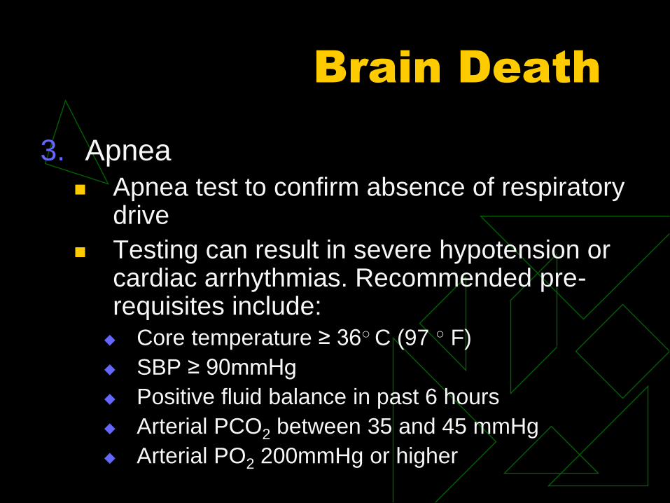
3. Apnea
Apnea test to confirm absence of respiratory drive
Testing can result in severe hypotension or cardiac arrhythmias. Recommended pre-requisites include: Core temperature ≥ 36○ C (97 ○ F)
SBP ≥ 90mmHg
Positive fluid balance in past 6 hours
Arterial PCO2 between 35 and 45 mmHg
Arterial PO2 200mmHg or higher

Arteriovenous
Malformation (AVM) (P)
Tangling of the high pressure arterial
flow with low pressure venous flow
without intervening capillary network
Creates a shunt that leads to ischemia
and atrophy of adjacent tissue
Leads to venous engorgement and
rupture

AVM
Congential
Most common cause of spontaneous
hemorrhage in children
Mortality 20%-60%
80% symptomatic AVM patients
between ages of 20 and 40 years

AVM: Clinical
Presentation
Hemorrhage
ICH most common presenting symptom
Sudden headache
Nausea and vomiting
Paresis or plegia
Decreased LOC
Seizures
Headache
Progressive focal deficits

AVM
Diagnostic Studies
CT
MRI/MRA
Angiography

Management of AVM
Symptom Control
Airway management
BP management
HTN: labetalol, hydralazine
Hypotension: phenylephrine
Seizure management
Phenytoin, fosphenytoin

Treatment of AVM
Following recovery from hemorrhage
Surgery Complete removal
May be embolized prior to resection
Radiotherapy Induces inflammatory process results in
thrombosis and obliteration of AVM
Endovascular treatment Embolozation to permanently occlusion of AVM
Indicated when lesion not accesible by surgery

Encephalopathy (P)
General category used to designate diffuse cerebral dysfunction
Manifested by alterations in cortical function and disturbances of consciousness ranging from mild confusion to coma
Caused by systemic disorder that has a diffuse effect on the brain- Infections, hypoxia, alcohol, liver failure, renal
failure, metabolic disease, brain tumors, toxic chemicals, intracranial hypertension,
malnourishment
Presentation, Diagnosis
and Treatment
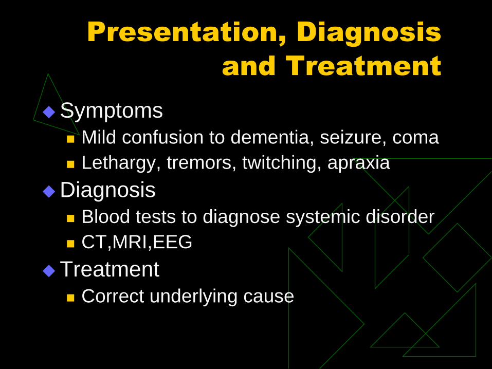
Symptoms
Mild confusion to dementia, seizure, coma
Lethargy, tremors, twitching, apraxia
Diagnosis
Blood tests to diagnose systemic disorder
CT,MRI,EEG
Treatment
Correct underlying cause

Break!

Head Trauma
Background:
External forces that impact with the body
resulting in structural and/or physiological
injuries
Leading cause of trauma related deaths in
people <45, predominantly male
1.5 -2 million/year, 50K deaths, 80-100K
significant disability, 300,000 sports related

Etiology
Blunt Trauma:
MVC, pedestrian vs. car, bicycle accidents, falls, sports related, assaults
Caused by contact, acceleration-deceleration, and rotational forces
Penetrating trauma
GSW, knives, sharp objects
Penetration of the skull producing focal damage

Primary and
Secondary Injuries
Primary injury results from direct trauma
to the brain
Secondary injuries follow the primary
Immediate and delayed
Due to hypoxia, cerebral edema, alterations
in cerebral blood flow, hypercapnia,
increased ICP

Secondary Injury
Cascade of ischemia and cellular
changes that lead to neuronal injury and
cell death
Ischemia
Cerebral edema (vasogenic, cytotoxic,
interstitial)
Rapid increase in release of excitatory
neurotransmitters

Types of Injuries
Scalp Lacerations
Often associated with skull fracture
May bleed profusely!
Scalp Abrasions
Top layers scraped away, may have small mount of bleeding
Scalp Contusions
Bruise with blood accumulation in SQ layer

Types of Injury
Skull Fractures
Extent of injury depends on thickness of
skull, point of impact and the mechanism of
injury
Stress waves radiate throughout entire
skull

Types of Skull
Fractures
Linear-single fracture line
Comminuted-bone splintered and fractures into pieces
Depressed-bone embedded in brain tissue, dura may be torn (watch for underlying intracranial hemorrhage)
Compound-open depressed-greater risk for infection
Basilar skull fracture-linear fracture at base of skull, associated with dural tear,high risk of infection

Basilar Skull
Fracture
Watch for
CSF leak
Racoon eyes
Battle sign
Signs & symptoms
of meningitis,
encephalitis, EDH

Brain Injuries
Concussion
Brief loss of consciousness, headache,
dizziness, possible nausea and vomiting
Altered LOC and focal deficits usually
resolve in 6-12 hrs
Post-concussion syndrome common
STM deficits, headaches, cognitive and visual
disturbances, poor coordination, lethargy
Brain Injuries
Contusions
Bleeding of small vessels, necrotic brain tissue
Acceleration-deceleration
Change in LOC, cranial nerve dysfunction, hemiparesis or hemiplegia, seizures, increased ICP
Clinical findings depend on size and location

Brain Injuries
Diffuse Axonal Injury
Acceleration-deceleration
Shearing at white-gray matter junction,
corpus callosum, brainstem and sometimes
cerebellum
Diffuse tearing of axons and small blood
vessels

Diffuse Axonal
Injury
Mild
Coma duration 6-24 hours, death uncommon,
often have cognitive and neurological deficits
Moderate
Coma duration >24 hours, no prominent brainstem
signs, incomplete neurological recovery
Severe
Prolonged coma, prominent brainstem signs,
results in death or severe disability

Epidural Hematoma (P)
85% arterial
Most often injury to middle
meningeal artery
Classic “talk and die”
Brief loss of consciousness
followed by lucid interval then
rapid deterioration

Epidural Hematoma
Presentation
Alteration in LOC
Headache
Nausea and vomiting
Seizures
Ipsilateral occulomotor paralysis
Contralateral motor paresis/plegia
Increased ICP (know these signs & symptoms)

Epidural Hematoma
Diagnosis
Skull x-ray
CT without contrast
LP contraindicated due to increased ICP
Treatment
ICP management
Surgical evacuation

Subdural Hematoma (P)
Bleeding from injury to veins that
bridge the dura and arachnoid
space
May occur with minimal trauma
especially in patients on
anticoagulants
Majority in elderly patients with
atrophy or alcoholosm
May be acute or chronic or both

Subdural Hematoma
Presentation
Acute
Gradual or rapid deteroriation in LOC
Pupillary changes
Hemiparesis/hemiplegia
Chronic
Headache, cognitively impaired, confusion, slow
pupillary response, seizures
Symptoms develop gradually

Subdural Hematoma
Diagnosis
Skull films
CT without contrast
MRI
Treatment
Acute: surgery or burr hole to remove clot
Chronic or small acute: observation, serial CT, allow clot to liquefy

Intracerebral
Hemorrhage (P)
Trauma-penetrating or acceleration-
deceleration
Also due to tumors, bleeding disorders,
anticoagulant or hypertension

Intracerebral
Hemorrhage
Clinical Presentation
Depends on location, size and rate of blood
accumulation
Headache, decreased LOC progressing to
coma, hemiplegia/hemiparesis progressing
to bilateral weakness/paralysis, dilated
pupil, increased ICP

Intracerebral
Hemorrhage
Diagnosis
Skull films
CT and MRI
Treatment
Supportive including management of ICP
Surgery if accesible, rarely improves
outcome

Hydrocephalus
Abnormal accumulation of CSF in
cranial vault; brain tissue squeezed
against skull
Caused by tumors, ICH, intraventricular
blood, malabsorption by arachnoid villi
Surgical correction with VP shunt

Neurological
Infections
Meningitis
Inflammation of meninges and CSF within
subarachnoid space caused by bacteria,
virus or fungus
Blood-brain barrier interrupted
With bacterial meningitis, purulent exudate
obstructs CSF flow
Viral meningitis (acute aseptic meningitis)

Signs and
Symptoms
Headache and fever
Meningeal irritation Nuchal rigidity
Kernig’s Sign: 90 degree hip flexion and attempt to extend knee, + with pain and spasm of hamstring
Brudzinski Sign: + when hip and knee flex with lifting the head and neck
Altered LOC
Photophobia
Meningococcal – rapid development of delirium and stupor, petechial rash on legs

CSF Findings in
Meningitis
CSF
Characteristics
Normal Acute Bacterial
Meningitis
Acute Viral
Meningitis
Appearance Clear Turbid, cloudy Clear
WBC 0-5 cells/mm3 1000-2000
cells/mm3
300 cells/mm3
Protein 15-50 mg/dL 100-500mg/dL Normal
Glucose 40-80 mg/dL
≈ 66% of blood
value
<40mg/dL
≈40% of blood
value
Normal
Culture Bacteria on gram
stain
Virus using
special
techniques
Pressure 80-100mmH20,
8-14mmHg
Elevated;
>180mmH2O
Variable

Neuromuscular
Disorders
Guillian-Barre Syndrome
Inflammation of nervous system, flu-like illness
Probably autoimmune triggered by infection, leads to demyelination of axons
Acute onset of lower extremity weakness, ascends rapidly and progresses to respiratory failure
Treatment: Supportive
IV Ig or plasmaphoresis

Neuromuscular
Disorders
Muscular Dystrophy
Group of inherited diseases in which the
muscles voluntary muscles progressively
weaken. In some forms of this disease, the
heart and other organs are also affected
9 Types….
Oculopharyngeal: Affects men and women in
later in life. Progresses slowly with weakness in
the eye and face muscles. Difficulty swallowing
and recurrent pneumonia

Neuromuscular
Disorders
Myesthenia Gravis
Autoimmune disease of neuromuscular
junction, destruction of acetylcholine receptor
sites
Characterized by abnormal muscle fatigue
brought on by activity and improving with rest
May be autoimmune
Thymectomy may improve symptoms

Myesthenia Gravis
Diagnosis
Tensilon Test Administration of edrophonium, an
anticholinesterase, produces rapid improvement in symptoms
Treatment
Mestinon (pyridostigmine)
Immunosuppresion
Plasmaphoresis and IV Ig

Myesthenic Crisis
Sudden relapse of symptoms
Rapidly develop res;piratory and swallowing difficulties
May require intubation
Anticholinesterase drugs ineffective during crisis
Management is supportive until crisis resolves

Cholinergic Crisis
Issue of overmedication!
Abdominal cramping and diarrhea
Generalized weakness, excessive pulmonary
secretions, impaired respiratory function
Tensilon test used to differentiate
Improvement with drugs suggests myesthenic
crisis
No improvement or deterioration suggests
cholinergic crisis

Neurosurgery
Craniotomy
Surgical opening to allow access to brain
Supratentorial for access to frontal, parietal,
temporal and occipital lobes
Infratentorial to access posterior fossa
(midbrain, pons, medulla and cerebellum)
Transsphenoidal approach to remove
pituitary tumors
Neurosurgery
Craniectomy Excision of portion of skull without replacement,
used for decompression or removal of bone fragments from skull fracture
Cranioplasty Repair of the skull using synthetic material
Burr holes are small holes drilled on the skull to access underlying structures Drainage of epidural or subdural hematomas,
insertion of EVD or ICP monitors
Neurosurgery
Complications
Intracranial hypertension
Ischemia or infarction
CSF leaks
CNS infection
Seizures
Fluid & electrolyte imbalances-DI following
pituitary resection, CSW

Seizure Disorders (P)
Sudden uncontrolled discharge of
electrical activity
Frequently a symptom of underlying
pathology
Etiology
CNS infections, AVM, genetic metabolic
disorders, TBI, stroke, following cranial
surgery

Clinical
Presentation
Aura
Auditory, visual, gustatory, visceral
Ictal phase-seizure activity
Post-ictal phase- period following the seizure
Sleepy, confused, amnesic
Generalized seizures originate in all regions of the cortex, partial seizures originate in a specific area

Generalized
Seizures
Absence-childhood, last 5-10 sec, eye blinking, lip smaking
Atypical absence-assocociated with mental retardation, same as absence with muscle spasm
Myoclonic-sudden brief muscle contractions (arms>legs), usually little change in LOC
Generalized
Seizures
Clonic-rhythmic, repetitive clonic movements of extremities (arms>legs), neck and face
Tonic-clonic-”grand mal”, most common, last 2-3 minutes,risk for injuries, tongue biting,head injury
Atonic-sudden loss of muscle control, fall to the floor, increased risk of injury
Partial Seizures
Clinical manifestations depend on region of brain involved
Simple partial-may have motor, sensory, autonomic or cognitive manifestations but no loss of consciousness
Complex partial-most common epileptic seizures in adults, similar to simple but there is loss of consciousness and awareness

Status Epilpticus
Continuous seizures lasting >5 minutes or
two or more seizures with incomplete
recovery of consciousness between
Most common cause is abrupt
discontinuation of AEDs
Characterized by tonic,clonic, or tonic-
clonic movements, may be subclinical

Status Epilpticus
Medical emergency
Accompanied by respiratory distress
Morbidity and mortality 20%
Increased cerebral metabolism leads to hypoglycemia, HTN, increased cardiac output, increased CVP, increased HR, fever, excessive salivation, vomiting, incontinence Elevated catecholamines and lactic acidosis
promote cardiac dysrhythmias and autonomic dysfunction
Develop electrolyte disturbances and dehydration

Status Epilpticus
Lack oxygen and glucose stimulates
production and release of glutamine,
changes in electrical balance eventually
leads to instability and injury of neurons
If unable to control seizures with AEDs,
use general anesthetics until seizure
activity on EEG activity stops

Stroke (P)
“Brain Attack”
3rd leading cause of death and leading
cause of disability in the US
Sudden development of focal neurological
deficits caused by interruption of blood flow
to brain tissue
Brain dependant on constant supply of oxygen
and glucose, insufficiency leads to cellular injury
Severity proportional to reduced blood flow
Infarction core surrounded by ischemic zone

Signs & Symptoms
FAST Acryonym
Facial asymmetry, droop, usually unilateral
Arm and/or leg weakness and/or numbness,
usually unilateral
Speech abnormal, slurred or unable to speak
Time-TPA window 3 hours
Headache, visual disturbances, confusion

Transient Ischemic
Attack (P)
Stroke symptoms lasting <24 hours
Last several minutes to few hours
Warning sign for stroke
5% risk for stroke within 48 hours
10% risk of stroke within 3 months
2% death within 3 months

Ischemic Stroke (P)
Due to thrombus or embolus
Risk factors:
Atrial fibrillation
CAD
Bacterial endocarditis
Valvular heart disease
DVT
Air and fat embolism

Treatment of Acute
Ischemic Stroke
Eligibility for thrombolytic therapy (rtPA)
Age 18 or older
Clinical diagnosis of ischemic stroke
causing a measurable neurological deficit.
Time of symptom onset well established
to be less than 180 minutes (3 hours)
before treatment would begin.

Ischemic Stroke:
BP Management
Patient otherwise eligible for acute reperfusion therapy
except that BP is >185/110 mm Hg:
Labetalol 10–20 mg IV over 1–2 minutes, may repeat
1 time; or
Nicardipine 5 mg/h IV, titrate up by 2.5 mg/h every
5–15 minutes, maximum15 mg/h; when desired BP
reached, adjust to maintain proper BP limits; or other
agents (hydralazine, enalaprilat, etc) may be
considered when appropriate
If BP is not maintained at or below 185/110 mm Hg, do
not administer rtPA

Ischemic Stroke: BP
Management
Management of BP during and after rtPA or other acute
reperfusion therapy to maintain BP at or below 180/105
mm Hg: Monitor BP every 15 minutes for 2 hours from the start of rtPA therapy, then every 30
minutes for 6 hours, and then every hour for 16 hours
If systolic BP >180–230 mm Hg or diastolic BP >105–
120 mm Hg: Labetalol 10 mg IV followed by continuous IV infusion 2–8 mg/min; or
Nicardipine 5 mg/h IV, titrate up to desired effect by 2.5 mg/h every 5–15 minutes,
maximum 15 mg/h
If BP not controlled or diastolic BP >140 mm Hg,
consider IV sodium nitroprusside
Administration of rtPa
2 peripheral IVs
Complete all invasive procedures
Dose:
0.9mg/kg up to max of 90 mg
10% of dose as bolus over 1-2 minutes with
remaining 90% over 60 minutes
Hold all antithrombotics for 24 hours to
prevent re-bleeding

Hemorrhagic Stroke (P)
Rupture of cerebral vessel, blood leaks into the brain tissue
Major risk factor is long-standing, poorly controlled hypertension
Most common sites
Basal ganglia (50%)
Thalamus (30%)
Cerebellum (10%)
Pons (10%)

Hemorrhagic Stroke:
BP Managment
Focus is to prevent rebleeding
Between time of bleed and aneurysm obliteration, BP controlled to balance risk of stroke, HTN related rebleeding and maintenance of CPP
Magnitude of BP control not established-decrease in systolic BP to <160mmHg is reasonable
Once aneurysm is secure, increased BP permitted to increase CBF
Goal is to maintain cerebral perfusion and prevent ischemia

Left Brain Stroke
Paralysis or weakness on right side
Impaired speech
Impaired right/left discrimination
Slow performance, cautious
Impaired comprehension related to
language and computation
Aware of deficits-depression, anxiety

Right Brain Stroke
Paralysis or weakness of left side
Left-sided neglect
Spatial-perceptual deficits
Denies or minimizes problems
Impulsive
Rapid performance, short attention span
Impaired judgement
Impaired concept of time

Cerebellar Stroke
Nausea and vomiting
Dysphagia and dysarthria
Nystagmus
Ipsilateral Horner’s Syndrome
Ataxia and vertigo
Loss of pain and temperature sensation
on opposite side

Safety
Fall prevention
Weakness, visual deficits, neglect/denial, impulsiveness, memory deficits, overestimating abilities and underestimating deficits
DVT
Compression stockings
Aspiration
Swallow screening
Seizures

Motor Deficits
Mobility
Respiratory function
Swallowing and speech
Gag reflex
Self-care

Affect
Difficulty controlling
emotions
Exaggerated or
unpredictable
Depression
Frustration

Intellectual
Function
Impaired memory and judgment
Left
Memory problems related to language
Extremely cautious
Right
Impulsive
Moves quickly
Underestimates limitations

Spatial-Perceptual
Alterations
Can occur with both but more
common in right-sided stroke
Altered perception of self and
limitations
Deny illness or body parts
Neglect input from affected
side
Erroneous perception of self
related to space
Worsened by homonymous
hemianopsia, agnosia and/or
apraxia

Urinary Elimination
Present initially but usually resolves
over time
Prognosis for normal bladder function
best if stroke affects only one side
Intact sensation of bladder filling
Voluntary urination present
UTI most likely due to presence of
indwelling catheter

Bowel Elimination
Motor control usually intact
Problems associated with constipation
Immobility
Weak abdominal muscles
Dehydration
Diminished response to defecation reflex
Inability to express needs
Difficulty managing clothing

Airway Management
Optimal positioning to maintain a clear
airway and maximize intrathoracic
pressure during cough
Deep breaths, several huffs against open
glottis and lean forward to expectorate
secretions
Incentive spirometry

Mobility
Active and/or passive ROM
Positioning Prevent dependant edema
Preserve function
Careful positioning and moving to avoid injury and pain Trochanter roll
Hand cones
Arm supports and slings
Do not pull up by arms
Rest periods after to exercise

Neglect
Teach patient to scan the environment
At first, approach from unaffected side, later approach
patient from affected side
Place objects in field of vision
Provide physical and verbal cues
Teach patient & family to stimulated affected limbs to
promote reintegration with whole body
Remind patient to survey whole body for position,
cleanliness and appropriate dress
Nutrition
NPO until swallow screening completed and oral
intake approved by SLP or physician
Out of bed for meals if possible, HOB as close to
90° as possible
Maintain sitting position for 30 min after meal
Teach patient to take small bites, place in
unaffected side of mouth
Check oral cavity for pocketing
Consult OT for assistive devices

Urinary
Incontinence
Avoid indwelling catheter, remove ASAP
Establish toileting schedule based on
voiding patterns

Self-Esteem
Establish achievable
goals
Involve patient in
planning
Praise every success
Encourage
independence
Identify stressors and
situations that trigger
emotional response

Body Image
Monitor patient’s ability
to look at affected body
part
Help patient to
determine extent of
actual changes
Prevent
misinterpretation of
function