cd133 cxcr4 colon cancer cells exhibit metastatic potential and predict poor prognosis of patients
TRANSCRIPT
RESEARCH ARTICLE Open Access
CD133+CXCR4+ colon cancer cells exhibitmetastatic potential and predict poor prognosisof patientsShan-shan Zhang1†, Zhi-peng Han1†, Ying-ying Jing1†, Shuang-fen Tao1, Tie-jun Li2, Hao Wang3, Yang Wang3, Rong Li1,Yang Yang1, Xue Zhao1, Xiao-dong Xu3, En-da Yu3, Yao-cheng Rui2, Hou-jia Liu3, Li Zhang3* and Li-xin Wei1*
Abstract
Background: Colorectal cancer (CRC), which frequently metastasizes to the liver, is one of the three leading causesof cancer-related deaths worldwide. Growing evidence suggests that a subset of cells exists among cancer stemcells. This distinct subpopulation is thought to contribute to liver metastasis; however, it has not been fullyexplored in CRC yet.
Methods: Flow cytometry analysis was performed to detect distinct subsets with CD133 and CXCR4 markers inhuman primary and metastatic CRC tissues. The ‘stemness’ and metastatic capacities of different subpopulationsderived from the colon cancer cell line HCT116 were compared in vitro and in vivo. The roles of epithelial-mesenchymal transition (EMT) and stromal-cell derived factor-1 (SDF-1) in the metastatic process were alsoinvestigated. A survival curve was used to explore the correlation between the content of CD133+CXCR4+ cancercells and patient survival.
Results: In human specimens, the content of CD133+CXCR4+ cells was higher in liver metastases than in primarycolorectal tumors. Clonogenic and tumorigenic cells were restricted to CD133+ cells in the HCT116 cell line, withCXCR4 expression having no impact on the ‘stemness’ properties. We found that CD133+CXCR4+ cancer cells had ahigh metastatic capacity in vitro and in vivo. Compared with CD133+CXCR4- cells, CD133+CXCR4+ cancer cellsexperienced EMT, which contributed partly to their metastatic phenotype. We then determined that SDF-1/CXCL12treatment could further induce EMT in CD133+CXCR4+ cancer cells and enhance their invasive behavior, while thiscould not be observed in CD133+CXCR4- cancer cells. Blocking SDF-1/CXCR4 interaction with a CXCR4 antagonist,AMD3100 (1,10-[1,4-phenylenebis(methylene)]bis-1,4,8,11 -tetraazacyclotetradecane octahydrochloride), inhibitedmetastatic tumor growth in a mouse hepatic metastasis model. Finally, a high percentage of CD133+CXCR4+ cellsin human primary CRC was associated with a reduced two-year survival rate.
Conclusions: Strategies targeting the SDF-1/CXCR4 interaction may have important clinical applications in thesuppression of colon cancer metastasis. Further investigations on how high expression of CXCR4 and EMT occur inthis identified cancer stem cell subset are warranted to provide insights into our understanding of tumor biology.
Keywords: colorectal cancer, cancer stem cell, CXCR4, epithelial-mesenchymal transition, liver metastasis
* Correspondence: [email protected]; [email protected]† Contributed equally1Tumor Immunology and Gene Therapy Center, Eastern HepatobiliarySurgery Hospital, Second Military Medical University, 225 Changhai Road,Shanghai 200438, China3Changhai Hospital, Second Military Medical University, 168 Changhai Road,Shanghai 200433, ChinaFull list of author information is available at the end of the article
Zhang et al. BMC Medicine 2012, 10:85http://www.biomedcentral.com/1741-7015/10/85
Clinical Biomarkers
© 2012 Zhang et al; licensee BioMed Central Ltd. This is an Open Access article distributed under the terms of the Creative CommonsAttribution License (http://creativecommons.org/licenses/by/2.0), which permits unrestricted use, distribution, and reproduction inany medium, provided the original work is properly cited.
BackgroundColorectal cancer (CRC) is among the three leadingcauses of cancer-related deaths worldwide. Nearly 50%of patients with CRC develop liver metastases synchro-nously or metachronously, and in advanced disease themortality of CRC is principally attributable to the devel-opment of hepatic metastases [1,2]. Therefore, it isimportant to uncover the biological mechanisms under-lying liver metastasis of CRC and accelerate the develop-ment of new treatment strategies.Cancer stem cells (CSCs) have moved to the center
stage in cancer research in recent years and have beenviewed as the origin of cancer formation, developmentand metastasis. CSCs possess the ability to self-renewand to differentiate into phenotypically diverse progeny, asubpopulation within a tumor that could also be labeledtumor-initiating cells [3-5]. Investigation into hemato-poietic stem cells has led the way for CSC research [6],and has been followed by studies showing the existenceof CSCs in various types of tumors, including colon can-cer [7-12]. Recently, Brabletz and colleagues proposed aconcept that CSCs may represent a heterogeneous popu-lation consisting of two forms of CSCs during tumorprogression, namely stationary and migrating CSCs. Thelatter is a small subpopulation that combines the twomost decisive traits, stemness and mobility, and thusholds important clues for the further understanding ofmalignant progression [13].Recent studies have highlighted the role of chemokines
in cancer metastasis. According to the signaling/homingtheory, target organs produce and release specific chemo-kines and attract nearby or distant cancer cells bearingcorresponding receptors [14]. These studies have sug-gested that the stromal cell-derived factor-1 (SDF-1/CXCR4) axis plays a key role in tumor invasiveness lead-ing to local progression and tumor metastasis in lung,pancreatic, and breast cancers, as well as CRCs [15-20].Hermann et al. found that in human pancreatic cancers,a distinct subpopulation of CD133+CXCR4+ CSCs wasidentified that determines the metastatic phenotype ofthe individual tumor. Depletion of this specific stem cellpopulation virtually abrogated the tumor metastatic phe-notype without affecting their tumorigenic potential [21].However, the existence of a migrating subpopulationexpressing CD133 and CXCR4 has not been reported inCRC.The acquisition of the mesenchymal phenotype by
epithelial cells, known as the epithelial-mesenchymaltransition (EMT), is a key process that is required dur-ing embryonic development. Epithelial cells have tightcell-cell contact via various junctions, which only allowlimited movement of epithelial cells. In contrast, with anelongated spindle shape, mesenchymal cells interactwith neighboring cells to a limited extent (and only at
focal points) and have increased motility [22,23]. EMTis associated with cancer cell migration and metastasis,and cancer cells acquire a more aggressive phenotypevia EMT, indicating that it is a crucial event in malig-nancy [24-27]. Some studies have reported a correlationbetween CSCs and EMT [27-30]. We hypothesized thatEMT plays an essential role in endowing migratoryCSCs with metastatic capacity. In this study, we haveprovided evidence for the existence of a distinct migrat-ing CSC subpopulation of CD133+CXCR4+ cells inhuman CRC specimens as well as in the human coloncancer cell line, HCT116. We found that EMT and theSDF-1/CXCR4 axis are involved in the metastaticprocess.
MethodsTissue samplesPrimary CRC and metastatic liver cancer tissue sampleswere obtained from 29 patients undergoing surgicalresection of primary CRC and/or liver metastasis at theDepartment of Surgery, Changhai Hospital and EasternHepatobiliary Surgery Hospital of the Second MilitaryMedical University from February 2007 to May 2008.After resection, patients were followed up every threemonths. Sections were reviewed by two experiencedpathologists to verify the histologic assessment. All thespecimens were adenocarcinoma. Prior informed con-sent was obtained and the study protocol was approvedby the Ethics Committee of the Second Military MedicalUniversity.
Cell culture and animalsThe human colon cancer cell line, HCT116, was main-tained in McCoy’s 5A Medium (GIBCO, Invitrogen,Carlsbad, CA, USA) supplemented with 10% fetal bovineserum (FBS; GIBCO, Invitrogen), 100 units/ml penicillinand 100 mg/ml streptomycin in a humidified incubatorunder 95% air and 5% CO2 at 37°C.Male nude mice (BALB/c strain), six to eight weeks
old, were purchased from the Shanghai ExperimentalAnimal Center of the Chinese Academy of Sciences(Shanghai, China). Mice in this study were housedunder pathogen-free conditions, and all procedures wereperformed in accordance with the institutional animalwelfare guidelines of the Second Military MedicalUniversity.
Flow cytometry and FACSFresh specimens from primary CRC, hepatic metastaticcancer and their corresponding normal tissues weretransferred to a petri dish, where the tissue was gentlyminced and filtered (100 mm) to remove large aggre-gates. This was followed by incubation for 45 minutes at37°C in 50 ml of Hank’s balanced salt solution containing
Zhang et al. BMC Medicine 2012, 10:85http://www.biomedcentral.com/1741-7015/10/85
Page 2 of 14
0.05% collagenase, with continuous stirring. DNAase(0.5 mg) in 1.0 ml of PBS was added 20 to 40 minutesafter this incubation period. The cell suspension was fil-tered (40 mm), and non-parenchymal cells were sepa-rated by discontinuous density gradients of Percoll(Pharmacia Biotech, Piscataway, NJ, USA) at 1.044 g/mland 1.07 g/ml. The final cell suspension was washedtwice, and CD133 (Miltenyi Biotech, Bergisch Gladbach,Germany) and/or CXCR4 antibody (eBioscience, SanDiego, CA, USA) was added and incubated at 4°C for20 minutes before washing. Stained cells were analyzedusing flow cytometry.The CD133+CXCR4+ cancer cell content determined by
flow cytometry was utilized to investigate the correlationbetween CD133+CXCR4+ cancer cells and clinical charac-teristics and two-year survival. Suspensions of HCT-116cells (107/ml) were sorted according to the expression ofCD133 and CXCR4 with a fluorescence activated cell sort-ing system (FACS, Becton Dickinson, San Jose, CA, USA)following multicolor staining as described for flow cyto-metric analyses. Separated subpopulations were reanalyzedfor purity and then used in subsequent experiments.
Standard tail vein metastatic assayTumor cells (5 × 10⁵) were injected into the lateral tailvein using a 27-gauge needle, more experimental detailswere performed as previously described [17]. At 120 dayspost-injection, mice were sacrificed and tissues wereexamined macroscopically and microscopically for occur-rence of metastases.
Clonogenic assayAbout 5 × 10² cells were added into each well of a six-well culture plate (three wells for each group). Afterincubation at 37°C for 14 days, the cells were washedtwice with PBS and stained with 0.1% crystal violet solu-tion. The number of colonies containing ≥20 cells wascounted under a microscope.
Subcutaneous tumorigenic assaySubcutaneous administration of colon tumor cells wasperformed in the armpit area of nude mice. Approxi-mately 1 × 10⁶ cells were injected at each site. Mice werekilled 30 days later, and tumorigenic incidence wasassessed. The xenografts were excised for weightevaluation.
Real-time RT-PCRAfter FACS isolation, cells (3 × 10⁵ cells per well) werecultured in six-well plates to 50% to 60% confluence. Thetreatment group was subjected to SDF-1 (Peprotech,Rocky Hill, NJ, USA) at a concentration of 100 ng/ml for12 hours. The cells were collected to extract total cellular
mRNA with Trizol reagent (Invitrogen, Carlsbad, CA,USA). Expression of mRNA was determined by real-timeRT-PCR using SYBR Green Master Mix (AppliedBiosystems, Foster City, CA, USA). Total sample RNAwas normalized to endogenous GADPH mRNA. Thesequences of primers used in this study are shown inTable 1. Thermal cycling conditions included an initialhold period at 95°C for four minutes; this was followedby a two-step PCR program of 95°C for 20 seconds and72°C for 30 seconds repeated for 40 cycles on anMx4000 system (Stratagene, La Jolla, CA, USA).
Boyden chamber invasion assayA Boyden chamber was separated into two compart-ments by a polycarbonate membrane with an 8-mmpore, over which a thin layer of extracellular matrix(ECM) was dried. The ECM layer occluded membranepores, blocking noninvasive cells from migrating. TheBoyden chamber invasion assay was performed as pre-viously described [31]. For the experiment that did notrequire SDF-1 treatment, 1 × 10⁵ cancer cells in 200 μlof serum-free medium were added to the top chamber.McCoy’s 5A Medium containing 10% FBS was added tothe lower chamber. For the experiment subjected toSDF-1 treatment, both upper and lower chambers werefilled with McCoy’s 5A Medium containing 1% FBS forthe control group, while SDF-1 at a concentration of100 ng/ml was added to the lower chamber for thetreatment group. After incubation for 48 hours, thenoninvasive cells were removed with a cotton swab. Thecells that had migrated through the membrane andadhered to the lower surface of the membrane werefixed with methanol for ten minutes and stained withcrystal violet solution (0.1%). For quantification, the cellswere counted using a microscope from five randomizedfields at ×200 magnification.
Table 1 Oligonucleotide sequences of primers used inreal-time RT-PCR.
Gene Sequence (5’ ® 3’)
E-cadherin F TGAAGGTGACAGAGCCTCTGGA
R TGGGTGAATTCGGGCTTGTT
Vimentin F TGGCCGACGCCATCAACACC
R CACCTCGACGCGGGCTTTGT
N-cadherin F GCGCGTGAAGGTTTGCCAGTG
R CCGGCGTTTCATCCATACCACAA
b-catenin F AGCCGACACCAAGAAGCAGAGATG
R CGGCGCTGGGTATCCTGATGT
Snail F CCTCCCTGTCAGATGAGGAC
R CCAGGCTGAGGTATTCCTTG
GAPDH F TGCCAAATATGATGACATCAAGAA
R GGAGTGGGTGTCGCTGTTG
Zhang et al. BMC Medicine 2012, 10:85http://www.biomedcentral.com/1741-7015/10/85
Page 3 of 14
Transwell cell migration assaysTranswell cell migration assays were performed using aprotocol similar to that used for the invasive assaydescribed above. A Boyden chamber lacking a thin layer ofECM and a higher density of cells (2.5 × 10⁵ cells) was used.
Nude mouse hepatic metastasis assayCells (5 × 10⁵) were intrasplenically administered, and fiveminutes later the spleen was resected and more experi-ments were performed as previously described [32]. Thethree groups for injection were: CD133+CXCR4- cells;CD133+CXCR4+ cells; and CD133+CXCR4+ cells withAMD3100 (Sigma, St. Louis, MO, USA) administration.AMD3100 (2.5 mg/kg) or PBS was intraperitoneally admi-nistered twice a day over 20 days. Mice were sacrificed45 days later and livers were harvested to observe meta-static tumor formation.
Statistical analysisAll of the in vitro experiments were repeated at leastthree times. The data were expressed as means ± SD.Statistical analysis was performed by Student’s t test.The comparison of the incidence of tumor/metastasisformation by in vivo mouse models was performed withFisher’s exact test. For analyzing the association betweenCD133+CXCR4+ cell content and various clinical factors,the ‘age’ as a continuous variable was expressed as mean(SD) and comparison between groups was done byusing the Student’s t-test. Categorical variables includinggender, location, N status and M status were analyzedby Fisher’s exact test and ranked variables includingTNM (tumor-node-metastasis) staging, T status andgrading were analyzed with the permutation test . TheKaplan-Meier method was used to estimate the mediansfor time-to-event parameters and to construct the survi-val curve. The equality of the two curves was comparedby the permutation test. The permutation test was con-ducted by randomly permuting samples’ labels (forexample, high versus low CD133+CXCR4+ cell content)and recomputing the two-sample statistic (for example,Log Rank c2 for survival) for 50,000 times. The permu-tation P value was determined by the proportion of ran-dom permuted data sets which resulted in the same ormore extreme statistic as observed in the actual data[33-35]. The permutation test was performed using SASand all other analyses were performed using SPSS, ver-sion 17.0 (SPSS Inc, Chicago, Illinois) and tests weretwo-sided with a significance level <0.05 [36,37].
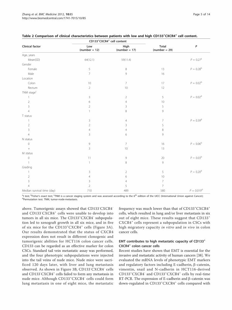
ResultsCD133+CXCR4+ cancer cell content is higher in hepaticmetastasis than in human primary colorectal tumorsWe collected tissue samples from 29 patients with CRC(patient characteristics are given in Table 2). First, we
aimed to identify CSCs with the widely recognized sur-face marker CD133 in primary CRCs, hepatic metastasisand their corresponding normal tissues. Flow cytometryanalysis demonstrated that a rare population of CSCswas present in primary CRCs, while they were hardlydetected in corresponding normal colorectal tissues.Furthermore, an increased number of CSCs were pre-sent in metastatic liver tumors, and the amount of CSCsin metastatic liver tumors was almost four times greaterthan those in primary colorectal tumors (Figure 1A).Next, because recent data have demonstrated that insome cancers there exists a subpopulation of migratingCSCs responsible for cancer metastasis and CXCR4 hasbeen reported to be associated with the cancer cellmetastasis phenotype, CD133+CXCR4+ cells were alsodetected by flow cytometry. Results showed that thecontent of CD133+CXCR4+ CSCs in metastatic livertumors was more than seven times higher than that inprimary CRCs (Figure 1B). These data demonstrate anenrichment of CD133+CXCR4+ cells in metastatic can-cers, which indicates that these cells may play a poten-tial role in hepatic metastasis of CRC.
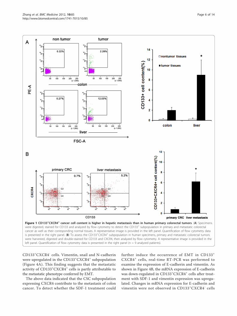
CD133+CXCR4+ colon cancer cells show higher migratorycapacity than CD133+CXCR4- cancer cells in vitroAs Figure 1 showed that CD133+CXCR4+cells wereincreased in hepatic metastasis, in order to investigate theunderlying mechanism of the phenomenon, we employedthe human colon cancer cell line HCT-116 for in vitro andin vivo studies. Representative staining of CD133 andCXCR4 via flow cytometry is shown in Figure 2A. Foursubgroups of cells were isolated using a high-speedFlowAria (Becton Dickinson) including CD133-CXCR4-;CD133-CXCR4+; CD133+CXCR4-; and CD133+CXCR4+
subgroups. We performed clonogenic assays to detect theclonogenic capacity of the four phenotypic subpopulations.As shown in Figure 2B, much lower percentages ofCD133-CXCR4- and CD133-CXCR4+ cells could formclones compared with CD133+CXCR4- and CD133+
CXCR4+ cells. However, there was no significant differ-ence in clone number between the CD133-CXCR4- andCD133-CXCR4+ groups, and between the CD133+CXCR4-
and CD133+CXCR4+ groups. Next we performed transwellmigration and invasion assays to compare the migratoryand invasive capacities between CD133+CXCR4- andCD133+CXCR4+ cells. Our results showed that the num-bers of migratory and invasive cells in the lower chamberof the CD133+CXCR4+ group were greater than those inthe CD133+CXCR4- group (Figure 2C and 2D).
CD133+CXCR4+ colon cancer cells have higher metastaticpotential in the nude mice modelTumorigenic and standard tail vein metastatic assayswere employed to validate the in vitro findings reported
Zhang et al. BMC Medicine 2012, 10:85http://www.biomedcentral.com/1741-7015/10/85
Page 4 of 14
above. Tumorigenic assays showed that CD133-CXCR4-
and CD133-CXCR4+ cells were unable to develop intotumors in all six mice. The CD133+CXCR4- subpopula-tion led to xenograft growth in all six mice, and in fiveof six mice for the CD133+CXCR4+ cells (Figure 3A).Our results demonstrated that the status of CXCR4expression does not result in different clonogenic andtumorigenic abilities for HCT116 colon cancer cells.CD133 can be regarded as an effective marker for colonCSCs. Standard tail vein metastatic assay was performed,and the four phenotypic subpopulations were injectedinto the tail veins of nude mice. Nude mice were sacri-ficed 120 days later, with liver and lung metastasisobserved. As shown in Figure 3B, CD133-CXCR4- cellsand CD133-CXCR4+ cells failed to form any metastasis innude mice. Although CD133+CXCR4- cells could formlung metastasis in one of eight mice, the metastatic
frequency was much lower than that of CD133+CXCR4+
cells, which resulted in lung and/or liver metastasis in sixout of eight mice. These results suggest that CD133+
CXCR4+ cells represent a subpopulation in CSCs withhigh migratory capacity in vitro and in vivo in coloncancer cells.
EMT contributes to high metastatic capacity of CD133+
CXCR4+ colon cancer cellsRecent studies have shown that EMT is essential for theinvasive and metastatic activity of human cancers [38]. Weevaluated the mRNA levels of phenotypic EMT markersand regulatory factors including E-cadherin, b-catenin,vimentin, snail and N-cadherin in HCT116-derivedCD133+CXCR4- and CD133+CXCR4+ cells by real-timeRT-PCR. The expression of E-cadherin and b-catenin wasdown-regulated in CD133+CXCR4+ cells compared with
Table 2 Comparison of clinical characteristics between patients with low and high CD133+CXCR4+ cell content.
CD133+CXCR4+ cell content
Clinical factor Low(number = 12)
High(number = 17)
Total(number = 29)
P
Age, years
Mean(SD) 64(12.1) 59(11.4) P = 0.27a
Gender
Female 5 8 13 P = 0.28b
Male 7 9 16
Location
Colon 10 7 17 P = 0.02b
Rectum 2 10 12
TNM stagec
1 3 2 5 P = 0.02d
2 6 4 10
3 2 3 5
4 1 8 9
T status
1 3 4 7 P = 0.39d
2 2 3 5
3 4 4 8
4 3 6 9
N status
0 9 7 16 P = 0.06b
+ 3 10 13
M status
0 11 9 20 P = 0.03b
1 1 8 9
Grading
1 3 2 5 P = 0.20d
2 4 6 10
3 3 4 7
4 2 5 7
Median survival time (day) 710 489 580 P = 0.019d
at test; bFisher’s exact test; cTNM is a cancer staging system and was assessed according to the 6th edition of the UICC (International Union against Cancer);dPermutation test. TNM, tumor-node-metastasis.
Zhang et al. BMC Medicine 2012, 10:85http://www.biomedcentral.com/1741-7015/10/85
Page 5 of 14
CD133+CXCR4- cells. Vimentin, snail and N-cadherinwere upregulated in the CD133+CXCR4+ subpopulation(Figure 4A). This finding suggests that the metastaticactivity of CD133+CXCR4+ cells is partly attributable tothe metastatic phenotype conferred by EMT.The above data indicated that the CSC subpopulation
expressing CXCR4 contribute to the metastasis of coloncancer. To detect whether the SDF-1 treatment could
further induce the occurrence of EMT in CD133+
CXCR4+ cells, real-time RT-PCR was performed toexamine the expression of E-cadherin and vimentin. Asshown in Figure 4B, the mRNA expression of E-cadherinwas down-regulated in CD133+CXCR4+ cells after treat-ment with SDF-1 and vimentin expression was upregu-lated. Changes in mRNA expression for E-cadherin andvimentin were not observed in CD133+CXCR4- cells
Figure 1 CD133+CXCR4+ cancer cell content is higher in hepatic metastasis than in human primary colorectal tumors. (A) Specimenswere digested, stained for CD133 and analyzed by flow cytometry to detect the CD133+ subpopulation in primary and metastatic colorectalcancer as well as their corresponding normal tissues. A representative image is provided in the left panel. Quantification of flow cytometry datais presented in the right panel. (B) To assess the CD133+CXCR4+ subpopulation in human specimens, primary and metastatic colorectal tumorswere harvested, digested and double-stained for CD133 and CXCR4, then analyzed by flow cytometry. A representative image is provided in theleft panel. Quantification of flow cytometry data is presented in the right panel (n = 9 analyzed patients).
Zhang et al. BMC Medicine 2012, 10:85http://www.biomedcentral.com/1741-7015/10/85
Page 6 of 14
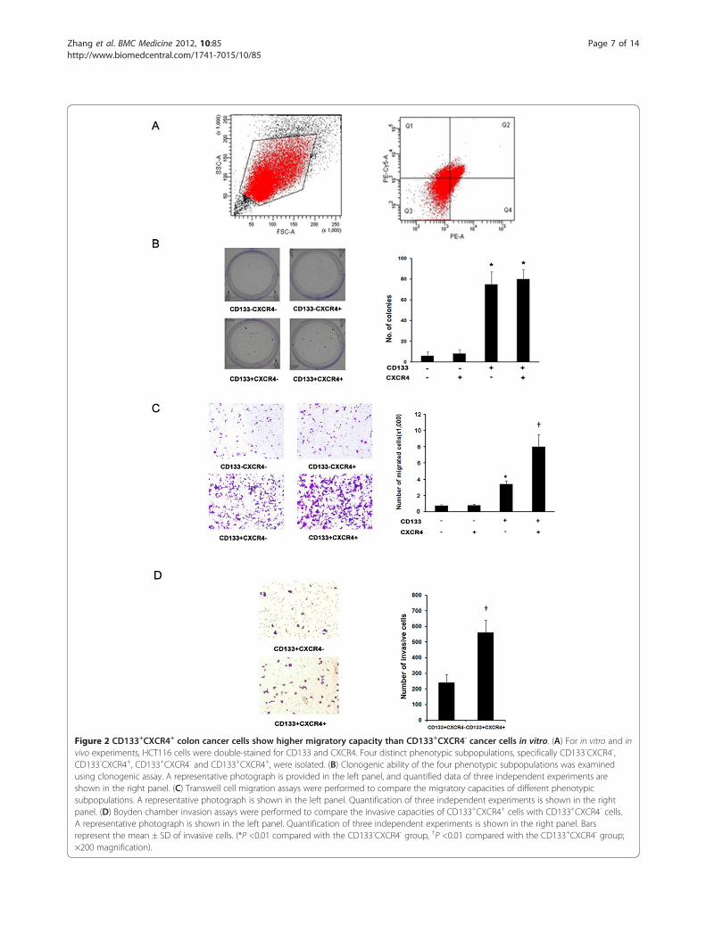
Figure 2 CD133+CXCR4+ colon cancer cells show higher migratory capacity than CD133+CXCR4- cancer cells in vitro. (A) For in vitro and invivo experiments, HCT116 cells were double-stained for CD133 and CXCR4. Four distinct phenotypic subpopulations, specifically CD133-CXCR4-,CD133-CXCR4+, CD133+CXCR4- and CD133+CXCR4+, were isolated. (B) Clonogenic ability of the four phenotypic subpopulations was examinedusing clonogenic assay. A representative photograph is provided in the left panel, and quantified data of three independent experiments areshown in the right panel. (C) Transwell cell migration assays were performed to compare the migratory capacities of different phenotypicsubpopulations. A representative photograph is shown in the left panel. Quantification of three independent experiments is shown in the rightpanel. (D) Boyden chamber invasion assays were performed to compare the invasive capacities of CD133+CXCR4+ cells with CD133+CXCR4- cells.A representative photograph is shown in the left panel. Quantification of three independent experiments is shown in the right panel. Barsrepresent the mean ± SD of invasive cells. (*P <0.01 compared with the CD133-CXCR4- group, †P <0.01 compared with the CD133+CXCR4- group;×200 magnification).
Zhang et al. BMC Medicine 2012, 10:85http://www.biomedcentral.com/1741-7015/10/85
Page 7 of 14
Figure 3 CD133+CXCR4+ colon cancer cells show higher migratory capacity than CD133+CXCR4- cancer cells in vivo. (A) To assess thetumorigenic capacity of the four phenotypic subpopulations, cells were injected subcutaneously into nude mice. At 30 days post-injection, micewere killed and xenografts excised for evaluation. A representative photograph of four mice from four groups is shown in the upper panel. Thetumorigenesis data from all groups are shown in the lower panel. Data represent the mean ± SD of tumor weight. (B) Standard tail veinmetastatic assays were used to confirm that CD133+CXCR4+ cells were responsible for metastatic cancer formation. Four phenotypicsubpopulations isolated from HCT116 cells were injected into the tail veins of nude mice. After 120 days, the mice were sacrificed, and livers andlungs were harvested to observe metastatic tumor formation. A representative photograph of harvested lungs from the four groups is shown inthe upper panel, with corresponding hematoxylin and eosin staining of metastatic lung tumor tissue. Arrows indicate metastatic lung nodules.The metastatic status of four groups is shown in the lower table. Comparisons between each group were made by Fisher’s exact test orStudent’s t-test (*P <0.05 compared with the CD133+CXCR4- group).
Zhang et al. BMC Medicine 2012, 10:85http://www.biomedcentral.com/1741-7015/10/85
Page 8 of 14

after SDF-1 treatment. The transwell invasion assay wasperformed to examine whether SDF-1 treatment couldalso enhance the invasive properties of the CD133+
CXCR4+ subpopulation rather than CD133+CXCR4-
cells. There was no significant difference in the numberof invasive cells in the lower chamber between untreated-and SDF-1-treated cells in the CD133+CXCR4- group,while SDF-1 treatment almost doubled the number of
Figure 4 EMT contributes to high metastatic capacity of CD133+CXCR4+ colon cancer cells. (A) Expression levels of mRNAs encoding E-cadherin, b-catenin, vimentin, Snail, and N-cadherin in CD133+CXCR4+ cells and CD133+CXCR4- cells, as determined by real-time RT-PCR. GAPDHmRNA was used to normalize the variability in template loading. The data are reported as mean ± SD. (*P <0.05; **P <0.01 compared with theCD133+CXCR4- group). (B) Real-time RT-PCR was performed to determine the mRNA expression levels of E-cadherin (left panel) and vimentin(right panel) in CD133+CXCR4- and CD133+CXCR4+ cells with or without SDF-1 treatment. GAPDH mRNA was used to normalize the variability intemplate loading. The data are reported as mean ± SD. (*P <0.05 compared with the control CD133+CXCR4- group). (C) A Boyden chamberassay was performed to compare the invasive capacities of CD133+CXCR4- and CD133+CXCR4+ cells with or without SDF-1 treatment. Arepresentative photograph is shown in the left panel. Quantification of three independent experiments is shown in the right panel. Barsrepresent the mean ± SD of invasive cells. (*P <0.05 compared with the control CD133+CXCR4+group). EMT, epithelial-mesenchylal transition;SDF-1, stromal cell-derived factor-1.
Zhang et al. BMC Medicine 2012, 10:85http://www.biomedcentral.com/1741-7015/10/85
Page 9 of 14
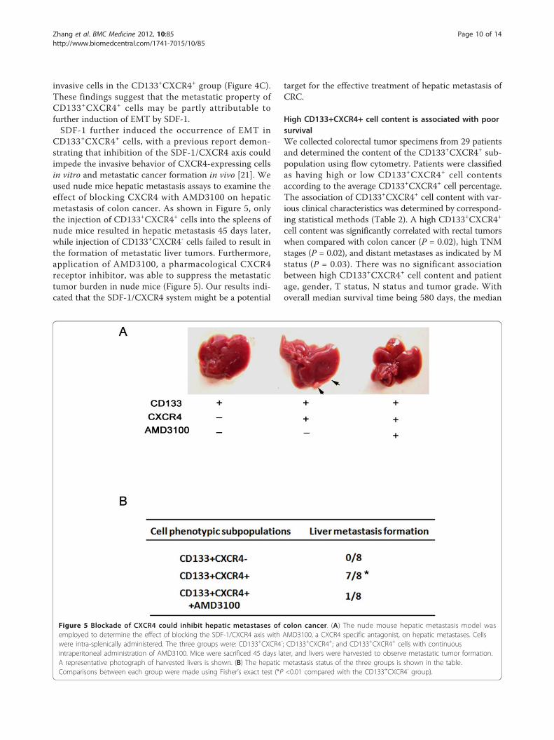
invasive cells in the CD133+CXCR4+ group (Figure 4C).These findings suggest that the metastatic property ofCD133+CXCR4+ cells may be partly attributable tofurther induction of EMT by SDF-1.SDF-1 further induced the occurrence of EMT in
CD133+CXCR4+ cells, with a previous report demon-strating that inhibition of the SDF-1/CXCR4 axis couldimpede the invasive behavior of CXCR4-expressing cellsin vitro and metastatic cancer formation in vivo [21]. Weused nude mice hepatic metastasis assays to examine theeffect of blocking CXCR4 with AMD3100 on hepaticmetastasis of colon cancer. As shown in Figure 5, onlythe injection of CD133+CXCR4+ cells into the spleens ofnude mice resulted in hepatic metastasis 45 days later,while injection of CD133+CXCR4- cells failed to result inthe formation of metastatic liver tumors. Furthermore,application of AMD3100, a pharmacological CXCR4receptor inhibitor, was able to suppress the metastatictumor burden in nude mice (Figure 5). Our results indi-cated that the SDF-1/CXCR4 system might be a potential
target for the effective treatment of hepatic metastasis ofCRC.
High CD133+CXCR4+ cell content is associated with poorsurvivalWe collected colorectal tumor specimens from 29 patientsand determined the content of the CD133+CXCR4+ sub-population using flow cytometry. Patients were classifiedas having high or low CD133+CXCR4+ cell contentsaccording to the average CD133+CXCR4+ cell percentage.The association of CD133+CXCR4+ cell content with var-ious clinical characteristics was determined by correspond-ing statistical methods (Table 2). A high CD133+CXCR4+
cell content was significantly correlated with rectal tumorswhen compared with colon cancer (P = 0.02), high TNMstages (P = 0.02), and distant metastases as indicated by Mstatus (P = 0.03). There was no significant associationbetween high CD133+CXCR4+ cell content and patientage, gender, T status, N status and tumor grade. Withoverall median survival time being 580 days, the median
Figure 5 Blockade of CXCR4 could inhibit hepatic metastases of colon cancer. (A) The nude mouse hepatic metastasis model wasemployed to determine the effect of blocking the SDF-1/CXCR4 axis with AMD3100, a CXCR4 specific antagonist, on hepatic metastases. Cellswere intra-splenically administered. The three groups were: CD133+CXCR4-; CD133+CXCR4+; and CD133+CXCR4+ cells with continuousintraperitoneal administration of AMD3100. Mice were sacrificed 45 days later, and livers were harvested to observe metastatic tumor formation.A representative photograph of harvested livers is shown. (B) The hepatic metastasis status of the three groups is shown in the table.Comparisons between each group were made using Fisher’s exact test (*P <0.01 compared with the CD133+CXCR4- group).
Zhang et al. BMC Medicine 2012, 10:85http://www.biomedcentral.com/1741-7015/10/85
Page 10 of 14
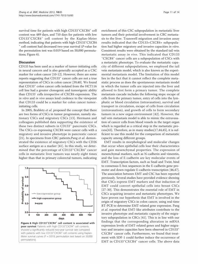
survival time for patients with high CD133+CXCR4+ cellcontent was 489 days, and 710 days for patients with lowCD133+CXCR4+ cell content by the Kaplan-Meiermethod, indicating that patients with high CD133+CXCR4+ cell content had decreased two-year survival (P-value forthe permutation test was 0.019 based on 50,000 permuta-tions; Figure 6).
DiscussionCD133 has been used as a marker of tumor-initiating cellsin neural cancers and is also generally accepted as a CSCmarker for colon cancer [10-12]. However, there are somereports suggesting that CD133+ cancer cells are not a truerepresentation of CSCs in colon cancer [39,40]. We foundthat CD133+ colon cancer cells isolated from the HCT116cell line had a greater clonogenic and tumorigenic abilitythan CD133- cells irrespective of CXCR4 expression. Thein vitro and in vivo assays lend credence to the viewpointthat CD133 could be a marker for colon cancer tumor-initiating cells.In 2005, Brabletz et al. proposed the concept that there
are two forms of CSCs in tumor progression, namely sta-tionary CSCs and migratory CSCs [13]. Hermann andcolleagues published data supporting the existence ofthese two distinct subsets in CD133+ pancreatic CSCs.The CSCs co-expressing CXCR4 were cancer cells with amigratory and invasive phenotype in pancreatic cancer[21]. In specimens from CRC patients,Pang et al. demon-strated the existence of migratory CSCs with the CD26surface antigen as a marker [41]. In this study, we deter-mined that the percentage of CD133+CXCR4+ cancercells in metastatic liver tumors was nearly eight timeshigher than that in primary colorectal tumors, indicating
enrichment of this CSC subpopulation in metastatic livertumors and their potential involvement in CRC metasta-sis to the liver. Transwell migration and invasion assayresults indicated that the CD133+CXCR4+ subpopula-tion had higher migratory and invasive capacities in vitro.Consistent results were obtained by the standard tail veinmetastatic assay in vivo. This indicated that CD133+CXCR4+ cancer cells are a subpopulation of CSCs witha metastatic phenotype. To evaluate the metastatic capa-city of different subpopulations, we employed the tailvein metastasis model, which is also known as the experi-mental metastasis model. The limitation of this modellies in the fact that it cannot reflect the complete meta-static process as does the spontaneous metastasis modelin which the tumor cells are injected into the liver andallowed to first form a primary tumor. The completemetastasis cascade includes the following steps: escape ofcells from the primary tumor, entry of cells into the lym-phatic or blood circulation (intravasation), survival andtransport in circulation, escape of cells from circulation(extravasation), and growth of cells to form secondarytumors in a new organ environment [42]. However, thetail vein metastasis model is able to mimic the extravasa-tion of cancer cells from blood vessels in the target organwhich is regarded as a critical step in the metastatic pro-cess[43]. Therefore, as in many studies[17,44,45], it is suf-ficient to use this model for the comparison of metastaticcapacity among different groups.EMT results in morphological and molecular changes
that occur when epithelial cells lose their characteristicsand gain mesenchymal properties. The expression ofmesenchymal markers, such as N-cadherin and vimentin,and the loss of E-cadherin are key molecular events ofEMT. Transcription factors, such as Snail and Twist, bindto consensus E-box sequences in the E-cadherin gene pro-moter and down-regulate E-cadherin transcription [46,47].The association between EMT and CSC has been reportedpreviously. Several studies have provided evidence showingthat CSCs express EMT markers and that induction ofEMT could convert epithelial cells into breast CSCs[27-30]. This demonstrates the essential role of EMT inCSCs acquiring invasive and metastatic phenotypes. Wehave proven our hypothesis that EMT is involved in theorigin of migratory CSCs in colon cancer, using real-timeRT-PCR to determine EMT-related gene expression. Panget al. reported that EMT-like attributes contribute to theinvasive phenotype and metastatic capacity of the migra-tory subpopulation in CRCs [41]. This is in line with ourfindings that the corresponding alteration in mRNAexpression levels of EMT-related genes and higher migra-tory and invasive capacities have been observed in CD133+
CXCR4+ cancer cells. Furthermore, we found that treat-ment with SDF-1 could further induce the occurrence ofEMT in CD133+CXCR4+ cancer cells. The above data
Figure 6 High CD133+CXCR4+ cell content is associated withpoor survival. Patients with high CD133+CXCR4+ cell contentsshowed a significantly reduced two-year survival rate comparedwith patients with low CD133+CXCR4+ cell contents using Kaplan-Meier survival curves (P = 0.019, permutation test based on 50,000permutations).
Zhang et al. BMC Medicine 2012, 10:85http://www.biomedcentral.com/1741-7015/10/85
Page 11 of 14
indicate that the CD133+CXCR4+ subpopulation contri-butes to liver metastasis of colorectal cancer via EMT.Consistent with our findings, Esther and colleagues
demonstrated that transforming growth factor-b (TGF-b)induced the EMT process and de-differentiation in Fao rathepatoma cells. This process coincided with upregulatedCXCR4 expression and also sensitization of these cells torespond to SDF-1, which mediated migration [48]. Similarresults were observed in oral squamous cell carcinoma[26,49]. However, the reason cancer cells that have under-gone EMT have a higher expression of CXCR4 is far fromclear. Exploring the origin of migratory CSCs warrantsfurther research and requires integration of current tumorinitiation and progression concepts, including CSC, EMT,accumulation of genetic alterations and the tumor envir-onment as driving forces [13]. A deeper understanding ofthese factors could provide further insights into tumorbiology.The CSC hypothesis suggests that CSCs are a minority
population that has the potential to self-renew, differenti-ate and regenerate a phenocopy of the original tumor.They would seem the most probable candidates that areresistant to chemotherapy, and they have been investigatedpreviously [3,5,50-52]. Novel treatments targeting CSCsmay result in the complete eradication of tumor growth,and furthermore, based on the migratory CSC theory, iftreatment targeting migratory CSCs can be developed, itmight be possible to prevent tumor metastasis. Wehypothesized that blockade of the SDF-1/CXCR4 axismight suppress colon cancer metastasis to the liver, withthe knowledge that the liver secretes high amounts ofSDF-1 [53]. This is also in line with the theory that organsproducing SDF-1 attract CXCR4+ tumor cells and formmetastatic tumors analogous to the directed homing ofleukocytes. In our study, a nude mouse hepatic metastasismodel was employed, and the results indicated that chemi-cal inhibition of CXCR4 with AMD3100 could inhibitcolon cancer metastasis to the liver. The anti-metastasiseffect caused by the blockade of the SDF-1/CXCR4 axis issupported by another report [54]. This finding providesimportant clues for the development of a targeted therapyin the treatment of CRC.To validate the above findings in in vitro experimental
and in animal models, we carried out a prospective studyto investigate whether CD133+CXCR4+ cancer cell con-tent was associated with disease progression and prog-nosis. Statistical analysis showed that high CD133+CXCR4+ cell content is associated with poor 2-year survival ofcolorectal cancer patients. The clinical data provide evi-dence to support our hypothesis that double positive can-cer cells might be involved in the metastatic process. Ourdata showed that cancer located in the rectum was asso-ciated with a high content of CD133+CXCR4+ cancer cell
compared with colon cancer. This might be due to higherCXCR4 expression in rectal cancer than in colon cancer[20], suggesting that the percentage of CD133+CXCR4+
cancer cells in future studies should be investigated sepa-rately in colon and rectal cancer rather than in a mixedway.
ConclusionsTaken together, our data demonstrate that CD133+
CXCR4+ cancer cells are possible migratory CSC subtypesin CRC. EMT is partly involved in these cells acquiring aninvasive phenotype and metastatic behavior. Blockade ofthe SDF-1/CXCR4 axis could be developed for targetedtherapy to control CRC metastasis.
AbbreviationsCRC: colorectal cancer; CSC: cancer stem cell; EMT: epithelial-mesenchymaltransition; PBS: phosphate buffered saline; RT-PCR: reverse transcriptase-polymerase chain reaction; SDF-1: stromal cell-derived factor-1; TNM: tumor-node-metastasis.
AcknowledgementsThis project was supported by Key project of the National Natural ScienceFoundation of China(Grant NO: 81030041); Key Basic Research Project ofChina (Grant NO: 2010CB945600, 2011CB966200); National Natural ScienceFoundation of China (Grant NO: 30870974, 30801347, 30901722, 31171321,81000970, 81101622, 30973433, 30801094); Special Funds for National keySci-Tech Special Project of China (Grant NO: 2008ZX10002-019,2008ZX10002-025); Shanghai Science and Technology Committee (Grant NO:10ZR1439600, 11ZR1449500, 10411963100, 10ZR1439900, 11nm0504700,09QA1407200, 07zR14143, 2008B009); Shanghai Municipal Health Bureau(Grant NO: XYQ2011044); Science Fund for Creative Research Groups, NSFC,China(Grant NO: 30921006); Stem Cell and Medicine Research Center’sInnovation Research Program(NO:SCMRC1102) and 2011 Ph.D. GraduateInnovation Fund Project(NO:27).
Author details1Tumor Immunology and Gene Therapy Center, Eastern HepatobiliarySurgery Hospital, Second Military Medical University, 225 Changhai Road,Shanghai 200438, China. 2Department of Pharmacology, Second MilitaryMedical University, 325 Guohe Road, Shanghai 200433, China. 3ChanghaiHospital, Second Military Medical University, 168 Changhai Road, Shanghai200433, China.
Authors’ contributionsParticipated in the conception and design of the study and the criticalrevision of the manuscript for important intellectual content: LZ, LW, SZ, YJ,ZH. Performed the data collection and analysis: ST, HW, YW, RL, YY, XZ, XX,EY, YR, HL. Interpreted the data and produced the draft of the manuscript:SZ, ZH, ST, TL, HW, YW, RL, YY, XZ, XX, EY, YR, HL. Obtained funding for thestudy: LZ, LW. All authors read and approved the final version of themanuscript.
Competing interestsThe authors declare that they have no competing interests.
Received: 31 October 2011 Accepted: 7 August 2012Published: 7 August 2012
References1. Ries LAG, Eisner MP, Kosary CL: SEER Cancer Statistics Review. Bethesda,
MD: National Cancer Institute; 2002.2. Jemal A, Siegel R, Ward E, Hao Y, Xu J, Murray T, Thun MJ: Cancer statistics.
CA Cancer J Clin 2008, 58:71-96.
Zhang et al. BMC Medicine 2012, 10:85http://www.biomedcentral.com/1741-7015/10/85
Page 12 of 14
3. Clarke MF, Dick JE, Dirks PB, Eaves CJ, Jamieson CHM, Jones DL, Visvader J,Weissman IL, Wahl GM: Cancer stem cells–perspectives on current statusand future directions: AACR Workshop on Cancer Stem Cells. Cancer Res2006, 66:9339-9344.
4. Tannishtha R, Sean JM, Michael FC, Irving LW: Stem cells, cancer, andcancer stem cells. Nature 2001, 414:105-111.
5. Tang C, Ang BT, Pervaiz S: Cancer stem cell: target for anti-cancertherapy. FASEB J 2007, 21:3777-85.
6. Bonnet D, Dick JE: Human acute myeloid leukemia is organized as ahierarchy that originates from a primitive hematopoietic cell. Nat Med1997, 3:730-737.
7. Al-Hajj M, Wicha MS, Benito-Hernandez A: Prospective identification oftumorigenic breast cancer cells. Proc Natl Acad Sci USA 2003,100:3983-3988.
8. Collins AT, Berry PA, Hyde C: Prospective identification of tumorigenicprostate cancer stem cells. Cancer Res 2005, 65:10946-10951.
9. Prince ME, Sivanandan R, Kaczorowski A: Identification of a subpopulationof cells with cancer stem cell properties in head and neck squamouscell carcinoma. Proc Natl Acad Sci USA 2007, 104:973-978.
10. Singh SK, Hawkins C, Clarke ID: Identification of human brain tumourinitiating cells. Nature 2004, 432:396-401.
11. O’Brien CA, Pollett A, Gallinger S: A human colon cancer cell capable ofinitiating tumour growth in immunodeficient mice. Nature 2007,445:106-110.
12. Ricci-Vitiani L, Lombardi DG, Pilozzi E: Identification and expansion ofhuman colon cancer-initiating cells. Nature 2007, 445:111-115.
13. Brabletz T, Jung A, Spaderna S: Opinion: migrating cancer stem cells – anintegrated concept of malignant tumour progression. Nat Rev Cancer2005, , 5:: 744-749.
14. Stetler-Stevenson WG, Kleiner DE Jr: Molecular biology of cancer: invasionand metastasis. In Cancer: Principles and Practice of Oncology. Edited by:DeVita VT Jr, Hellman S, Rosenberg SA. Philadelphia, PA: Lippincott Williams2001:123-136.
15. Kucia M, Reca R, Miekus K: Trafficking of normal stem cells and metastasisof cancer stem cells involve similar mechanisms: pivotal role of the SDF-1-CXCR4 axis. Stem Cells 2005, 23:879-894.
16. Muller A, Homey B, Soto H: Involvement of chemokine receptors inbreast cancer metastasis. Nature 2001, 410:50-56.
17. Saur D, Seidler B, Schneider G: CXCR4 expression increases liver and lungmetastasis in a mouse model of pancreatic cancer. Gastroenterology 2005,129:1237-1250.
18. Zeelenberg IS, Ruuls-Van Stalle L, Roos E: The chemokine receptor CXCR4is required for outgrowth of colon carcinoma micrometastases. CancerRes 2003, 63:3833-3839.
19. Kim J, Takeuchi H, Lam ST: Chemokine receptor CXCR4 expression incolorectal cancer patients increases the risk for recurrence and for poorsurvival. J Clin Oncol 2005, 23:2744-2753.
20. Schimanski CC, Schwald S, Simiantonaki N: Effect of chemokine receptorsCXCR4 and CCR7 on the metastatic behavior of human colorectalcancer. Clin Cancer Res 2005, 11:1743-1750.
21. Hermann PC, Huber SL, Herrler T: Distinct populations of cancer stemcells determine tumor growth and metastatic activity in humanpancreatic cancer. Cell Stem Cell 2007, 1:313-323.
22. Gavert N, Ben-Ze’ev A: Epithelial-mesenchymal transition and the invasivepotential of tumors. Trends Mol Med 2008, 14:199-209.
23. Thiery JP, Sleeman JP: Complex networks orchestrate epithelial-mesenchymal transitions. Nat Rev Mol Cell Biol 2006, 7:131-142.
24. Gilles C, Thompson EW: The epithelial to mesenchymal transition andmetastatic progression in carcinoma. Breast J 1996, 2:83-96.
25. Thiery JP: Epithelial-mesenchymal transitions in tumour progression. NatRev Cancer 2002, 2:442-454.
26. Onoue T, Uchida D, Begum NM: Epithelial-mesenchymal transitioninduced by the stromal cell-derived factor-1/CXCR4 system in oralsquamous cell carcinoma cells. Int J Oncol 2006, 29:1133-1138.
27. Na DC, Lee JE, Yoo JE: Invasion and EMT-associated genes are up-regulated in B viral hepatocellular carcinoma with high expression ofCD133-human and cell culture study. Exp Mol Pathol 2011, 90:66-73.
28. Mani SA, Guo W, Liao MJ: The epithelial-mesenchymal transitiongenerates cells with properties of stem cells. Cell 2008, 133:704-715.
29. Morel AP, Lievre M, Thomas C: Generation of breast cancer stem cellsthrough epithelial-mesenchymal transition. PLoS One 2008, 3:e2888.
30. Lo JF, Yu CC, Chiou SH: The epithelial-mesenchymal transition mediatorS100A4 maintains cancer initiating cells in head and neck cancers.Cancer Res 2011, 71:1912-1923.
31. Li G, Liu C, Yuan J: CD133+ single cell-derived progenies of colorectalcancer cell line SW480 with different invasive and metastatic potential.Clin Exp Metastasis 2010, 27:517-527.
32. Hiraoka K, Kimura T, Logg CR: Therapeutic efficacy of replication-competent retrovirus vector-mediated suicide gene therapy in amultifocal colorectal cancer metastasis model. Cancer Res 2007,67:5345-5353.
33. Ludbrook J, Dudley H: Why permutation tests are superior to t and Ftests in biomedical research. Am Stat 1998, 52:127-132.
34. Hesterberg T, Moore DS, Monaghan S, Clipson A, Epstein R: Bootstrapmethods and permutation tests. In Introduction to the Practice of Statistics.Volume 14.. 5 edition. Edited by: Moore DS, MacCabe GP. New York: WHFreeman 2005:1-70.
35. Marozzi M: A bi-aspect nonparametric test for the two-sample locationproblem. Comput Stat Data Anal 2004, 44:639-648.
36. Dunn OJ, Clark VA: Basic Statistics: A Primer for the Biomedical Sciences. 4edition. (Wiley Series in Probability and Statistics). Hoboken, NJ: John Wiley& Sons; 2009.
37. He J, Jin ZC, Yu DH: Statistical reporting in Chinese biomedical journals.Lancet 2009, 373:2091-2093.
38. Thiery JP: Epithelial-mesenchymal transition in tumour progression. NatRev Cancer 2002, 2:442-454.
39. LaBarge MA, Bissell MJ: Is CD133 a marker of metastatic colon cancerstem cells? J Clin Invest 2008, 118:2021-2024.
40. Zhi-Li Y, Qi Z, Jun Y: Upregulated CD133 expression in tumorigenesis ofcolon cancer cells. World J Gastroenterol 2011, 17:932-937.
41. Pang R, Law WL, Chu AC: A subpopulation of CD26+ cancer stem cellswith metastatic capacity in human colorectal cancer. Cell Stem Cell 2010,6:603-615.
42. Ruoslahti E: How cancer spreads. Sci Am 1996, 275:72-77.43. Elkin M, Vlodavsky I: Tail vein assay of cancer metastasis. Curr Protoc Cell
Biol 2001, Chapter 19:Unit 19.2.44. Johnson JL, Pillai S, Pernazza D, Sebti SM, Lawrence NJ, Chellappan SP:
Regulation of matrix metalloproteinase genes by E2F transcriptionfactors: Rb-Raf-1 interaction as a novel target for metastatic disease.Cancer Res 2012, 72:516-526.
45. Yang CH, Yue J, Pfeffer SR, Handorf CR, Pfeffer LM: MicroRNA miR-21regulates the metastatic behavior of B16 melanoma cells. J Biol Chem2011, 286:39172-39178.
46. Batlle E, Sancho E, Franci C: The transcription factor snail is a repressor ofE-cadherin gene expression in epithelial tumour cells. Nat Cell Biol 2000,2:84-89.
47. Gavert N, Ben-Ze’ev A: Epithelial-mesenchymal transition and the invasivepotential of tumors. Trends Mol Med 2008, 14:199-209.
48. Bertran E, Caja L, Navarro E: Role of CXCR4/SDF-1α in the migratoryphenotype of hepatoma cells that have undergone epithelial-mesenchymal transition in response to the transforming growth factor-β. Cell Signal 2009, 21:1595-1606.
49. Taki M, Higashikawa K, Yoneda S: Up-regulation of stromal cell-derivedfactor-1α and its receptor CXCR4 expression accompanied withepithelial-mesenchymal transition in human oral squamous cellcarcinoma. Oncol Rep 2008, 19:993-998.
50. Hirschmann-Jax C, Foster AE, Wulf GG: A distinct “side population” of cellswith high drug efflux capacity in human tumor cells. Proc Natl Acad SciUSA 2004, 101:14228-14233.
51. Zhou S, Schuetz JD, Bunting KD: The ABC transporter Bcrp1/ABCG2 isexpressed in a wide variety of stem cells and is a molecular determinantof the side-population phenotype. Nat Med 2001, 7:1028-1034.
52. Liu G, Yuan X, Zeng Z: Analysis of gene expression and chemoresistanceof CD133+ cancer stem cells in glioblastoma. Mol Cancer 2006, 5:67.
53. Muller A, Homey B, Soto H, Ge N, Catron D, Buchanan ME, McClanahan T,Murphy E, Yuan W, Wagner SN, Barrera JL, Mohar A, Verastegui E, Zlotnik A:Involvement of chemokine receptors in breast cancer metastasis. Nature2001, 410:50-56.
Zhang et al. BMC Medicine 2012, 10:85http://www.biomedcentral.com/1741-7015/10/85
Page 13 of 14
54. Saur D, Seidler B, Schneider G: CXCR4 expression increases liver and lungmetastasis in a mouse model of pancreatic cancer. Gastroenterology 2005,129:1237-1250.
Pre-publication historyThe pre-publication history for this paper can be accessed here:http://www.biomedcentral.com/1741-7015/10/85/prepub
doi:10.1186/1741-7015-10-85Cite this article as: Zhang et al.: CD133+CXCR4+ colon cancer cellsexhibit metastatic potential and predict poor prognosis of patients.BMC Medicine 2012 10:85.
Submit your next manuscript to BioMed Centraland take full advantage of:
• Convenient online submission
• Thorough peer review
• No space constraints or color figure charges
• Immediate publication on acceptance
• Inclusion in PubMed, CAS, Scopus and Google Scholar
• Research which is freely available for redistribution
Submit your manuscript at www.biomedcentral.com/submit
Zhang et al. BMC Medicine 2012, 10:85http://www.biomedcentral.com/1741-7015/10/85
Page 14 of 14