cd150-mediated akt signalling pathway in normal and malignant b
TRANSCRIPT

Experimental Oncology ��� ����� ���� ��arc����� ����� ���� ��arc�� ��arc�� �
CD150-MEDIATED AKT SIGNALLING PATHWAY IN NORMAL AND MALIGNANT B CELLS
M. Yurchenko1,*, L.M. Shlapatska1, O.L. Romanets 1, D. Ganshevskiy 1, E. Kashuba1,2, A. Zamoshnikova1, Yu.V. Ushenin3, B.A. Snopok3, S.P. Sidorenko1
1R.E. Kavetsky Institute of Experimental Pathology, Oncology and Radiobiology NAS of Ukraine, Kyiv 03022, Ukraine
2Microbiology and Tumorbiology Center, Karolinska Institutet, Stockholm 171 77, Sweden3V.E. Lashkarev Institute of Semiconductors Physics NAS of Ukraine, Kyiv 03028, Ukraine
Aim: To study upstream and downstream events in CD150-mediated Akt signaling pathway in normal human B cells, EBV-transformed lymphoblastoid (LCL) and malignant Hodgkin’s lymphoma (HL) B cell lines. Methods: To access protein-protein interaction we applied immunoprecipitation, Western blot analysis and surface plasmon resonance (SPR) technique. A novel modification of SPR technique using reduced glutathione bound to golden surface was proposed. Immunostaining and isolation of cytoplasmic fractions and nuclear extracts were performed to detect proteins’ localization in cells. Western blot analysis was performed to follow up the phosphorylation of proteins on specific sites and proteins’ expression level. Results: It was shown that CD150 ligation induced Akt activation in normal tonsillar B cells (TBC), SH2D1A positive LCL and HL B cell lines. The p85α subunit of PI3K co-precipitated with CD150 cytoplasmic tail. This direct association depends on tyrosine phosphorylation and is mediated by N terminal SH2 domain of p85α. CD150 initiated phosphorylation of FoxO1 transcription factor in normal B cells as well as in LCL MP-1 and HL cell line L1236. At the same time, CD150 ligation triggered GSK-3β kinase phosphorylation only in immortalized LCL MP-1 and HL cell line L1236. Conclusions: We have demonstrated that CD150 receptor could trigger PI3K-mediated Akt signaling pathway in normal, EBV-transformed and malignant B cells. CD150-mediated phosphorylation of Akt downstream targets GSK-3β and FoxO1 in EBV-transformed and HL cells could be one of the mechanisms to avoid apoptosis and support survival program in these immortalized B cells. Key Words: CD150 receptor, PI3 kinase, Akt, GSK-3β, FoxO1, immortalized B cells, Hodgkin’s lymphoma.
CD�5� �IPO-�/SLA�� is a member of CD� family of t�e immonoglobulin �Ig� superfamily of surface receptors [��5]. It is expressed on activated T and B lymp�ocytes� dendritic cells and monocytes [�� 6]. Low level of CD�5� expression was also found on natural killer T cells� platelets� basop�iles and ma-ture dendritic cells [7� �]. T�is receptor is upregulated via antigen receptors� CD4�� Toll-like receptors and by mitogenes or cytokines �reviewed in [�]�. T�ere are experimental evidences t�at CD�5� and SH�D�A adaptor protein are co-expressed during a narrow window of B cell maturation� and SH�D�A may be in-volved in regulation of B cell differentiation via switc�-ing of CD�5�-mediated signaling pat�ways [��� ��]. CD�5� is co-expressed wit� SH�D�A adaptor protein in some B-lymp�oblastoid cell lines and lymp�oma cells wit� activated B cell p�enotype� suc� as Hodg-kin’s lymp�oma �HL� and ABC-type diffuse large B cell lymp�oma [��� ��].
Tumor cells of classical HL are known as Hodgkin and Reed-Sternberg cells �HRS�� and originate from preapoptotic germinal center B cells t�at do not ex-press functional B cell receptor �BCR� [��� �4]. HRS cells are able to escape t�e regulation mec�anisms aimed to eliminate B cells lacking functional BCR. Sev-eral aberrantly activated signaling pat�ways �ave been identified t�at contribute to t�e rescue of HRS cells from apoptosis: JAK-STAT [�5� �6]� �APK/ERK initiated by CD��-� CD4�-� RANK [�7] and CD�5� [��] signaling� PI�K/AKT linked to CD4� [��]� L�P� and L�P�a in EBV-positive cases [�����]; NF-kappaB due to aberrant expression� mutations� CD4�� CD�� stimu-lation �reviewed in [��� �4� ��]�.
Activation of t�e p�osp�atidylinositol �-kinase �PI�K� pat�way �as been linked wit� tumor cell growt�� survival and resistance to t�erapy in several cancer types [�4]. T�e main downstream PI�K effector� w�ic� controls t�e survival of normal and malignant cells� is Akt/PKB [�5]. Ligation of tumor necrosis factor �TNF� family receptors could induce Akt p�osp�orylation/ac-tivation in normal [�6] and HRS cells [��]. Previously� we �ave demonstrated t�at Akt could be activated via CD�5� in DT4� model system� lymp�oblastoid cell line �P-� [��� �7] and HL cells L���6 [��]. T�e �ypot�esis was proposed t�at CD�5�-SH�D�A association could play decisive role in CD�5�-mediated Akt signaling. In current study we furt�er explored t�e upstream and downstream events in CD�5�-mediated Akt pat�way in normal and malignant B cells.
Received: November 15, 2010. *Correspondence: Fax: +380442581656; E-mail: [email protected] Abbreviations used: BCR – B cell receptor; CD150ct – CD150 cy-toplasmic tail; Cyt – cytoplasmic �raction; �� – �od�kin�s lym-Cyt – cytoplasmic �raction; �� – �od�kin�s lym-�� – �od�kin�s lym-phoma; �RS – �od�kin�s and Reed-Sternber�; ��S� – immu-��S� – immu-notyrosine based switch moti�; �C� - lymphoblastoid cell line; NE – nuclear extract; NS�2 – N-terminal S�2 domain; P�3K - phosphatidylinositol 3-kinase; S�2D1� – S�2 domain contain-S�2D1� – S�2 domain contain-in� protein 1�; SPR – sur�ace plasmon resonance; �BC – tonsillar B cells; �C� – total cell lysate; �NF – tumor necrosis �actor.
Exp Oncol ������� �� ����
ORIGINAL CONTRIBUTIONS
�� Experimental Oncology ��� ����� ���� ��arc��
MATERIALS AND METHODSCell lines. T�e B lymp�oblastoid cell lines �B-LCL�
�P-�� CESS� T5-�� fres�ly infected LCLs from pe-rip�eral blood B cells and HL cell line of B cell origin L���6 were maintained in RP�I �64� medium con-taining ��% FBS� � m� L-glutamine� and antibiotics.
Human tissue specimens. After receiving informed consent in accordance wit� t�e Declara-tion of Helsinki� fres� tonsils were obtained from �� patients undergoing tonsillectomy. Tonsillar B cells were isolated by density fractionation on discontinu-ous Lymp�oprep �Axis-S�ield PoC AS� Norway� and Percoll �Sigma� USA� gradients as described [��]. �6.5���.6% of cells in CD�- populations were of B cell lineage since expressed CD�� and/or CD��� antigens.
GST proteins preparation. GST-fusion con-structs of CD�5� cytoplasmic tail �CD�5�ct� were prepared earlier and described in [��� �7]. cDNA of p�5α regulatory subunit was a kind gift of Dr. I. Gout �University College� London� UK�. N-terminal SH� do-main of p�5α regulatory subunit was cloned into pGEX-�T vector using specific primers� and sequenced �we t�ank for t�e �elp Dr. Vladimir Kas�uba� �TC� Karolinska institutet� Sweden�. T�e plasmid encoding GST-SH�D�A was kindly provided by Dr. Kim Nic�ols �T�e C�ildren’s Hospital of P�iladelp�ia� P�iladelp�ia� PA� USA�. Plasmids containing GST-CD�5�ct� GST-SH�D�A and GST-NSH�-p�5α were transformed into t�e Escherichia coli strains BL��DE� �Invitrogen� USA� to produce non-p�osp�orylated fusion proteins� and GST-CD�5�ct wit� and wit�out point mutations were transformed to TKX� strain �Stratagene� USA� for pro-duction of tyrosine-p�osp�orylated fusion proteins.
Surface plasmon resonance (SPR) instrumen-tation and SPR analysis. T�e substrates and cell design of scanning SPR spectrometer “BioSuplar” �“PLAS�ON-��5”� �V. Las�karev Institute of Semicon-ductor P�ysics� NAS of Ukraine� Kyiv� Ukraine� as well as its ot�er c�aracteristics are described in [��]. T�is spectrometer� w�ic� �as open measurement arc�itec-ture� was used to study protein�protein interactions. T�e peristaltic pump provided a constant solution flow �> 5� μl/min� t�roug� t�e experimental cell. SPR kinet-ics was analyzed wit� a model t�at takes into account t�e �eterogeneous processes at t�e interface using a stretc�ed exponential function [��].
Surface modification and protein immobiliza-tion. T�e surface cleaning and activation procedure �for planar metal electrodes of SPR c�ips� eac� wit� a metal layer 5�� Å t�ick consisting of a 45�-Å layer of gold on a 5�-Å c�romium ad�esive layer� was carried out at room temperature as follows. To remove organic con-tamination� t�e c�ips were was�ed in a fres�ly prepared mixture of aqueous �NH4��S�O� ��.�� �� and H�O� ���%� ��5:6 vol:vol� for �� min. Traces of �eavy metal ions were removed wit� a fres�ly prepared mixture H�O� ���%� and HCl ��7%� in water ��:�:�� vol:vol:vol� for �� min. T�e surface was modified by t�iols СOOH-�CH����-SH ��.� μМ� and CH�-�CH���-SH �� μМ�� �Sigma� USA� in ratio �:��� ��� �� �7 °С� [��]. C�ips were was�ed
by et�anol. Cadmium acetate ��� m� solution� room temperature� was used for activation of carboxyl groups at least for � � prior experiment. Cadmium acetate was used as an immobilization bridge� w�ic� �as selectiv-ity to t�iogroup of glutat�ione �as ligand� and interacts wit� carboxyl groups on c�arged t�iols. Glutat�ione immobilization: � m� solution of reduced glutat�i-one �Sigma� USA� in TBE buffer �рН �.�� was applied on c�ips’ surface for � � at room temperature. Proteins were immobilized in PBS buffer.
Biochemical methods and antibodies. Cell ly-sis� SDS-PAGE� Western blotting� stimulation of cells by anti-CD4� �G��-5� kind gift of Prof. Edward Clark� University of Was�ington� Seattle� WA� USA�� anti-Ig� �AffiniPure F�ab’�� fragment goat anti-�uman Ig� Fc5μ fragment specific� Jackson ImmunoResearc� laboratories� West Grove� PA� USA�� anti-CD�5� mAb �IPO-�� produced in IEPOR NASU� Kiev� Ukraine�� CD�5� immunoprecipitations were performed as de-scribed earlier [��� �7]. Western blot results were visualized using a ��x LumiGLO® Reagent and ��x Per-oxide �Cell Signaling Tec�nology� Beverly� �A� USA�. Goat antisera against actin� was purc�ased from Santa Cruz Biotec�nology �Santa Cruz� CA� USA�. Rabbit anti-SH�D�A serum was a kind gift of Dr. Kim Nic�ols �T�e C�ildren’s Hospital of P�iladelp�ia� P�iladelp�ia� PA� USA�. Rabbit anti-pAkt �Ser47�� and �T�r����� anti-Akt� anti-pFoxO��T�r�4�/Fox��a�T�r���� anti-FoxO�� anti-FoxO�a� anti-pGSK-�β �Ser��� anti-PI�K p�5α antibody� anti-PARP� anti-Bcl-� were purc�ased from Cell Signaling Tec�nology �Beverly� �A� USA�.
Immunostaining. T�e double staining for CD�5� receptor and SH�D�A adaptor protein was done as follows: cells were spun on glass slides ��� x ��4 per slide�; fixed in cold met�anol-acetone ��:�� at ��� oC for � �; re�ydrated wit� PBS for � �; stained wit� anti-CD�5� �IPO-�� mAb �5 μg/ml� for � �� followed by � was�es in PBS; incubated wit� �orse anti-mouse Texas Red-conjugated antibodies �VectorLab� CA� USA�� and was�ed � times. Cells were stained wit� anti-SH�D�A antibodies �dilution �:���� for � �� t�ree was�es in PBS� incubated wit� mouse anti-rabbit FITC-conjugated antibodies �DAKO� Den-mark�� and mounted wit� ��% glycerol solution in PBS t�at contained �.5% ��4-diazabicyclo-��.�.��octane �Sigma�. Bisbenzimide �Hoec�st ���5�� was added at a concentration of �.4 μg/ml to t�e last secondary antibody for DNA staining. T�e images were recorded on a DAS microscope Leitz D� RB �Leica Inc.� Deer-field� IL� USA� wit� a Hamamatsu dual mode cooled CCD camera C4��� �Hamamatsu City� Japan�.
Isolation of cytoplasmic fraction and nuclear extracts. Total cell lysates �TCL� were prepared from 5 x ��6 of cells in RIPA lysis buffer ��5� m� NaCl� �� m� Tris-HCl �pH �.��� � m� EGTA� � m� EDTA �pH �.��� �% Tryton X-���� protease in�ibitors’ cocktail �Sigma� USA�� � m� P�SF�� and sonicated on ice for �5 s �Sonicator �SE Soniprep �5� Plus� �SE �UK� Limited� UK�� spun for �5 min� �7��� g� +4 oC� su-pernatants transferred to fres� tubes. For subcellular

Experimental Oncology ��� ����� ���� ��arc����� ����� ���� ��arc�� ��arc�� ��
fractionation� control and stimulated cells ��� x ��6� were lysed in buffer A �5� m� NaCl� �� m� HEPES �pH �.��� 5�� m� sucrose� � m� EDTA �pH �.��� �.�% Tryton X-���� protease in�ibitors’ cocktail �Sigma� USA�� � m� P�SF�� vortexed and spun for � min ��75 g� +4 oC�. Cytoplasmic fractions �Cyt� of supernatant were collected to fres� tubes. Pellets �nuclei� were was�ed twice by �.5 ml of cold buffer B �5� m� NaCl� �� m� HEPES �pH �.��� �5% glycerol� �.� m� EDTA �pH �.��� and spun for � min ��75 g� +4 oC�. Pellets were resuspended in ��� μl of buffer C ��5� m� NaCl� �� m� HEPES �pH �.��� �5 % glycerol� �.� m� EDTA �pH �.��� protease in�ibitors’ cocktail �Sigma� USA�� � m� P�SF�� and sonicated on ice for �5 s� spun for �5 min� �7��� g� +4 oC� and superna-tants were transferred to fres� tubes. TCL� NE and Cyt fractions were stored at -�� oC. Protein concentration was measured in all samples �TCL� NE� Cyt�� and total amount of �� μg of protein was loaded to eac� well for Western blot analysis.
RESULTSp85α subunit of PI3K co-precipitates with
CD150 cytoplasmic tail. Previously� we �ave s�own t�at CD�5�-mediated signals lead to Akt S47� p�os-p�orylation in �uman B-lymp�oblastoid cell line �P-� [�7] and c�icken B cell line DT4� [��]. �oreover� using DT4� model system it was s�own t�at CD�5�-mediated Akt pat�way was dependent on SH�D�A binding to CD�5�� was negatively regulated by Lyn and Btk� and positively - by Syk [��]. Also� anot�er member of CD� family� CD�44 ��B4�� upon tyrosine p�osp�ory-lation was s�own to recruit not only SH�D�A� but also associate wit� t�e p�5α regulatory subunit of PI�K [��]. To answer t�e question w�et�er CD�5� could form a complex wit� PI�K in B cells� we performed immunoprecipitations of CD�5� by specific mAb �IPO-�� from tonsillar B cell �TBC� and �P-� lysates. Western blot analysis of t�ese immunoprecipitates demonstrated t�at p�5α regulatory subunit of PI�K co-precipitated wit� CD�5� from bot� examined lysates �Fig. ��.
MP-1
TBC
p85α PI3K
p85α PI3K
lysa
te
anti-
MO
PC
anti-
CD15
0
Fig. 1. PI�K p�5α regulatory subunit precipitated wit� CD�5� re-ceptor bot� from �P-� and tonsillar B cells �TBC�. Western blot analysis wit� anti-p�5α of CD�5� immunoprecipitates. Ig isotype matc�ed �OPC�� antibody served as negative control
CD150ct directly interacts with p85α subunit N terminal SH2 domain of PI3K. To test w�et�er p�5α regulatory subunit of PI�K directly binds CD�5� cytoplasmic tail we applied surface plasmon resonance �SPR� approac�. For SPR-based analy-sis of protein-protein interactions we used tyrosine
p�osp�orylated and non-p�osp�orylated GST-fused cytoplasmic tail of CD�5� �GST-CD�5�ct� wit� point mutations �Y/F substitution� in ITS� motifs� described earlier [��]. Also� fusion proteins of GST-SH�D�A� GST-fused N-terminal SH� domain of p�5α regula-tory subunit of PI�K �GST-NSH�-p�5alp�a� were used in t�is study. GST-NSH�-p�5alp�a was c�osen since it was demonstrated for p�5-associated receptors EGF and PDGF t�at t�e affinity of t�e N-SH� domain of p�5α for t�e receptors correlated wit� t�e steady-state level of p�5-receptor complex [�4]. T�e quantitative ratio of protein partners was calculated based on molecular weig�t of interacting proteins.
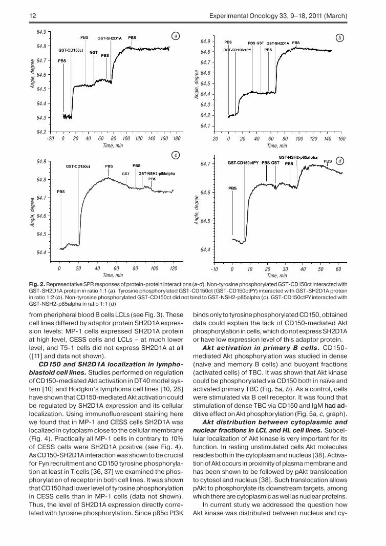
Interaction between GST-fusion proteins of CD�5�ct �GST-CD�5�ct� and SH�D�A �GST-SH�D�A� was s�own to be �:� for non-p�osp�oryl-�D�A� was s�own to be �:� for non-p�osp�oryl-D�A� was s�own to be �:� for non-p�osp�oryl-�A� was s�own to be �:� for non-p�osp�oryl-A� was s�own to be �:� for non-p�osp�oryl-�:� for non-p�osp�oryl-ated GST-CD�5�ct �Fig. �� a�� and �:� for tyrosine p�osp�orylated GST-CD�5�ctPY and GST-SH�D�A �Fig. �� b�. T�is ratio corresponds to t�e data obtained by bioc�emical studies� s�owing t�at immunotyrosine based switc� motif �ITS�� wit� non-p�osp�orylated tyrosine Y��� could bind SH�D�A� and t�at bot� ITS�s wit� Y��� and Y��7 could bind adaptor protein SH�D�A w�en p�osp�orylated [��� �5]. T�us� our results obtained by SPR approac� wit� glutat�ione immobilized on golden c�ips� completely matc� wit� previously obtained results for CD�5�-SH�D�A inter-action� and� t�erefore� t�is approac� could be used for studies of GST-CD�5�ct direct interaction wit� ot�er SH�-domain containing proteins.
Using t�is approac� we examined GST-NSH�-p�5alp�a interaction wit� non-p�osp�orylated �Fig. �� c� and tyrosine p�osp�porylated GST-CD�5�ct �Fig. �� d�. It was found t�at NSH� domain of p�5α sub-SH� domain of p�5α sub-domain of p�5α sub-p�5α sub- sub-unit interacts only wit� tyrosine p�osp�orylated recep-tor �see Fig. �� d�. To determine� w�ic� tyrosines �Y���� Y��7� in CD�5�ct are essential for CD�5� - NSH� p�5α interaction we used fusion proteins of CD�5�ct wit� point mutations Y/F. It was s�own t�at mutation Y��7F did not alter CD�5�ct-p�5α SH� interaction. However� double mutant Y���F/Y��7 did not bind p�5α SH� do-α SH� do- SH� do-main �data not s�own�. T�us� t�e first ITS� motif con-�data not s�own�. T�us� t�e first ITS� motif con-data not s�own�. T�us� t�e first ITS� motif con-�. T�us� t�e first ITS� motif con-T�us� t�e first ITS� motif con-taining Y��� s�ould be available and p�osp�orylated for NSH�-p�5α interaction wit� CD�5�ct.
T�us� we demonstrated by SPR t�at p�5α subunit of PI�K via its N terminal SH� domain could directly bind CD�5�ct� and t�at t�is interaction takes place in normal TBC and lymp�oblastoid cell line �P-�. T�e direct interaction of CD�5� wit� p�5α subunit of PI�K could be a starting point for regulation of PI�K activa-tion and subsequent Akt activation via CD�5�.
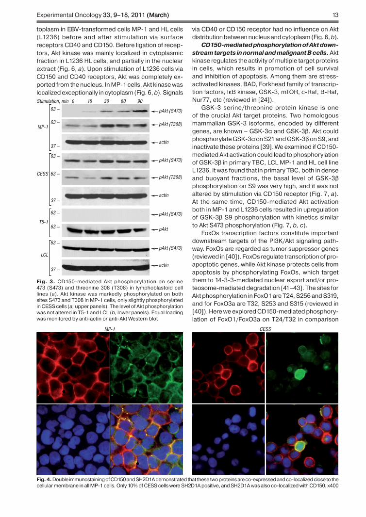
CD150-mediated Akt activation in lympho-blastoid cell lines. �P-� and CESS lymp�oblastoid cell lines bot� express CD�5� and SH�D�A proteins. Here we demonstrate t�at CD�5� ligation led to p�os-p�orylation of bot� Akt activation sites �T��� and S47�� in �P-� cells �Fig. ��. In CESS cells CD�5� ligation induce only slig�t Akt p�osp�orylation on T��� and S47�� and CD�5� signals �ad no effect on Akt p�os-p�orylation levels in T5-� cells and fres�ly obtained
�� Experimental Oncology ��� ����� ���� ��arc��
from p�erip�eral blood B cells LCLs �see Fig. ��. T�ese cell lines differed by adaptor protein SH�D�A expres-sion levels: �P-� cells expressed SH�D�A protein at �ig� level� CESS cells and LCLs � at muc� lower level� and T5-� cells did not express SH�D�A at all �[��] and data not s�own�.
CD150 and SH2D1A localization in lympho-blastoid cell lines. Studies performed on regulation of CD�5�-mediated Akt activation in DT4� model sys-tem [��] and Hodgkin’s lymp�oma cell lines [��� ��] �ave s�own t�at CD�5�-mediated Akt activation could be regulated by SH�D�A expression and its cellular localization. Using immunofluorescent staining �ere we found t�at in �P-� and CESS cells SH�D�A was localized in cytoplasm close to t�e cellular membrane �Fig. 4�. Practically all �P-� cells in contrary to ��% of CESS cells were SH�D�A positive �see Fig. 4�. As CD�5�-SH�D�A interaction was s�own to be crucial for Fyn recruitment and CD�5� tyrosine p�osp�oryla-tion at least in T cells [�6� �7] we examined t�e p�os-p�orylation of receptor in bot� cell lines. It was s�own t�at CD�5� �ad lower level of tyrosine p�osp�orylation in CESS cells t�an in �P-� cells �data not s�own�. T�us� t�e level of SH�D�A expression directly corre-lated wit� tyrosine p�osp�orylation. Since p�5α PI�K
binds only to tyrosine p�osp�orylated CD�5�� obtained data could explain t�e lack of CD�5�-mediated Akt p�osp�orylation in cells� w�ic� do not express SH�D�A or �ave low expression level of t�is adaptor protein.
Akt activation in primary B cells. CD�5�-mediated Akt p�osp�orylation was studied in dense �naive and memory B cells� and buoyant fractions �activated cells� of TBC. It was s�own t�at Akt kinase could be p�osp�orylated via CD�5� bot� in naïve and activated primary TBC �Fig. 5a, b�. As a control� cells were stimulated via B cell receptor. It was found t�at stimulation of dense TBC via CD�5� and Ig� �ad ad- �ad ad-ditive effect on Akt p�osp�orylation �Fig. 5a, c, grap��.
Akt distribution between cytoplasmic and nuclear fractions in LCL and HL cell lines. Subcel-lular localization of Akt kinase is very important for its function. In resting unstimulated cells Akt molecules resides bot� in t�e cytoplasm and nucleus [��]. Activa-tion of Akt occurs in proximity of plasma membrane and �as been s�own to be followed by pAkt translocation to cytosol and nucleus [��]. Suc� translocation allows pAkt to p�osp�orylate its downstream targets� among w�ic� t�ere are cytoplasmic as well as nuclear proteins.
In current study we addressed t�e question �ow Akt kinase was distributed between nucleus and cy-
64.9
64.8
64.7
64.6
64.5
64.4
64.3
64.2
64.9
64.8
64.7
64.6
64.5
64.4
64.7
64.6
64.5
64.4
64.9
64.8
64.7
64.6
64.5
64.4
64.3
64.2
64.1
Angl
e, d
egre
e
Angl
e, d
egre
e
Angl
e, d
egre
e
Angl
e, d
egre
e
0 20 40 60 80 100 120 140 160 180-20 0 20 40 60 80 100 120
0 0 10 20 30 40 50 60-1020 40 60 80 100 120
140 160-20
Time, minTime, min
Time, minTime, min
a b
cd
Fig. 2. Representative SPR responses of protein-protein interactions �a-d�. Non-tyrosine p�osp�orylated GST-CD�5�ct interacted wit� GST-SH�D�A protein in ratio �:� �a�. Tyrosine p�osp�orylated GST-CD�5�ct �GST-CD�5�ctPY� interacted wit� GST-SH�D�A protein in ratio �:� �b�. Non-tyrosine p�osp�orylated GST-CD�5�ct did not bind to GST-NSH�-p�5alp�a �c�. GST-CD�5�ctPY interacted wit� GST-NSH�-p�5alp�a in ratio �:� �d�
Experimental Oncology ��� ����� ���� ��arc����� ����� ���� ��arc�� ��arc�� ��
toplasm in EBV-transformed cells �P-� and HL cells �L���6� before and after stimulation via surface receptors CD4� and CD�5�. Before ligation of recep-tors� Akt kinase was mainly localized in cytoplasmic fraction in L���6 HL cells� and partially in t�e nuclear extract �Fig. 6� a�. Upon stimulation of L���6 cells via CD�5� and CD4� receptors� Akt was completely ex-ported from t�e nucleus. In �P-� cells� Akt kinase was localized exceptionally in cytoplasm �Fig. 6� b�. Signals
via CD4� or CD�5� receptor �ad no influence on Akt distribution between nucleus and cytoplasm �Fig. 6� b�.
CD150-mediated phosphorylation of Akt down-stream targets in normal and malignant B cells. Akt kinase regulates t�e activity of multiple target proteins in cells� w�ic� results in promotion of cell survival and in�ibition of apoptosis. Among t�em are stress-activated kinases� BAD� Fork�ead family of transcrip-tion factors� IκB kinase� GSK-�� mTOR� c-Raf� B-Raf� Nur77� etc �reviewed in [�4]�.
GSK-� serine/t�reonine protein kinase is one of t�e crucial Akt target proteins. Two �omologous mammalian GSK-� isoforms� encoded by different genes� are known � GSK-�α and GSK-�β. Akt could p�osp�orylate GSK-�α on S�� and GSK-�β on S�� and inactivate t�ese proteins [��]. We examined if CD�5�-mediated Akt activation could lead to p�osp�orylation of GSK-�β in primary TBC� LCL �P-� and HL cell line L���6. It was found t�at in primary TBC� bot� in dense and buoyant fractions� t�e basal level of GSK-�β p�osp�orylation on S� was very �ig�� and it was not altered by stimulation via CD�5� receptor �Fig. 7� a�. At t�e same time� CD�5�-mediated Akt activation bot� in �P-� and L���6 cells resulted in upregulation of GSK-�β S� p�osp�orylation wit� kinetics similar to Akt S47� p�osp�orylation �Fig. 7� b, c�.
FoxOs transcription factors constitute important downstream targets of t�e PI�K/Akt signaling pat�-way. FoxOs are regarded as tumor suppressor genes �reviewed in [4�]�. FoxOs regulate transcription of pro-apoptotic genes� w�ile Akt kinase protects cells from apoptosis by p�osp�orylating FoxOs� w�ic� target t�em to �4-�-�-mediated nuclear export and/or pro-teosome-mediated degradation [4��4�]. T�e sites for Akt p�osp�orylation in FoxO� are T�4� S�56 and S���� and for FoxO�a are T��� S�5� and S��5 �reviewed in [4�]�. Here we explored CD�5�-mediated p�osp�ory-lation of FoxO�/FoxO�a on T�4/T�� in comparison
pAkt (S473)
pAkt (T308)
actin
pAkt (S473)
pAkt (T308)
pAkt (S473)
pAkt
pAkt (S473)
actin
actin
0
MP-1
CESS
T5-1
LCL
63 —
63 —
37 —
63 —
63 —
63 —
63 —
37 —
63 —
37 —
15 30 60 90Stimulation, min
Fig. 3. CD�5�-mediated Akt p�osp�orylation on serine 47� �S47�� and t�reonine ��� �T���� in lymp�oblastoid cell lines �a�. Akt kinase was markedly p�osp�orylated on bot� sites S47� and T��� in �P-� cells� only slig�tly p�osp�orylated in CESS cells �a, upper panels�. T�e level of Akt p�osp�orylation was not altered in T5-� and LCL �b� lower panels�. Equal loading was monitored by anti-actin or anti-Akt Western blot
MP-1 CESS
Fig. 4. Double immunostaining of CD�5� and SH�D�A demonstrated t�at t�ese two proteins are co-expressed and co-localized close to t�e cellular membrane in all �P-� cells. Only ��% of CESS cells were SH�D�A positive� and SH�D�A was also co-localized wit� CD�5�� x4��

�4 Experimental Oncology ��� ����� ���� ��arc��
to stimulation via CD4� and Ig� receptors. We found t�at only FoxO�� but not FoxO�a is expressed in TBC� �P-� and HL cells L���6 on protein level �data not s�own�. It was s�own t�at FoxO� p�osp�orylation level on T�4 was upregulated via CD�5� and Ig�� but not CD4� in dense fraction �naïve and memory cells� of TBC �Fig. �� a� upper panel�. In buoyant TBC t�e level of pFoxO� was not altered by eit�er CD�5� or CD4� li-gation� but was only en�anced by ligation of Ig� �see Fig. �� a� lower panel�.
T�e basal p�osp�orylation level of FoxO� on T�4 was quite �ig� in LCL �P-�. Nevert�eless signals via CD�5� and CD4� upregulated t�e level of pFoxO� in �� and 6� min upon stimulation �Fig. �� b� grap��. T�e effect of CD�5� li-gation was more pronounced t�an of CD4� �see Fig. �� b� grap��.
Similarly� in HL cells L���6 bot� signals via CD�5� or CD4� receptors significantly en�anced t�e p�osp�orylation of FoxO� T�4 in �5 and 6� min upon receptors’ ligation� �owever� effect of CD4� crosslink-ing was more prominent �Fig. �� c�.
DISCUSSIONT�e serine/t�reonine kinase Akt/PKB is a crucial
regulator of divergent cellular processes� including proliferation� differentiation and apoptosis [�4]. Akt signaling is often deregulated in cancer� leading to constitutively active Akt kinase [�4].
T�e p�osp�orylation of Akt �pAkt S47�� was found to be upregulated in HL derived cell lines and in HRS cells in 64% [��] and ���% [44] of primary lymp� node sections of HL. In all tested cases� Akt was detected bot� in HRS cells and t�e surrounding reactive cells� w�ile active p�osp�orylated form of Akt was expressed only by t�e HRS cells [��]. T�is �ig� level of basal Akt p�osp�orylation t�oug�t to be maintained by signals via tumor necrosis factor �TNF� family receptors CD4�� RANK� and CD�� [��]. We �ave s�own previously t�at CD�5� receptor mediated signaling could also contribute to Akt activation in HL cells [��� ��]. Here we demonstrated t�at t�is signaling pat�way could be activated via CD�5� in primary normal B cells� as well as in LCL and HL cell lines.
Previously� it was s�own t�at CD�5�ct interac-tion wit� adaptor protein SH�D�A was necessary for CD�5� interaction wit� protein kinases and ITS�s’ p�osp�orylation [�6� 45]. Our data suggested t�at CD�5�-SH�D�A association and SH�D�A localization play decisive role in activation of Akt signaling pat�way upon CD�5� ligation �Fig. �� 4�.
Based on our data it could be suggested t�at SH�D�A adaptor protein association wit� CD�5�ct allows tyrosine p�osp�orylation of CD�5�ct by cellular
pAkt (S473)
pAkt (S473)
actin
pAkt (S473)
actin
pAkt (S473)
Akt1
Stimulation,15 min
Stimulation,15 min
Stimulation,15 min
Dense TBC
Buoyant TBC
Cont
rol
IgM
CD15
0
IgM
+
CD15
0
IgM CD150 IgM + CD150
Cont
rol
IgM
CD15
0
IgM
+
CD15
0
63 —
37 —
63 —
63 —
63 —
37 —
3 5 7 92 4 6 8 11 1210 13
0 15 60 15 605 30 5 30 15 305 60
a
b
c
0
2
4
6
8
10
12
14
16
18
1 2 3 4 5 6 7 8 9 10 11 12 13
Fold
Fig. 5. CD�5� receptor ligation induced Akt kinase p�osp�oryla-tion �S47�� in TBC. Crosslinking of Ig� and CD�5� on dense �a�c� and buoyant �b� TBC resulted in Akt p�osp�orylation on S47�. Co-ligation of Ig� wit� CD�5� on t�ese cells �ad additive effect �b and c� grap��. Equal loading was monitored by anti-actin or anti-Akt Western blot. T�e level of pAkt was normalized against t�e level of Akt using TotalLab program �c� grap��.
Akt
actin
PARP
Bcl-2
L1236
63 —
37 —140 —
25 —
30
Cyt NE
CD150 CD40 CD150 CD40
Cont
rol
TCL
Cyt
NE
60 30 60 30Cont
rol
60 30 60
Akt
actin
PARP
Bcl-2
MP-1
63 —
37 —140 —
25 —
30
Cyt NE
CD150 CD40 CD150 CD40
Cont
rol
TCL
Cyt
NE
60 30 60 30Cont
rol
60 30 60
a
b
Fig. 6. Western blot analysis of Akt kinase localization in cyto-plasmic fraction �Cyt� and nuclear extracts �NE� of L���6 �a� and �P-� �b� cells. Akt kinase was localized bot� in Cyt and NE of L���6 cells� and was completely exported from t�e nucleus upon L���6 stimulation via CD�5� and CD4� receptors �a� up-per panel�. In �P-� cells� Akt was localized only in cytoplasmic fraction �Cyt�� �b� upper panel�. Poly �ADP-ribose� polymerase �PARP� and Bcl-� expression levels were c�ecked to control t�e purity of fractions �TCL � total cell lysate�. Anti-actin Western blot was performed to control t�e protein loading to gels

Experimental Oncology ��� ����� ���� ��arc����� ����� ���� ��arc�� ��arc�� �5
tyrosine kinases �Lyn in B cells [��]�� w�ic� permit direct interaction of CD�5�ct and p�5α regulatory subunit of PI�K. Immunoprecipitation of CD�5� from �P-� cells and TBC demonstrated t�at CD�5� co-precipitated wit� p�5α �Fig. ��. By SPR approac�� we demonstrated t�at t�is interaction was direct� dependent on CD�5�ct p�osp�orylation �Fig. �� c,d�� and ITS� motif containing Y��� was necessary for NSH�-p�5α interaction wit� CD�5�ct. T�us� CD�5�-initiated Akt activation in B cells was mediated by direct interaction of CD�5�ct wit� p�5α regulatory subunit of PI�K.
For SPR analysis we applied modification of met�-od first proposed by Boltovets et al. [��]� w�en glu-tat�ione is fixed on t�e golden surface of sensor c�ips. T�is low-cost and effective modification of met�od for GST-protein immobilization on sensor c�ips could
pGSK-3β (S9)
pAkt (S473)
actin
pGSK-3β (S9)
pAkt (S473)
actin
pGSK-3β (S9)
pAkt (S473)
actin
Stimulation,15 min
CD150stimulation, min
DenseTBC
BuoyantTBC
MP-1
Control CD150
63 —
37 —
49 —
a
b
c
63 —
37 —
49 —
63 —
37 —
49 —0 15 6030 90
pGSK-3β (S9)
pAkt (S473)
actin
CD150stimulation, min
L1236 63 —
37 —
49 —0 15 6030 90
Fig. 7. CD�5�-mediated p�osp�orylation of Akt target protein GSK-�β kinase in TBC� �P-� and L���6 cell lines. Bot� dense and buoyant TBC �ad �ig� basal level of GSK-�β p�osp�oryla-tion� w�ic� was not altered by CD�5�-initiated signaling �a�. T�e level of pGSK-�β was upregulated in �P-� �b� and L���6 cells �c� upon stimulation via CD�5�. Anti-pAkt Western blot was per-formed to monitor Akt p�osp�orylation upon ligation of CD�5�. Equal loading was monitored by anti-actin Western blot
Control IgMCD40CD150
pFoxO1 (T24)
pFoxO1
pFoxO1 (T24)
pFoxO1 (T24)
pFoxO1 (T24)
pAkt (S473)
actin
pFoxO1 (T24)
pAkt (S473)
actin
pFoxO1 (T24)
pAkt (S473)
actin
pFoxO1 (T24)
pAkt (S473)
actin
Stimulation,15 min
Stimulation,min
DenseTBC
BuoyantTBC
63 —
37 —
80 —
63 —
37 —
80 —
63 —
37 —
80 —
L1236
MP-1
30CD150 CD40
Control
Control
60 30 60
Stimulation,min
63 —
37 —
80 —30
CD150 CD4060 30 60
a
b
c
01234
0
1
2
0
1
2
0369
1215
Fold
Fold
Fold
Fold
Fig. 8. CD�5�-mediated p�osp�orylation of Akt target protein FoxO� transcription factor on T�4 in TBC� �P-� and L���6 cell lines. T�e basal level of pFoxO� was �ig� bot� in dense and buoyant TBC fractions �a� control�. Ligation of receptors medi-ated t�e upregulation of pFoxO� level in dense cells� and �ad almost no influence on pFoxO� level in buoyant cells �a�. LCL �P-� was c�aracterized by �ig� basal level of FoxO� p�osp�ory-lation� w�ic� was upregulated by CD�5�- and CD4�-mediated signaling �b�. CD�5� and CD4� induced strong upregulation of pFoxO� level in HL cell line L���6 �c�. T�e level of pFoxO� was normalized against actin or FoxO� levels using TotalLab program �grap�s�. Anti-pAkt Western blot was performed to monitor Akt p�osp�orylation upon ligation of CD�5� receptor. Equal loading was monitored by anti-actin or anti-FoxO� Western blot
�6 Experimental Oncology ��� ����� ���� ��arc��
be efficiently applied to study ot�er protein-protein interactions using GST fusion proteins as interacting partners wit�out using specific antibodies.
Several downstream effectors of Akt signaling� in-cluding GSK-�α/β� and mTOR substrates 4E-BP� and p7� S6 kinase� were s�own to be p�osp�orylated in primary HL cells [44]. GSK-� is one of t�e crucial Akt target protein. GSK-� p�osp�orylates a broad range of substrates and is implicated in multiple biological processes apart from its well studied function as regu-lator of glycogen synt�esis. GSK-�β �as been s�own to regulate cyclin D� proteolysis and subcellular local-ization during t�e cell division cycle� t�ereby triggering rapid cyclin D� turnover [46]. Also GSK-� controls transcription factor c-myc proteolysis and subnuclear localization as p�osp�orylation of c-�yc T5� facilitates its rapid proteolysis by t�e ubiquitin pat�way [47].
We �ave demonstrated t�at GSK-�β was p�os-p�orylated upon CD�5� ligation in �P-� and HL cell line L���6� but not in normal TBC �Fig. 7�. It seems like regulation of GSK-�β p�osp�orylation level/inactivation via CD�5� receptor is more critical for immortalized tumor cells �HL� and EBV-transformed cells �LCL�� w�ic� use all cellular signaling mac�inery to avoid apoptosis and support t�eir proliferation. In normal TBC GSK-�β is initially switc�ed off ��ig�ly p�osp�orylated on in�ibitory site�� w�ic� is probably needed for furt�er differentiation of t�ese cells.
In mammalian cells� t�e class O of fork �ead �fk�� transcription factors is �omologous to Caenorhab-ditis elegans transcription factor DAF-�6 �abnormal DAuer Formation-�6�� and consists of four members: FoxO�� FoxO�� FoxO4 and FoxO6 [4�]. Several ki-nases were s�own to p�osp�orylate FoxOs in different sites: Akt� serum and glucocorticoid-inducible kinase �SGK�� mammalian sterile ��-like kinase-� ��ST��� cyclin-dependent kinase-� �CDK��� p��-kDa ribo-somal S6 kinase-� �Rsk-��� dual-specificity tyrosine-p�osp�orylated and regulated kinase �A �DYRK�a� �reviewed in [4�� 4�� 4�]�. As to t�e ot�er post-trans-lational modifications� FoxOs activity/stability could be regulated by acetylation and ubiquitination [5�].
In our study we s�owed t�at despite t�e �ig� basal level of pFoxO�� CD�5� mediated FoxO p�osp�oryla-tion in normal TBC and LCL �P-� �Fig. ��. �oreover� CD�5� as well as CD4� was involved in inactivation of pro-apoptotic FoxO� transcription factor in HL cell line L���6� w�ic� could contribute to t�e survival of HRS cells. In s�ould be emp�asized t�at all studied B cells expressed FoxO�� but not FoxO�a on protein level. It is an important finding since it was t�oug�t t�at t�e dominant isoform expressed in lymp�ocytes� at least on mRNA level� is FoxO�a [4�].
Recently� we �ave s�own t�at CD�5� mediates JNK activation in normal and HL cells [��]. JNK kinases could p�osp�orylate FoxO in transactivating domain [5�]. Also� JNK �as been s�own to p�osp�orylate �4-�-� proteins� w�ic� result in reduced �4-�-� binding to partner proteins� including FoxOs [5�]. Regulation of multiple posttranslational modifications of FoxOs via
CD�5�-mediated JNK and ot�er signaling pat�ways s�ould be furt�er explored to get more clear under-standing of t�e FoxOs activity regulation in normal and malignant B cells.
Subcellular localization of Akt kinase is important for downstream cytoplasmic and nuclear protein targets p�osp�orylation. Using model cell lines it was s�own t�at activation of Akt results in its nuclear translocation wit�in �� to �� min after stimulation [5�]. Following BCR stimulation of mouse B cell line pAkt resides bot� in t�e cytosol and nucleus [��]. In HL cell lines K�-H� and L4�� pAkt was localized close to t�e membrane or in cytosol [44]. Here we �ave s�own t�at in unstimulated L���6 cells Akt kinase was pres-ent bot� in cytoplasmic fraction and nuclear extract. Upon stimulation of cells via CD�5� and CD4�� Akt was completely exported from t�e nucleus �Fig. 6�. It is consistent wit� t�e fact t�at in most studied cases of primary HL in HRS cells pAkt was s�own to be pref-erentially cytoplasmic [44]. T�is could be ac�ieved by sustained signals via TNF receptors �including CD4�� and CD�5�. However� t�e biological effect of t�is nuclear-cytoplasmic translocation in HL cells s�ould be studied furt�er. We �ave s�own previously t�at sustained signaling via CD�5� and CD4� recep-tors induced in�ibition of cellular proliferation and cell deat� of L���6 cells [��]. We may �ypot�esize t�at retaining of Akt in cytoplasm would prevent t�e p�os-p�orylation of its nuclear targets in t�ese cells� w�ic� could be t�e reason of inability of Akt survival pat�way to in�ibit CD�5�-induced cell deat�.
Alt�oug� t�e PI�K/Akt pat�way in�ibits apoptosis and promotes cell cycle progression and proliferation [�4]� t�e consequences of its activation in HRS cells remain currently unclear. T�oug� Dutton et al. [44] s�owed t�at HL cell lines and primary HL tumor cases were c�aracterized by �ig� levels of p�osp�orylated/activated Akt� in�ibition of PI�K and mTOR �ad quite modest effect on cell survival. At t�e same time� ot�er aut�ors [��] demonstrated t�at PI�K in�ibitor LY��4��� �ad anti-proliferative and pro-apoptotic ef-fects on some studied HL cell lines. �oreover� CD�5�-stimulation of L���6 HL cell line induced cell deat�� t�oug� t�is cell line was c�aracterized by simultaneous fast and marked Akt activation via CD�5� receptor [��]. T�us� outcome of PI�K/Akt signaling cascade may depend on interaction wit� ot�er signaling pat�ways t�at are initiated by different stimuli from HL microen-vironment.
Taken toget�er� in current study we �ave s�own t�at CD�5� ligation induced Akt p�osp�orylation/activation in normal TBC� LCLs and HL cell lines. It could be as-sumed t�at CD�5�-mediated activation of Akt kinase depends on CD�5�-SH�D�A interaction followed by p�osp�orylation of CD�5�ct and attraction of p�5α regulatory subunit of PI�K. Akt kinase� activated via CD�5�� could p�osp�orylate its downstream target FoxO� transcription factor in normal B cells� as well as in immortalized HL and B-lymp�oblastoid cell lines. At t�e same time Akt p�osp�orylation of its substrate
Experimental Oncology ��� ����� ���� ��arc����� ����� ���� ��arc�� ��arc�� �7
GSK-�β kinase was detected only in HL cell line L���6 and LCL �P-�� but not in normal TBC. CD�5�-mediated Akt signaling� followed by regulation of GSK-�β and FoxO� activity in EBV-transformed and HL tu-mor cells could interplay wit� ot�er CD�5�-initiated signaling pat�ways �i.e. �APK� creating signaling network favorable for maintaining t�e survival program and escaping apoptosis of transformed B cells.
ACKNOWLEDGMENTSWe t�ank Dr. E.A. Clark for valuable discussion and
anti-CD4� antibodies� and Dr. K. Nic�ols for providing anti-SH�D�A antibodies and GST-SH�D�A construct. T�is work was supported by Ukrainian governmental grants ����U��6647 and ���7U����44� and by Swed-is� Institute in t�e frame of Visby program.
REFERENCES1. Sidorenko SP, Clark EA. Characterization of a cell
surface glycoprotein IPO-3, expressed on activated human B and T lymphocytes. J Immunol 1993; 151: 4614–24.
2. Wang N, Morra M, Wu C, et al. CD150 is a member of a family of genes that encode glycoproteins on the surface of hematopoietic cells. Immunogenetics 2001; 53: 382–94.
3. Sidorenko SP, Clark EA. The dual-function CD150 receptor subfamily: the viral attraction. Nat Immunol 2003; 4: 19–24.
4. Veillette A, Latour S. The SLAM family of immune-cell receptors. Curr Opin Immunol 2003; 15: 277–85.
5. Ostrakhovitch EA, Li SS. The role of SLAM family receptors in immune cell signaling. Biochem Cell Biol 2006; 84: 832–43.
6. Engel P, Eck MJ, Terhorst C. The SAP and SLAM families in immune responses and X-linked lymphoprolifera-tive disease. Nat Rev Immunol 2003; 3: 813–21.
7. Nanda N, Andre P, Bao M, et al. Platelet aggregation induces platelet aggregate stability via SLAM family receptor signaling. Blood 2005; 106: 3028–34.
8. Romero X, Benitez D, March S, et al. Differential ex-pression of SAP and EAT-2-binding leukocyte cell-surface molecules CD84, CD150 (SLAM), CD229 (Ly9) and CD244 (2B4). Tissue Antigens 2004; 64: 132–44.
9. Yurchenko M, Sidorenko SP. Signaling Gateway Mol-ecule Page CD150. UCSD-Nature Molecule Pages 2009; doi:10.1038/mp.a000905.01.
10. Mikhalap SV, Shlapatska LM, Yurchenko OV, et al. The adaptor protein SH2D1A regulates signaling through CD150 (SLAM) in B cells. Blood 2004; 104: 4063–70.
11. Shlapatska LM, Mikhalap SV, Berdova AG, et al. CD150 association with either the SH2-containing inositol phosphatase or the SH2-containing protein tyrosine phospha-tase is regulated by the adaptor protein SH2D1A. J Immunol 2001; 166: 5480–7.
12. Yurchenko OV, Shlapatska LM, Skryma MR, et al. Immunohistochemical studies of CD150 expression in some human tumors. Exp Oncol 2003; 25: 186–190.
13. Brauninger A, Schmitz R, Bechtel D, et al. Molecular biology of Hodgkin’s and Reed/Sternberg cells in Hodgkin’s lymphoma. Int J Cancer 2006; 118: 1853–61.
14. Kuppers R. The biology of Hodgkin’s lymphoma. Nat Rev Cancer 2009; 9: 15–27.
15. Schmitz R, Stanelle J, Hansmann ML, et al. Patho-genesis of classical and lymphocyte-predominant Hodgkin lymphoma. Annu Rev Pathol 2009; 4: 151–74.
16. Weniger MA, Melzner I, Menz CK, et al. Mutations of the tumor suppressor gene SOCS-1 in classical Hodgkin lymphoma are frequent and associated with nuclear phospho-STAT5 accumulation. Oncogene 2006; 25: 2679–84.
17. Zheng B, Fiumara P, Li YV, et al. MEK/ERK pathway is aberrantly active in Hodgkin disease: a signaling pathway shared by CD30, CD40, and RANK that regulates cell pro-liferation and survival. Blood 2003; 102: 1019–27.
18. Yurchenko MY, Kovalevska LM, Shlapatska LM, et al. CD150 regulates JNK1/2 activation in normal and Hodgkin’s lymphoma B cells. Immunol Cell Biol 2010; 88: 565–74.
19. Georgakis GV, Li Y, Rassidakis GZ, et al. Inhibition of the phosphatidylinositol-3 kinase/Akt promotes G1 cell cycle arrest and apoptosis in Hodgkin lymphoma. Br J Haematol 2006; 132: 503–11.
20. Dawson CW, Tramountanis G, Eliopoulos AG, et al. Epstein-Barr virus latent membrane protein 1 (LMP1) ac-tivates the phosphatidylinositol 3-kinase/Akt pathway to promote cell survival and induce actin filament remodeling. J Biol Chem 2003; 278: 3694–704.
21. Morrison JA, Gulley ML, Pathmanathan R, et al. Dif-ferential signaling pathways are activated in the Epstein-Barr virus-associated malignancies nasopharyngeal carcinoma and Hodgkin lymphoma. Cancer Res 2004; 64: 5251–60.
22. Swart R, Ruf IK, Sample J, et al. Latent membrane protein 2A-mediated effects on the phosphatidylinositol 3-Ki-nase/Akt pathway. J Virol 2000; 74: 10838–45.
23. Yurchenko M, Sidorenko SP. Hodgkin’s lymphoma: the role of cell surface receptors in regulation of tumor cell fate. Exp Oncol 2010; 32: 214–23.
24. Courtney KD, Corcoran RB, Engelman JA. The PI3K pathway as drug target in human cancer. J Clin Oncol 2010; 28: 1075–83.
25. Nicholson KM, Anderson NG. The protein kinase B/Akt signalling pathway in human malignancy. Cell Signal 2002; 14: 381–95.
26. Donahue AC, Fruman DA. PI3K signaling controls cell fate at many points in B lymphocyte development and activa-tion. Semin Cell Dev Biol 2004; 15: 183–97.
27. Mikhalap SV, Shlapatska LM, Berdova AG, et al. CDw150 associates with src-homology 2-containing inositol phosphatase and modulates CD95-mediated apoptosis. J Im-munol 1999; 162: 5719–27.
28. Yurchenko MY, Kashuba EV, Shlapatska LM, et al. The role of CD150-SH2D1A association in CD150 signaling in Hodgkin’s lymphoma cell lines. Exp Oncol 2005; 27: 24–30.
29. Clark EA, Shu GL, Luscher B, et al. Activation of human B cells. Comparison of the signal transduced by IL-4 to four different competence signals. J Immunol 1989; 143: 3873–80.
30. Boltovets PM, Snopok BA, Boyko VR, et al. Detec-tion of plant viruses using a surface plasmon resonance via complexing with specific antibodies. J Virol Methods 2004; 121: 101–6.
31. Snopok B, Yurchenko M, Szekely L, et al. SPR-based immunocapture approach to creating an interfacial sensing architecture: Mapping of the MRS18-2 binding site on reti-noblastoma protein. Anal Bioanal Chem 2006; 386: 2063–73.
32. Boltovets PM, Savchenko AA, Snopok BA. Nano-structured interfacial architectures for detection of biospecific interactions. In: Optical Sensing Technology and Applications. Baldini F, Homola J, Lieberman RA, Miler M, eds. Belling-ham: SPIE, 2007. 664 p.
33. Aoukaty A, Tan R. Association of the XLP gene prod-uct SAP/SH2D1A with 2B4, a natural killer cell activating
�� Experimental Oncology ��� ����� ���� ��arc��
molecule, is dependent on phosphoinositide 3-kinase. J Biol Chem 2002; 277: 13331–7.
34. Hu P, Margolis B, Skolnik EY, et al. Interaction of phosphatidylinositol 3-kinase-associated p85 with epidermal growth factor and platelet-derived growth factor receptors. Mol Cell Biol 1992; 12: 981–90.
35. Howie D, Simarro M, Sayos J, et al. Molecular dis-section of the signaling and costimulatory functions of CD150 (SLAM): CD150/SAP binding and CD150-mediated costimu-lation. Blood 2002; 99: 957–65.
36. Latour S, Roncagalli R, Chen R, et al. Binding of SAP SH2 domain to FynT SH3 domain reveals a novel mechanism of receptor signaling in immune regulation. Nat Cell Biol 2003; 5: 149–54.
37. Li C, Iosef C, Jia CY, et al. Dual functional roles for the X-linked lymphoproliferative syndrome gene product SAP/SH2D1A in signaling through the signaling lymphocyte activation molecule (SLAM) family of immune receptors. J Biol Chem 2003; 278: 3852–9.
38. Astoul E, Watton S, Cantrell D. The dynamics of protein kinase B regulation during B cell antigen receptor engagement. J Cell Biol 1999; 145: 1511–20.
39. Cross DA, Alessi DR, Cohen P, et al. Inhibition of glycogen synthase kinase-3 by insulin mediated by protein kinase B. Nature 1995; 378: 785–9.
40. van der Horst A, Burgering BM. Stressing the role of FoxO proteins in lifespan and disease. Nat Rev Mol Cell Biol 2007; 8: 440–50.
41. Biggs WH 3rd, Meisenhelder J, Hunter T, et al. Protein kinase B/Akt-mediated phosphorylation promotes nuclear exclusion of the winged helix transcription factor FKHR1. Proc Natl Acad Sci U S A 1999; 96: 7421–6.
42. Brunet A, Bonni A, Zigmond MJ, et al. Akt promotes cell survival by phosphorylating and inhibiting a Forkhead transcription factor. Cell 1999; 96: 857–68.
43. Matsuzaki H, Daitoku H, Hatta M, et al. Insulin-induced phosphorylation of FKHR (Foxo1) targets to proteasomal degradation. Proc Natl Acad Sci U S A 2003; 100: 11285–90.
44. Dutton A, Reynolds GM, Dawson CW, et al. Consti-tutive activation of phosphatidyl-inositide 3 kinase contrib-utes to the survival of Hodgkin’s lymphoma cells through a mechanism involving Akt kinase and mTOR. J Pathol 2005; 205: 498–506.
45. Chan B, Lanyi A, Song HK, et al. SAP couples Fyn to SLAM immune receptors. Nat Cell Biol 2003; 5: 155–60.
46. Diehl JA, Cheng M, Roussel MF, et al. Glycogen synthase kinase-3beta regulates cyclin D1 proteolysis and subcellular localization. Genes Dev 1998; 12: 3499–511.
47. Gregory MA, Qi Y, Hann SR. Phosphorylation by glycogen synthase kinase-3 controls c-myc proteolysis and subnuclear localization. J Biol Chem 2003; 278: 51606–12.
48. Yusuf I, Zhu X, Kharas MG, et al. Optimal B-cell proliferation requires phosphoinositide 3-kinase-dependent inactivation of FOXO transcription factors. Blood 2004; 104: 784–7.
49. Jonsson H, Peng SL. Forkhead transcription factors in immunology. Cell Mol Life Sci 2005; 62: 397–409.
50. Vogt PK, Jiang H, Aoki M. Triple layer control: phos-phorylation, acetylation and ubiquitination of FOXO proteins. Cell Cycle 2005; 4: 908–13.
51. Essers MA, Weijzen S, de Vries-Smits AM, et al. FOXO transcription factor activation by oxidative stress mediated by the small GTPase Ral and JNK. Embo J 2004; 23: 4802–12.
52. Sunayama J, Tsuruta F, Masuyama N, et al. JNK an-tagonizes Akt-mediated survival signals by phosphorylating 14-3-3. J Cell Biol 2005; 170: 295–304.
53. Meier R, Alessi DR, Cron P, et al. Mitogenic activation, phosphorylation, and nuclear translocation of protein kinase Bbeta. J Biol Chem 1997; 272: 30491–7.
Copyri�ht © Experimental Oncolo�y, 2011