cdk5 regulation of the grab-mediated rab8-rab11 cascade in ... · cellular/molecular...
TRANSCRIPT

Cellular/Molecular
Cdk5 Regulation of the GRAB-Mediated Rab8-Rab11Cascade in Axon Outgrowth
Kotaro Furusawa,1 Akiko Asada,1 Pamela Urrutia,2,3 Christian Gonzalez-Billault,2,3,4 XMitsunori Fukuda,5
and X Shin-ichi Hisanaga1
1Department of Biological Sciences, Graduate School of Science, Tokyo Metropolitan University, Minami-Osawa, Hachioji, Tokyo 192-0397, Japan,2Department of Biology, Faculty of Sciences, Universidad de Chile, 7800024 Nunoa, Chile, 3FONDAP Geroscience Center for Brain Health and Metabolism,7500922 Santiago, Chile, 4Buck Institute for Research on Aging, Novato, California 94945, and 5Department of Developmental Biology and Neurosciences,Graduate School of Life Sciences, Tohoku University, Aoba-ku, Sendai, Miyagi 980-8578, Japan
Neurons communicate with each other through their axons and dendrites. However, a full characterization of the molecular mechanismsinvolved in axon and dendrite formation is still incomplete. Neurite outgrowth requires the supply of membrane components for surfaceexpansion. Two membrane sources for axon outgrowth are suggested: Golgi secretary vesicles and endocytic recycling endosomes. Innon-neuronal cells, trafficking of secretary vesicles from Golgi is regulated by Rab8, a member of Rab small GTPases, and that of recyclingendosomes is by Rab11, another member of Rabs. However, whether these vesicles are coordinately or independently transported ingrowing axons is unknown. Herein, we find that GRAB, a guanine nucleotide exchange factor for Rab8, is a novel regulator of axonoutgrowth. Knockdown of GRAB suppressed axon outgrowth of cultured mouse brain cortical neurons. GRAB mediates the interactionbetween Rab11A and Rab8A, and this activity is regulated by phosphorylation at Ser169 and Ser180 by Cdk5-p35. The nonphosphorylat-able GRAB mutant S169/180A promoted axonal outgrowth to a greater extent than did the phosphomimetic GRAB mutant S169/180D.Phosphorylation of GRAB suppressed its guanine nucleotide exchange factor activity and its ability to recruit Rab8A- to Rab11A-positiveendosomes. In vivo function of GRAB and its Cdk5-phophorylation were shown in migration and process formation of developingneurons in embryonic mouse brains. These results indicate that GRAB regulates axonal outgrowth via activation and recruitment ofRab8A- to Rab11A-positive endosomes in a Cdk5-dependent manner.
Key words: axon outgrowth; Cdk5; GRAB; phosphorylation; Rab; vesicle transport
IntroductionNeurons communicate with each other through their axons anddendrites, which are structurally and functionally distinct. Theproper elongation timing, speed, and direction of these processes
need to be tightly controlled. Dysregulation of process formationcauses detrimental brain diseases because of improper neuronalconnections (Bellon, 2007; Bakos et al., 2015). Because axon anddendrite formation can be recapitulated in cultured neurons(Dotti et al., 1988), many studies have attempted to elucidate theunderlying molecular mechanisms of process formation. Al-though the roles of cytoskeletal proteins have been well docu-Received July 11, 2016; revised Nov. 21, 2016; accepted Nov. 28, 2016.
Author contributions: K.F. and S.-i.H. designed research; K.F. and P.U. performed research; A.A. and M.F. contrib-uted unpublished reagents/analytic tools; K.F., A.A., C.G.-B., M.F., and S.-i.H. analyzed data; K.F., C.G.-B., and S.-i.H.wrote the paper.
This work was supported in part by MEXT in Japan Grants-in-Aid for Scientific Research on Priority Area 25290024and 26117004 to S.H. and 15H04367 and 15H01198 to M.F., Grants Fondecyt 1140325 and FONDAP 15150012 toC.G.-B., and postdoctoral Fondecyt Grant 3160630 to P.U.
The authors declare no competing financial interests.
Correspondence should be addressed to either Dr. Kotaro Furusawa or Dr. Shin-ichi Hisanaga, Department ofBiological Sciences, Graduate School of Science, Tokyo Metropolitan University, Minami-Osawa, Hachioji, Tokyo192-0397, Japan. E-mail: [email protected] or [email protected].
DOI:10.1523/JNEUROSCI.2197-16.2016Copyright © 2017 the authors 0270-6474/17/370790-17$15.00/0
Significance Statement
While axon outgrowth requires membrane supply for surface expansion, the molecular mechanisms regulating the membranetransport in growing axons remain unclear. Here, we demonstrate that GRAB, a guanine nucleotide exchange factor for Rab8, is anovel regulator of axon outgrowth. GRAB promotes the axonal membrane transport by mediating the interaction between Rab11and Rab8 in neurons. The activity of GRAB is regulated by phosphorylation with Cdk5. We describe an in vivo role for GRAB andits Cdk5 phosphorylation during neuronal migration and process formation in embryonic brains. Thus, the membrane supply foraxonal outgrowth is regulated by Cdk5 through the Rab11-GRAB-Rab8 cascade.
790 • The Journal of Neuroscience, January 25, 2017 • 37(4):790 – 806

mented during process formation (Conde and Caceres, 2009;Lewis et al., 2013), only recently the regulation of membranetransport at the molecular level has become a research target(Sann et al., 2009). Although some evidence is available suggest-ing that membrane transport is important for neuronal differen-tiation (Villarroel-Campos et al., 2014, 2016a), the underlyingdelivery mechanisms responsible for specific axonal or dendriticmembrane formation have not been elucidated.
Membrane trafficking is a dynamic process involving vesicleformation in donor compartments for transport and their deliv-ery, tethering, and docking to their acceptor membranes (Takaiet al., 2001; Zerial and McBride, 2001; Stenmark, 2009). Thesmall GTPases of the Rab family regulate these processes. Morethan 60 Rabs are known, each of which is located and functions ina distinct membrane compartment (Klopper et al., 2012). Theiractivity cycles between an activated form bound to GTP and aninactive form bound to GDP (Stenmark, 2009). Interconversionof the active and inactive states is mediated by guanine nucleotideexchange factors (GEFs), which exchange GDP for GTP andGTPase-activating proteins (GAPs), which stimulate GTP hydro-lysis. Therefore, the regulation mechanism of each GEF or GAPfor each Rab must be individually described, but only a few caseshave been reported (Xu et al., 2015).
Membranes are required for surface expansion during axongrowth and are supplied by exocytic (secretory) and endocyticpathways (Horton and Ehlers, 2003; Pfenninger, 2009). In thesecretory pathway, Rab8 regulates the transport of exocytic vesi-cles from the trans-Golgi network to peripheral membranes(Stenmark, 2009). A deficiency in Rab8 activity results in de-creased neurite outgrowth in embryonic hippocampal neurons(Huber et al., 1995). In the endocytic pathway, which requiresseveral Rabs (e.g., Rab5 and Rab11), plasma membranes are en-docytosed and then processed as recycling vesicles, which aredelivered back to the cell surface (Takai et al., 2001; Zerial andMcBride, 2001). Rab11 regulates endosome transport to axontips (Ascano et al., 2009; Eva et al., 2010; Takano et al., 2012).However, whether these endocytic and exocytic Rabs act inde-pendently or cooperatively during axon outgrowth is not known.We had an interest in GRAB, a GEF for Rab8 (Yoshimura et al.,2010; Guo et al., 2013), which was proposed to have a role insynaptic vesicle recycling in the presynaptic region (Luo et al.,2001), because GRAB also interacts with Rab11 (Horgan et al.,2013). However, the role of GRAB in axon outgrowth has notbeen addressed at all.
Cdk5 is a proline-directed Ser/Thr protein kinase that canbe activated by binding the regulatory subunit of neuronal,membrane-bound p35 or p39 (Hisanaga and Endo, 2010). Cdk5is active mainly in postmitotic neurons and is involved in a vari-ety of neuronal functions (e.g., neuronal migration and axonaloutgrowth) (Smith and Tsai, 2002; Kawauchi, 2014). Recently,we have shown that Cdk5-p35 regulates trafficking of Rab11A-positive recycling endosomes via lemur kinase 1A (LMTK1A)during axon outgrowth (Takano et al., 2012, 2014). However,other molecular mechanisms regulated by Cdk5-p35 and involv-ing membrane delivery to supoport axonal outgrowth are cur-rently unknown.
Herein, we first validate that GRAB is a GEF of Rab8 and thenshow that it mediates the interaction between Rab11 and Rab8 inneurons. We find that Cdk5-p35 regulates GRAB activity byphosphorylation, thereby regulating axonal outgrowth via aRab11-GRAB-Rab8 cascade.
Materials and MethodsAntibodies and plasmids. Anti-p35 C19 (Santa Cruz Biotechnology, cat-alog #sc-820, RRID:AB_632137), anti-Cdk5 C-8 (Santa Cruz Biotech-nology, catalog #sc-173, RRID:AB_631224) or DC17 (Millipore, catalog#05-364, RRID:AB_2229170), and control IgG (Santa Cruz Biotechnol-ogy, catalog #sc-2025, RRID:AB_737182) were obtained from SantaCruz Biotechnology. Anti-Rab8 (catalog #6975S, RRID:AB_10827742)was purchased from Cell Signaling Technology. Anti-Rab11 (catalog#610656, RRID:AB_397983) was from BD Biosciences. Anti-myc 4A6(catalog #05-724, RRID:AB_309938) was purchased from Millipore.Mouse monoclonal anti-GFP antibody (catalog #11814460001, RRID:AB_390913) was obtained from Roche Diagnostics. Rabbit polyclonalanti-GFP antibody (catalog #598, RRID:AB_591819) was from MBL In-ternational. Anti-actin (catalog #A2066, RRID:AB_476693) was pur-chased from Sigma-Aldrich. Biotinylated anti-rabbit IgG (catalog#BA-1000, RRID:AB_2313606) was from Vector Laboratories. Anti-GRAB rabbit polyclonal antibody was obtained using purified GST-GRAB as the antigen and affinity purifying it from the resultingantiserum using antigen-immobilized beads (Fukuda and Mikoshiba,1999). Secondary antibodies conjugated with AlexaFluor-546 (ThermoFisher Scientific, catalog #A-11010, RRID:AB_2534077) or AlexaFluor-647 (Thermo Fisher Scientific, catalog #A-21244, RRID:AB_2535812)were purchased from Invitrogen.
The plasmid encoding HA-Cdk5, kinase-negative HA-Cdk5, or myc-p35,was described previously (Asada et al., 2008; Kaminosono et al., 2008). Theplasmids, pEGFP-Rab8A, pEGFP-Rab11A, their constitutively active (ca)and constitutively inactive (ci)-containing mutants, and pmStr-Rab11Awere described previously (Fukuda, 2003; Sano et al., 2008; Mori et al., 2012).pEGFP-GRAB, pmStr-GRAB, and pEF-GRAB were prepared as describedpreviously (Mori et al., 2013). pGEX-MICAL-L2-CC was isolated as de-scribed previously (Sano et al., 2008). The nonphosphorylatable Ala (A) andthe phosphorylation-mimic Asp (D) mutants of GRAB were constructedusing pmStr-GRAB or pEGFP-GRAB as the template as follows: 5�-ACACCAGCCGCTCCCAACCGT-3� and 5�-ACGGTTGGGAGCGGCTGGTGT-3� as forward and reverse primers for the S169A mutant; 5�-ACACCAGCCGATCCCAACCGT-3� and 5�-ACGGTTGGGATCGGCTGGTGT-3� as forward and reverse primers for the S169D mutant; 5�-CAGCTGCTGGCCCCCACCAAA-3� and 5�-TTTGGTGGGGGCCAGCAGCTG-3� as forward and reverse primers for the S180A mutant; 5�-CAGCTGCTGGACCCCACCAAA-3� and 5�-TTTGGTGGGGTCCAGCAGCTG-3� as forward and reverse primers for the S180D mutant; 5�-CATATCCCTGCCCCTGACAAA-3� and 5�-TTTGTCAGGGGCAGGGATATG-3� as forward and reverse primers for the T213A mutant; 5�-ACACCAGCCGATCCCAACCGT-3� and 5�-ACGGTTGGGATCGGCTGGTGT-3� as forward and reverse primers for the S169A mutant; and 5�-CAGCTGCTGGACCCCACCAAA-3� and 5�-TTTGGTGGGGTCCAGCAGCTG-3� as forward and reverse primers for the S180A mutant. The GRABknockdown shRNA sequences were 5�-GTATGCAACTTCTTCACCTAT-3�for shGRAB #1; 5�-CTTCTTCACCTATATTCGCTA-3� for shGRAB #2; 5�-GAGCTGAAGCTAAAGGATGAA-3� for shGRAB #3; 5�-GCCCACTGTTGAGTGTAACAA-3� for shGRAB #4; and 5�-ATCCTTATAGGCCATCTCTTA-3� for the scramble sequence (SC). Each knockdown sequence wasindividually inserted into a pSUPER.puro vector (Oligoengine).
Antibodies against GRAB phospho-Ser169 and phospho-Ser180. Thephospho-Ser169 peptide CTSTPA(pS)PNRE, phospho-Ser180 peptideHPQLL(pS)PTKA, non-phospho-Ser169 peptide CTSTPASPNRE,non-phospho-Ser180 peptide HPQLLSPTKA, the keyhole limpethemocyanin-conjugated phospho-Ser169 peptide, and the keyhole lim-pet hemocyanin-conjugated phospho-Ser180 peptide were obtainedfrom GenScript. Female rabbits (New Zealand White, 14 –15 weeksold; Sankyo Labo) were inoculated with one of the keyhole limpethemocyanin-conjugated peptides using TiterMax Gold (Funakoshi) asthe adjuvant. Anti-phospho-Ser180 was purified from the rabbit antise-rum using the phospho-Ser180 peptide-affinity column after removingnon-phospho-antibody over the corresponding nonphospho-peptide-affinity column. The phospho-Ser169 antiserum was used for immuno-blotting without purification because the anti-phospho-Ser169 lostactivity upon purification.
Furusawa et al. • Cdk5 Regulates Axon Outgrowth via GRAB J. Neurosci., January 25, 2017 • 37(4):790 – 806 • 791

COS-7 cell culture and transfection. COS-7 cells (RCB catalog#RCB0539, RRID:CVCL_0224) were maintained in DMEM, 10% (v/v)FBS, 100 U/ml penicillin, 0.1 mg/ml streptomycin, and cultures weretransfected with plasmids using Lipofectamine 2000 (Invitrogen) orHilyMax (Dojindo) according to the manufacturer’s instructions.
Mouse, primary neuron culture and transfection. Pregnant female ICRmice were obtained from Sankyo Laboratory Service. Cdk5-knock-outmice (RRID:IMSR_JAX:003536) were described previously (Contreras-Vallejos et al., 2014). All mouse experiments were performed accordingto the guidelines for animal experimentation of Tokyo MetropolitanUniversity or in compliance with the National Institute of Health’sGuidelines on the care and use of laboratory and experimental animals. Thestudies were approved by the Research Ethics Committee of TokyoMetropolitan University (approval no. 24 – 45). All efforts were made toreduce the suffering of animals used. Primary cortical neurons were pre-pared from mouse brains of either sex at embryonic day 15 or 16 asdescribed previously (Furusawa et al., 2014). Neuron cultures were indi-vidually transfected with indicated plasmids using Lipofectamine 2000 at0 or 2 d in vitro (DIV) and maintained in neurobasal medium (Invitrogen),2% (v/v) B27 (Invitrogen), 0.5 �M L-glutamine. To assess GRAB-kno-ckdown efficiency, neurons were nucleofected with each of the pSuperknockdown vectors by electroporation using an Amaxa Nucleofector device(Lonza).
In utero electroporation and tissue processing. Plasmid vectors wereinjected into lateral ventricle of ICR mouse brain of either sex at embry-onic day 14 (E14), as reported previously (Tabata and Nakajima, 2003).For GRAB knockdown, shGRAB SC, #1, or #4 (3 �g/�l) was coinjectedwith pCAGGS-EGFP (1 �g/�l). For the GRAB overexpression experi-ments, pEGFP-GRAB wt, 2A, or 2D (5 �g/�l) was coinjected withpCAGGS-EGFP (1 �g/�l). The brains were dissected at E17 and fixed in4% PFA (w/v) overnight at 4°C and then changed to PBS containing 30%sucrose (w/v). Cerebral cortex was sliced into 20 �m cortical sections andobserved with a confocal laser microscope LSM710 (Carl Zeiss) or macrozoom fluorescence microscope MVX10 (Olympus).
Immunofluorescence staining. COS-7 cells were fixed with 4% (w/v)PFA in PBS for 20 min and then treated with 0.1% (v/v) Triton X-100 and5% (w/v) skim milk in PBS for permeabilization and blocking. The cellswere probed with anti-phospho-Ser180 in PBS, 0.1% (v/v) Triton X-100,5% (w/v) skim milk for 1 h, and then incubated with a secondary anti-body and DAPI (Dojindo) in PBS, 0.1% (v/v) Triton X-100, 5% (w/v)skim milk for 1 h.
Primary neurons fixed in 4% (w/v) PFA in PBS were treated with PBS,0.1% (v/v) Triton X-100, 5% (w/v) normal goat serum for 30 min. Thecells were incubated with the primary antibody, anti-pS180, in PBS, 0.1%(v/v) Triton X-100, 1% (w/v) normal goat serum, followed by incubationwith a secondary antibody, and were observed using a confocal laserscanning microscope Exciter or LSM710.
Rat hippocampal neurons at 3 DIV were fixed with 4% (w/v) PFA, 4%(w/v) sucrose for 30 min at 37°C, permeabilized with PBS, 0.1% (v/v)Triton X-100 for 5 min, and blocked with 5% (w/v) BSA, PBS for 1 h.Cells were then incubated with primary antibodies anti-Rab11 (1:150dilution) or anti-Rab8 (1:100 dilution) in 1% (w/v) BSA overnight at4°C; anti-rabbit (1:200 dilution) or Alexa-543 anti-mouse (1:500 dilu-tion), for 1 h at room temperature. Subsequently, the cells were washedwith PBS, incubated with streptavidin-Alexa-488 (1:100, Invitrogen)for 30 min, washed, and mounted on slides using FluorSave reagent(Calbiochem). Images were captured using the LSM710 confocal micro-scope. ImageJ (http://imagej.nih.gov/ij/) was used for colocalizationexperiments.
Live-cell imaging. COS-7 cells were observed in a Greiner Bio-Oneglass-bottom dish containing DMEM, 10% (v/v) FBS. Mouse brain cor-tical neurons were observed in a polyethylenimine-coated glass-bottomdish containing neurobasal medium supplemented with B27. Fluores-cent images were captured every 1 or 2 s using the LSM710 confocalmicroscope.
Laemmli SDS-PAGE, Phos-tag SDS-PAGE, and immunoblotting.Laemmli SDS-PAGE and immunoblotting were as described previously(Furusawa et al., 2014). Phos-tag SDS-PAGE gels contained 5% (w/v)polyacrylamide, 50 �M Phos-tag acrylamide (Wako Pure Chemical), and100 �M MnCl2, and electrophoresis was performed as described previ-ously (Kinoshita et al., 2006; Hosokawa et al., 2010).
Immunoprecipitation. COS-7 cells were disrupted in 50 mM Tris-HCl,pH 7.4, 100 mM NaCl, 1 mM MgCl2, 1 mM EDTA, 0.5 mM EGTA, 0.5%(v/v) Nonidet P-40, 0.4 mM Pefabloc, 10 �g/ml leupeptin, 0.1 mM DTT,10 mM NaF, and 10 mM �-glycerophosphate on ice for 10 min. Aftercentrifugation at 17,000 � g for 5 min, each supernatant was incubatedwith anti-GFP or control IgG for 1 h on ice and then with Protein GSepharose beads (GE Healthcare). After washing four times with theaforementioned buffer, proteins that had been bound to the beads weresubjected to SDS-PAGE.
Pull-down of Rab8A by GST-MICAL-L2. Recombinant GST-MICAL-L2-CC was produced in and purified from Escherichia coli, and then
Figure 1. Interaction of GRAB with Rab8A or Rab11A. A, Preferential association of ci Rab8A with GRAB. The extracts (input) of COS-7 cells expressing mStr-GRAB and wt Rab8A, ca Rab8A, or ciRab8A (EGFP-tagged) were immunoprecipitated with control IgG (Cont) or anti-GFP, and GRAB and Rab8A were detected in the immunoprecipitates (IP) by immunoblotting with anti-GRAB andanti-GFP, respectively. B, Ratio of GRAB bound to each Rab8A isoform and normalized to the GRAB to wt Rab8A ratio. Data are mean�SEM; n�3. *p�0.05. C, Association of ca Rab11A with GRAB.GRAB was coexpressed in COS-7 cells with wt Rab11A, ca Rab11A, or ci Rab11A (EGFP-tagged). The cell extracts (input) were immunoprecipitated with control IgG (Cont) or anti-GFP, and GRAB andRab11A were detected in the immunoprecipitates (IP) by immunoblotting with anti-GRAB and anti-GFP, respectively. D, Quantification of GRAB bound to each Rab11A isoform. Data are mean �SEM; n � 3. **p � 0.01.
792 • J. Neurosci., January 25, 2017 • 37(4):790 – 806 Furusawa et al. • Cdk5 Regulates Axon Outgrowth via GRAB

Figure 2. Effect of GRAB knockdown on axon outgrowth. A, GRAB knockdown by shRNAs. Cortical neurons were transfected with shGRAB #1– 4, SC, or an empty vector (EV) at 0 DIV, and celllysates were immunoblotted with anti-GRAB at 3 DIV. Actin is the control. B, Effect of GRAB knockdown on axon outgrowth. Cortical neurons were transfected with SC, shGRAB #1, or #4 with EGFPfor visualization at 0 DIV, and axon length was measured at 3 DIV. Scale bar, 20 �m. C, Axon lengths of neurons expressing EGFP. Data are mean � SEM. SC, n � 56; shGRAB #1, n � 62; shGRAB#4, n � 60. D, Effect of Rab8A overexpression on axonal outgrowth in primary neurons. EGFP (Cont), EGFP-Rab8A wt, ci (T22N), or ca (Q67L) was individually transfected into mouse brain corticalneurons at 0 DIV, and axonal outgrowth was observed by fluorescence of EGFP at 3 DIV. Scale bar, 20 �m. E, Quantification of axon length in EGFP-expressing neurons. Data are mean � SEM; n �71 for Cont, n � 75 for Rab8A wt, n � 70 for Rab8A ci, and n � 67 for Rab8A ca. F, Effect of Rab11A overexpression on axonal outgrowth in primary neurons. EGFP (Cont), EGFP-Rab11A wt, ci (S25N),or ca (Q70L) was individually transfected into mouse brain cortical neurons at 0 DIV, and axonal outgrowth was observed by fluorescence of EGFP at 3 DIV. Scale bar, 20 �m. G, Quantification of axonlength in EGFP-expressing neurons. Data are mean � SEM; n � 60 for each of Cont, Rab11A wt, Rab11A ci, and Rab11A ca. H, Effect of GRAB knockdown on Rab8A-dependent axonal outgrowth.Cortical neurons were cotransfected with shGRAB #1, #4, or SC, and wt Rab8A, ci Rab8A, or ca Rab8A (EGFP-tagged) at 0 DIV, and axon length was measured at 3 DIV. Scale bar, 20 �m. I, Axonlengths for neurons expressing EGFP. Data are mean � SEM. SC and wt Rab8A, ci Rab8A, or ca Rab8A, n � 51, n � 54, and n � 50, respectively; shGRAB #1 (Figure legend continues.)
Furusawa et al. • Cdk5 Regulates Axon Outgrowth via GRAB J. Neurosci., January 25, 2017 • 37(4):790 – 806 • 793
bound to Glutathione-Sepharose beads (GE Healthcare) (Sano et al.,2008). The beads were incubated with COS-7 cell extracts that had ex-pressed wt EGFP-Rab8A and wt EGFP-GRAB, EGFP-GRAB 2A, orEGFP-GRAB 2D. After washing with 50 mM Tris-HCl, pH 7.4, 150 mM
NaCl, 1 mM MgCl2, 0.2% (v/v) Triton X-100, 0.4 mM Pefabloc, 10 �g/mlleupeptin, 0.1 mM DTT, 10 mM NaF, and 10 mM �-glycerophosphate,proteins bound to the beads were subjected to immunoblotting withanti-EGFP to assess the presence of active EGFP-Rab8A.
Quantification and statistical analysis. Immunoreactions were visual-ized by exposing the blots to x-ray film or acquiring a digital image witha Fusion SL4 imager (Vilber Lourmat). Band intensities were quantifiedusing ImageJ (RRID:SCR_003070). The longest neurite of the primaryneurons at 3 DIV was defined as the axon, and its length was measuredusing ZEN imaging software (ZEN Digital Imaging for Light Microscopy,RRID:SCR_013672) (Carl Zeiss). The length of the leading process wasmeasured using ImageJ. Total GRAB and the ratio of phosphorylatedGRAB to total GRAB were obtained by quantifying fluorescence intensityin the whole-cell body, a part of axon and the whole growth cone usingZEN imaging software. Quantitative data are expressed as the mean �SEM and were subjected to the Student’s t test for single comparisons orone-way ANOVA analysis, followed by Tukey’s multiple-comparisonstest for multiple comparisons.
ResultsGRAB interacts preferably with constitutively inactive (ci)Rab8A and constitutively active (ca) Rab11AGRAB was originally reported to be a GEF for Rab3A (Luo et al.,2001), but recent reports indicate that it is a GEF for Rab8 instead(Yoshimura et al., 2010; Guo et al., 2013) and bind Rab11 (Hor-gan et al., 2013). We first confirmed that GRAB interacted withRab8 and Rab11 using coimmunoprecipitation. When wt Rab8A,ca Rab8A (Q67L mutant), or ci Rab8A (T22N mutant) was coex-pressed with GRAB, a larger amount of GRAB was detected in theimmunoprecipitate of ci Rab8A, compared with wt or ca Rab8A(Fig. 1A,B). When GRAB was coexpressed with wt Rab11A, caRab11A (Q70L mutant), or ci Rab11A (S25N mutant), and im-munoprecipitated with Rab11A, a larger amount of GRAB copre-cipitated with ca Rab11A than with wt or ci Rab11A (Fig. 1C,D).These results confirmed that GRAB is an effector of Rab11A(Horgan et al., 2013).
GRAB regulates axon outgrowth upstream of Rab8A anddownstream of Rab11ABecause nothing is known concerning a role for GRAB duringaxonal outgrowth, we assessed its possible role by knocking downGRAB expression in cultured neurons. We prepared four differ-ent shRNA sequences for GRAB, shGRAB #1– 4, and found thatshGRAB #1, #3, and #4 decreased GRAB expression (Fig. 2A).shGRAB #1 and #4 were then used to evaluate the role of GRAB inaxon outgrowth. Knockdown by either shRNA decreased axonallengths (Fig. 2B,C), indicating the involvement of GRAB in axonoutgrowth.
Possibly GRAB promotes axon outgrowth via activation ofRab8, although only one report has investigated the role of Rab8in neurite outgrowth (Huber et al., 1995). We therefore exam-ined whether Rab8A was involved in axon outgrowth by overex-
pressing its isoforms in mouse cortical neurons. Wild-typeRab8A, ca Rab8A, and ci Rab8A, each as an EGFP construct, wereindividually transfected into cultured neurons at 0 DIV, and ax-onal length was measured at 3 DIV. Transfected wt Rab8A in-creased axonal outgrowth by �60%. ca Rab8A enhanced it morethan twofold, but ci Rab8A did not enhance outgrowth comparedwith the other two isoforms (Fig. 2D,E). Therefore, Rab8A has apositive role in axonal elongation. To confirm the role of Rab11in axon outgrowth, we overexpressed wt Rab11A, ca Rab11A, orci Rab11A in mouse cortical neurons. wt Rab11A or ca Rab11Aoverexpression induced approximately a twofold increase in ax-onal length, whereas ci Rab11A did not (Fig. 2F,G), consistentwith our previous report (Takano et al., 2012).
We next asked whether GRAB acts upstream or downstreamof Rab8A. GRAB was knocked down in the neurons in the pres-ence of transfected wt, ci, or ca Rab8A. GRAB knockdown re-duced axon length regardless of which Rab8A construct wasexpressed. In particular, the axon lengths of neurons expressingwt Rab8A were similar to that of ci Rab8A-expressing neurons.However, neurons expressing ca Rab8A had longer axons thanthose expressing wt or ci Rab8A, indicating that GRAB acts onaxon growth upstream of Rab8A. Nonetheless, ca Rab8A did notreverse the knockdown effect of GRAB to that of control scramble(CA) shRNA (Fig. 2H, I), which suggests that, in addition to theRab8A pathway, a second GRAB-related pathway for axon out-growth exists.
We showed, as described above, that GRAB interacts prefera-bly with the ca form of Rab11A (Fig. 1C,D). To determinewhether GRAB is an effector of Rab11A in axon outgrowth, weknocked down GRAB with shGRAB #1 or #4 in cultured corticalneurons expressing wt, ca, or ci Rab11A (Fig. 2 J,K). In controlexperiments in the presence of SC shRNA, ca Rab11A enhancedaxon outgrowth and ci Rab11A suppressed it (Fig. 2 J,K). Theseresults are consistent with those for which an shRNA was absence(Takano et al., 2012). GRAB knockdown decreased the axonlength of neurons cotransfected with all Rab11A isoforms (Fig.2 J,K), which is unlike that found for the Rab8A isoforms. Theseresults suggest that GRAB acts downstream of Rab11A duringaxon elongation.
Cdk5-p35 phosphorylates GRAB at Ser169 and Ser180We next searched for phosphorylation sites on GRAB that mightbe involved in its regulation. We identified three possible Cdk5-phosphorylation sites at Ser169, Ser180, and Thr213, which arenear the GEF domain of GRAB (Fig. 3A). We expressed GRABwith Cdk5 and p35 in COS-7 cells to determine the phosphory-lation state, which was assessed using Phos-tag SDS-PAGE forwhich phosphorylated proteins have markedly decreased mobil-ities compared with those found for conventional SDS-PAGE(Kinoshita et al., 2006; Hosokawa et al., 2010). GRAB migrated asa single band in an SDS-PAGE gel whether or not it was expressedwith Cdk5-p35 (Fig. 3B, bottom). In a Phos-tag SDS-PAGE gel,GRAB appeared as multiple bands even when expressed alone(Fig. 3B, top), suggesting that GRAB had been phosphorylated byan endogenous kinase(s). When coexpressed with active Cdk5-p35, but not when expressed with a kinase negative (kn) Cdk5-p35, all GRAB molecules migrated more slowly and as a singleband (Fig. 3B, top), indicating that it had been phosphorylated byCdk5-p35.
To identify the GRAB phosphorylation sites, we mutatedSer169, Ser180, or Thr213 in GRAB to an Ala to form S169A,S180A, and T213A, and double Ala mutant of S169/180A. Theseconstructs were expressed individually in the presence or absence
4
(Figure legend continued.) and wt Rab8A, ci Rab8A, or ca Rab8A, n�59, n�58, and n�55,respectively; shGRAB #4 and wt Rab8A, ci Rab8A, or ca Rab8A, n � 52, n � 51, and n � 51,respectively. J, Effect of GRAB knockdown on Rab11A-dependent axonal outgrowth. Corticalneurons were cotransfected with SC, shGRAB #1, or #4 and wt Rab11A, ci Rab11A, or ca Rab11A(EGFP-tagged) at 0 DIV, and axon length was measured at 3 DIV. Scale bar, 20 �m. K, Axonlengths of neurons expressing EGFP. Data are mean � SEM. Each set of an shRNA and a Rab11Aisoform; n � 60. *p � 0.05. **p � 0.01. ***p � 0.001.
794 • J. Neurosci., January 25, 2017 • 37(4):790 – 806 Furusawa et al. • Cdk5 Regulates Axon Outgrowth via GRAB

Figure 3. Cdk5-p35 phosphorylates GRAB at Ser169 and Ser180. A, Schematic of the GRAB molecule showing the GEF domain and three possible Cdk5-phosphorylation sites, which are conservedin mouse, rat, and human GRAB. B, Phosphorylation of GRAB by Cdk5-p35. EGFP-GRAB was cotransfected with Cdk5-p35 (�) or a kinase negative Cdk5-p35 (kn) into COS-7 cells. Cell lysateswere subjected to Laemmli (bottom) or Phos-tag SDS-PAGE (top) followed by immunoblotting as indicated. C, Phosphorylation of GRAB at Ser169 and Ser180 by Cdk5-p35. wt GRAB, GRAB S169A,GRAB S180A, GRAB S169/180A, or GRAB T213A (all EGFP constructs) was expressed with (�) or without () Cdk5-p35. Immunoblotting was performed as indicated after Laemmli (bottom) orPhos-tag SDS-PAGE (top). D, E, EGFP-GRAB wt or S169A (D) or S180A (E) was coexpressed with or without Cdk5-p35 in COS-7 cells. The cell lysates were blotted with anti-pS169 (D) or anti-pS180(E), and with anti-GFP (GRAB), anti-p35, and anti-Cdk5. F, EGFP-GRAB wt (top) or S180A (bottom) was coexpressed with Cdk5-p35, and cells were stained with anti-pS180. GRAB is shown with EGFP.G, Phosphorylation of GRAB at Ser169 and Ser180 in neurons. Primary cortical neurons at 5 DIV were treated with the Cdk5 inhibitor roscovitine (Ros) for 4 h. Cell lysates were then immunoblottedwith anti-pS169, anti-pS180, and anti-GRAB. Quantification is shown on right. *p � 0.05. ***p � 0.001.
Furusawa et al. • Cdk5 Regulates Axon Outgrowth via GRAB J. Neurosci., January 25, 2017 • 37(4):790 – 806 • 795

of Cdk5-p35, and their Phos-tag SDS-PAGE migration patternswere examined. The migration rates seen for S169A and S180A,and S169/180A were increased in comparison with wt GRAB inthe Phos-tag SDS-PAGE gel, and the upper bands were com-pletely absent for S169/180A (Fig. 3C). Conversely, the migrationrates for T213A were mostly unaffected by the absence or pres-ence of Cdk5-p35. These results clearly indicate that GRABSer169 and Ser180 are Cdk5-phosphorylation sites.
To confirm the in vivo phosphorylation of GRAB, we indi-vidually generated antibodies against GRAB phospho-Ser169(anti-pS169) and phospho-Ser180 (anti-pS180). Anti-pS169 andanti-pS180 reacted strongly with wt GRAB, but not with S169A orS180A, respectively, when coexpressed with Cdk5-p35 (Fig.3D,E). Weak detection of wt GRAB in the absence of Cdk5-p35suggested that the sites were phosphorylated to a certain extent byan endogenous kinase(s). The specificity of pS180 for immuno-fluorescence staining was tested by expression of each GRABisoform with Cdk5-p35 in COS-7 cells. Anti-pS180 stained cellsexpressing wt GRAB but not GRAB 2A-expressing cells (Fig. 3F).Although total GRAB, observed as EGFP, was found evenlythroughout the cytoplasm, anti-pS180 staining was detectedmore strongly at the cell periphery (Fig. 3F). (We could not useanti-pS169 for immunostaining because the antibody lost activityupon purification.)
Phosphorylation at Ser169 and Ser180 by Cdk5 was con-firmed for primary neurons using the antibodies. EndogenousGRAB migrated at �48 kDa in an SDS-PAGE gel and reacted onblots with these antibodies, indicating that phosphorylation ofGRAB had occurred in the neurons. When the neurons weretreated with the Cdk5 inhibitor, roscovitine, the amounts ofphosphorylated GRAB S169 and S180 was reduced to 72 � 7%and 52 � 4%, respectively (Fig. 3G), indicating the phosphoryla-tion of GRAB at Ser169 and Ser180 by Cdk5 in neurons.
Phosphorylation of GRAB regulates axonal outgrowthvia Rab8AWe next investigated whether the axon outgrowth activity ofGRAB is regulated by its phosphorylated state. Axonal length wasmeasured in primary neurons expressing wt GRAB, its nonphos-phorylatable mutant GRAB 2A (S169/180A), or the phosphomi-metic mutant, GRAB 2D (S169/180D). Wild-type GRAB and 2Dmutant increased the axonal length 1.7- and 1.5-fold, respec-tively, compared with control neurons, which had been trans-fected with only EGFP (Fig. 4A,B). GRAB 2A increased axonallength 2.4-fold compared with control neurons. Thus, when notphosphorylated (i.e., in the form of GRAB 2A), GRAB morestrongly increased axon outgrowth than when phosphorylated(i.e., in the form of GRAB 2D).
Next, we tested whether the phosphorylation of GRAB occursupstream of Rab8A by coexpressing GRAB wt, GRAB 2A, orGRAB 2D with wt, ca, or ci Rab8A. Neurons expressing GRAB 2Ahad longer axons than those expressing GRAB 2D when coex-pressed with wt Rab8A (Fig. 4C,D) as was observed when GRAB2A or 2D was expressed alone (Fig. 4A,B). However, a differencein axon length for GRAB 2A and GRAB 2D was absent when theywere coexpressed with ca or ci Rab8A, with longer neuronal ax-ons found when ca Rab8A was expressed compared with ciRab8A expression (Fig. 4C,D). Thus, mutations of GRAB Ser169and Ser180 to Ala or Asp did not impact the effect of ca or ciRab8A on axon elongation. These results also confirm that GRABregulates axonal outgrowth upstream of Rab8A.
Phosphorylation of GRAB regulates its GEF activity but doesnot affect its interaction with Rab11AGRAB Ser169 and Ser180 are located downstream of its GEFdomain (Fig. 3A). We investigated the effect of phosphorylationon its GEF activity for Rab8A using MICAL-L2, an effector ofRab8 (Fukuda et al., 2008; Yamamura et al., 2008). We expressedwt GRAB, GRAB 2A, or GRAB 2D, and wt Rab8A in COS-7 cellsand quantified the levels of active Rab8A bound to the GST-MICAL-L2-CC-beads. A larger amount of Rab8A was pulleddown when it was coexpressed with GRAB 2A than with wt GRABor GRAB 2D (Fig. 5A,B), indicating that Cdk5-p35 phosphory-lation of GRAB suppresses its GEF activity for Rab8A.
GRAB has a Rab11A-binding site in its C-terminal region,which contains the essential sequence, ILFAEF, at positions 223–228 (Horgan et al., 2013). We wondered whether phosphoryla-tion at S169 and/or S180 affects association of GRAB andRab11A. To answer this question, we cotransfected wt GRAB, 2Aor 2D, with wt Rab11A and assessed their binding by immuno-precipitation. Wild-type GRAB, GRAB 2A, and GRAB 2D boundwt Rab11A to a similar extent (Fig. 5C,D). To evaluate whetherGRAB isoforms have a preferential binding to ca Rab11A, asexpected for a small GTPase effector protein, we immunoprecipi-tated ca Rab11A in the presence of wt GRAB, GRAB 2A, or GRAB2D. We observed that GRAB binding to ca Rab11A was similar towt Rab11A (Fig. 5E, F). Therefore, we concluded that GRABbinds to Rab11A, irrespective of its phosphorylation status.
Phosphorylation of GRAB regulates its recruitment of Rab8Ato Rab11A-positive endosomesCertain GEFs activate their target Rabs and then recruit them to aspecific membrane compartment (Blumer et al., 2013). We askedwhether phosphorylation of GRAB affected its association withRab8A by coexpressing wt Rab8A with wt GRAB, GRAB 2A, orGRAB 2D and assessing whether the GRAB isoforms had boundRab8A in the Rab8A immunoprecipitates. GRAB 2A preferen-tially bound to wt Rab8A, whereas GRAB 2D showed a decreasedinteraction (Fig. 6A,B). Wild-type GRAB showed no significantdifferences from GRAB 2A, likely because Cdk5 is not active inCOS-7 cells, leading to a higher ratio of unphosphorylatedGRAB. On the other hand, GRAB 2D might mimic the phosphor-ylated form only partially, probably because of its less minuscharge. Considering that GEF binds to ci Rab, we performed thebinding experiment using ci Rab8A (Fig. 6C,D). The results weresimilar to those of wt Rab8A, indicating that the substantialamount of wt Rab8A is present as the inactive form.
Concerning endocytic Rab cascades, a coupling effect betweentwo different Rabs is found such that a Rab effector for oneRab is a GEF for a second Rab (Hutagalung and Novick, 2011).The aforementioned results suggested that GRAB mediates theactivation and recruitment of Rab8A- to Rab11A-positive endo-somes, with the recruitment being regulated by Cdk5 phosphor-ylation of GRAB. When we coexpressed EGFP-Rab8A andmStr-Rab11A in the absence of GRAB transfection, the Rabs weremostly found in different vesicles, as evidenced by the near ab-sence of yellow vesicles (Fig. 6E, left column). In the peripheralcytoplasm, most of the vesicles were labeled by Rab8A or Rab11A,but not by both. At the perinuclear region, both Rabs accumu-lated, but not to a great extent, in the same vesicle. When wtGRAB was transfected with Rab8A and Rab11A, the colocaliza-tion of the two Rabs was �76%, a threefold increase over thatfound in the absence of transfected GRAB (Fig. 6F).
Recruitment of EGFP-Rab8A to mStr-Rab11A-positive vesi-cles was also observed by dual-color live-cell imaging in the
796 • J. Neurosci., January 25, 2017 • 37(4):790 – 806 Furusawa et al. • Cdk5 Regulates Axon Outgrowth via GRAB

presence of transfected wt GRAB. A gradualaccumulation of EGFP-Rab8A on mStr-Rab11A-positive endosomes was observedwith only a slight change in mStr fluores-cence intensity during that time (Fig.6G,H). Conversely, we did not observe ac-cumulation of Rab11A on Rab8A-positiveendosomes. We hypothesized that this re-cruitment is regulated by phosphoryla-tion of GRAB. To test this hypothesis,we characterized the colocalization ofRab8A and Rab11A in the presence ofGRAB 2A or GRAB 2D. In the presenceof GRAB 2A, colocalization was 88%(Fig. 6 E, F, “2A”), whereas colocaliza-tion was only 43% for GRAB 2D (Fig.6 E, F, “2D”), indicating that, whenphosphorylated, GRAB has a decreasedability to recruit Rab8A onto Rab11A-positive endosomes.
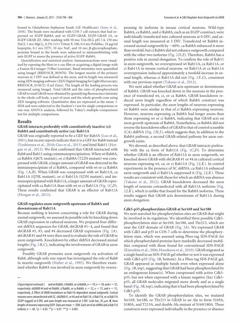
Association of GRAB with Rab8A andRab11A-positive endosomes in neuronsGiven that phosphorylation suppressedthe GEF and recruiting activities of GRABfor Rab8A, we next evaluated the subcel-lular location(s) of phosphorylated GRABin neurons. Cultured neurons were thentransfected with mStr-GRAB and thelocation of phosphorylated GRAB wasobserved by immunostaining with anti-pS180. Total GRAB, observed as mStr,was found throughout the cell-body cyto-plasm (Fig. 7A). Anti-pS180 stained cyto-plasmic vesicles but more strongly stainedthose at the cytoplasm periphery (Fig.7A). The axon (Fig. 7B) and growth cone(Fig. 7C) vesicles that mStr fluorescedwere also usually stained by anti-pS180.The ratio of total and phosphorylatedGRAB was measured in the above threeregions of neurons and shown in Figure7D. Phospho-GRAB was increased in theaxon and growth cone compared withthat in the cell body (Fig. 7D; pS180/GRAB). This result suggests that phos-phorylated GRAB is selectively trans-ported from the cell body to the growthcone through the axon or GRAB is phos-phorylated during transportation in theaxon. Nevertheless, the amount of de-phosphorylated GRAB appeared to in-crease in the growth cone because the totalGRAB was more abundant in the growthcone than the axon (Fig. 7D; GRAB).
Endogenous Rab11 accumulated at theperinuclear region of rat hippocampalneurons, whereas Rab8 was more dif-fusely distributed in the cell body (Fig.7E). We found that these endogenousRab8 and Rab11 partially colocalized incell bodies (Pearson’s colocalization coef-ficient, 0.30) and weakly in axons (Pear-
Figure 4. Effect of phosphorylation of GRAB by Cdk5 on axon outgrowth. A, Effect of GRAB overexpression on axonal outgrowthin primary neurons. EGFP (Cont), wt GRAB, GRAB 2A (S169/180A), or GRAB 2D (S169/180D) (EGFP-tagged) was individuallytransfected into cortical neurons at 0 DIV, and axonal outgrowth was observed by EGFP fluorescence at 3 DIV. Scale bar, 20 �m.B, Quantification of axonal lengths in EGFP-expressing neurons. Data are mean � SEM. Cont, n � 57; wt GRAB, n � 66; GRAB 2A,n � 68; GRAB 2D, n � 53. C, Effect of phosphorylation of GRAB on Rab8A-stimulated axonal outgrowth. wt Rab8A, ci Rab8A, or caRab8A (EGFP-tagged) was individually cotransfected with GRAB wt, 2A, or 2D into cortical neurons at 0 DIV, and axonal outgrowthwas observed by EGFP fluorescence at 3 DIV. Left panels, Control neurons expressing EGFP alone (). Scale bar, 20 �m. D, GRABregulates axonal outgrowth upstream of Rab8A. Quantification of axonal length in EGFP-expressing neurons. Data are mean �SEM. wt Rab8A and GRAB wt, GRAB 2A, or GRAB 2D, n � 60, n � 61, or n � 65, respectively; ci Rab8A and GRAB wt, GRAB 2A, orGRAB 2D, n � 60, n � 66, or n � 62, respectively; ca Rab8A and GRAB wt, GRAB 2A, or GRAB 2D, n � 60, n � 60, or n � 61,respectively. Control was taken in a respective set of experiments (n � 60 for each). **p � 0.01. ***p � 0.001.
Furusawa et al. • Cdk5 Regulates Axon Outgrowth via GRAB J. Neurosci., January 25, 2017 • 37(4):790 – 806 • 797

son’s colocalization coefficient 0.08; Fig. 7F). As shown above,Cdk5 phosphorylation of GRAB decreased its interaction withRab8A but not with ca Rab11A Q70L (Figs. 5C,D, 6A,B), suggest-ing that Rab11A-positive vesicles bind phosphorylated GRAB. Toidentify the location of phosphorylated GRAB, we transfectedmStr-GRAB with EGFP-Rab8A or EGFP-Rab11A, and then im-munostained those neurons with anti-pS180. In the cell body,anti-pS180 stained vesicles labeled with Rab11A but not withRab8A (Fig. 7G, bottom right, arrows). This dotted-type stainingwas found in axons and growth cones. In the axon, most GRAB-positive vesicles were also stained with anti-pS180, and thosevesicles were also labeled with Rab11A (Fig. 7H, bottom right)but not with Rab8A (Fig. 7H, bottom left). There were a smallnumber of GRAB-positive and Rab8A-positive vesicles in the ax-ons, but they were not stained with anti-pS180 (Fig. 7H, bottomleft, arrowhead). Thus, Rab8A associated primarily with vesicleswith nonphosphorylated GRAB in neurons. Two vesicles con-taining Rab8A and GRAB are shown in growth cone (Fig. 7I, leftpanels). Anti-pS180 stained them differently. These vesiclesmight represent a transition involving phosphorylated to de-
phosphorylated GRAB, which can activate and associate withRab8A. Expression of Rab11A was mainly found in the shafts ofaxon, which was also strongly labeled with phosphorylated GRABand total GRAB (Fig. 7I, right panels). It appears, therefore, thatphosphorylated GRAB is conveyed on endosomes to the tip of theaxon by active Rab11A.
GRAB is transported on Rab11A-positive endosomes in axonsThe aforementioned results suggested that phosphorylatedGRAB is transported on Rab11A-positive endosomes in the axonto the growth cone. We observed the movements of EGFP-caRab11A and mStr-GRAB in axons by live-cell imaging (Fig. 8A).An example is shown in Figure 8A, where three vesicles areonly ca Rab11A-labeled and the other three are GRAB/ca Rab11Adouble-labeled. Thus, GRAB-positive vesicles also containRab11A, and GRAB in these vesicles would be phosphorylated asshown by our immunostaining data. Several patterns of themovement were observed (Fig. 8A). Although one doubly labeledvesicle did not move during the 32 s observation period (Fig. 8A,asterisk), the other two GRAB/ca Rab11A-double-positive vesi-
Figure 5. Effect of phosphorylation of GRAB on its GEF activity and interaction with Rab11A. A, Effect of phosphorylation on GRAB GEF activity. COS-7 cells were cotransfected with Rab8A and wtGRAB, GRAB 2A, or GRAB 2D (EGFP-tagged), and active Rab8A was pulled down by GST-MICAL-L2-CC. Bottom, Cell lysate. Top, Rab8A pulled down. B, Quantification of active Rab8A. Relative ratiosto the EGFP-expressing cells are expressed as mean�SEM; n�4. **p�0.01. ***p�0.001. C, E, Effect of mutated Cdk5-phosphorylation sites in GRAB in association with Rab11A. wt GRAB, GRAB2A, or GRAB 2D (all mStr constructs) was coexpressed in COS-7 cells with EGFP-wt Rab11A (C) or EGFP-ca Rab11A (E). Rab11A was immunoprecipitated with anti-GFP, and GRAB and Rab11 weredetected in the immunoprecipitates by immunoblotting with anti-GRAB and anti-EGFP, respectively. D, F, Relative amount of each GRAB isoform bound to wt Rab11A (D) or ca Rab11A (F) andnormalized to wt GRAB. Data are mean � SEM. Each GRAB isoform; n � 3.
798 • J. Neurosci., January 25, 2017 • 37(4):790 – 806 Furusawa et al. • Cdk5 Regulates Axon Outgrowth via GRAB

cles moved toward the growth cone; the vesicle indicated bythe arrows moved in the distal direction along the axon until8 s and then stopped, and the second vesicle, indicated byarrowheads was initially stationary and began to move at 8 s(Fig. 8A). On the other hand, whereas one vesicle labeled withonly ca Rab11A moved bidirectionally, the other two caRab11A-labeled vesicles did not move (Fig. 8A). The observa-tion indicates clearly that GRAB is transported on Rab11A-positive endosomes in the axon.
The above observation suggested a possibility that the associationof GRAB alters the movement of Rab11-positive endosomes. Then,we analyzed the movement of GRAB-positive or negative and caRab11A-positive vesicles quantitatively. GRAB/ca Rab11A-doublepositive vesicles were �86% mobile, and GRAB-negative vesicleswere 76% (Fig. 8B), suggesting that the GRAB-binding increases theratio of moving vesicles. Among mobile vesicles, 27% of ca Rab11A-labeled vesicles moved anterogradely, 49% moved retrogradely, and24% were bidirectional. The association of GRAB increased bidirec-
Figure 6. Effect of phosphorylation of GRAB on the recruitment of Rab8A- to Rab11A-positive endosomes. A, C, Effect of Cdk5-phosphorylatable sites in GRAB on its association with wt Rab8A (A)or ci Rab8A (C). The extract (input) of cells expressing wt GRAB, GRAB 2A, or GRAB 2D (mStr-tagged) and EGFP-wt Rab8A (A) or EGFP-ci Rab8A (C). EGFP-Rab8A was immunoprecipitated withanti-GFP or control IgG (Cont), and GRAB and Rab8 were detected in the immunoprecipitate (IP) by immunoblotting with anti-GRAB and anti-GFP, respectively. B, D, Quantification of GRAB boundto wt Rab8A (B) or ci Rab8A (D). Data are mean � SEM. Each Rab8 isoform; n � 3. E, Effect of phosphorylation of GRAB on the localization of Rab8A- into Rab11A-positive vesicles. EGFP-Rab8A andmStr-Rab11A were coexpressed with wt GRAB, GRAB 2A, or GRAB 2D. Control (), no transfected GRAB. Scale bar, 10 �m. F, Percentage of Rab8A localized in Rab11A-containing vesicles. Data aremean � SEM. Each experiment, n � 10. G, Time course of EGFP-Rab8A accumulation on mStr-Rab11A-positive endosomes when coexpressed with wt GRAB in COS-7 cells. Scale bar, 1 �m.H, Quantification with time of EGFP-Rab8A and mStr-Rab11A accumulation on mStr-Rab11A-positive endosomes when coexpressed with wt GRAB in COS-7 cells. Data are mean � SEM; n � 5.*p � 0.05. **p � 0.01. ***p � 0.001.
Furusawa et al. • Cdk5 Regulates Axon Outgrowth via GRAB J. Neurosci., January 25, 2017 • 37(4):790 – 806 • 799
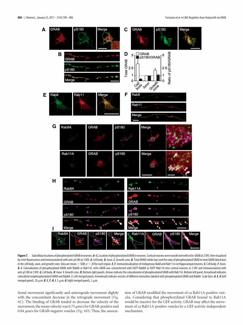
tional movement significantly and anterograde movement slightlywith the concomitant decrease in the retrograde movement (Fig.8C). The binding of GRAB tended to decrease the velocity of themovement; the mean velocity was 0.75 �m/s for GRAB-positive and0.84 �m/s for GRAB-negative vesicles (Fig. 8D). Thus, the associa-
tion of GRAB modified the movement of ca Rab11A-positive vesi-cles. Considering that phosphorylated GRAB bound to Rab11Awould be inactive for the GEF activity, GRAB may affect the move-ment of ca Rab11A-positive vesicles by a GEF activity-independentmechanism.
Figure 7. Subcellular locations of phosphorylated GRAB in neurons. A–C, Location of phosphorylated GRAB in neurons. Cortical neurons were transfected with mStr-GRAB at 2 DIV, then visualizedby mStr fluorescence and immunostained with anti-pS180 at 3 DIV. A, Cell body. B, Axon. C, Growth cone. D, Total GRAB (white bar) and the ratio of phosphorylated GRAB to total GRAB (black bar)in the cell body, axon, and growth cone. Data are mean � SEM; n � 20 for each region. E, F, Immunolocalization of endogenous Rab8 and Rab11 in rat hippocampal neurons. E, Cell body. F, Axon.G–I, Colocalization of phosphorylated GRAB with Rab8A or Rab11A. mStr-GRAB was cotransfected with EGFP-Rab8A or EGFP-Rab11A into cortical neurons at 2 DIV and immunostained withanti-pS180 at 3 DIV. G, Cell body. H, Axon. I, Growth cone. H, Bottom right panels, Arrows indicate the colocalization of phosphorylated GRAB with Rab11A. Bottom left panel, Arrowhead indicatescolocalized nonphosphorylated GRAB and Rab8A. I, Left merged panel, Arrowheads indicate vesicles of different intensities labeled with phosphorylated GRAB and Rab8A. Scale bars: A, E, G (leftmerged panel), 20 �m; B, C, F, H, I, 5 �m; G (right merged panel), 2 �m.
800 • J. Neurosci., January 25, 2017 • 37(4):790 – 806 Furusawa et al. • Cdk5 Regulates Axon Outgrowth via GRAB
Cdk5-dependent GRAB phosphorylation regulates in vivoneuronal migration and process formationTo demonstrate in vivo function of GRAB and its phosphorylation, weused in utero electroporation approach for the study of neuronal migra-tion. At first, we knocked GRAB down by electroporation of shGRAB#1, #4, or SC into neural progenitors present in the ventricular zone atembryonic day 14 (E14), and the migration of differentiated neuronswas scored at E17 (Fig. 9A–D). In control experiments with SC, 90%of neurons exit the ventricular zone/subventricular zone and 58% ofneurons reached the cortical plate (CP). In contrast, in neurons electro-porated with shGRAB #1 or #4, most neurons remained in lower re-
gions of the cerebral cortex, such as the intermediate zone (IZ),without further migration into the CP (Fig. 9A,B). These resultsindicate that GRAB has a role in neuronal migration during braindevelopment. Neurons in IZ displayed a multipolar morphology,and their movement was characterized by extending and retract-ing short processes (Tabata and Nakajima, 2003). We assessed therole of GRAB in the process formation of neurons in IZ. Whereas80% of control neurons display a short process oriented towardthe marginal zone, such percentage was reduced to �60% whenthe knockdown vectors were introduced (Fig. 9C,D), indicatingthe involvement of GRAB in the process formation in vivo.
Figure 8. Cotransport of GRAB with ca Rab11A in an axon. A, mStr-GRAB was cotransfected with EGFP-ca Rab11A into cortical neurons at 2 DIV, and the transport of GRAB and ca Rab11A wasobserved in the axons at 4 DIV. ca Rab11A was visualized as EGFP fluorescence (middle panels) and GRAB as mStr fluorescence (left panels). Arrows, arrowheads, and asterisks indicate the threevesicles labeled by GRAB and Rab11A. Scale bar, 5 �m. Bottom panels, Kymographs for a 32 s observation period. The cell body is to the left. B, Percentage of moving or stationary Rab11A-positivevesicles in the axon. Data are mean � SEM. ca Rab11A-labeled vesicles, n � 205; GRAB/ca Rab11-labeled vesicles, n � 200. **p � 0.01. C, Percentage of Rab11A-positive endosomes movinganterogradely, retrogradely, or bidirectionally. ca Rab11A-labeled vesicles, n � 156; GRAB/ca Rab11-labeled vesicles, n � 171. *p � 0.05. D, Velocity of Rab11A-positive endosomes in the axon.ca Rab11A-labeled vesicles, n � 75; GRAB/ca Rab11-labeled vesicles, n � 88.
Furusawa et al. • Cdk5 Regulates Axon Outgrowth via GRAB J. Neurosci., January 25, 2017 • 37(4):790 – 806 • 801

Figure 9. Role of GRAB and its phosphorylation in neuronal migration and process formation in vivo. A, Effect of GRAB knockdown on migration of cortical projection neurons. shGRAB #1, #4, orSC was coelectroporated into neural progenitor neurons in mouse embryonic brains at E14 with EGFP. Migration of neurons was observed at E17. The nucleus of neurons was stained with DAPI. CP,IZ, and ventricular zone/subventricular zone (VZ/SVZ) are indicated. Scale bar, 100 �m. B, Quantification of EGFP-expressing neurons in respective cortical layers. The percent ratio of total neuronswas expressed as the mean � SEM; n 60 neurons from four brains injected with respective shRNA. ***p � 0.001. C, Representative images of neurons in IZ of GRAB knocked-down mouse braincortex. Scale bar, 50 �m. D, Quantification of neurons with or without processes. Data are mean � SEM; n 40 neurons from four brains injected with respective shRNA. ***p � 0.001. E, Effectof phosphorylation of GRAB on neuronal migration. wt GRAB, GRAB 2A, or GRAB 2D was coelectroporated into brains of mouse at E14 with EGFP. Migration of neurons was observed at E17. Thenucleus was stained with DAPI. Scale bar, 100 �m. F, Quantification of EGFP-expressing neurons in each cortical layer. The percent ratio was expressed as mean � SEM. Control or wt GRAB; n 40 neurons from four brains; GRAB 2A or 2D; n 60 neurons from five brains. *p � 0.05. **p � 0.01. G, Representative images of locomotive neurons in lower CP at E17. Scale bar, 10 �m. H, Thelengths of a leading process. Data are mean � SEM. Each GRAB isoform; n 100. ***p � 0.001. I, Phosphorylation of GRAB at Ser180 in brains of Cdk5-deficient mouse (/). An asterisk in GRABindicates nonspecific bands. Actin is the control.
802 • J. Neurosci., January 25, 2017 • 37(4):790 – 806 Furusawa et al. • Cdk5 Regulates Axon Outgrowth via GRAB

Next, we examined the role of phosphorylation of GRAB byexpression of GRAB wt, 2A, or 2D in migrating neurons (Fig.9E–H). GRAB 2D as well as wt increased the number of neuronsthat reach the CP. In contrast, GRAB 2A did not affect the migra-tion; the number of neurons counted in three regions was similarto those of control mice expressing EGFP (Fig. 9E,F). The resultsindicate that the phosphorylation regulates the GRAB activity inneuronal migration. We measured the length of a leading processof locomotive neurons in CP. Typical examples of them areshown in Figure 9G. Expression of GRAB wt or GRAB 2D in-creased the length of the leading process slightly compared withcontrol neurons, and GRAB 2A increased the length more signif-icantly, �1.6-fold of control neurons (Fig. 9H). Finally, we con-firmed the in vivo phosphorylation of GRAB by Cdk5 usingCdk5/ mouse brains. Anti-pS180 reactive GRAB in wild-typemouse was greatly reduced in Cdk5/ mouse brain (Fig. 9I).Thus, GRAB is a substrate for Cdk5 in mouse brains. These re-sults indicate that GRAB plays a role in the migration of cerebralcortical neurons likely through the process formation and phos-phorylation by Cdk5 regulates the GRAB activity.
DiscussionHere we have shown that the GRAB-mediated Rab11A-Rab8Ainteraction constitutes a novel Rab cascade regulating axonaloutgrowth and that Cdk5 is an upstream regulator of the path-way. Rab8 is involved in the secretory exocytic pathway from thetrans Golgi network to the surface membrane (Ang et al., 2003;Sato et al., 2007). Rab8 has been reported to promote neuriteelongation (Huber et al., 1995), but its function(s) has not beenfurther assessed. Herein, we confirmed those results by showingthat Rab8A promoted axonal outgrowth in cortical neurons.Conversely, Rab11 functions as a conventional Rab that regulatesthe transport of recycling endosomes from sorting endosomes tosurface membranes via the perinuclear endosomal compartmentin cultured cells (Li and DiFiglia, 2012). Rab11 participation inaxon elongation was recently reported (Ascano et al., 2009; Eva etal., 2010; Takano et al., 2012, 2014). We observed that Rab11A-labeled vesicles bud from the perinuclear endocytic region lo-cated at the axon base (Takano et al., 2014). Because the Golgiapparatus is also found there, both endocytic and exocytic vesi-cles could travel from the perinuclear region of neurons to theiraxon tips. In contrast to cultured nonpolarized cells, both endo-cytic and secretory vesicles in axons are conveyed along a lim-ited number of microtubules. Thus, it was intriguing to knowwhether the two types of vesicles are transported independentlyor together. Our observations indicate that Rab11-positive vesi-cles are transported separately from Rab8-positive vesicles in ax-ons. Although it is not clear how Rab8 is transported, it appears tobe inactive during its transport in axon and activated at the axonterminus.
We showed that GRAB stimulates axons outgrowth by medi-ating the interaction between Rab11 and Rab8. GRAB is knownto be a Rab8 GEF and a Rab11-binding partner (Yoshimura et al.,2010; Guo et al., 2013; Horgan et al., 2013). However, the GRABRab8-GEF and Rab11-binding activities were independently ex-amined in the aforementioned reports, leaving open the questionof whether these activities are linked. Further, GRAB had notbeen examined for the role in neurite formation. Our study is thefirst to determine a biological relationship between GRAB and itsrelated Rabs, even though GRAB had previously been proposedto regulate synaptic vesicle movement in the presynaptic region(Luo et al., 2001). Moreover, we have shown that Cdk5 regu-lates GRAB GEF activity, which is the first demonstration of
phosphorylation-dependent regulation of GRAB GEF activity.Cdk5-p35 is a protein kinase preferentially expressed in neurons,and it regulates neurite outgrowth via cytoskeletal reorganizationpartly by targeting a number of GEFs for Rac1 (Nikolic et al.,1998; Rashid et al., 2001; Mokalled et al., 2010; Fang et al., 2011;Tang et al., 2014). In contrast, recent studies indicated that Cdk5-p35 also regulates membrane transport in axon and dendrites(Ou et al., 2010; Goodwin et al., 2012; Klinman and Holzbaur,2015). That Cdk5-p35 is membrane bound and accumulates atthe perinuclear membrane region (Patrick et al., 1999; Asada etal., 2008) supports its role in membrane trafficking. Its associa-tion with the Golgi apparatus may also suggest participation inexocytic membrane delivery (Paglini et al., 2001). In particular,its close relationship to Rab11 function seemed obvious. Weshowed that lemur kinase 1A (LMTK1A), a Cdk5-p35 substrate,suppresses transport of Rab11-positive vesicles in axons and den-drites in a Cdk5-dependent manner (Takano et al., 2012, 2014).Herein, we showed that Cdk5-p35 regulates the Rab11-effectorGRAB by phosphorylation. Thus, Cdk5 modulates the transportof Rab11-dependent endosomes upstream and downstream,suggesting that Cdk5 activity mediates Rab11-dependent vesicletransport.
Nonphosphorylated GRAB, which can bind active Rab11A,activates Rab8A and recruits it to Rab11A-positive vesicles. Thishandoff seems to occur in the growth cone, allowing vesiclesdriven by Rab11A to be passed off to Rab8A for fusion withplasma membranes (Fig. 10). This pass-forward interaction hasbeen shown for Rab5-Rab7 endocytic trafficking in the lysosomalpathway (Hutagalung and Novick, 2011). Although a Rab11A-Rab8A cascade has not been found for canonical endosomal re-cycling or exocytic pathways, its occurrence was suggested forAMPA-receptor transport involving Rab11-dependent transportin the dendritic shaft to Rab8-dependent insertion into thepostsynaptic membrane in spines (Brown et al., 2007). TheRab11-Rab8 cascade also operates during ciliogenesis. The ciliaryRab11-Rab8 cascade is regulated by Rabin8 GEF-Rab8 activity(Knodler et al., 2010; Westlake et al., 2011; Wang et al., 2012;Chiba et al., 2013). Very recently, it is shown that Rabin8 alsoregulates neurite outgrowth by coordinating Rab8 with Rab11 inPC12 cells (Homma and Fukuda, 2016). The sequence of Rabin8is 46% identical to that of GRAB, and it also can bind Rab11.However, its activation mechanism is different from that ofGRAB as discussed below.
GRAB GEF activity is suppressed by Cdk5-p35 phosphoryla-tion. In contrast, ERK1/2 phosphorylation of Rabin8 activates it(Wang et al., 2015). ERK1/2 is also a proline-directed kinase withoverlapping phosphorylation consensus sequences with Cdk5.ERK1/2 phosphorylates Rabin8 at four sites: Ser16 and Ser19 nearthe N terminus, and Ser247 and Ser250 C-terminal to the Rabin8GEF domain. The two N-terminal phosphorylation sequencesare not present in GRAB, whereas the other two are conserved inGRAB, although the sequences are not identical. The Ser250ERK-phosphorylation site in Rabin8 corresponds to the Ser169Cdk5-phosphorylation site in GRAB. Given the opposing effectsof phosphorylation that these two kinases have on GEF activitiessuggests that the N-terminal phosphorylation sites in Rabin8 maybe involved in its activation. Regulation of their kinase activitiesalso differs for ERK1/2 and Cdk5-p35. ERK1/2 is activated tran-siently by growth factors, whereas Cdk5 is constitutively active inthe form of Cdk5-p35 (Hisanaga and Endo, 2010). Ciliogenesis,involving ERK1/2-Rabin8, is a cell cycle-dependent event, whichis promoted by growth factors. Therefore, “activation” of Rabin8is a dynamic regulatory mechanism controlled by ERK1/2. In
Furusawa et al. • Cdk5 Regulates Axon Outgrowth via GRAB J. Neurosci., January 25, 2017 • 37(4):790 – 806 • 803

contrast, GRAB needs to be inactive while it is transported withinlong neuronal axons to avoid inappropriate activation of Rab8.To fulfill this requirement, a constitutive active and membrane-bound kinase (i.e., Cdk5-p35) would be used.
We have shown here that GRAB regulates the neuronal mi-gration in embryonic mouse brains. The results have several, atleast triple, meanings: (1) in vivo function of GRAB, (2) the role ofthe exocytic membrane transport in neuronal migration, and (3)Cdk5-dependent regulation of neuronal migration via mem-brane trafficking. (1) This is the first evidence indicating physio-logical function of GRAB in brains. In particular, the effects ofGRAB expression on short process formation in IZ neurons andof the phosphorylation on the length of the leading process inlocomotive neurons substantiate its in vivo role in axon out-growth. GRAB may play a role in the formation of various typesof processes through supplying membrane components to grow-ing tip. (2) The involvement of endocytic Rabs, Rab5, Rab7, andRab11 in neuronal migration is already shown (Kawauchi et al.,2010). However, there is no report on exocytic Rabs. GRAB is aGEF of exocytic Rab8. Together, the data indicate that endocyticand exocytic cycles of membrane component are necessary forneuronal migration. (3) Layer formation of cortical neurons isdisrupted in Cdk5/ mouse brains (Ohshima et al., 1996;Gilmore et al., 1998). Several Cdk5 substrates, such as FAK, dou-blecortin, p27 kip1, drebrin, and Mst3, have been reported as pos-sible downstream pathways (Xie et al., 2003; Tanaka et al., 2004;Kawauchi et al., 2006; Tanabe et al., 2014; Tang et al., 2014).Considering that Cdk5 is a membrane-bound kinase, however, itis also likely that Cdk5 regulates neuronal migration throughmembrane proteins and/or membrane trafficking. GRAB is aCdk5 substrate, which regulates membrane transport.
Axon outgrowth accompanies surface expansion, which takesplace by addition of new membrane components at the tips ofneurites. The membranes to be inserted are transported as smallvesicles in axons from the cell bodies. Even though axons appearto be narrow tubes, their trafficking system is complicated. Theirmembrane supplies are derived from endocytic and secretoryvesicles, and many Rabs (e.g., Rab6, Rab8, Rab10, Rab11, Rab27,Rab33, and Rab35) are involved (Huber et al., 1995; Arimura etal., 2009; Schlager et al., 2010; Wang et al., 2011; Kobayashi andFukuda, 2012; Nakazawa et al., 2012; Takano et al., 2012). Rab35supports Cdc42 activation, an essential feature during axon spec-ification and elongation (Villarroel-Campos et al., 2016b). Be-cause most studies concerning membrane transport have focused
on one or a few Rabs, little is known concerning the interactionsamong Rabs and their regulatory mechanisms, although a recentstudy showed that several Rabs form clusters in the neurites ofNGF-treated PC12 cells (Kobayashi et al., 2014). In these clusters,Rab35 recruits Rab8, Rab13, and Rab36 to Arf6-positive recyclingendosomes in conjunction with an MICAL-L1 scaffold. We havenow extended the knowledge of Rab interactions by showing theordered interaction between Rab11 and Rab8 in the axons ofdeveloping neurons. Other Rab-Rab interactions would also beused for proper delivery of the distinct membrane componentsduring axon outgrowth.
ReferencesAng AL, Folsch H, Koivisto UM, Pypaert M, Mellman I (2003) The Rab8
GTPase selectively regulates AP-1B-dependent basolateral transport inpolarized Madin-Darby canine kidney cells. J Cell Biol 163:339 –350.CrossRef Medline
Arimura N, Kimura T, Nakamuta S, Taya S, Funahashi Y, Hattori A, ShimadaA, Menager C, Kawabata S, Fujii K, Iwamatsu A, Segal RA, Fukuda M,Kaibuchi K (2009) Anterograde transport of TrkB in axons is mediatedby direct interaction with Slp1 and Rab27. Dev Cell 16:675– 686. CrossRefMedline
Asada A, Yamamoto N, Gohda M, Saito T, Hayashi N, Hisanaga S (2008)Myristoylation of p39 and p35 is a determinant of cytoplasmic or nuclearlocalization of active cyclin-dependent kinase 5 complexes. J Neurochem106:1325–1336. CrossRef Medline
Ascano M, Richmond A, Borden P, Kuruvilla R (2009) Axonal targeting ofTrk receptors via transcytosis regulates sensitivity to neurotrophin re-sponses. J Neurosci 29:11674 –11685. CrossRef Medline
Bakos J, Bacova Z, Grant SG, Castejon AM, Ostatnikova D (2015) Are mol-ecules involved in neuritogenesis and axon guidance related to autismpathogenesis? Neuromolecular Med 17:297–304. CrossRef Medline
Bellon A (2007) New genes associated with schizophrenia in neurite forma-tion: a review of cell culture experiments. Mol Psychiatry 12:620 – 629.CrossRef Medline
Blumer J, Rey J, Dehmelt L, Mazel T, Wu YW, Bastiaens P, Goody RS, Itzen A(2013) RabGEFs are a major determinant for specific Rab membranetargeting. J Cell Biol 200:287–300. CrossRef Medline
Brown TC, Correia SS, Petrok CN, Esteban JA (2007) Functional compart-mentalization of endosomal trafficking for the synaptic delivery of AMPAreceptors during long-term potentiation. J Neurosci 27:13311–13315.CrossRef Medline
Chiba S, Amagai Y, Homma Y, Fukuda M, Mizuno K (2013) NDR2-mediated Rabin8 phosphorylation is crucial for ciliogenesis by switchingbinding specificity from phosphatidylserine to Sec15. EMBO J 32:874 –885. CrossRef Medline
Conde C, Caceres A (2009) Microtubule assembly, organization and dy-namics in axons and dendrites. Nat Rev Neurosci 10:319 –332. CrossRefMedline
Figure 10. Schematic of the forward handoff mechanism by GRAB in endosomal vesicles at the axonal tip. Cdk5-p35 increases the association of GRAB with Rab11A by phosphorylating GRAB.Phosphorylated GRAB is transported with Rab11A on vesicles through the axon from the cell body to its growth cone where the GEF activity of GRAB is activated by dephosphorylation so as to recruitRab8A to Rab11A-positive endosomes.
804 • J. Neurosci., January 25, 2017 • 37(4):790 – 806 Furusawa et al. • Cdk5 Regulates Axon Outgrowth via GRAB

Contreras-Vallejos E, Utreras E, Borquez DA, Prochazkova M, Terse A, JaffeH, Toledo A, Arruti C, Pant HC, Kulkarni AB, Gonzalez-Billault C(2014) Searching for novel Cdk5 substrates in brain by comparativephosphoproteomics of wild type and Cdk5 / mice. PLoS One 9:e90363.CrossRef Medline
Dotti CG, Sullivan CA, Banker GA (1988) The establishment of polarity byhippocampal neurons in culture. J Neurosci 8:1454 –1468. Medline
Eva R, Dassie E, Caswell PT, Dick G, ffrench-Constant C, Norman JC, FawcettJW (2010) Rab11 and its effector Rab coupling protein contribute to thetrafficking of beta 1 integrins during axon growth in adult dorsal rootganglion neurons and PC12 cells. J Neurosci 30:11654 –11669. CrossRefMedline
Fang WQ, Ip JP, Li R, Ng YP, Lin SC, Chen Y, Fu AK, Ip NY (2011) Cdk5-mediated phosphorylation of Axin directs axon formation during cere-bral cortex development. J Neurosci 31:13613–13624. CrossRef Medline
Fukuda M (2003) Distinct Rab binding specificity of Rim1, Rim2, rabphilin,and Noc2: identification of a critical determinant of Rab3A/Rab27A rec-ognition by Rim2. J Biol Chem 278:15373–15380. CrossRef Medline
Fukuda M, Mikoshiba K (1999) A novel alternatively spliced variant of syn-aptotagmin VI lacking a transmembrane domain. Implications for dis-tinct functions of the two isoforms. J Biol Chem 274:31428 –31434.CrossRef Medline
Fukuda M, Kanno E, Ishibashi K, Itoh T (2008) Large scale screening fornovel rab effectors reveals unexpected broad Rab binding specificity. MolCell Proteomics 7:1031–1042. CrossRef Medline
Furusawa K, Asada A, Saito T, Hisanaga S (2014) The effect of Cyclin-dependent kinase 5 on voltage-dependent calcium channels in PC12 cellsvaries according to channel type and cell differentiation state. J Neuro-chem 130:498 –506. CrossRef Medline
Gilmore EC, Ohshima T, Goffinet AM, Kulkarni AB, Herrup K (1998)Cyclin-dependent kinase 5-deficient mice demonstrate novel develop-mental arrest in cerebral cortex. J Neurosci 18:6370 – 6377. Medline
Goodwin PR, Sasaki JM, Juo P (2012) Cyclin-dependent kinase 5 regulatesthe polarized trafficking of neuropeptide-containing dense-core vesiclesin Caenorhabditis elegans motor neurons. J Neurosci 32:8158 – 8172.CrossRef Medline
Guo Z, Hou X, Goody RS, Itzen A (2013) Intermediates in the guaninenucleotide exchange reaction of Rab8 protein catalyzed by guanine nu-cleotide exchange factors Rabin8 and GRAB. J Biol Chem 288:32466 –32474. CrossRef Medline
Hisanaga S, Endo R (2010) Regulation and role of cyclin-dependent kinaseactivity in neuronal survival and death. J Neurochem 115:1309 –1321.CrossRef Medline
Homma Y, Fukuda M (2016) Rabin8 regulates neurite outgrowth in both aGEF-activity-dependent and -independent manner. Mol Biol Cell 27:2107–2118.
Horgan CP, Hanscom SR, McCaffrey MW (2013) GRAB is a binding part-ner for the Rab11a and Rab11b GTPases. Biochem Biophys Res Commun411:214 –219. CrossRef Medline
Horton AC, Ehlers MD (2003) Neuronal polarity and trafficking. Neuron40:277–295. CrossRef Medline
Hosokawa T, Saito T, Asada A, Fukunaga K, Hisanaga S (2010) Quantitativemeasurement of in vivo phosphorylation states of Cdk5 activator p35 byPhos-tag SDS-PAGE. Mol Cell Proteomics 9:1133–1143. CrossRefMedline
Huber LA, Dupree P, Dotti CG (1995) A deficiency of the small GTPase rab8inhibits membrane traffic in developing neurons. Mol Cell Biol 15:918 –924. CrossRef Medline
Hutagalung AH, Novick PJ (2011) Role of Rab GTPases in membrane trafficand cell physiology. Physiol Rev 91:119 –149. CrossRef Medline
Kaminosono S, Saito T, Oyama F, Ohshima T, Asada A, Nagai Y, Nukina N,Hisanaga S (2008) Suppression of mutant Huntingtin aggregate forma-tion by Cdk5/p35 through the effect on microtubule stability. J Neurosci28:8745– 8755. CrossRef Medline
Kawauchi T (2014) Cdk5 regulates multiple cellular events in neural devel-opment, function and disease. Dev Growth Differ 56:335–348. CrossRefMedline
Kawauchi T, Chihama K, Nabeshima Y, Hoshino M (2006) Cdk5 phos-phorylates and stabilizes p27kip1 contributing to actin organization andcortical neuronal migration Nat Cell Biol 8:17–26. CrossRef Medline
Kawauchi T, Sekine K, Shikanai M, Chihama K, Tomita K, Kubo K, Na-kajima K, Nabeshima Y, Hoshino M (2010) Rab GTPases-dependent
endocytic pathways regulate neuronal migration and maturationthrough N-cadherin trafficking. Neuron 67:588 – 602. CrossRefMedline
Kinoshita E, Kinoshita-Kikuta E, Takiyama K, Koike T (2006) Phosphate-binding tag, a new tool to visualize phosphorylated proteins. Mol CellProteomics 5:749 –757. CrossRef Medline
Klinman E, Holzbaur EL (2015) Stress-induced CDK5 activation disruptsaxonal transport via Lis1/Ndel1/Dynein. Cell Rep 12:462– 473. CrossRefMedline
Klopper TH, Kienle N, Fasshauer D, Munro S (2012) Untangling the evo-lution of Rab G proteins: implications of a comprehensive genomic anal-ysis. BMC Biol 10:71. CrossRef Medline
Knodler A, Feng S, Zhang J, Zhang X, Das A, Peranen J, Guo W (2010)Coordination of Rab8 and Rab11 in primary ciliogenesis. Proc Natl AcadSci U S A 107:6346 – 6351. CrossRef Medline
Kobayashi H, Fukuda M (2012) Rab35 regulates Arf6 activity throughcentaurin-�2 (ACAP2) during neurite outgrowth. J Cell Sci 125:2235–2243. CrossRef Medline
Kobayashi H, Etoh K, Ohbayashi N, Fukuda M (2014) Rab35 promotes therecruitment of Rab8, Rab13 and Rab36 to recycling endosomes throughMICAL-L1 during neurite outgrowth. Biol Open 3:803– 814. CrossRefMedline
Lewis TL Jr, Courchet J, Polleux F (2013) Cell biology in neuroscience: cel-lular and molecular mechanisms underlying axon formation, growth, andbranching. J Cell Biol 202:837– 848. CrossRef Medline
Li X, DiFiglia M (2012) The recycling endosome and its role in neurologicaldisorders. Prog Neurobiol 97:127–141. CrossRef Medline
Luo HR, Saiardi A, Nagata E, Ye K, Yu H, Jung TS, Luo X, Jain S, Sawa A,Snyder SH (2001) GRAB: a physiologic guanine nucleotide exchangefactor for Rab3A, which interacts with inositol hexakisphosphate kinase.Neuron 31:439 – 451. CrossRef Medline
Mokalled MH, Johnson A, Kim Y, Oh J, Olson EN (2010) Myocardin-related transcription factors regulate the Cdk5/Pctaire1 kinase cascade tocontrol neurite outgrowth, neuronal migration and brain development.Development 137:2365–2374. CrossRef Medline
Mori Y, Matsui T, Furutani Y, Yoshihara Y, Fukuda M (2012) Small GTPaseRab17 regulates dendritic morphogenesis and postsynaptic developmentof hippocampal neurons. J Biol Chem 287:8963– 8973. CrossRef Medline
Mori Y, Matsui T, Fukuda M (2013) Rabex-5 protein regulates dendriticlocalization of small GTPase Rab17 and neurite morphogenesis in hip-pocampal neurons. J Biol Chem 288:9835–9847. CrossRef Medline
Nakazawa H, Sada T, Toriyama M, Tago K, Sugiura T, Fukuda M, Inagaki N(2012) Rab33a mediates anterograde vesicular transport for membraneexocytosis and axon outgrowth. J Neurosci 32:12712–12725. CrossRefMedline
Nikolic M, Chou MM, Lu W, Mayer BJ, Tsai LH (1998) The p35/Cdk5kinase is a neuron-specific Rac effector that inhibits Pak1 activity. Nature395:194 –198. CrossRef Medline
Ohshima T, Ward JM, Huh CG, Longenecker G, Veeranna, Pant HC, BradyRO, Martin LJ, Kulkarni AB (1996) Targeted disruption of the cyclin-dependent kinase 5 gene results in abnormal corticogenesis, neuronalpathology and perinatal death. Proc Natl Acad Sci U S A 93:11173–11178.CrossRef Medline
Ou CY, Poon VY, Maeder CI, Watanabe S, Lehrman EK, Fu AK, Park M, FuWY, Jorgensen EM, Ip NY, Shen K (2010) Two cyclin-dependent kinasepathways are essential for polarized trafficking of presynaptic compo-nents. Cell 141:846 – 858. CrossRef Medline
Paglini G, Peris L, Diez-Guerra J, Quiroga S, Caceres A (2001) The Cdk5–p35 kinase associates with the Golgi apparatus and regulates membranetraffic. EMBO Rep 2:1139 –1144. CrossRef Medline
Patrick GN, Zukerberg L, Nikolic M, de la Monte S, Dikkes P, Tsai LH (1999)Conversion of p35 to p25 deregulates Cdk5 activity and promotes neuro-degeneration. Nature 402:615– 622. CrossRef Medline
Pfenninger KH (2009) Plasma membrane expansion: a neuron’s Herculeantask. Nat Rev Neurosci 10:251–261. CrossRef Medline
Rashid T, Banerjee M, Nikolic M (2001) Phosphorylation of Pak1 by thep35/Cdk5 kinase affects neuronal morphology. J Biol Chem 276:49043–49052. CrossRef Medline
Sann S, Wang Z, Brown H, Jin Y (2009) Roles of endosomal trafficking inneurite outgrowth and guidance. Trends Cell Biol 19:317–324. CrossRefMedline
Sano H, Roach WG, Peck GR, Fukuda M, Lienhard GE (2008) Rab10 in
Furusawa et al. • Cdk5 Regulates Axon Outgrowth via GRAB J. Neurosci., January 25, 2017 • 37(4):790 – 806 • 805

insulin-stimulated GLUT4 translocation. Biochem J 411:89 –95. CrossRefMedline
Sato T, Mushiake S, Kato Y, Sato K, Sato M, Takeda N, Ozono K, Miki K,Kubo Y, Tsuji A, Harada R, Harada A (2007) The Rab8 GTPase regulatesapical protein localization in intestinal cells. Nature 448:366 –369.CrossRef Medline
Schlager MA, Kapitein LC, Grigoriev I, Burzynski GM, Wulf PS, Keijzer N, deGraaff E, Fukuda M, Shepherd IT, Akhmanova A, Hoogenraad CC(2010) Pericentrosomal targeting of Rab6 secretory vesicles by Bicaudal-D-related protein 1 (BICDR-1) regulates neuritogenesis. EMBO J 29:1637–1651. CrossRef Medline
Smith DS, Tsai LH (2002) Cdk5 behind the wheel: a role in trafficking andtransport? Trends Cell Biol 12:28 –36. CrossRef Medline
Stenmark H (2009) Rab GTPases as coordinators of vesicle traffic. Nat RevMol Cell Biol 10:513–525. CrossRef Medline
Tabata H, Nakajima K (2003) Multipolar migration: the third mode of ra-dial neuronal migration in the developing cerebral cortex. J Neurosci23:9996 –10001. Medline
Takai Y, Sasaki T, Matozaki T (2001) Small GTP-binding proteins. PhysiolRev 81:153–208. Medline
Takano T, Tomomura M, Yoshioka N, Tsutsumi K, Terasawa Y, Saito T,Kawano H, Kamiguchi H, Fukuda M, Hisanaga S (2012) LMTK1/AATYK1 is a novel regulator of axonal outgrowth that acts via Rab11 in aCdk5-dependent manner. J Neurosci 32:6587– 6599. CrossRef Medline
Takano T, Urushibara T, Yoshioka N, Saito T, Fukuda M, Tomomura M,Hisanaga S (2014) LMTK1 regulates dendritic formation by regulatingmovement of Rab11A-positive endosomes. Mol Biol Cell 25:1755–1768.CrossRef Medline
Tanabe K, Yamazaki H, Inaguma Y, Asada A, Kimura T, Takahashi J, TaokaM, Ohshima T, Furuichi T, Isobe T, Nagata K, Shirao T, Hisanaga S(2014) Phosphorylation of drebrin by cyclin-dependent kinase 5 and itsrole in neuronal migration. PLoS One 9:e92291. CrossRef Medline
Tanaka T, Serneo FF, Tseng HC, Kulkarni AB, Tsai LH, Gleeson JG (2004)Cdk5 phosphorylation of doublecortin ser297 regulates its effect on neu-ronal migration Neuron 41:215–227. CrossRef
Tang J, Ip JP, Ye T, Ng YP, Yung WH, Wu Z, Fang W, Fu AK, Ip NY (2014)Cdk5-dependent Mst3 phosphorylation and activity regulate neuronalmigration through RhoA inhibition. J Neurosci 34:7425–7436. CrossRefMedline
Villarroel-Campos D, Gastaldi L, Conde C, Caceres A, Gonzalez-Billault C
(2014) Rab-mediated trafficking role in neurite formation. J Neurochem120:240 –248. CrossRef Medline
Villarroel-Campos D, Bronfman FC, Gonzalez-Billault C (2016a) RabGTPase signaling in neurite outgrowth and axon specification. Cytoskel-eton 73:498 –507. CrossRef Medline
Villarroel-Campos D, Henríquez DR, Bodaleo FJ, Oguchi ME, Bronfman FC,Fukuda M, Gonzalez-Billault C (2016b) Rab35 functions in axon elon-gation are regulated by P53-related protein kinase in a mechanism thatinvolves Rab35 protein degradation and the microtubule-associated pro-tein 1B. J Neurosci 36:7298 –7313. CrossRef Medline
Wang J, Morita Y, Mazelova J, Deretic D (2012) The Arf GAP ASAP1 pro-vides a platform to regulate Arf4- and Rab11-Rab8-mediated ciliary re-ceptor targeting. EMBO J 75:5–10. CrossRef Medline
Wang J, Ren J, Wu B, Feng S, Cai G, Tuluc F, Peränen J, Guo W (2015)Activation of Rab8 guanine nucleotide exchange factor Rabin8 by ERK1/2in response to EGF signaling. Proc Natl Acad Sci U S A 112:148 –153.Medline
Wang T, Liu Y, Xu XH, Deng CY, Wu KY, Zhu J, Fu XQ, He M, Luo ZG (2011)Lgl1 activation of rab10 promotes axonal membrane trafficking underlyingneuronal polarization. Dev Cell 21:431–444. CrossRef Medline
Westlake CJ, Baye LM, Nachury MV, Wright KJ, Ervin KE, Phu L, ChalouniC, Beck JS, Kirkpatrick DS, Slusarski DC, Sheffield VC, Scheller RH,Jackson PK (2011) Primary cilia membrane assembly is initiated byRab11 and transport protein particle II (TRAPPII) complex-dependenttrafficking of Rabin8 to the centrosome. Proc Natl Acad Sci U S A 108:2759 –2764. CrossRef Medline
Xie Z, Sanada K, Samuels BA, Shih H, Tsai LH (2003) Serine 732 phosphor-ylation of FAK by Cdk5 is important for microtubule organization, nu-clear movement, and neuronal migration Cell 114:469 – 482. CrossRef
Xu J, Fotouhi M, McPherson PS (2015) Phosphorylation of the exchangefactor DENND3 by ULK in response to starvation activates Rab12 andinduces autophagy. EMBO Rep 16:709 –718. CrossRef Medline
Yamamura R, Nishimura N, Nakatsuji H, Arase S, Sasaki T (2008) The in-teraction of JRAB/MICAL-L2 with Rab8 and Rab13 coordinates theassembly of tight junctions and adherens junctions. Mol Biol Cell 19:971–983. CrossRef Medline
Yoshimura S, Gerondopoulos A, Linford A, Rigden DJ, Barr FA (2010)Family-wide characterization of the DENN domain Rab GDP-GTP ex-change factors. J Cell Biol 191:367–381. CrossRef Medline
Zerial M, McBride H (2001) Rab proteins as membrane organizers. Nat RevMol Cell Biol 2:107–117. CrossRef Medline
806 • J. Neurosci., January 25, 2017 • 37(4):790 – 806 Furusawa et al. • Cdk5 Regulates Axon Outgrowth via GRAB