celia e. shiau nih public access 1,2 na hu , and marianne...
TRANSCRIPT

Altering Glypican-1 levels modulates canonical Wnt signalingduring trigeminal placode development
Celia E. Shiau1,2, Na Hu1, and Marianne Bronner-Fraser1
1Division of Biology 139-74, California Institute of Technology Pasadena, CA 91125, USA
Abstract
Glypicans are conserved cell surface heparan sulfate proteoglycans expressed in a
spatiotemporally regulated manner in many developing tissues including the nervous system.
Here, we show that Glypican-1 (GPC1) is expressed by trigeminal placode cells as they ingress
and contribute to trigeminal sensory neurons in the chick embryo. Either expression of full-length
or truncated GPC1 in vivo causes defects in trigeminal gangliogenesis in a manner that requires
heparan sulfate side chains. This leads to either abnormal placodal differentiation or organization,
respectively, with near complete loss of the ophthalmic (OpV) trigeminal ganglion in the most
severe cases after over-expression of full-length GPC1. Interestingly, modulating GPC1 alters
levels of endogenous Wnt signaling activity in the forming trigeminal ganglion, as indicated by
Wnt-reporter expression. Accordingly, GPC1 over-expression phenocopies Wnt inhibition in
causing loss of OpV placodal neurons. Furthermore, increased Wnt activity rescues the effects of
GPC1 over-expression. Taken together, these results suggest that appropriate levels of GPC1 are
essential for proper regulation of canonical Wnt signaling during differentiation and organization
of trigeminal placodal cells into ganglia.
Keywords
glypican; trigeminal ganglion; placode; Wnt
Introduction
Glypicans (GPCs) are a conserved family of cell surface heparan sulfate proteoglycans
(HSPGs) that modulate major signaling pathways during embryonic development of fruit
flies to mammals (Fico et al., 2007; Filmus et al., 2008). Heparan sulfate side chains,
attached to the core protein at conserved serine residues near the membrane anchored
glycosyl-phosphatidylinositol (GPI) linkage, are thought to facilitate binding of growth
factors and ligands, including Wingless/Wnt, Dpp/BMP, Fgf and Hh, and are considered
ligand carriers or co-receptors. Accordingly, functional perturbation of glypicans has been
shown to cause significant defects in cell fate, cell movements, survival and proliferation in
mice, Xenopus laevis, Drosophila, and zebrafish (Filmus and Song, 2000; De Cat and
David, 2001; Lin, 2004; Fico et al., 2007).
Correspondence to: Marianne Bronner-Fraser.2Present address: Department of Developmental Biology, Stanford University School of Medicine, Stanford, CA 94305, USA
NIH Public AccessAuthor ManuscriptDev Biol. Author manuscript; available in PMC 2014 July 06.
Published in final edited form as:Dev Biol. 2010 December 1; 348(1): 107–118. doi:10.1016/j.ydbio.2010.09.017.
NIH
-PA
Author M
anuscriptN
IH-P
A A
uthor Manuscript
NIH
-PA
Author M
anuscript

During vertebrate development, glypicans are expressed in a spatiotemporally regulated
manner in the nervous system and other tissues (Niu et al., 1996; Saunders et al., 1997;
Litwack et al., 1998; Luxardi et al., 2007). Their expression changes in pathological
conditions, such as cancer. GPC3 and/or GPC1 are down-regulated in some ovarian cancer
and mesothelioma cell lines (Lin et al., 1999; Murthy et al., 2000) while upregulated in
others (e.g. pancreatic tumors) (Kleeff et al., 1998; Filmus, 2001; Matsuda et al., 2001; Su et
al., 2006). Six glypican (GPC1–6) family members have been identified in mammals, two in
Drosophila melanogaster (Dally and Dally-like), and two in Caenorhabiditis elegans (gpn-1
and lon-2) (Fico et al., 2007). Loss of function mutations in OCI-5/GPC3 in humans cause
Simpson-Golabi-Behmel syndrome (SGBS), characterized by pre- and post-natal
overgrowth, visceral and skeletal defects, and an increased risk of tumors (Pilia et al., 1996).
A GPC3 deficient mouse model exhibits a similar phenotype (Cano-Gauci et al., 1999). Of
the six glypicans in mammals, GPC1 is the most abundantly expressed in the developing
brain, in both neuroepithelial precursors and differentiated neurons (Karthikeyan et al.,
1994; Litwack et al., 1994; Litwack et al., 1998) and functions in neurogenesis of the central
nervous system (Jen et al., 2009). However, its potential role in patterning and formation of
the peripheral nervous system has yet to be explored.
Here, we show that Glypican-1 is expressed by ectodermal and ingressing chick trigeminal
placode cells at the time they differentiate into neurons and assemble into ganglia.
Modulation of GPC1 levels by either expression of full-length GPC1 or a truncated form
that is thought to act in a dominant-negative fashion prevents proper placodal differentiation
and formation of the trigeminal ganglion, with particularly strong effects on the ophthalmic
lobe. Consistent with studies showing that Wnt signaling is important for differentiation of
ophthalmic trigeminal placodes (Lassiter et al., 2007; Canning et al., 2008), we find that
GPC1 regulates Wnt activity in OpV ganglion formation. The results suggest that proper
levels of GPC1 are critical for appropriate modulation of canonical Wnt signaling for
differentiation and assembly of trigeminal placodes into ganglia.
Materials and Methods
Embryos
Fertilized chicken (Gallus gallus domesticus) eggs were obtained from local commercial
sources and incubated at 37°C to the desired stages.
In situ hybridization
cDNA plasmid obtained from BBSRC (ChickEST clone 418p2) was used to transcribe
antisense riboprobe against chick Glypican-1. The plasmid was sequenced and found to
contain the coding sequence of the chick Glypican-1 gene (NCBI accession number:
XM_422590.1) corresponding to nucleotides 1233–2107. Whole mount chick in situ
hybridization was performed as described (Shiau et al., 2008). Embryos were imaged and
subsequently sectioned at 12 μm.
Shiau et al. Page 2
Dev Biol. Author manuscript; available in PMC 2014 July 06.
NIH
-PA
Author M
anuscriptN
IH-P
A A
uthor Manuscript
NIH
-PA
Author M
anuscript
Immunohistochemistry
Primary antibodies used were: anti-TuJ1 (Covance; 1:250), anti-HNK-1 (American Type
Culture; 1:3–1:5), anti-GFP to recognize GFP signal after in situ hybridization (Molecular
Probes; 1:150–1:250), anti-Islet1 (DSHB, clone 40.2D6; 1:150–1:250) and anti-active-
caspase-3 (Promega; 1:150–250). Appropriate secondary antibodies against the subtype of
the primary antibodies were conjugated to Alexa Fluor 488, 568, or 350 dyes (Molecular
Probes). Images were taken on a Zeiss Axioskop2 plus fluorescence microscope, and
processed using Adobe Photoshop CS3.
In ovo electroporation of the trigeminal ectoderm
Plasmid constructs were targeted to the presumptive trigeminal placodal ectoderm at the
approximate axial level between the posterior forebrain and anterior hindbrain at 5 somites
stage (stage 8+) up to stage 11. Immediately after injection, platinum electrodes were placed
vertically across the chick embryo delivering current pulses of 5 × 8 V in 50 ms at 100 ms
intervals as described (Shiau et al., 2008). Targeting DNA to the ectoderm resulted in
transfection of the trigeminal placodes in the ectoderm and subsequently placode-derived
cells that detach from the ectoderm and migrate into the ganglion anlage. The operated eggs
were sealed and incubated at 37°C for later analysis. Incubation times were ~16–24 hours to
reach stages 13–14, ~ 24–36 hours to reach stages 15–16, ~ 40–48 hours to reach stages 17–
18, and ~ 50–60 hours to reach stage 19.
Plasmid constructs
Full length chick Glypican-1 cDNA (clone CS5) was isolated from a 4– to 12– somites stage
chick macroarray library (Gammill and Bronner-Fraser, 2002). To create cytopcig-FL-
GPC1, the full-length coding sequence (1.65 kb) was amplified from the library clone CS5
by PCR using forward and reverse primers corresponding to the coding sequence with
flanking XhoI and ClaI site respectively. Gene fragment was directionally inserted into the
cytopcig vector (Shiau et al., 2008) at the XhoI/ClaI sites. Three mutant GPC1 constructs
were made by a single-step or fusion PCR amplification off the full-length sequence, and
modified gene fragment was directionally cloned into the XhoI and ClaI sites in the cytopcig
vector. These are: 1) cytopcig-GPC1-ΔGPI which encodes the first 517 amino acids (1.55
kb) with a premature stop codon which eliminates the GPI-anchoring domain, 2) cytopcig-
GPC1-ΔHS which has a 18bp deletion that spans a coding region of six residues, SGSGSG
(483–488 aa), containing three tandemly positioned putative glycosaminoglycan (GAG)
attachment sites (serine residues 483, 485, 487) based on sequence annotation in UniProtKB
for chick GPC1 protein (accession P50593), and 3) cytopcig-GPC1- ΔGPI ΔHS which has
deletions of both the GPI-anchor domain and the putative HS sites. Two versions of full-
length and mutant constructs were made: one without myc-tag fusion and one with a 6x
myc-tag inserted at ClaI/EcoRV sites in frame with the coding sequence at the C-terminus,
which was used to validate protein expression of the constructs. Both versions were tested
and determined to give the same effects on ganglion development, albeit FL-GPC1 was
more potent without the myc-tag. Thus, most experiments with the full-length and mutant
expressions were conducted with the construct lacking the myc-tag. Dominant active ß-
catenin construct was made in the pCIG vector with IRES-nuclear localized GFP as
Shiau et al. Page 3
Dev Biol. Author manuscript; available in PMC 2014 July 06.
NIH
-PA
Author M
anuscriptN
IH-P
A A
uthor Manuscript
NIH
-PA
Author M
anuscript

previously described (Megason and McMahon, 2002; Lassiter et al., 2007), and RFP-Wnt-
Reporter (also named pTOP-nDSRed2) (gift from Dr. Andy Groves;(Lassiter et al., 2007))
was a modified version of the TOPGAL construct (DasGupta and Fuchs, 1999) where the
reporter gene was replaced with RFP.
BrdU treatment
Electroporated embryos were screened and selected for broad GFP expression in the
trigeminal region at stage 14 for both control and experimental cases. Each embryo was
explanted into an individual well and treated with 0.1 mM BrdU in Ringer's solution at 37°C
for 30 minutes. Embryos were then fixed in 4% paraformaldehyde overnight at 4°C, washed
in PBS, incubated in 2N HCl in PBS for 30 minutes, followed by 0.1M perboric acid
(H3BO4) for 10 minutes, washed in PBS several times, and processed for cryosectioning and
immunostaining with the mouse anti-BrdU (Sigma, B2531; 1:150–1:250).
Quantification of the area of trigeminal ganglion
An outline of the ganglion as marked by TuJ1 staining was made by the freehand selection
tool on whole mount grayscale fluorescent images in the ImageJ software. All images were
taken with the same setup using a 5x objective on the Zeiss Axioskop2 plus microscope and
at the same image size, with the entire ganglion in focus. The area of the ganglion outline
was determined by the area measurement function in ImageJ with the scale calibrated to the
actual length.
Results
Expression of Glypican-1 mRNA in the trigeminal placodes and other embryonic tissuesduring early chick development
As a first step in examining the possible developmental role of Glypican-1 (GPC1), we first
characterized its mRNA expression in the chick embryo at stages 9–18 by whole mount in
situ hybridization (Fig. 1 and data not shown). This corresponds to the time window of early
trigeminal development starting from neural crest migration to ganglion condensation. No
expression of GPC1 mRNA was noted in the trigeminal neural crest cells, derived from the
midbrain and anterior hindbrain (rhombomeres 1 and 2) levels of the neural tube, from stage
9 through gangliogenesis (Fig. 1, A–C, F, L). Interestingly, we find that GPC1 is expressed
by the presumptive trigeminal placodal ectoderm starting at approximately stage 12,
coincident with the beginning of placodal differentiation and ingression, but not earlier (Fig.
1C, D). GPC1 expression persists later as placodal cells assemble and condense into
ganglion at stages 14–16 (Fig. 1F, L) and later at stage 18 (data not shown). GPC1 is
expressed by both the ophthalmic (OpV) and maxillo-mandibular (MmV) placodes that form
the trigeminal ganglion (Fig. 1, F, L). To confirm that these GPC1 expressing cells are in
fact placode-derived, we labeled the placodal ectoderm with GFP by in ovo electroporation
prior to placodal ingression. Embryos then were collected at later stages after placodal cells
have begun to delaminate from the ectoderm. GPC1 expression was detected by in situ
hybridization in the GFP expressing placodal cells that had ingressed into the mesenchyme.
Results show that all GFP expressing placode-derived cells and discrete regions of the
placodal ectoderm express GPC1 (Fig. 1, G–J), while the interacting HNK-1 positive
Shiau et al. Page 4
Dev Biol. Author manuscript; available in PMC 2014 July 06.
NIH
-PA
Author M
anuscriptN
IH-P
A A
uthor Manuscript
NIH
-PA
Author M
anuscript

trigeminal neural crest cells do not. Not all placode-derived cells are GFP-labeled as the
transfection of the ectodermal region was mosaic in some cases.
In addition to placodal cells, GPC1 mRNA is expressed in other tissues. In contrast to the
midbrain neural crest, GPC1 is detected in the migrating hindbrain neural crest cells from
rhombomeres 4 and 6 during migration at stage 12 (Fig. 1C, E), albeit transiently, being
down-regulated by stage 14 (Fig. 1M, N). In the more posterior placodes, GPC1 is expressed
in the epibranchial placodal ectoderm at stages 14–16 (Fig. 1F, L–M). The otic placode
expresses GPC1 by stage 12, albeit weakly; by stage 14, its expression is strong in the
invaginating and forming otic vesicle (Fig. 1F, M). Furthermore, GPC1 was weakly
expressed in the forming cranial neural tube and notochord throughout these stages (Fig. 1,
A–M). The developing mesoderm and forming limb bud also express GPC1 (Fig. 1K).
Expression is particularly dynamic in the developing somites. Through stages 9–18, the
GPC1 mRNA appears to be expressed in a gradient in the presomitic mesoderm (PSM)
highest at the newly forming somites and decreasing both rostrally toward the more anterior
somites and caudally toward the tail (Fig. 1B, O). The expression patterns of GPC1 in the
forming neural tube, somite, and limb are consistent with those described previously (Niu et
al., 1996). This study analyzed developmental stages at st.7–12 and 20–25, but not the time
window (st.13–18) or tissues involved in gangliogenesis as demonstrated here.
The multiple tissue specific expression patterns of GPC1 in the early developing chick
embryo are consistent with possible roles for GPC1 patterning several different embryonic
regions. The GPC1 expression in the trigeminal placodes at the time of neuronal
differentiation and ganglion assembly, after specification, raises the intriguing possibility
that GPC1 may have a role in regulating later events of trigeminal development.
Over-expression of Glypican-1 in placodes causes loss of trigeminal ganglia
Glypicans are thought to act as ligand carriers or co-receptors for several major families of
signaling molecules (Wnt, FGF, BMP, Hh) (Lin, 2004; Fico et al., 2007); some of these
have been implicated previously in trigeminal placode formation, most notably Wnts and
Fgfs (Stark et al., 2000; Lassiter et al., 2007; Canning et al., 2008; Lassiter et al., 2009).
Since modulation of glypican expression can differentially affect cellular behavior (Qiao et
al., 2008) as well as the distribution and signaling of these growth factors (De Cat and
David, 2001; Hufnagel et al., 2006), we asked whether changing levels of GPC1 expression
in the trigeminal placodes would also affect the signaling events required for normal
gangliogenesis. To test this, we expressed higher levels of GPC1 in the trigeminal placodal
tissue at or just prior to its endogenous expression in the placodes. Full-length chick GPC1
expression construct (cytopcig-FL-GPC1), or the empty vector (cytopcig) as control, were
introduced into the placodal ectoderm by in ovo electroporation at stages 8+ – 11 (5 – 14 ss
[somites stage]) before placodal ingression and after expression of the earliest known
trigeminal placode fate marker Pax3 mRNA which begins by 4ss (Stark et al., 1997).
Embryos with efficient electroporation of OpV and MmV lobes were analyzed at three time
points corresponding to early ganglion formation (stages 15 – 16), after condensation (stages
17 – 18), and in the mature ganglion (stage 19). Neuronal components of the ganglion were
analyzed by immunostaining with the neuronal marker, ß-neurotubulin, TuJ1. Since neural
Shiau et al. Page 5
Dev Biol. Author manuscript; available in PMC 2014 July 06.
NIH
-PA
Author M
anuscriptN
IH-P
A A
uthor Manuscript
NIH
-PA
Author M
anuscript

crest cells differentiate into neurons much later (beginning at embryonic day 4; ~ stages 22–
24)(D'Amico-Martel and Noden, 1980), only placodal cells express this marker at the stages
examined, as shown previously by the co-labeling of neuronal markers (TuJ1 and Islet1)
with GFP expressed by transfected placodal cells whereas neural crest cells (Sox10 and
HNK-1 positive) lack these markers (Shiau et al., 2008; Shiau, 2009).
The results show that over-expression of GPC1 in the placodal ectoderm causes a dramatic
loss of the trigeminal ganglion, with nearly the entire OpV lobe missing in many cases (Fig.
2). The penetrance of this effect was categorized according to severity: “reduced” ganglia
were clearly smaller by overall size in at least one lobe for all stages, whereas “severely
reduced” were those that lost all or most of the OpV lobe at st. 15 – 18 (Fig. 2, A–F) or had
both lobes markedly reduced at st. 19 (Fig. 2, G–L). At st. 15 – 18, 45% (n=42) of the cases
had markedly “reduced” ganglia whereas ~ 10% were “severely reduced”. By st. 19, 25%
(n=12) were “reduced” and 8% were “severely reduced”, while no control GFP embryos
showed a reduced phenotype (n=23 at st. 15 – 18, and n= 7 at st. 19) (Fig. 2M). The slight
recovery with time may be caused by dilution of the construct over time, though we cannot
rule out the possibility that there also may be some compensation by other mechanisms. The
striking loss of trigeminal placodal neurons in the ganglia caused by alteration in GPC1
expression suggests that appropriate regulation of GPC1 expression is essential for
gangliogenesis.
GPC1 over-expression blocks proliferation and differentiation of the placodal ectodermthat leads to loss of ganglia
The severely reduced ganglia caused by GPC1 over-expression prompted us to examine
whether this is mediated by defects in cell survival and/or proliferation in the placodal
tissue. To analyze changes in cell death, stage-matched FL-GPC1 and control GFP embryos
at stage 14 were sectioned and immunostained for active caspase-3 (casp-3), which is a
robust marker for apoptotic cells. Given that we can detect a phenotype as early as stages
15–16, embryos were examined at stage 14 which allows analysis of both ingression and
coalescence of placodal neurons into ganglion. Only well-transfected FL-GPC1 and control
embryos were selected for analysis. We counted transfected GFP+ only and GFP+/casp-3+
double positive cells (apoptotic cells) in the placodal ectoderm as well as in the
mesenchyme. The latter correspond to ingressing placode cells and those already in the
forming ganglion. The percentage of dying cells was then calculated by dividing the number
of GFP+/casp-3+ cells over the total number of GFP+ cells in each region (ectoderm versus
mesenchyme). At least ten serial sections through the trigeminal ganglion anlage were
analyzed per n. Using this assay, our data show that there is no significant difference in
levels of cell death between FL-GPC1 and control embryos (in the ectoderm, 2.2 +/− 0.7 %
cell death in FL-GPC1, n=4 compared with 0.85 +/− 0.3 % cell death in control cases, n=4;
p-value= 0.22 using a two-tailed student's t-test, Fig. 3A). No cell death was detected in
ingressing placodal cells in the mesenchyme for either control or experimental cases. Taken
together, data suggest that increased cell death cannot account for the loss of the OpV lobe.
Interestingly, however, we found that transfected FL-GPC1, but not control, placodal
ectoderm cells tended to abnormally cluster and formed clumps of cells in the ectoderm
Shiau et al. Page 6
Dev Biol. Author manuscript; available in PMC 2014 July 06.
NIH
-PA
Author M
anuscriptN
IH-P
A A
uthor Manuscript
NIH
-PA
Author M
anuscript
instead of forming a normally organized epithelial sheet from which placodes delaminate
(Fig. 3B, C).
We next analyzed whether proliferation in the placodal ectoderm is affected, using the same
criteria and stages as for analysis of cell death. To determine the percentage of proliferating
cells in the placodal tissue at stage 14, we briefly treated electroporated embryos for 0.5
hour at the time of collection at stage 14 with bromodeoxyuridine (BrdU), an analog of
thymidine that gets incorporated into dividing cells during S phase. The short treatment
provided a snapshot of all proliferating cells at the time point of collection, presumably
within the 0.5 hour of treatment. At least six serial sections through the trigeminal ganglion
anlage were counted and analyzed per n. Interestingly, BrdU analysis shows a significant
reduction in proliferation in placodal ectoderm cells of FL-GPC1 embryos (12.9 +/− 1.3%
proliferation in FL-GPC1 (n=6) versus 29.7 +/− 2.7 % in control (n=4) cases; p-value =
0.00061), with relatively normal levels of proliferation in the mesenchymally located
placodal cells (8.0 +/− 2.7% in FL-GPC1 and 11.2 +/− 3.8% in controls, p-value = 0.54
using a two-tailed student's t-test) (Fig. 3A, C). This difference between ectodermal and
mesenchyme-residing cells suggests that GPC1 over-expression predominantly inhibits
proliferation in the placodal ectoderm only.
Since proliferation was significantly blocked in the placodal ectoderm and cell death was not
significantly higher, the reduced ganglia phenotype may be due to a decrease in generation
of placodal neurons in the surface ectoderm and thus less of them ingressing to form
ganglion. In support of this, we found significantly less ingression based on the average
number of ectoderm-derived GFP+ cells in the mesenchyme of the ganglion region analyzed
in FL-GPC1 (36 +/− 8 cells, n=6) compared with controls (95 +/− 17 cells, n=4; p-value =
0.015 using a two-tailed student's t-test) (Fig. 3A). However, there was no significant
difference in the average number of GFP+ in the ectoderm (300 +/− 36 cells in FL-GPC1
versus 342 +/− 5 cells in control GFP cases; p-value = 0.38 using a two-tailed student's t-
test)(Fig. 3A). The numbers of ingressed GFP+ cells and those in the ectoderm between
control and experimental cases were counted from the same sections as those used for the
BrdU analysis.
The onset of GPC1 expression at the beginning of placodal differentiation in the ectoderm
(Fig. 1D) is consistent with its possible role in differentiation of placodal cells.
Differentiation of neurons appears to occur in the surface placodal ectoderm prior to
ingression. This observation is based on the fact that placodal cells in the ectoderm already
express neuronal markers (Islet1 and TuJ1) and all, if not most, of them that have ingressed
express Islet1 and TuJ1 (as determined by labeling ingressing placodal cells using ectoderm
electroporation with a GFP vector and examining whether all ectoderm-derived GFP+
placodal cells express neuronal markers) ((Shiau, 2009) and data not shown). In sections
through the ganglion region of experimental and control embryos, we consistently observed
fewer placodal neurons in the ectoderm and in the mesenchyme in FL-GPC1 embryos,
particularly in the OpV region (Fig. 3D). GPC1 over-expression significantly inhibits
proliferation in the ectoderm but not in ingressed placodal cells, and subsequently leads to
loss of placodal neurons. This suggests that appropriate regulation of cell division in the
ectoderm is crucial for placodal differentiation.
Shiau et al. Page 7
Dev Biol. Author manuscript; available in PMC 2014 July 06.
NIH
-PA
Author M
anuscriptN
IH-P
A A
uthor Manuscript
NIH
-PA
Author M
anuscript

Specification and commitment of the first placode cells to the ophthalmic (OpV) fate occurs
by 8 ss (Stark et al., 1997; Baker et al., 1999). We found no difference in the effect of over-
expressing GPC1 on ganglion formation at the different stages of electroporation (5–14 ss),
before or after 8 ss, suggesting the effects of GPC1 over-expression occur after initial fate
specification. Furthermore, no changes were noted in expression of the early OpV placode
fate marker Pax3 mRNA expression in FL-GPC1 (n=5/5) embryos compared with controls
(n=2) at stages 12–14 (data not shown).
Taken together, the data suggest that the level of GPC1 expression is critical for the
appropriate regulation of cell division in the placodal ectoderm, following their specification
but prior to differentiation. GPC1 expression correlates with the onset of neuronal
differentiation in the placode and its over-expression causes a specific proliferation defect in
the differentiating ectoderm. This in turn leads to loss of placodal neurons, mostly those of
the OpV lobe. Thus, problems with placodal differentiation likely explain the phenotype
caused by GPC1 over-expression.
Altering GPC1 expression causes ganglion disorganization and both truncated and full-length forms require heparan sulfate modification
Several conserved domains of glypicans are important for their function, including the
conserved heparan sulfate (HS) chains (long unbranched dissacharides) near the cell surface
that attach to specific serine residues near the carboxyl terminus of the core protein and GPI
membrane attachment site (De Cat and David, 2001) (see simplified schematic in Fig. 4A).
Although a number of studies have highlighted the importance of HS modification for GPC
function in regulation of several signaling pathways (including Wnt, Fgf, Hh, BMP) (Hacker
et al., 2005; Fico et al., 2007), they are not required in all cases (Gonzalez et al., 1998;
Capurro et al., 2005; Kirkpatrick et al., 2006; Williams et al., 2010). GPI anchorage may be
important for cell autonomous functions of GPC, such as being co-receptors for stabilizing
ligand-receptor interaction or for regulating ligand levels by endocytosis. The GPI linkage
also potentially plays a role in post-translational modifications leading to cleavage of GPC
to yield soluble forms of glypican that can affect distribution, spreading, or levels of ligand
signaling (Fico et al., 2007; Gallet et al., 2008).
To better define the mechanism by which full-length GPC1 (FL-GPC1) affects signaling
during trigeminal development, we tested the functional requirement of HS modification and
membrane anchoring. Three mutant GPC1 expression constructs were designed and tested in
the trigeminal placodes: cytopcig-GPC1-ΔGPI which encodes a soluble truncated form of
GPC1 where the GPI anchoring domain and remaining C-terminal sequence are removed,
cytopcig-GPC1-ΔHS that has a deletion of all three putative HS attachment sites near the
carboxyl terminus, and cytopcig-GPC1- ΔGPI ΔHS which has deletion of both GPI
anchoring and glycanation sites (Fig. 4B). We introduced these constructs into the
trigeminal placodal ectoderm by in ovo electroporation during the same time window and in
parallel with FL-GPC1 and control GFP as discussed above. Ganglia were scored at stages
15–19 for the reduced ganglia phenotype with only well-electroporated embryos selected for
analysis.
Shiau et al. Page 8
Dev Biol. Author manuscript; available in PMC 2014 July 06.
NIH
-PA
Author M
anuscriptN
IH-P
A A
uthor Manuscript
NIH
-PA
Author M
anuscript

The results show that electroporation of either the mutant with deletion of the GPI
anchoring, HS attachment sites or both fail to cause reduced ganglia observed with the wild
type construct (Fig. 4C). To determine the size differences, we quantified the area of the
ganglion using the ImageJ area function on outlines of TuJ1 stained ganglia. As expected,
the size of the ganglion increases as it continues to grow from stage 15 –16 to 19
(Supplementary Fig. 1), albeit the area value at stages 15–16 is larger due to the fact that the
cells are less condensed and more spread out. Compared with controls, the ganglion area
was on average decreased by 39.4 +/− 3.7 % after electroporation with FL-GPC1 over all
stages. The area of FL-GPC1 electroporated embryos with phenotype (0.066 +/− 0.017 mm2
at stages 15–16, 0.064 +/− 0.006 mm2 at stage 17–18, 0.11 +/− 0.011 mm2 at stage 19) was
markedly reduced compared with those electroporated with control GFP constructs (0.10 +/
− 0.009 mm2 at stages 15–16, 0.11+/− 0.005 mm2 at stages 17–18, 0.18 +/− 0.012 mm2 at
stage 19) or mutant forms of GPC1 expressing placodal ganglia at all stages (Supplementary
Fig. 1). In contrast to that of FL-GPC1 (49%, n=53), expression of mutant GPC1 constructs
in the trigeminal placodes resulted in significantly fewer ganglia of reduced size (GPC1-
ΔGPI, 8.7% (n=23); GPC1- ΔHS, 9% (n=11); GPC1- ΔGPI ΔHS, 5.6%, n=18)
(Supplementary Fig. 1). Control GFP ganglia exhibited no apparent ganglion reduction
(n=30) over all stages.
Previous studies have shown that the truncated soluble form of GPC that lacks GPI
membrane anchorage can act as a dominant-negative inhibitor (Zittermann et al., 2009),
presumably by competing with endogenous GPC for binding to signaling factors. Consistent
with this idea, we found that expression of GPC1- ΔGPI did not cause the same level of
reduced ganglia phenotype as that of FL-GPC1. Instead, it caused a distinct phenotype of
disrupting ganglion morphology (26.1%, n=23) at a significantly higher frequency than full-
length construct (11.3 %, n=53); other mutant forms did not cause these effects
(Supplementary Fig. 1). Furthermore, the subcellular localization of GPC1- ΔGPI is
concentrated at or near the membrane. In contrast, the full-length construct is expressed in
both the cytoplasm and on the membrane (Supplementary Fig. 2). This suggests that the
GPC1- ΔGPI protein is properly processed for secretion to act in a soluble form. This is
consistent with previous reports suggesting that a significant portion of secreted glypicans
lacking the GPI domain remain associated with the cell membrane due to electrostatic
interactions (Carey and Evans, 1989; Gonzalez et al., 1998).
These cumulative results show that both the GPI anchoring and HS GAG chains are required
for the effects observed after electroporation of Glypican-1 into the placodal ectoderm.
Furthermore, GPC1 may have a role in ganglion organization as expression of a putative
secreted antagonist (GPC1-ΔGPI) leads to aberrant ganglia formation without reducing
ganglion size. For either full-length or truncated GPC1 phenotype, we find that HS
modification appears to be critical.
Manipulating levels of GPC1 alters endogenous activity of Wnt signaling
Given that glypicans interact with growth factors, it is intriguing to speculate that GPC1 may
interact with Wnt signaling in the trigeminal placodes. Blocking canonical Wnt signaling
inhibits placodal differentiation and OpV ganglion formation (Lassiter et al., 2007) in a
Shiau et al. Page 9
Dev Biol. Author manuscript; available in PMC 2014 July 06.
NIH
-PA
Author M
anuscriptN
IH-P
A A
uthor Manuscript
NIH
-PA
Author M
anuscript
fashion reminiscent of the effects observed with GPC1 over-expression. Furthermore, Wnts
have been shown to bind and interact with glypican (De Cat and David, 2001; Ohkawara et
al., 2003; Baeg et al., 2004). Consistent with the possibility that Wnt signaling may be
involved in various steps of trigeminal placode development, the trigeminal placode
expresses Wnt receptors Frizzled-2 and -7, whereas several different Wnt ligands are
expressed in the adjacent chick neural tube (Hollyday et al., 1995; Marcelle et al., 1997;
Stark et al., 2000).
To address if there is a link between GPC1 and Wnt signaling in the trigeminal placodes, we
tested whether increasing GPC1 expression or its mutant truncated form (GPC1-ΔGPI)
would modulate endogenous levels of canonical Wnt signaling in vivo. To assay the activity
of Wnt signaling, we used the RFP version of the TOPGAL Wnt-Reporter (DasGupta and
Fuchs, 1999; Lassiter et al., 2007) which drives RFP expression under the control of
LEF/TCF consensus binding sites. Wnt signaling leads to stabilization and nuclear
localization of β-catenin which transactivates LEF/TCF transcription factors that bind to
target LEF/TCF sequences to drive expression of Wnt downstream genes, and in the case of
the reporter, to drive RFP expression. The Wnt-Reporter was co-electroporated with the
various glypican constructs: empty GFP vector as control, FL-GPC1 to over-express GPC1,
or GPC1-ΔGPI to block GPC1 at stages 9–11. Electroporated embryos with broad
transfection in the trigeminal region were collected at stages 14–16 for analysis.
In control GFP embryos in which there is broad transfection of the entire trigeminal ganglia,
we found Wnt signaling activity restricted to the OpV region. In general, few to no ganglion
cells were RFP+ in the MmV (Fig. 5A). At early stage 14, Wnt-Reporter expression was
observed in the dorsal ectoderm (including ectoderm overlying the dorsal neural tube) and
OpV placodes at stage 14 (data not shown;(Lassiter et al., 2007)). RFP expression was
restricted to the OpV lobe of the ganglion at stages 15–16 and surrounding dorsal ectoderm
(generally near the neural tube and above the OpV branch) (Fig. 5A). The area of cells in the
OpV ganglion that expressed the RFP Wnt-Reporter appeared to increase over time,
suggesting the RFP expression is reflective of continuing addition of OpV placodal neurons
to the ganglion and not merely from residual RFP expression of placodal cells from earlier
stages (Fig. 5A). Over-expression of GPC1 in the trigeminal placodal ectoderm caused a
reduction in Wnt signaling activity in the OpV ganglion cells, but not in the dorsal ectoderm
cells external to the ganglion (22.2%, n=18, Fig. 5A,B). The knockdown of Wnt signaling
by GPC1 over-expression suggests that GPC1 may negatively modulate Wnt signaling.
Further support for this idea stems from the finding that GPC1 gain-of-function caused loss
of OpV ganglion which is the same phenotype previously reported for inhibition of Wnt
signaling (Lassiter et al., 2007).
Consistent with the possibility that GPC1 modulates Wnt signaling, the truncated GPC1
construct (GPC1-ΔGPI) has the reciprocal effect in causing some expansion of the RFP Wnt
reporter expression domain in the trigeminal ganglia (36%, n=25, Fig. 5A,B). Not only was
there an apparent increase in RFP expression in the OpV placodal ganglion, but, strikingly,
there also was an expansion of RFP expression to the MmV domain near the border of the
OpV lobe, as well as in sporadic placodal cells in the MmV ganglion distinct from controls
(Fig. 5A).
Shiau et al. Page 10
Dev Biol. Author manuscript; available in PMC 2014 July 06.
NIH
-PA
Author M
anuscriptN
IH-P
A A
uthor Manuscript
NIH
-PA
Author M
anuscript

These results show that manipulating GPC1 expression or function causes a change in
endogenous Wnt signaling levels, suggesting a potential role for Glypican-1 as a regulator of
canonical Wnt signaling in trigeminal placodes.
Activation of Wnt signaling reverses the effects of GPC1 over-expression but phenocopieseffects of truncated GPC1
If the effects of GPC1 over-expression are mediated by reduced Wnt signaling, we predict
that activation of Wnt signaling should rescue the ganglion loss. To test this, we used a
dominant-active form of ß-catenin (DA-ßcat) in which the phosphorlyation sites required for
APC-mediated degradation are mutated, thus allowing ß-catenin to constitutively activate
Wnt target genes (Tetsu and McCormick, 1999; Lassiter et al., 2007). We co-electroporated
DA-ßcat with FL-GPC1 or control GFP in the trigeminal placodal ectoderm at stages 9–11
and examined formation of trigeminal placodal ganglion at stages 15–18 using TuJ1
antibody staining. The results show that activation of canonical Wnt signaling in these FL-
GPC1 electroporated embryos eliminated the reduced ganglia phenotype observed after
GPC1 over-expression (Fig. 6A). Furthermore, constitutive activation of Wnt-signaling
alone (by DA-ßcat plus GFP expression) in the placodal tissue produced disorganized
ganglia (42.9%, n=7)(Fig. 6A,B), similar to the effects of expressing the GPC1- ΔGPI
construct. In addition, DA-ßcat and FL-GPC1 caused ganglion disorganization (33.3%,
n=6), perhaps due to abnormally high levels of Wnt signaling which not only reversed the
inhibitory effects of GPC1 on Wnt signaling but also induced a gain-of-function phenotype.
This effect is similar to expression of DA-ßcat alone. Thus, increased Wnt signaling through
DA-ßcat expression reversed the reduced ganglia phenotype of FL-GPC1 and the effects of
constitutively activated Wnt signaling phenocopied that of the truncated GPC1 that may act
in a dominant-negative manner. Taken together, these data suggest a potential negative
regulation of Wnt signaling by GPC1 in the trigeminal placodes. GPCs are also known to
interact with other signaling pathways (Fico et al., 2007; Filmus et al., 2008), raising the
possibility that GPC1 not only interact with Wnts but perhaps also with other signaling
pathways functioning during placode differentiation.
Discussion
Our data provide novel insights into the expression and function of Glypican-1 as a potential
modulator of Wnt signaling in the trigeminal placodes during neuronal differentiation and
ganglion assembly. Wnt-glypican interactions in vivo have been best-studied in the
Drosophila wing imaginal disc (Baeg and Perrimon, 2000; Franch-Marro et al., 2005; Gallet
et al., 2008; Yan et al., 2009), but are poorly understood in vertebrate development. Dally
(division abnormally delayed), orthologue of vertebrate glypican 3/5, was found in a screen
for defects in cell division patterning in the forming Drosophila CNS (Nakato et al., 1995;
Filmus and Song, 2000). Dally mutants have delayed G2–M transition in dividing cells in
the eye disc and lamina as well as defects in morphogenesis of adult tissues (i.e. the eye,
antenna, wing, and genitalia) and in viability. Dally appears to act as a classical co-receptor
that facilitates or enhances Wingless (Wg) signaling (Franch-Marro et al., 2005). In contrast,
Dally-like (Dlp), the Drosophila orthologue of vertebrate GPC 1/2/4/6 (Filmus et al., 2008),
has the opposite effects to Dally. It inhibits local but facilitates long range Wnt/Wingless
Shiau et al. Page 11
Dev Biol. Author manuscript; available in PMC 2014 July 06.
NIH
-PA
Author M
anuscriptN
IH-P
A A
uthor Manuscript
NIH
-PA
Author M
anuscript
(Wg) signaling by transporting the signal to neighboring cells (Franch-Marro et al., 2005;
Gallet et al., 2008; Yan et al., 2009). Over-expression of GPC1 homologue Dlp causes
reduced local Wg signaling and loss of imaginal disc tissue in a cell autonomous manner,
whereas Dlp knock-down causes increased local Wg signaling (Baeg and Perrimon, 2000;
Franch-Marro et al., 2005).
Similar to the effects of Dlp, we observed a decrease in Wnt signaling with GPC1 over-
expression and an increase with the expression of a putative dominant-negative form of
GPC1, GPC1-ΔGPI, which lacks the GPI anchorage, in the trigeminal placodal tissue. Our
data show that the mutant GPC1-ΔGPI has the reciprocal effect to full-length GPC1 by
causing 1) an increase in Wnt signaling, and 2) ganglion disorganization instead of a
decrease in Wnt signaling and ganglion reduction as is the case after FL-GPC1 transfection.
This suggests that the truncated GPC1 form acts in a dominant-negative manner to disrupt
normal GPC1 function, or at least in a way that is distinct from the function of the full-
length glycoprotein. In light of the alteration of endogenous Wnt signaling by manipulating
GPC1 expression in the trigeminal placodes, the results suggest that GPC1 acts similarly to
Dlp in that it can negatively regulate Wnt signaling. Similar negative regulation by GPC3 on
canonical Wnt signaling has also been shown in mouse and culture studies (Song et al.,
2005).
The effects of altering GPC1 expression on endogenous Wnt signaling in trigeminal
placodes appear to occur at later times during differentiation and assembly of placodal
ganglia and not during Wnt's role in early ophthalmic (OpV) fate specification (Lassiter et
al., 2007; Canning et al., 2008). We found that GPC1 expression begins at ~stage 12 which
is well after placode induction, and that GPC1 over-expression does not affect expression of
the early placode marker Pax3, but rather blocks later steps in neuronal differentiation.
Interestingly, the most severe effect of GPC1 over-expression was the loss of OpV placodal
neurons, whereas MmV neurons were less affected. The OpV neurons are the same cells in
which endogenous Wnt signaling is detected. We found that expression of the TOPGAL
Wnt-Reporter was largely restricted to the OpV area, and mostly absent from the MmV.
These findings are consistent with the possibility that GPC1 regulates Wnt signaling in the
forming OpV lobe of the trigeminal ganglion.
How might GPC1 regulate Wnt signaling? In Drosophila, genetic evidence on Dally
mutants clearly shows that Dally has a positive influence on Wg signaling (Lin and
Perrimon, 1999; Fujise et al., 2001; Franch-Marro et al., 2005; Han et al., 2005). However,
the mechanism of Dlp is more complicated and several models have been proposed to
explain its negative regulation. This includes cleavage of Dlp at the GPI anchor to create
secreted antagonist for Wg ligands (Kreuger et al., 2004), endocytosis of Dlp through its
GPI anchor with Wg (as exchange of GPI anchor for a transmembrane domain blocks this
function) (Gallet et al., 2008), functioning of Dlp as a competitor for ligand binding, and
positive or negative action of Dlp based on the ratio of Wg, Wg receptor, and Dlp (Yan et
al., 2009) or based on modification of Dlp by cleavage (Kreuger et al., 2004).
Shiau et al. Page 12
Dev Biol. Author manuscript; available in PMC 2014 July 06.
NIH
-PA
Author M
anuscriptN
IH-P
A A
uthor Manuscript
NIH
-PA
Author M
anuscript

Here, we find that excess GPC1 expression inhibited Wnt signaling, similar to that of the
Dlp, and expression of a mutant truncated GPC1 enhanced Wnt signaling. Distinct
phenotypes, either loss or disorganization of the ganglion, were observed for the two types
of perturbations respectively. One possibility is that GPC1 may act as a negative regulator of
Wnt signaling in trigeminal placodes. Alternatively, GPC1 may act as both a negative and a
positive regulator of Wnt such that different modes of GPC1 function (e.g., either as full-
length or cleaved soluble form) or different levels of GPC1 expression might differentially
influence Wnt signaling. For example, full-length GPC1 is capable of negatively regulating
Wnt by reducing ligand levels by endocytosis via its GPI anchor whereas truncated soluble
GPC1 competes with full-length for ligand binding and therefore blocks endocytosis but
promotes ligand distribution. Although the detailed mechanism is not yet known, it is clear
from our findings that an appropriate level of GPC1 expression is required for normal
formation of trigeminal placode-derived neurons, such that elevated GPC1 levels causes
dramatic ganglion loss and leads to changes to endogenous Wnt activity.
GPCs interact with several major signaling pathways in addition to Wnts. We have
previously shown that interaction between Slit1 from neural crest cells and its cognate
receptor Robo2 on trigeminal placodes mediates proper assembly of the trigeminal ganglion
in part through regulation of N-cadherin protein distribution on placodal neurons for
ganglion aggregation (Shiau et al., 2008; Shiau and Bronner-Fraser, 2009). Direct
interactions of heparan sulfate proteoglycans (HSPGs) with Slit have been suggested to be
important for its function (Hussain et al., 2006; Fukuhara et al., 2008; Hohenester, 2008).
Consistent with this, recombinant vertebrate Glypican-1 has been shown to bind specifically
to Slit1 and Slit2 in rat brain extracts (Liang et al., 1999; Ronca et al., 2001). In light of this
and the effect of disorganized ganglia caused by GPC1 inhibition using the soluble truncated
form of GPC1, we cannot rule out the possibility that GPC1 may also regulate aspects of
Slit-Robo signaling during trigeminal gangliogenesis. Similarly, GPC1 may affect Fgf
signaling, which has been implicated in differentiation and ingression of trigeminal placodes
(Canning et al., 2008; Lassiter et al., 2009). Loss of Fgf signaling leads to failure of placodes
to delaminate from the ectoderm and contribute to ganglion formation in the mesenchyme
(Lassiter et al., 2009), similar to the effects we have shown of GPC1 over-expression.
Regulation of Fgf signaling by glypicans has been demonstrated previously in other systems,
including the mammalian brain and Drosophila tracheal morphogenesis (Su et al., 2006;
Yan and Lin, 2007; Jen et al., 2009).
In summary, we identify the heparan sulfate proteoglyan, GPC1, as a novel molecular player
in trigeminal gangliogenesis. It is distinctively expressed by the trigeminal placodal
ectoderm and placode-derived sensory neurons. Importantly, we show that it can act as a
regulator of Wnt signaling during placodal differentiation and ganglion formation, and that
proper levels of GPC1 expression are essential for these processes in vivo. Our results
represent an important entry point into dissecting the regulation of signaling mechanisms
involved in early formation of the trigeminal sensory system in chick, potentially revealing
how signaling is modulated at proper levels.
Shiau et al. Page 13
Dev Biol. Author manuscript; available in PMC 2014 July 06.
NIH
-PA
Author M
anuscriptN
IH-P
A A
uthor Manuscript
NIH
-PA
Author M
anuscript

Supplementary Material
Refer to Web version on PubMed Central for supplementary material.
Acknowledgments
We thank Andy Groves for the pTOP-nDSRed2 plasmid and members of M.B.-F. lab for discussions and technicaladvice, in particular Meyer Barembaum, Tatjana Sauka-Spengler, and Pablo Strobl. This work was supported byUS National Institutes of Health (NIH) National Research Service Award 5T32 GM07616 to C. E. S. and NIHgrant DE16459 to M.B.-F.
References
Baeg GH, Perrimon N. Functional binding of secreted molecules to heparan sulfate proteoglycans inDrosophila. Curr Opin Cell Biol. 2000; 12:575–580. [PubMed: 10978892]
Baeg GH, Selva EM, Goodman RM, Dasgupta R, Perrimon N. The Wingless morphogen gradient isestablished by the cooperative action of Frizzled and Heparan Sulfate Proteoglycan receptors. DevBiol. 2004; 276:89–100. [PubMed: 15531366]
Baker CV, Stark MR, Marcelle C, Bronner-Fraser M. Competence, specification and induction ofPax-3 in the trigeminal placode. Development. 1999; 126:147–156. [PubMed: 9834194]
Canning CA, Lee L, Luo SX, Graham A, Jones CM. Neural tube derived Wnt signals cooperate withFGF signaling in the formation and differentiation of the trigeminal placodes. Neural Dev. 2008;3:35. [PubMed: 19077309]
Cano-Gauci DF, Song HH, Yang H, McKerlie C, Choo B, Shi W, Pullano R, Piscione TD, Grisaru S,Soon S, Sedlackova L, Tanswell AK, Mak TW, Yeger H, Lockwood GA, Rosenblum ND, Filmus J.Glypican-3-deficient mice exhibit developmental overgrowth and some of the abnormalities typicalof Simpson-Golabi-Behmel syndrome. J Cell Biol. 1999; 146:255–264. [PubMed: 10402475]
Capurro MI, Xiang YY, Lobe C, Filmus J. Glypican-3 promotes the growth of hepatocellularcarcinoma by stimulating canonical Wnt signaling. Cancer Res. 2005; 65:6245–6254. [PubMed:16024626]
Carey DJ, Evans DM. Membrane anchoring of heparan sulfate proteoglycans by phosphatidylinositoland kinetics of synthesis of peripheral and detergent-solubilized proteoglycans in Schwann cells. JCell Biol. 1989; 108:1891–1897. [PubMed: 2523890]
D'Amico-Martel A, Noden DM. An autoradiographic analysis of the development of the chicktrigeminal ganglion. J Embryol Exp Morphol. 1980; 55:167–182. [PubMed: 6966308]
DasGupta R, Fuchs E. Multiple roles for activated LEF/TCF transcription complexes during hairfollicle development and differentiation. Development. 1999; 126:4557–4568. [PubMed: 10498690]
De Cat B, David G. Developmental roles of the glypicans. Semin Cell Dev Biol. 2001; 12:117–125.[PubMed: 11292377]
Fico A, Maina F, Dono R. Fine-tuning of cell signalling by glypicans. Cell Mol Life Sci. 2007
Filmus J. Glypicans in growth control and cancer. Glycobiology. 2001; 11:19R–23R.
Filmus J, Capurro M, Rast J. Glypicans. Genome Biol. 2008; 9:224. [PubMed: 18505598]
Filmus, J.; Song, HH. Glypicans. In: Iozzo, RV., editor. Proteoglycans: Structure, Biology, andMolecular Interactions. CRC Press; 2000. p. 161-176.
Franch-Marro X, Marchand O, Piddini E, Ricardo S, Alexandre C, Vincent JP. Glypicans shunt theWingless signal between local signalling and further transport. Development. 2005; 132:659–666.[PubMed: 15647318]
Fujise M, Izumi S, Selleck SB, Nakato H. Regulation of dally, an integral membrane proteoglycan,and its function during adult sensory organ formation of Drosophila. Dev Biol. 2001; 235:433–448. [PubMed: 11437449]
Fukuhara N, Howitt JA, Hussain SA, Hohenester E. Structural and functional analysis of slit andheparin binding to immunoglobulin-like domains 1 and 2 of Drosophila Robo. J Biol Chem. 2008;283:16226–16234. [PubMed: 18359766]
Shiau et al. Page 14
Dev Biol. Author manuscript; available in PMC 2014 July 06.
NIH
-PA
Author M
anuscriptN
IH-P
A A
uthor Manuscript
NIH
-PA
Author M
anuscript
Gallet A, Staccini-Lavenant L, Therond PP. Cellular trafficking of the glypican Dally-like is requiredfor full-strength Hedgehog signaling and wingless transcytosis. Dev Cell. 2008; 14:712–725.[PubMed: 18477454]
Gammill LS, Bronner-Fraser M. Genomic analysis of neural crest induction. Development. 2002;129:5731–5741. [PubMed: 12421712]
Gonzalez AD, Kaya M, Shi W, Song H, Testa JR, Penn LZ, Filmus J. OCI-5/GPC3, a glypicanencoded by a gene that is mutated in the Simpson-Golabi-Behmel overgrowth syndrome, inducesapoptosis in a cell line-specific manner. J Cell Biol. 1998; 141:1407–1414. [PubMed: 9628896]
Hacker U, Nybakken K, Perrimon N. Heparan sulphate proteoglycans: the sweet side of development.Nat Rev Mol Cell Biol. 2005; 6:530–541. [PubMed: 16072037]
Han C, Yan D, Belenkaya TY, Lin X. Drosophila glypicans Dally and Dally-like shape theextracellular Wingless morphogen gradient in the wing disc. Development. 2005; 132:667–679.[PubMed: 15647319]
Hohenester E. Structural insight into Slit-Robo signalling. Biochem Soc Trans. 2008; 36:251–256.[PubMed: 18363568]
Hollyday M, McMahon JA, McMahon AP. Wnt expression patterns in chick embryo nervous system.Mech Dev. 1995; 52:9–25. [PubMed: 7577679]
Hufnagel L, Kreuger J, Cohen SM, Shraiman BI. On the role of glypicans in the process of morphogengradient formation. Dev Biol. 2006; 300:512–522. [PubMed: 17074313]
Hussain SA, Piper M, Fukuhara N, Strochlic L, Cho G, Howitt JA, Ahmed Y, Powell AK, TurnbullJE, Holt CE, Hohenester E. A molecular mechanism for the heparan sulfate dependence of slit-robo signaling. J Biol Chem. 2006; 281:39693–39698. [PubMed: 17062560]
Jen YH, Musacchio M, Lander AD. Glypican-1 controls brain size through regulation of fibroblastgrowth factor signaling in early neurogenesis. Neural Dev. 2009; 4:33. [PubMed: 19732411]
Karthikeyan L, Flad M, Engel M, Meyer-Puttlitz B, Margolis RU, Margolis RK. Immunocytochemicaland in situ hybridization studies of the heparan sulfate proteoglycan, glypican, in nervous tissue. JCell Sci. 1994; 107(Pt 11):3213–3222. [PubMed: 7699018]
Kirkpatrick CA, Knox SM, Staatz WD, Fox B, Lercher DM, Selleck SB. The function of a Drosophilaglypican does not depend entirely on heparan sulfate modification. Dev Biol. 2006; 300:570–582.[PubMed: 17055473]
Kleeff J, Ishiwata T, Kumbasar A, Friess H, Buchler MW, Lander AD, Korc M. The cell-surfaceheparan sulfate proteoglycan glypican-1 regulates growth factor action in pancreatic carcinomacells and is overexpressed in human pancreatic cancer. J Clin Invest. 1998; 102:1662–1673.[PubMed: 9802880]
Kreuger J, Perez L, Giraldez AJ, Cohen SM. Opposing activities of Dally-like glypican at high andlow levels of Wingless morphogen activity. Dev Cell. 2004; 7:503–512. [PubMed: 15469839]
Lassiter RN, Dude CM, Reynolds SB, Winters NI, Baker CV, Stark MR. Canonical Wnt signaling isrequired for ophthalmic trigeminal placode cell fate determination and maintenance. Dev Biol.2007; 308:392–406. [PubMed: 17604017]
Lassiter RN, Reynolds SB, Marin KD, Mayo TF, Stark MR. FGF signaling is essential for ophthalmictrigeminal placode cell delamination and differentiation. Dev Dyn. 2009; 238:1073–1082.[PubMed: 19347953]
Liang Y, Annan RS, Carr SA, Popp S, Mevissen M, Margolis RK, Margolis RU. Mammalianhomologues of the Drosophila slit protein are ligands of the heparan sulfate proteoglycanglypican-1 in brain. J Biol Chem. 1999; 274:17885–17892. [PubMed: 10364234]
Lin H, Huber R, Schlessinger D, Morin PJ. Frequent silencing of the GPC3 gene in ovarian cancer celllines. Cancer Res. 1999; 59:807–810. [PubMed: 10029067]
Lin X. Functions of heparan sulfate proteoglycans in cell signaling during development. Development.2004; 131:6009–6021. [PubMed: 15563523]
Lin X, Perrimon N. Dally cooperates with Drosophila Frizzled 2 to transduce Wingless signalling.Nature. 1999; 400:281–284. [PubMed: 10421372]
Lindahl U, Kusche-Gullberg M, Kjellen L. Regulated diversity of heparan sulfate. J Biol Chem. 1998;273:24979–24982. [PubMed: 9737951]
Shiau et al. Page 15
Dev Biol. Author manuscript; available in PMC 2014 July 06.
NIH
-PA
Author M
anuscriptN
IH-P
A A
uthor Manuscript
NIH
-PA
Author M
anuscript

Litwack ED, Ivins JK, Kumbasar A, Paine-Saunders S, Stipp CS, Lander AD. Expression of theheparan sulfate proteoglycan glypican-1 in the developing rodent. Dev Dyn. 1998; 211:72–87.[PubMed: 9438425]
Litwack ED, Stipp CS, Kumbasar A, Lander AD. Neuronal expression of glypican, a cell-surfaceglycosylphosphatidylinositol-anchored heparan sulfate proteoglycan, in the adult rat nervoussystem. J Neurosci. 1994; 14:3713–3724. [PubMed: 8207484]
Luxardi G, Galli A, Forlani S, Lawson K, Maina F, Dono R. Glypicans are differentially expressedduring patterning and neurogenesis of early mouse brain. Biochem Biophys Res Commun. 2007;352:55–60. [PubMed: 17107664]
Marcelle C, Stark MR, Bronner-Fraser M. Coordinate actions of BMPs, Wnts, Shh and noggin mediatepatterning of the dorsal somite. Development. 1997; 124:3955–3963. [PubMed: 9374393]
Matsuda K, Maruyama H, Guo F, Kleeff J, Itakura J, Matsumoto Y, Lander AD, Korc M. Glypican-1is overexpressed in human breast cancer and modulates the mitogenic effects of multiple heparin-binding growth factors in breast cancer cells. Cancer Res. 2001; 61:5562–5569. [PubMed:11454708]
Megason SG, McMahon AP. A mitogen gradient of dorsal midline Wnts organizes growth in the CNS.Development. 2002; 129:2087–2098. [PubMed: 11959819]
Murthy SS, Shen T, De Rienzo A, Lee WC, Ferriola PC, Jhanwar SC, Mossman BT, Filmus J, TestaJR. Expression of GPC3, an X-linked recessive overgrowth gene, is silenced in malignantmesothelioma. Oncogene. 2000; 19:410–416. [PubMed: 10656689]
Nakato H, Futch TA, Selleck SB. The division abnormally delayed (dally) gene: a putative integralmembrane proteoglycan required for cell division patterning during postembryonic developmentof the nervous system in Drosophila. Development. 1995; 121:3687–3702. [PubMed: 8582281]
Niu S, Antin PB, Akimoto K, Morkin E. Expression of avian glypican is developmentally regulated.Dev Dyn. 1996; 207:25–34. [PubMed: 8875073]
Ohkawara B, Yamamoto TS, Tada M, Ueno N. Role of glypican 4 in the regulation of convergentextension movements during gastrulation in Xenopus laevis. Development. 2003; 130:2129–2138.[PubMed: 12668627]
Pilia G, Hughes-Benzie RM, MacKenzie A, Baybayan P, Chen EY, Huber R, Neri G, Cao A,Forabosco A, Schlessinger D. Mutations in GPC3, a glypican gene, cause the Simpson-Golabi-Behmel overgrowth syndrome. Nat Genet. 1996; 12:241–247. [PubMed: 8589713]
Qiao D, Yang X, Meyer K, Friedl A. Glypican-1 regulates anaphase promoting complex/cyclosomesubstrates and cell cycle progression in endothelial cells. Mol Biol Cell. 2008; 19:2789–2801.[PubMed: 18417614]
Ronca F, Andersen JS, Paech V, Margolis RU. Characterization of Slit protein interactions withglypican-1. J Biol Chem. 2001; 276:29141–29147. [PubMed: 11375980]
Saunders S, Paine-Saunders S, Lander AD. Expression of the cell surface proteoglycan glypican-5 isdevelopmentally regulated in kidney, limb, and brain. Dev Biol. 1997; 190:78–93. [PubMed:9331333]
Shiau, CE. Biology. Caltech; Pasadena: 2009. Formation of cranial sensory ganglia: role of neuralcrest–placode interactions, Slit–Robo, and Cadherins.
Shiau CE, Bronner-Fraser M. N-cadherin acts in concert with Slit1-Robo2 signaling in regulatingaggregation of placode-derived cranial sensory neurons. Development. 2009; 136:4155–4164.[PubMed: 19934013]
Shiau CE, Lwigale PY, Das RM, Wilson SA, Bronner-Fraser M. Robo2-Slit1 dependent cell-cellinteractions mediate assembly of the trigeminal ganglion. Nat Neurosci. 2008; 11:269–276.[PubMed: 18278043]
Song HH, Shi W, Xiang YY, Filmus J. The loss of glypican-3 induces alterations in Wnt signaling. JBiol Chem. 2005; 280:2116–2125. [PubMed: 15537637]
Stark MR, Biggs JJ, Schoenwolf GC, Rao MS. Characterization of avian frizzled genes in cranialplacode development. Mech Dev. 2000; 93:195–200. [PubMed: 10781956]
Stark MR, Sechrist J, Bronner-Fraser M, Marcelle C. Neural tube-ectoderm interactions are requiredfor trigeminal placode formation. Development. 1997; 124:4287–4295. [PubMed: 9334277]
Shiau et al. Page 16
Dev Biol. Author manuscript; available in PMC 2014 July 06.
NIH
-PA
Author M
anuscriptN
IH-P
A A
uthor Manuscript
NIH
-PA
Author M
anuscript
Su G, Meyer K, Nandini CD, Qiao D, Salamat S, Friedl A. Glypican-1 is frequently overexpressed inhuman gliomas and enhances FGF-2 signaling in glioma cells. Am J Pathol. 2006; 168:2014–2026. [PubMed: 16723715]
Tetsu O, McCormick F. Beta-catenin regulates expression of cyclin D1 in colon carcinoma cells.Nature. 1999; 398:422–426. [PubMed: 10201372]
Williams EH, Pappano WN, Saunders AM, Kim MS, Leahy DJ, Beachy PA. Dally-like core proteinand its mammalian homologues mediate stimulatory and inhibitory effects on Hedgehog signalresponse. Proc Natl Acad Sci U S A. 2010; 107:5869–5874. [PubMed: 20231458]
Yan D, Lin X. Drosophila glypican Dally-like acts in FGF-receiving cells to modulate FGF signalingduring tracheal morphogenesis. Dev Biol. 2007; 312:203–216. [PubMed: 17959166]
Yan D, Wu Y, Feng Y, Lin SC, Lin X. The core protein of glypican Dally-like determines its biphasicactivity in wingless morphogen signaling. Dev Cell. 2009; 17:470–481. [PubMed: 19853561]
Zittermann SI, Capurro MI, Shi W, Filmus J. Soluble glypican 3 inhibits the growth of hepatocellularcarcinoma in vitro and in vivo. Int J Cancer. 2009; 126:1291–1301. [PubMed: 19816934]
Shiau et al. Page 17
Dev Biol. Author manuscript; available in PMC 2014 July 06.
NIH
-PA
Author M
anuscriptN
IH-P
A A
uthor Manuscript
NIH
-PA
Author M
anuscript

Figure 1. Expression of Glypican-1 mRNA in the early chick embryo(A) Stage 9 and (B) stage 10 embryos showing GPC1 mRNA expression in the neural tube
(arrow) but not in the migrating midbrain neural crest cells, and in the presomitic mesoderm
(PSM) and the most caudal somites in a gradient fashion, high (black arrowhead) to low
(gray arrowhead) expression. (C) Stage 12 embryo showing onset of GPC1 expression in the
forming trigeminal placodes (arrow) and migrating hindbrain neural crest cells at
rhombomere 4 (r4) (gray arrow). Cross sections as indicated in C showing GPC1 expression
in (D) placodal ectoderm and spurs of ingressing placodal cells (arrow), (E) and hindbrain
migratory neural crest cells (arrow). (F) GPC1 expression in the ophthalmic (OpV) and
maxillo-mandibular (MmV) placodes of the forming trigeminal ganglion and in the otic
vesicle at stage 14. (G) Cross section through the OpV region of F showing GPC1
expression in the placodal ectoderm and ingressing placodes (dotted box), and weakly in the
neural tube. (H–J) Higher magnification of the dotted box in G showing GFP-labeled
ingressing trigeminal placodal cells express GPC1 (arrows) but HNK-1 expressing neural
crest cells do not. (K) Stage 16 embryo showing expression in the forming limb bud
(arrowhead). (L) GPC1 expression persists in the OpV and MmV placodes and the otic
vesicle. (M–N) Cross section of F showing GPC1 expression in the otic placode,
epibranchial ectoderm (arrow), and r4 HNK-1 expressing hindbrain neural crest cells
(arrowhead). (O) GPC1 expression persists in the PSM and newly formed somites at stage
14 (arrowhead). OpV, ophthalmic; MmV, maxillo-mandibular; nt, neural tube; ot, otic; no,
notochord; nf, neural folds.
Shiau et al. Page 18
Dev Biol. Author manuscript; available in PMC 2014 July 06.
NIH
-PA
Author M
anuscriptN
IH-P
A A
uthor Manuscript
NIH
-PA
Author M
anuscript

Figure 2. Increased Glypican-1 expression in the placodal ectoderm leads to significant loss ofplacodal gangliaEctoderm-electroporated embryos analyzed at stage 17 showing (A) normal trigeminal
ganglion formation with control GFP and (B–C) reduced or severely reduced ganglia with
full-length GPC1 (FL-GPC1). Color overlay images show area of transfection by GFP
expression in green and placodal neurons by TuJ1 antibody staining in red. (D–F) Single
channel images of TuJ1 of the above overlay images in A–C; insets, GFP expression only.
Ganglia assessed at stage 19 showing (G) normal ganglion in controls but (H–I) markedly
reduced ganglia in FL-GPC1 embryos. (J–L) Single channel images of TuJ1 of G–I and
insets show GFP expression of the color overlay. (M) Histogram showing frequency of
reduced and severely reduced ganglia phenotypes. n represents the number of ganglia
analyzed.
Shiau et al. Page 19
Dev Biol. Author manuscript; available in PMC 2014 July 06.
NIH
-PA
Author M
anuscriptN
IH-P
A A
uthor Manuscript
NIH
-PA
Author M
anuscript

Figure 3. GPC1 over-expression blocks cell proliferation and differentiation of the placodalectoderm, but does not induce significant cell death(A) Top, histogram showing no significant difference in cell death level between control
GFP and FL-GPC1 in the ectoderm. Bottom, graph showing compared with controls, FL-
GPC1 embryos had significantly reduced percentage of proliferation in the placodal
ectoderm and decreased total number of ingressing placodal cells. Dotted line marks the
50% level. Bars indicate s.e.m. (B) Cross sections through the OpV region at stage 15
showing few caspase-3 positive transfected cells in the ectoderm (arrow) and none in the
mesenchyme (arrowhead, which are ingressed placodal cells) in both control GFP and FL-
GPC1 embryos. FL-GPC1 transfected ganglia had regions of strikingly aberrant clustering
of cells in the ectoderm distinct from controls (asterisk). (C) Electroporated embryos
collected at stage 14 were treated with a short half hour pulse of BrdU to detect S-phase
mitotic cells at the time of analysis. Frontal plane sections at stage 14 showing (top panels)
many proliferating cells in the ectoderm in control (BrdU +, arrows). By contrast, (bottom
panels) most FL-GPC1 transfected ectodermal cells are non-mitotic (BrdU − and GFP +,
arrows) and tend to cluster (asterisk). Most ingressed placodal cells in both control and FL-
GPC1 transfections were not mitotic (BrdU −, arrowheads) though occasionally a very few
number of them are (red arrowhead, top panel). (D) Frontal plane sections through the
ganglion as shown by the dotted line in the schematic (left) showing strikingly less placodal
neurons (Islet1+/TuJ1+) particularly in the OpV region in FL-GPC1 embryo (arrows)
compared with control. OpV, ophthalmic; MmV, maxillo-mandibular.
Shiau et al. Page 20
Dev Biol. Author manuscript; available in PMC 2014 July 06.
NIH
-PA
Author M
anuscriptN
IH-P
A A
uthor Manuscript
NIH
-PA
Author M
anuscript
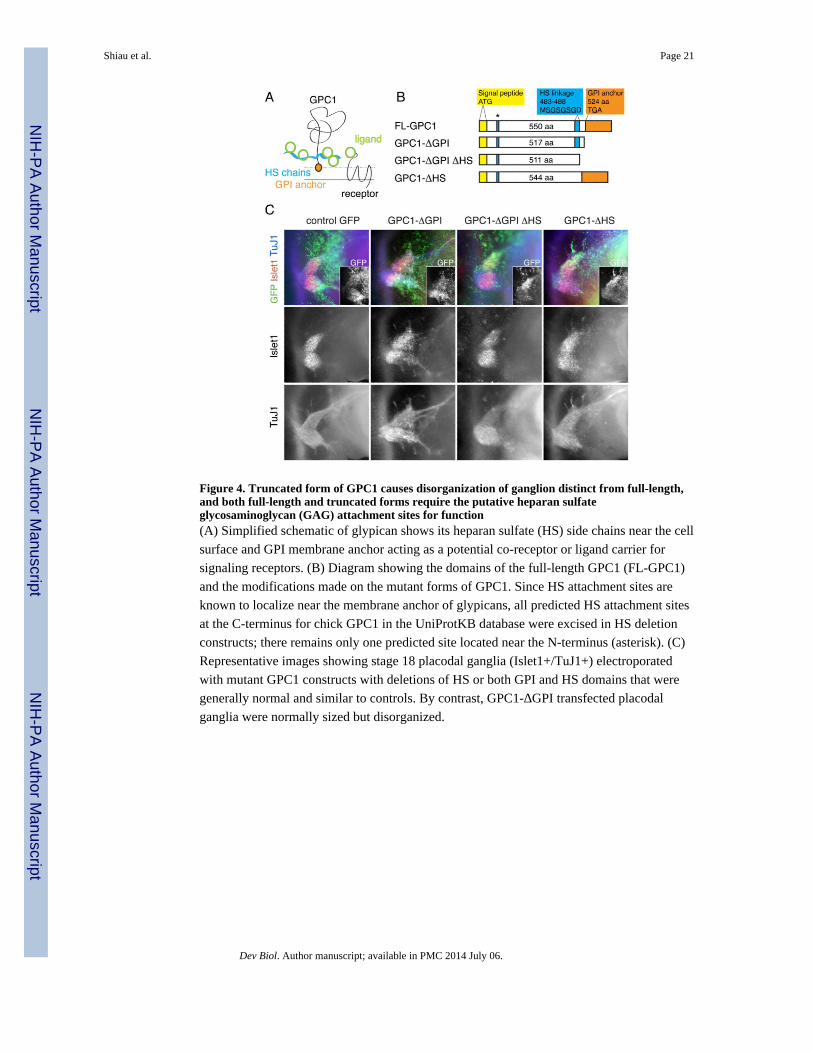
Figure 4. Truncated form of GPC1 causes disorganization of ganglion distinct from full-length,and both full-length and truncated forms require the putative heparan sulfateglycosaminoglycan (GAG) attachment sites for function(A) Simplified schematic of glypican shows its heparan sulfate (HS) side chains near the cell
surface and GPI membrane anchor acting as a potential co-receptor or ligand carrier for
signaling receptors. (B) Diagram showing the domains of the full-length GPC1 (FL-GPC1)
and the modifications made on the mutant forms of GPC1. Since HS attachment sites are
known to localize near the membrane anchor of glypicans, all predicted HS attachment sites
at the C-terminus for chick GPC1 in the UniProtKB database were excised in HS deletion
constructs; there remains only one predicted site located near the N-terminus (asterisk). (C)
Representative images showing stage 18 placodal ganglia (Islet1+/TuJ1+) electroporated
with mutant GPC1 constructs with deletions of HS or both GPI and HS domains that were
generally normal and similar to controls. By contrast, GPC1-ΔGPI transfected placodal
ganglia were normally sized but disorganized.
Shiau et al. Page 21
Dev Biol. Author manuscript; available in PMC 2014 July 06.
NIH
-PA
Author M
anuscriptN
IH-P
A A
uthor Manuscript
NIH
-PA
Author M
anuscript

Figure 5. Changes to GPC1 expression alters endogenous Wnt signaling activity(A) Top row, color overlay images showing trigeminal ganglia in whole mount chick
embryos at stages 15–16 after electroporation with control GFP, FL-GPC1, or GPC1-ΔGPI
constructs. GFP in green showing area of transfection (also shown in second row panels).
TuJ1 in blue showing placodal neurons (also shown in third row panels). RFP version of
TOPGAL Wnt-reporter expression in red showing endogenous activity of Wnt signaling
(also shown in fourth row panels). Outline of the trigeminal ganglion is demarcated by the
dotted lines. Fourth row panels, controls show Wnt-Reporter expression restricted to the
OpV region at stages 15 and 16 with generally absence of reporter in the MmV region
(asterisk), albeit occasionally one or a few cells were Wnt-Rep+. Reporter expression in the
OpV appears to increase over time. By contrast, FL-GPC1 expressing placodal ganglia had
markedly reduced Wnt-Reporter expression in the OpV region, and similar to controls, had
generally little to no reporter expression in the MmV region (lower asterisk). Conversely,
expression of GPC1-ΔGPI led to more expression of Wnt-Reporter in the OpV (arrowhead)
and expansion of reporter expression into the MmV region in more cells, though sparsely
(arrows). (B) Histogram showing percentages of cases of reduction of Wnt-Reporter
expression in the OpV and of expansion of Wnt-Reporter expression, which means an
increase in reporter expression in OpV as well as expression in more number of cells in
MmV, after electroporation with control GFP, FL-GPC1, or GPC1-ΔGPI. n shows number
of ganglia analyzed. Wnt-Rep, Wnt-Reporter. OpV, ophthalmic; MmV, maxillo-mandibular.
Shiau et al. Page 22
Dev Biol. Author manuscript; available in PMC 2014 July 06.
NIH
-PA
Author M
anuscriptN
IH-P
A A
uthor Manuscript
NIH
-PA
Author M
anuscript
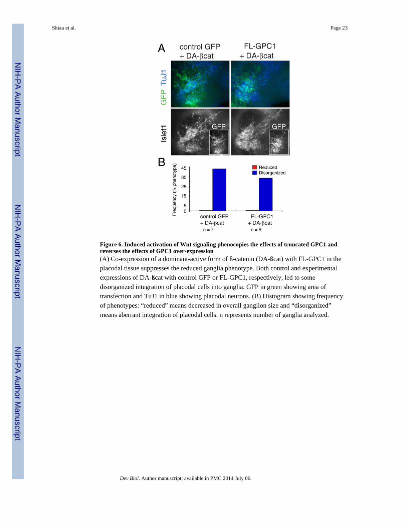
Figure 6. Induced activation of Wnt signaling phenocopies the effects of truncated GPC1 andreverses the effects of GPC1 over-expression(A) Co-expression of a dominant-active form of ß-catenin (DA-ßcat) with FL-GPC1 in the
placodal tissue suppresses the reduced ganglia phenotype. Both control and experimental
expressions of DA-ßcat with control GFP or FL-GPC1, respectively, led to some
disorganized integration of placodal cells into ganglia. GFP in green showing area of
transfection and TuJ1 in blue showing placodal neurons. (B) Histogram showing frequency
of phenotypes: “reduced” means decreased in overall ganglion size and “disorganized”
means aberrant integration of placodal cells. n represents number of ganglia analyzed.
Shiau et al. Page 23
Dev Biol. Author manuscript; available in PMC 2014 July 06.
NIH
-PA
Author M
anuscriptN
IH-P
A A
uthor Manuscript
NIH
-PA
Author M
anuscript