cell biology 1.2- ultrastructure of cells - fmfranco.com · 11/3/2015 1 cell biology 1.2-...
TRANSCRIPT
11/3/2015
1
Cell Biology
1.2- Ultrastructure of Cells
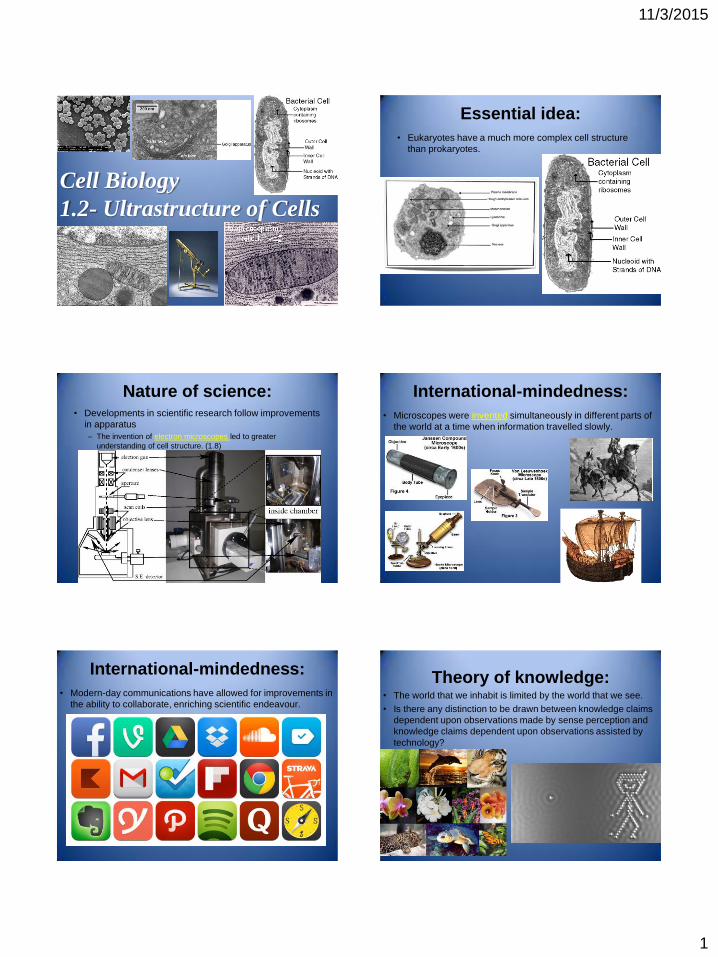
Essential idea:
• Eukaryotes have a much more complex cell structure
than prokaryotes.
Nature of science: • Developments in scientific research follow improvements
in apparatus
– The invention of electron microscopes led to greater
understanding of cell structure. (1.8)
International-mindedness:
• Microscopes were invented simultaneously in different parts of
the world at a time when information travelled slowly.
International-mindedness:
• Modern-day communications have allowed for improvements in
the ability to collaborate, enriching scientific endeavour.
Theory of knowledge: • The world that we inhabit is limited by the world that we see.
• Is there any distinction to be drawn between knowledge claims
dependent upon observations made by sense perception and
knowledge claims dependent upon observations assisted by
technology?
11/3/2015
2
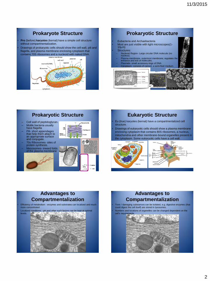
Prokaryote Structure
• Pro (before) karyotes (kernal) have a simple cell structure
without compartmentalization.
• Drawings of prokaryotic cells should show the cell wall, pili and
flagella, and plasma membrane enclosing cytoplasm that
contains 70S ribosomes and a nucleoid with naked DNA.
Prokaryotic Structure
• Eubacteria and Archaebacteria
• Most are just visible with light microscopes(1-10m)
• Structures: – Nucleoid Region- Large circular DNA molecule (no
nucleus).
– Plasma membrane- outermost membrane; regulates the entrance and exit of molecules.
– Plasmids- small accessory rings of DNA
– Cytoplasm- consists of cytosol, a semi-fluid medium.
Prokaryotic Structure
– Cell wall of peptidoglycan.
– Motile bacteria usually have flagella
– Pili- short appendages that help them attach to an appropriate surface and conjugate.
– 70s Ribosomes- sites of protein synthesis.
– Mesosomes- inward folds of the plasma membrane.
Eukaryotic Structure • Eu (true) karyotes (kernal) have a compartmentalized cell
structure.
• Drawings of eukaryotic cells should show a plasma membrane
enclosing cytoplasm that contains 80S ribosomes, a nucleus,
mitochondria and other membrane-bound organelles present in
the cytoplasm. Some eukaryotic cells have a cell wall.
Advantages to
Compartmentalization • Efficiency of metabolism - enzymes and substrates can localized and much
more concentrated
• Localized conditions - pH and other such factors can be kept at optimal
levels.
Advantages to
Compartmentalization • Toxic / damaging substances can be isolated, e.g. digestive enzymes (that
could digest the cell itself) are stored in lysosomes.
• Numbers and locations of organelles can be changed dependent on the
cell’s requirements
11/3/2015
3
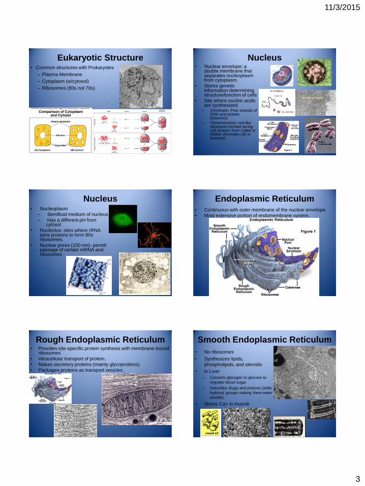
Eukaryotic Structure • Common structures with Prokaryotes
– Plasma Membrane
– Cytoplasm (w/cytosol)
– Ribosomes (80s not 70s)
Nucleus • Nuclear envelope: a
double membrane that separates nucleoplasm from cytoplasm.
• Stores genetic information determining structure/function of cells
• Site where nucleic acids are synthesized
– Chromatin: Fine strands of DNA and protein (histones)
– Chromosomes: rod-like structures formed during cell division from coiled or folded chromatin.(46 in humans)
Nucleus • Nucleoplasm
– Semifluid medium of nucleus
– Has a different pH from cytosol
• Nucleolus: sites where rRNA joins proteins to form 80s ribosomes.
• Nuclear pores (100 nm)- permit passage of certain mRNA and ribosomes
Endoplasmic Reticulum • Continuous with outer membrane of the nuclear envelope.
• Most extensive portion of endomembrane system.
Rough Endoplasmic Reticulum • Provides site-specific protein synthesis with membrane-bound
ribosomes
• Intracellular transport of protein.
• Makes secretory proteins (mainly glycoprotiens)
• Packages proteins as transport vesicles.
Smooth Endoplasmic Reticulum
• No ribosomes
• Synthesizes lipids,
phospholipids, and steroids
• In Liver
– Converts glycogen to glucose to
regulate blood sugar.
– Detoxifies drugs and poisons (adds
hydroxyl groups making them water
soluble).
• Stores Ca+ in muscle
11/3/2015
4
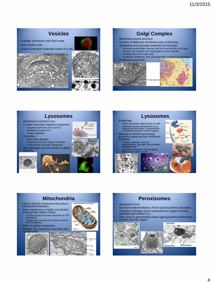
Vesicles
• A single membrane with fluid inside
• Very small in size
• Used to transport materials inside of a cell
Golgi Complex • Membrane-bound structure
• A series of flattened membrane sacs (cisternae).
• Synthesize and package materials for transport – Receives protein-filled vesicles that bud from the ER at cis face.
– Proteins are modified and repackaged as new vesicles.
– Vesicles leave from trans face.
– At plasma membrane, they discharge their contents as secretions.
• Produces lysosomes.
Lysosomes • Membrane-enclosed sacs
• Contain hydrolytic digestive enzymes. – Enzymes work best at low pH (5)
– Membrane pumps in H+
– Isolates digestion.
• Functions – Intracellular digestion
– Recycling of a cell’s organic materials
– Programmed cell death (apoptosis).
• Macrophages use lysosomes to digest bacteria.
Lysosomes • Autophagy
– When lysosomes digest parts of cells
– Important during development (e.g., tadpole tail absorption, degeneration of webbing between human fingers).
• Missing or inactive lysosomal enzymes cause serious childhood diseases.
– Pompe’s disease- missing carbohydrase, glycogen accumulates, damages liver.
– Tay-Sachs disease- missing lipase, lipids accumulate in brain.
Mitochondria • Have a double membrane that allows
compartmentalization
• Inner membrane is highly convoluted, forming folds called cristae. – Cristae contain enzymes important to ATP
production
– Cristae also increase the surface area for ATP production
• Sites of cellular respiration.
• Contain 70s ribosomes and their own circular DNA
Peroxisomes • Abundant in liver
• Not from endomembrane. From cytosol proteins and lipids.
• Transfers hydrogen (from substrates) to oxygen forming hydrogen peroxide(H2O2).
• Also occur in germinating seeds (glyoxysomes) where they convert oils into sugars
11/3/2015
5
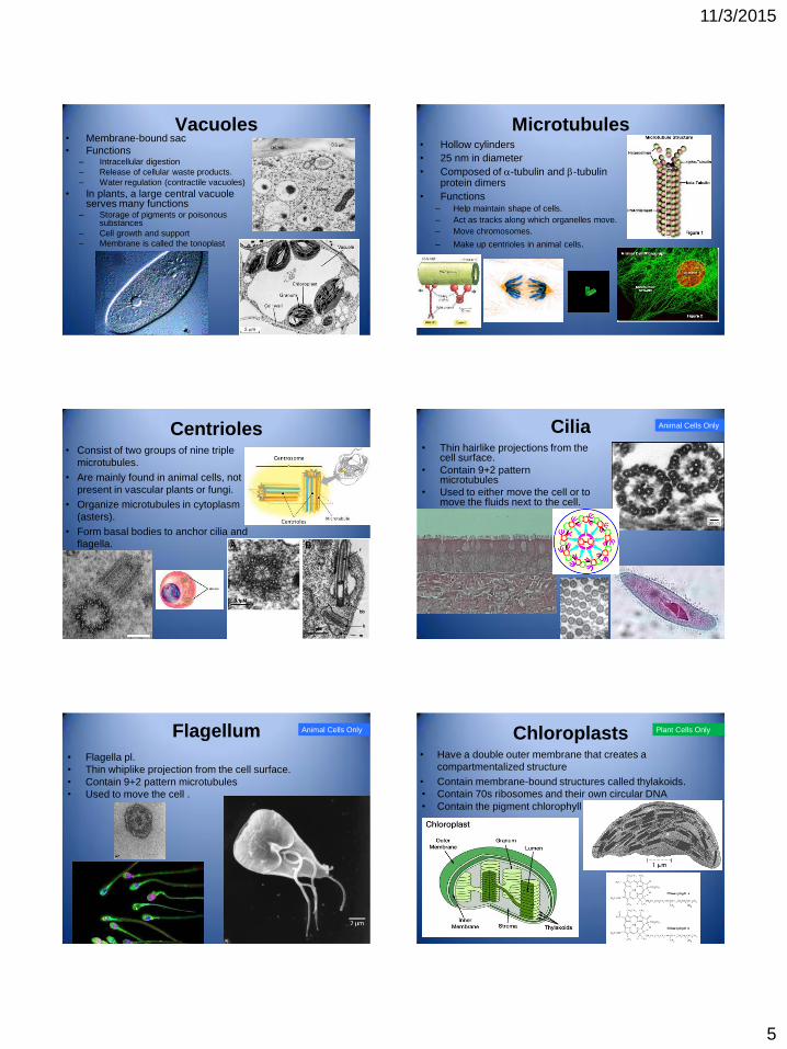
Vacuoles • Membrane-bound sac
• Functions – Intracellular digestion
– Release of cellular waste products.
– Water regulation (contractile vacuoles)
• In plants, a large central vacuole serves many functions
– Storage of pigments or poisonous substances
– Cell growth and support
– Membrane is called the tonoplast
Microtubules • Hollow cylinders
• 25 nm in diameter
• Composed of -tubulin and -tubulin protein dimers
• Functions – Help maintain shape of cells.
– Act as tracks along which organelles move.
– Move chromosomes.
– Make up centrioles in animal cells.
Centrioles • Consist of two groups of nine triple
microtubules.
• Are mainly found in animal cells, not
present in vascular plants or fungi.
• Organize microtubules in cytoplasm
(asters).
• Form basal bodies to anchor cilia and
flagella.
Cilia • Thin hairlike projections from the
cell surface.
• Contain 9+2 pattern microtubules
• Used to either move the cell or to move the fluids next to the cell.
Animal Cells Only
Flagellum
• Flagella pl.
• Thin whiplike projection from the cell surface.
• Contain 9+2 pattern microtubules
• Used to move the cell .
Animal Cells Only Chloroplasts • Have a double outer membrane that creates a
compartmentalized structure
• Contain membrane-bound structures called thylakoids.
• Contain 70s ribosomes and their own circular DNA
• Contain the pigment chlorophyll
Plant Cells Only
11/3/2015
6
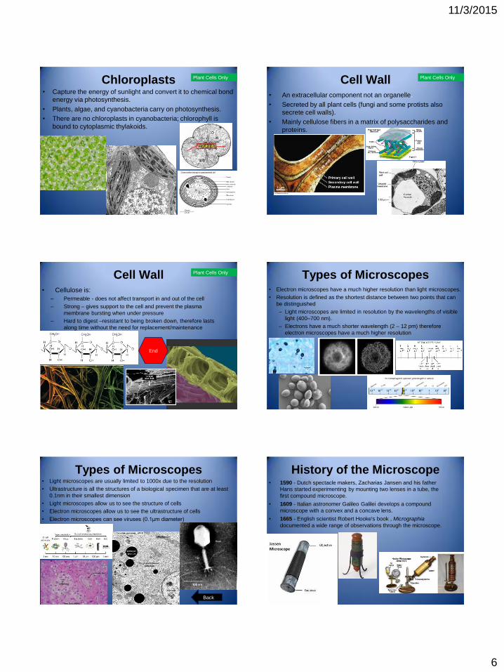
Chloroplasts • Capture the energy of sunlight and convert it to chemical bond
energy via photosynthesis.
• Plants, algae, and cyanobacteria carry on photosynthesis.
• There are no chloroplasts in cyanobacteria; chlorophyll is
bound to cytoplasmic thylakoids.
Plant Cells Only Cell Wall • An extracellular component not an organelle.
• Secreted by all plant cells (fungi and some protists also
secrete cell walls).
• Mainly cellulose fibers in a matrix of polysaccharides and
proteins.
Plant Cells Only
Cell Wall • Cellulose is:
– Permeable - does not affect transport in and out of the cell
– Strong – gives support to the cell and prevent the plasma
membrane bursting when under pressure
– Hard to digest –resistant to being broken down, therefore lasts
along time without the need for replacement/maintenance
Plant Cells Only
End
Types of Microscopes • Electron microscopes have a much higher resolution than light microscopes.
• Resolution is defined as the shortest distance between two points that can
be distinguished
– Light microscopes are limited in resolution by the wavelengths of visible
light (400–700 nm).
– Electrons have a much shorter wavelength (2 – 12 pm) therefore
electron microscopes have a much higher resolution
Types of Microscopes • Light microscopes are usually limited to 1000x due to the resolution
• Ultrastructure is all the structures of a biological specimen that are at least
0.1nm in their smallest dimension
• Light microscopes allow us to see the structure of cells
• Electron microscopes allow us to see the ultrastructure of cells
• Electron microscopes can see viruses (0.1μm diameter)
Back
History of the Microscope • 1590 - Dutch spectacle makers, Zacharias Jansen and his father
Hans started experimenting by mounting two lenses in a tube, the
first compound microscope.
• 1609 - Italian astronomer Galileo Galilei develops a compound
microscope with a convex and a concave lens.
• 1665 - English scientist Robert Hooke's book , Micrographia
documented a wide range of observations through the microscope.

11/3/2015
7
History of the Microscope • 1674 - Dutch scientist Anton van Leeuwenhoek used his
knowledge of grinding lenses to achieve greater magnification,
enabling detailed observations to be made of microorganisms.
• 1826 - British opticist Joseph Jackson Lister created an achromatic
lens to eradicate the chromatic effect caused by different
wavelengths of light.
Back