cell biology: structure and mechanism of mouse...
TRANSCRIPT

Golden, Olga Sokolova and Bruce L. GoodeSilvia Jansen, Agnieszka Collins, Leslie Regulating Actin DynamicsCyclase-associated Protein (CAP1) in Structure and Mechanism of MouseCell Biology:
doi: 10.1074/jbc.M114.601765 originally published online September 16, 20142014, 289:30732-30742.J. Biol. Chem.
10.1074/jbc.M114.601765Access the most updated version of this article at doi:
.JBC Affinity SitesFind articles, minireviews, Reflections and Classics on similar topics on the
Alerts:
When a correction for this article is posted•
When this article is cited•
to choose from all of JBC's e-mail alertsClick here
http://www.jbc.org/content/289/44/30732.full.html#ref-list-1
This article cites 36 references, 17 of which can be accessed free at
at BR
AN
DE
IS UN
IVE
RSIT
Y L
IBR
AR
Y on N
ovember 2, 2014
http://ww
w.jbc.org/
Dow
nloaded from
at BR
AN
DE
IS UN
IVE
RSIT
Y L
IBR
AR
Y on N
ovember 2, 2014
http://ww
w.jbc.org/
Dow
nloaded from

Structure and Mechanism of Mouse Cyclase-associatedProtein (CAP1) in Regulating Actin Dynamics*
Received for publication, July 31, 2014, and in revised form, September 14, 2014 Published, JBC Papers in Press, September 16, 2014, DOI 10.1074/jbc.M114.601765
Silvia Jansen‡, Agnieszka Collins‡, Leslie Golden‡, Olga Sokolova§, and Bruce L. Goode‡1
From the ‡Department of Biology, Rosenstiel Basic Medical Science Research Center, Brandeis University, Waltham, Massachusetts02454 and §Faculty of Biology, Moscow State University, GSP-1, 1 Leninskie Gory, Building 12, 119991 Moscow, Russia
Background: Mechanistic and structural conservation between evolutionarily distant Srv2/CAP homologs has remainedunclear.Results: Mouse CAP1 forms hexameric structures that autonomously bind F-actin, enhance cofilin-mediated severing, andcatalyze nucleotide exchange on actin.Conclusion: Yeast and mouse CAP share similar structures and functions.Significance: The role of CAP in actin regulation is remarkably well conserved.
Srv2/CAP is a conserved actin-binding protein with impor-tant roles in driving cellular actin dynamics in diverse animal,fungal, and plant species. However, there have been conflictingreports about whether the activities of Srv2/CAP are conserved,particularly between yeast and mammalian homologs. YeastSrv2 has two distinct functions in actin turnover: its hexamericN-terminal-half enhances cofilin-mediated severing of fila-ments, while its C-terminal-half catalyzes dissociation of cofilinfrom ADP-actin monomers and stimulates nucleotide ex-change. Here, we dissected the structure and function of mouseCAP1 to better understand its mechanistic relationship to yeastSrv2. Although CAP1 has a shorter N-terminal oligomerizationsequence compared with Srv2, we find that the N-terminal-halfof CAP1 (N-CAP1) forms hexameric structures with six protru-sions, similar to N-Srv2. Further, N-CAP1 autonomously bindsto F-actin and decorates the sides and ends of filaments, alteringF-actin structure and enhancing cofilin-mediated severing.These activities depend on conserved surface residues on thehelical-folded domain. Moreover, N-CAP1 enhances yeastcofilin-mediated severing, and conversely, yeast N-Srv2enhances human cofilin-mediated severing, highlighting themechanistic conservation between yeast and mammals. Fur-ther, we demonstrate that the C-terminal actin-binding�-sheet domain of CAP1 is sufficient to catalyze nucleotide-exchange of ADP-actin monomers, while in the presence ofcofilin this activity additionally requires the WH2 domain.Thus, the structures, activities, and mechanisms of mouseand yeast Srv2/CAP homologs are remarkably well con-served, suggesting that the same activities and mechanismsunderlie many of the diverse actin-based functions ascribedto Srv2/CAP homologs in different organisms.
While mechanisms for cellular actin assembly have beenextensively studied, the counter-balancing mechanisms under-lying actin filament disassembly and turnover are only nowbeginning to be understood. At the heart of the disassemblyprocess is the actin-binding protein cofilin, which severs fila-ments, thereby amplifying the number of filament ends fordepolymerization (1– 4). Cofilin is essential in vivo for rapidactin disassembly (5, 6), and three decades of biochemicalresearch have produced a rich understanding of its mechanism(7–9). Cofilin binds cooperatively to F-actin, alters the confor-mation of actin subunits, and induces filament twisting byabout 5 degrees per subunit, reducing the helical crossover dis-tance (10, 11). In addition, cofilin decoration changes themechanical properties of filaments, reducing filament persis-tence length by about 4-fold, and thus creates phase boundariesbetween decorated and undecorated regions, which inducessevering events (12, 13).
Although cofilin is sufficient to sever filaments in vitro, asvisualized in real time by TIRF microscopy (14), its activities invivo are further regulated and enhanced by additional factors,including Aip1, coronin, and Srv2/CAP (cyclase-associatedprotein)2 (15, 16). The physiological importance of these disas-sembly co-factors has been clearly demonstrated throughnumerous genetic studies; however, their underlying mecha-nisms remain only partially understood. Further, different pro-tein activities have been reported in some cases depending onwhich species is used, which has raised questions about howwell the mechanisms are conserved across distant species.Here, we address the mechanism and conservation of Srv2/CAP in regulating cofilin-mediated actin disassembly.
Yeast and mammalian Srv2/CAP proteins have two distinctfunctions in stimulating actin turnover. The C-terminal-half ofthe protein displaces cofilin from ADP-G-actin and catalyzesmonomer nucleotide exchange (17–19), whereas the N-termi-nal-half enhances cofilin-mediated severing of filaments, evenin the absence of actin monomers (20, 21). Recently, it was
* This work was supported, in whole or in part, by National Institutes of HealthGrant GM063691 (to B. G.).
1 To whom correspondence should be addressed: Rosenstiel Basic MedicalScience Research Center, Brandeis University, 415 South St., Waltham MA,02454. Tel.: 781-736-2464; Fax: 781-736-2405; E-mail: [email protected].
2 The abbreviations used are: CAP, cyclase-associated protein; TIRF, totalinternal reflection fluorescence; EM, electron microscopy; HsCof, humancofilin.
THE JOURNAL OF BIOLOGICAL CHEMISTRY VOL. 289, NO. 44, pp. 30732–30742, October 31, 2014© 2014 by The American Society for Biochemistry and Molecular Biology, Inc. Published in the U.S.A.
30732 JOURNAL OF BIOLOGICAL CHEMISTRY VOLUME 289 • NUMBER 44 • OCTOBER 31, 2014
at BR
AN
DE
IS UN
IVE
RSIT
Y L
IBR
AR
Y on N
ovember 2, 2014
http://ww
w.jbc.org/
Dow
nloaded from
shown that the two halves of yeast Srv2 can even be physi-cally separated and function in a largely autonomous manner invitro and in vivo (22). The N-terminal-half of yeast Srv2(N-Srv2) has also been shown to self-associate into hexamericstructures resembling shurikens (20), although it has beenunclear whether other species of Srv2/CAP adopt similar struc-tures. There is also some disagreement about the mechanism bywhich the C-terminal-half of Srv2/CAP catalyzes actin mono-mer recycling. Studies on yeast Srv2 show that the ability tocatalyze nucleotide exchange on cofilin-bound ADP-actinmonomers requires both the WH2 and �-sheet domains (23).In contrast, recent studies on mouse CAP1 reported that the�-sheet domain alone was sufficient to catalyze nucleotideexchange on ADP-actin monomers, and have called into ques-tion the role of the WH2 domain in C-CAP1 functions (24).Importantly, these assays were performed in the absence ofcofilin, precluding direct comparison of the two studies. Thishas left the role of the WH2 domain in C-CAP1 functionelusive.
To address the conservation of Srv2/CAP mechanismbetween yeast and mammals, here we dissected the structureand function of mouse CAP1 using a combination ofmutagenesis, bulk fluorescence assays, TIRF microscopy,and electron microscopy. Our results reveal that N-CAP1forms hexameric structures that bind autonomously to F-ac-tin using evolutionarily conserved surfaces, alter the twist ofF-actin, and enhance the severing effects of cofilin. More-over, the ability of C-CAP1 to catalyze nucleotide exchangeon cofilin-bound ADP-actin monomers requires both itsWH2 and �-sheet domains. These findings indicate that theactivities and mechanisms of distantly related Srv2/CAPhomologs are highly conserved, and suggest that the diversemembers of this protein family may have similar cellularfunctions in regulating the actin cytoskeleton.
EXPERIMENTAL PROCEDURES
Hydrodynamic Analysis of Endogenous Human CAP1-ActinComplex—HEK293T cells were maintained at 37 °C under ahumidified atmosphere containing 5% CO2 in Dulbecco’s mod-ified Eagle’s medium (DMEM), supplemented with 10% (v/v)heat-inactivated fetal bovine serum, glucose (4.5 g/liter), peni-cillin (100 units/ml), and streptomycin (100 �g/ml). Cells wereharvested in PBS, collected by centrifugation at 1000 � g for 5min, and lysed by douncing in 50 mM Tris/HCl, pH 7.5, 150 mM
NaCl, and 1% (v/v) Triton X-100. The lysate was cleared bycentrifugation at 14,000 rpm at 4 °C, then loaded on top of a12-ml sucrose gradient (3–30%) in PBS. Size standards werefractionated in parallel. After centrifugation in a SW40 Ti rotor(Beckman) at 30,000 rpm for 18 h at 4 °C, 500-�l fractions werecollected and analyzed by immunoblotting with CAP1 antibod-ies (MaxPab D01, Abnova, Taiwan) or Coomassie staining forprotein standards. Peak CAP1-positive fractions from sucrosegradients were concentrated and fractionated on a Superose 6gel filtration column, with protein standards fractionated inparallel. Using the sedimentation coefficient and Stokes radiusfor CAP1, obtained from the analyses above, the molecularweight of native CAP1 complex was calculated using the for-mula: M � (6��0Nas)/(1 � ��), with M � molecular weight, �0
(viscosity of water) � 1.002 � 10�2g/(cm*s), n � Avogadro’snumber, a � Stokes radius, s � sedimentation coefficient, �(partial specific volume of an average particle) � 0.725 cm3/g, �(density of water) � 0.998 g/cm2.
Plasmids—pHAT2-N-CAP1 and pGAT2-C-CAP1 were kindlyprovided by Pekka Lappalainen (Univ. Helsinki) and used topurify N-CAP1 (residues 1–217) and C-CAP1 (residues 216 –475) from Escherichia coli (24). Inserts from these plasmidswere combined to reconstitute a full-length CAP1 plasmid forexpression and purification from yeast. The �-sheet of CAP1was PCR amplified from a C-CAP1 plasmid and cloned into thepET28a vector. Mutant N-CAP1 and C-CAP1 constructs weregenerated by site-directed mutagenesis. All constructs wereverified by DNA sequencing. The plasmid for expressinghuman cofilin 1 (HsCof1) in E. coli was generously provided byDavid Kovar (Univ. Chicago). Plasmids for expressing yeastcofilin (yCof1) and N-Srv2 have been described elsewhere (20).
Protein Purification—Rabbit skeletal muscle actin was puri-fied as previously described in detail (25). His6-tagged polypep-tides (N-Srv2, N-CAP1, C-CAP1, B-CAP1, and mutants) wereexpressed in BL21 (pRARE) E. coli cells. Cultures were grown tolog phase at 37 °C and induced for 16 h with 0.4 mM isopropyl�-D-1-thiogalactopyranoside (IPTG) at 18 °C. Cells were har-vested by centrifugation and lysed by sonication in 20 mM phos-phate buffer pH 7.4, 300 mM NaCl, 1 mM DTT (lysis buffer)supplemented with 10 mM imidazole and a standard mixture ofprotease inhibitors. Clarified lysates were incubated with Ni2�-NTA beads (Qiagen, Valencia, CA) for 90 min at 4 °C and thentransferred to a poly-prep chromatography column (Bio-Rad).The resin was washed with 10 column volumes of lysis buffersupplemented with 50 mM imidazole. Proteins were eluted with5 column volumes of lysis buffer supplemented with 250 mM
imidazole, concentrated, and purified further on a Superose 6gel filtration column (GE Healthcare) equilibrated in 20 mM
Tris pH 8.0, 100 mM NaCl, and 1 mM DTT. Full-length CAP1was expressed under control of the GAL promotor in a prote-ase-deficient yeast strain (BGY502). Two liters of cells weregrown at 30 °C in synthetic medium without uracil and with 2%raffinose to an OD600 of 0.8 – 0.9, then expression was inducedfor 16 h at 30 °C by addition of 2% galactose. Cells were har-vested by centrifugation, washed twice with 100 ml water, andresuspended in 10 ml of water per 2.5 grams cells. The cellsuspension was drop frozen in liquid N2, then lysed by mechan-ical sheering in a coffee blender under liquid N2, and stored as alysed powder at �80 °C. For purifications, 20 g of yeast powderwas thawed in 20 ml of 2� lysis buffer supplemented with 20mM imidazole and protease inhibitors. The cleared lysate wasadded to Ni2�-NTA beads and purified as described above forthe other Srv2/CAP constructs. HsCof1 was expressed in E. colias above and purified as follows. Cells were lysed by sonicationin 20 mM Tris pH 8.0, 50 mM NaCl, 1 mM DTT, and proteaseinhibitors. Lysates were cleared and applied to a 5-ml HiTrapHP Q column (GE Healthcare). The flow-through fraction con-tained HsCof1, and was collected and dialyzed into 20 mM
Hepes pH 6.8, 25 mM NaCl, and 1 mM DTT. Then the proteinwas applied to a 5 ml of HiTrap SP FF column (GE Healthcare)and eluted with a linear gradient of 25 to 500 mM NaCl. Thefractions containing HsCof1 were concentrated and dialyzed to
Mammalian CAP1 Structure and Mechanism in Actin Regulation
OCTOBER 31, 2014 • VOLUME 289 • NUMBER 44 JOURNAL OF BIOLOGICAL CHEMISTRY 30733
at BR
AN
DE
IS UN
IVE
RSIT
Y L
IBR
AR
Y on N
ovember 2, 2014
http://ww
w.jbc.org/
Dow
nloaded from
20 mM Tris, pH 8.0, 50 mM NaCl, and 1 mM DTT, aliquoted,snap-frozen in liquid N2, and stored at �80 °C until use.
Electron Microscopy and Single Particle Analysis—To imageactin filaments by electron microscopy (EM), Ca2�-ATP-G-actin (24 �M) was polymerized by addition of 2 mM MgCl2 and50 mM KCl and incubation for 1 h at 25 °C. F-actin was dilutedto 2 �M in F-buffer (50 mM KCl, 2 mM MgCl2, 0.2 mM EGTA, 1mM DTT, 5 mM Tris, pH.8) and incubated for 15 min at 25 °Cwith control buffer (for undecorated filaments), or with 5 �M
HsCof1, N-CAP1, and/or N-CAP1–91. Samples were diluted2-fold in F-buffer and adsorbed to glow discharged formvar-carbon coated 200 mesh copper grids for 15–20 s, blotted toremove excess solution, negatively stained with 1% (w/v) uranylacetate for 1 min, blotted again, and allowed to air-dry. Forsingle particle analysis, 3 �M N-CAP1 or N-Srv2 was applied togrids and negative stained as above. For both analyses, imageswere captured using an FEI Morgani 268 transmission electronmicroscope at an acceleration voltage of 80 kV and magnifica-tions of 14,000, 18,000, or 22,000. For single particle analysis,4068 N-CAP1 particles were selected from the EM imagesusing Boxer (26) and windowed into 80 � 80 pixel images.These were then filtered and normalized to a standard devia-tion of 1, and processed for reference-free classification inIMAGIC (27). Iterative classification yielded 40 classes, whichwere used for the three-dimensional reconstruction by theangular reconstitution method (28) and the back-projectionalgorithm to obtain a first rough three-dimensional model. Thismodel was reprojected onto two-dimensional space for refiningusing iterative procedures. Further improvements in three-di-mensional reconstructions and correction for CTF were per-formed using Frealign program (29). 6-fold symmetry wasapplied to the final reconstruction, as the majority of class-sumaverages possessed 6-fold symmetry. Resolution of the finalstructure, defined by 0.5 FSC, was 25 Å. For scoring N-CAP1particles bound to actin filaments, particles were identified bytheir unique structure and size and categorized as bound if indirect contact with a filament. Densities of particles and fila-ments under these conditions were low, such that particles incontact with filaments are likely to represent specifically boundmolecules, in agreement with the specificity of binding ob-served in co-sedimentation assays (below).
Bulk F-actin Disassembly Assays—At time 0 in the assays,preassembled F-actin (2 �M final, 10% pyrene labeled) wasmixed with the indicated proteins or control buffers, 100 nM
CapZ, and 3 �M vitamin D-binding protein (VDBP)/humanplasma Gc-globulin (Sigma-Aldrich) in F-buffer (20 mM Tris,pH 7.5, 50 mM KCl, 0.2 mM ATP, 1 mM MgCl2, and 1 mM DTT).Decrease in fluorescence was monitored for 900 s at 25 °C at365-nm excitation and 407-nm emission in a fluorescencespectrophotometer (Photon Technology International, Law-renceville, NJ).
Total Internal Reflection Fluorescence (TIRF) Microscopy—Inall experiments, 24 � 60 mm coverslips (Fisher Scientific) werefirst cleaned by sonication in detergent for 60 min, followed bysuccessive sonications in 1 M KOH and 1 M HCl for 20 min each,then sonication in ethanol for at least 60 min. Coverslips werethen washed extensively with ddH2O, dried in an N2-stream,layered with 200 �l of 80% ethanol pH 2.0, 2 mg/ml methoxy-
poly (ethylene glycol)-silane and 2 �g/ml biotin-poly (ethyleneglycol)-silane (Laysan Bio Inc., Arab, AL), and incubated for16 h at 70 °C. Flow cells were assembled by rinsing PEG-coatedcoverslips extensively with ddH2O, then attaching it to a flowchamber (Ibidi, Martinsried, Germany) with double-sided tape(2.5 cm � 2 mm � 120 �m) and 5 min epoxy resin. OregonGreen (OG)-labeled actin was prepared as described (30). Forfilament severing assays, flow cells were incubated for 5 minwith HBSA (HEK buffer with 1% BSA), followed by 30 s incu-bation with 0.1 mg/ml streptavidin in PBS. Flow cells werewashed with 5 chamber volumes (�50 �l) HBSA, then equili-brated with 1� TIRF buffer (10 mM imidazole, 50 mM KCl, 1mM MgCl2, 1 mM EGTA, 0.2 mM ATP, 10 mM DTT, 15 mM
glucose, 20 �g/ml catalase, 100 �g/ml glucose oxidase, and0.5% methylcellulose (4000 cP), pH 7.5). Reactions were initi-ated by rapidly diluting actin monomers (1 �M final, 10% OG-labeled, 0.5% biotinylated) into 1� TIRF buffer and transferringthe mixture to a flow chamber. The filaments were left poly-merizing till 10 –15 �m, after which the reaction mixture wasreplaced with TIRF buffer containing Cof1 and/or CAP1 poly-peptides, and lacking actin monomers. Time-lapse TIRFM ofOG-actin filaments was performed using a Nikon-Ti200inverted microscope equipped with a 150 milliwatt Ar-Laser(Mellot Griot, Carlsbad, CA), a TIRF-objective with a N.A. of1.49 (Nikon Instruments Inc., New York, NY), and an EMCCDcamera (Andor Ixon, Belfast, Northern Ireland). During mea-surements, optimal focus was maintained using the perfectfocus system (Nikon Instruments Inc.). Images were capturedevery 5 s. The pixel size corresponded to 0.27 �m. Filamentsevering efficiency, expressed as severing events �m�1 s�1, wasdetermined by measuring the lengths of individual filamentsprior to Cof1 addition in ImageJ, and scoring severing eventsover time after flowing in cofilin and/or N-CAP1.
F-actin Cosedimentation Assays—Preformed actin filamentswere incubated with variable concentrations of N-CAP1 orN-CAP1–91 for 30 min at room temperature in F-buffer (20mM Tris, pH 7.5, 50 mM KCl, 0.2 mM ATP, 1 mM MgCl2, and 1mM DTT). Reactions were centrifuged at 350,000 � g for 30 minat 20 °C. Supernatant and pellet fractions were analyzed on gelsby Coomassie staining and quantified by scanning densitome-try. Each concentration of N-CAP1 and N-CAP1–91 was pro-cessed in parallel reactions lacking F-actin to control againstnonspecific pelleting. The binding affinity of N-CAP1 for F-ac-tin was determined in assays as above using F-buffer at twodifferent pH values (7.5 and 8.0). For each, a binding curve wasfit, and the Kd was determined by non-linear regression analysisusing Prism 5.0.
Nucleotide Exchange Assays—Nucleotide exchange rates onADP-G-actin were determined by measuring the increase influorescence upon incorporation of �-ATP (Sigma-Aldrich).ADP-G-actin was generated by removing bound ATP by dowextreatment and overnight incubation at 4 °C in the presence ofhexokinase and an excess of ADP. Next, 2 �M of ADP-G-actinwas mixed with proteins in CDT buffer (0.2 mM CaCl2, 0.2mM DTT, 10 mM Tris pH 8.0) or buffer alone and added to 50�M �-ATP. The reaction was monitored for 200 s at 350-nmexcitation and 410-nm emission at 25 °C in a fluorescence spec-trophotometer (Photon Technology International). Exchange
Mammalian CAP1 Structure and Mechanism in Actin Regulation
30734 JOURNAL OF BIOLOGICAL CHEMISTRY VOLUME 289 • NUMBER 44 • OCTOBER 31, 2014
at BR
AN
DE
IS UN
IVE
RSIT
Y L
IBR
AR
Y on N
ovember 2, 2014
http://ww
w.jbc.org/
Dow
nloaded from
rates were calculated from linear fitting of the first 50 s of eachreaction curve.
RESULTS
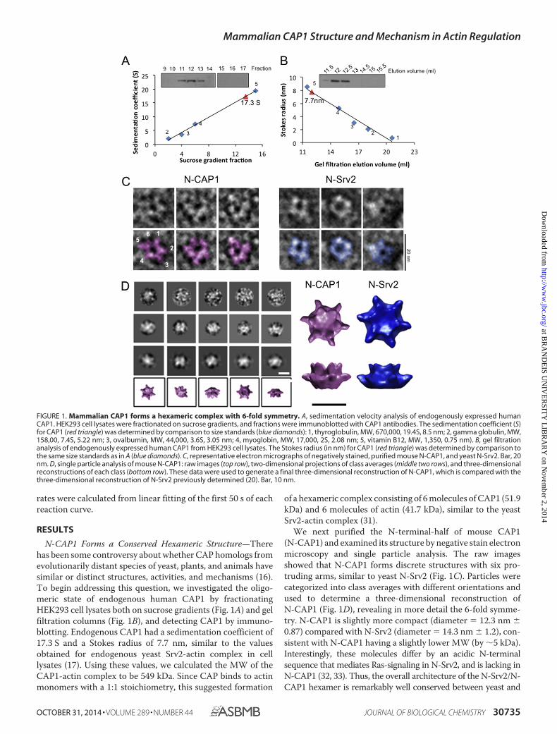
N-CAP1 Forms a Conserved Hexameric Structure—Therehas been some controversy about whether CAP homologs fromevolutionarily distant species of yeast, plants, and animals havesimilar or distinct structures, activities, and mechanisms (16).To begin addressing this question, we investigated the oligo-meric state of endogenous human CAP1 by fractionatingHEK293 cell lysates both on sucrose gradients (Fig. 1A) and gelfiltration columns (Fig. 1B), and detecting CAP1 by immuno-blotting. Endogenous CAP1 had a sedimentation coefficient of17.3 S and a Stokes radius of 7.7 nm, similar to the valuesobtained for endogenous yeast Srv2-actin complex in celllysates (17). Using these values, we calculated the MW of theCAP1-actin complex to be 549 kDa. Since CAP binds to actinmonomers with a 1:1 stoichiometry, this suggested formation
of a hexameric complex consisting of 6 molecules of CAP1 (51.9kDa) and 6 molecules of actin (41.7 kDa), similar to the yeastSrv2-actin complex (31).
We next purified the N-terminal-half of mouse CAP1(N-CAP1) and examined its structure by negative stain electronmicroscopy and single particle analysis. The raw imagesshowed that N-CAP1 forms discrete structures with six pro-truding arms, similar to yeast N-Srv2 (Fig. 1C). Particles werecategorized into class averages with different orientations andused to determine a three-dimensional reconstruction ofN-CAP1 (Fig. 1D), revealing in more detail the 6-fold symme-try. N-CAP1 is slightly more compact (diameter � 12.3 nm �0.87) compared with N-Srv2 (diameter � 14.3 nm � 1.2), con-sistent with N-CAP1 having a slightly lower MW (by �5 kDa).Interestingly, these molecules differ by an acidic N-terminalsequence that mediates Ras-signaling in N-Srv2, and is lacking inN-CAP1 (32, 33). Thus, the overall architecture of the N-Srv2/N-CAP1 hexamer is remarkably well conserved between yeast and
FIGURE 1. Mammalian CAP1 forms a hexameric complex with 6-fold symmetry. A, sedimentation velocity analysis of endogenously expressed humanCAP1. HEK293 cell lysates were fractionated on sucrose gradients, and fractions were immunoblotted with CAP1 antibodies. The sedimentation coefficient (S)for CAP1 (red triangle) was determined by comparison to size standards (blue diamonds): 1, thyroglobulin, MW, 670,000, 19.4S, 8.5 nm; 2, gamma globulin, MW,158,00, 7.4S, 5.22 nm; 3, ovalbumin, MW, 44,000, 3.6S, 3.05 nm; 4, myoglobin, MW, 17,000, 2S, 2.08 nm; 5, vitamin B12, MW, 1,350, 0.75 nm). B, gel filtrationanalysis of endogenously expressed human CAP1 from HEK293 cell lysates. The Stokes radius (in nm) for CAP1 (red triangle) was determined by comparison tothe same size standards as in A (blue diamonds). C, representative electron micrographs of negatively stained, purified mouse N-CAP1, and yeast N-Srv2. Bar, 20nm. D, single particle analysis of mouse N-CAP1: raw images (top row), two-dimensional projections of class averages (middle two rows), and three-dimensionalreconstructions of each class (bottom row). These data were used to generate a final three-dimensional reconstruction of N-CAP1, which is compared with thethree-dimensional reconstruction of N-Srv2 previously determined (20). Bar, 10 nm.
Mammalian CAP1 Structure and Mechanism in Actin Regulation
OCTOBER 31, 2014 • VOLUME 289 • NUMBER 44 JOURNAL OF BIOLOGICAL CHEMISTRY 30735
at BR
AN
DE
IS UN
IVE
RSIT
Y L
IBR
AR
Y on N
ovember 2, 2014
http://ww
w.jbc.org/
Dow
nloaded from

mice, yet in yeast Srv2 also allows the insertion of some residuesthat introduce new functional capabilities.
N-CAP1 Strongly Enhances Cofilin-mediated Severing of ActinFilaments—We next purified full-length CAP1, N-CAP1, andC-CAP1 (Fig. 2A) to compare their effects on cofilin-medi-ated F-actin disassembly in bulk assays (Fig. 2B). Both FL-CAP1 and N-CAP1, but not C-CAP1, enhanced cofilin-me-diated disassembly; however, they had no effect on actindisassembly in the absence of cofilin (Fig. 2B). Further, theability of N-CAP1 to enhance cofilin-mediated disassemblywas concentration-dependent (Fig. 2C). Mutant N-CAP1–91, which targets a conserved surface on the HFD domainrequired for yeast N-Srv2 activity (20, 31), abolishedN-CAP1 stimulatory effects on actin disassembly (Fig. 2B).These results indicate that the enhanced disassembly activ-ity of the N-terminal-half of CAP depends on surfaces in theHFD domain that are well conserved between yeast andmammals, and agrees with our results showing that N-Srv2and N-CAP1 form highly similar structures.
To better understand the mechanism by which CAP1enhances cofilin-mediated actin disassembly, we monitoredthe effects of cofilin and/or CAP1 on filaments in real timeusing TIRF microscopy (Fig. 2, D and E). Fluorescently labeledactin filaments (10% Oregon Green-labeled; 0.5% biotin-la-beled) were polymerized and tethered through biotin-strepta-vidin-biotin-PEG interactions to the coverslip surface. Humancofilin (HsCof1) and/or N-CAP1 were flowed in, and severingevents were monitored for 200 s (Fig. 2D). Quantificationrevealed that 50 s after flow-in, �8-fold more severing eventsper �m of filament had occurred in the presence of cofilin andN-CAP1 than in the presence of cofilin alone (Fig. 2E). N-CAP1alone failed to induce severing (see “Experimental Proce-dures”). Thus, N-CAP1 strongly increases the efficiency of cofi-lin-mediated severing. Importantly, there are no actin mono-mers present in these reactions when severing was beingmonitored, which excludes the possibility that enhanced disas-sembly stems from N-CAP1 recycling cofilin from actin mono-mers. These activities are similar to the effects reported for both
FIGURE 2. Effects of CAP1 on cofilin-mediated F-actin disassembly. A, schematic of domains in CAP and constructs used in these experiments. OD,oligomerization domain; HFD, helical folded domain; P, polyproline region; WH2, WASP-homology 2 domain. Arrow indicates position of CAP1–91 mutation inthe HFD. B, effects of 250 nM human cofilin (HsCof1) and/or 750 nM mouse N-CAP1 constructs on disassembly of 2 �M F-actin (10% pyrene-labeled) in thepresence of 100 nM CapZ and 3 �M vitamin D-binding protein. C, bulk F-actin disassembly assays, as in B, testing a range of concentrations of mouse N-CAP1both in the presence and absence of 250 nM HsCof1. D, TIRF microscopy analysis of actin filament severing by cofilin with and without N-CAP1. Filaments werepolymerized from 1 �M G-actin (10% Oregon Green, 0.5% biotinylated) and tethered through biotin-streptavidin conjugation. After filaments were polymer-ized to lengths of �15 �m, then 250 nM HsCof1 and/or 750 nM N-CAP1 was flowed in without actin monomers (indicated by black arrow), and filaments weremonitored for 200 s. In the montage shown, severing events are indicated by yellow arrows. Bar, 5 �m. E, average number of severing events per �m filamentat 50 s, quantified 50 s after flow in. Averages for each condition are from at least 40 individual filaments obtained from 3 independent trials. Error bars representS.D. (n � 3).
Mammalian CAP1 Structure and Mechanism in Actin Regulation
30736 JOURNAL OF BIOLOGICAL CHEMISTRY VOLUME 289 • NUMBER 44 • OCTOBER 31, 2014
at BR
AN
DE
IS UN
IVE
RSIT
Y L
IBR
AR
Y on N
ovember 2, 2014
http://ww
w.jbc.org/
Dow
nloaded from

yeast N-Srv2 (20) and full-length bovine CAP1 (21), and againsuggest a conserved mechanism.
N-CAP1 Hexamers Autonomously Bind F-actin—To probethe mechanism by which N-CAP1 enhances cofilin-mediatedsevering, we asked whether N-CAP1 and/or N-CAP1–91 binddirectly to F-actin. In the absence of cofilin, N-CAP1 co-sedi-mented with F-actin, in a concentration-dependent manner,demonstrating a direct interaction (Fig. 3, A and B). In contrast,N-CAP1–91 showed minimal association, indicating that bind-ing requires the conserved functional surface on the HFDdomain targeted by the CAP1–91 mutation. At a fixed concen-
tration of N-CAP1 (1 �M), the fraction of bound N-CAP1appears to saturate at 0.4 �M (Fig. 3, C and D), and the F-actinbinding affinity of N-CAP1 was measured (Kd � 7.6 �M at pH7.5; Kd � 1.6 �M at pH 8.0). Further, by performing these assaysat a higher concentration of F-actin (10 �M) and variable con-centrations of N-CAP1, we found that binding saturates at �0.3moles of N-CAP1 per mol of F-actin (Fig. 3D). It is not yet clearwhy binding of N-CAP1 saturates at 40%, but this may reflectheterogeneity in the population of N-CAP1 molecules, includ-ing the possibility of N-CAP1 existing in equilibrium betweendifferent conformations that do and do not bind F-actin.
FIGURE 3. N-CAP1 binding to F-actin. A, co-sedimentation of N-CAP1 constructs with F-actin. Reactions contained 2.5 �M F-actin and/or 10 �M N-CAP1 orN-CAP1-91. Pellets and supernatants were analyzed on Coomassie-stained gels. B, concentration-dependent binding of N-CAP1 to F-actin (2.5 �M) at pH 7.5.Co-sedimentation assays, as in A, were performed over a range of concentrations of N-CAP1 and N-CAP1-91. The concentration of N-CAP1 protein bound tofilaments was determined by scanning densitometry of bands on Coomassie-stained gels. C, binding affinity of N-CAP1 for F-actin. Co-sedimentation assayswere performed using 1 �M N-CAP1 and a range of F-actin concentrations at both pH 7.5 and pH 8.0. The concentration of N-CAP1 bound to filaments wasdetermined by densitometry of bands on Coomassie-stained gels. Each data point shown is an average from three independent trials. For each pH, the bindingcurve was fit, and the Kd was measured by non-linear regression analysis using Prism 5.0 (Rsquare at pH 7.5 � 0.97, Rsquare at pH 8.0 � 0.95). Error bars, S.D. D, molarratio of binding of N-CAP1 to F-actin. Co-sedimentation assays were performed using 10 �M F-actin and variable concentrations of N-CAP1 at pH 8.0. Themolar concentration of N-CAP1 bound to F-actin was determined by densitometry of bands on Coomassie-stained gels. Using a linear curve fit (shown), themolar ratio of binding was determined from the slope (0.312 � 0.018) E, representative electron micrograph of negatively stained actin filament decorated byN-CAP1 (top) or N-Srv2 (bottom). Shown for each is the raw micrograph and a replica micrograph with the N-CAP1 and N-Srv2 particles shaded (purple and blue,respectively). F, distribution of the distances measured between N-CAP1 particles on the actin filaments. G, distribution of N-CAP1 particle association withF-actin (n � 76). Representative images of each binding mode are shown, along with percentage of particles that fell into each class.
Mammalian CAP1 Structure and Mechanism in Actin Regulation
OCTOBER 31, 2014 • VOLUME 289 • NUMBER 44 JOURNAL OF BIOLOGICAL CHEMISTRY 30737
at BR
AN
DE
IS UN
IVE
RSIT
Y L
IBR
AR
Y on N
ovember 2, 2014
http://ww
w.jbc.org/
Dow
nloaded from
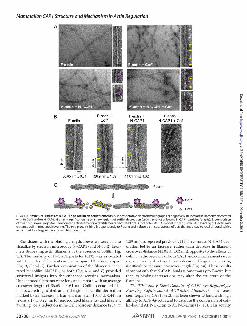
Consistent with the binding analysis above, we were able tovisualize by electron microscopy N-CAP1 (and N-Srv2) hexa-mers decorating actin filaments in the absence of cofilin (Fig.3E). The majority of N-CAP1 particles (81%) was associatedwith the sides of filaments and were spaced 35–54 nm apart(Fig. 3, F and G). Further examination of the filaments deco-rated by cofilin, N-CAP1, or both (Fig. 4, A and B) providedstructural insights into the enhanced severing mechanism.Undecorated filaments were long and smooth with an averagecrossover length of 36.65 � 0.61 nm. Cofilin-decorated fila-ments were fragmented, and had regions of cofilin-decorationmarked by an increase in filament diameter (10.07 � 0.44 nmversus 8.19 � 0.22 nm for undecorated filaments) and filament’twisting’, or a reduction in helical crossover distance (26.9 �
1.09 nm), as reported previously (11). In contrast, N-CAP1 dec-oration led to an increase, rather than decrease in filamentcrossover distance (41.01 � 1.02 nm), opposite to the effects ofcofilin. In the presence of both CAP1 and cofilin, filaments werereduced to very short and heavily decorated fragments, makingit difficult to measure crossover length (Fig. 4B). These resultsshow not only that N-CAP1 binds autonomously to F-actin, butthat its binding interactions may alter the structure of thefilament.
The WH2 and �-Sheet Domains of CAP1 Are Required forRecycling Cofilin-bound ADP-actin Monomers—The yeastcounterpart of CAP1, Srv2, has been shown to bind with highaffinity to ADP-G-actin and to catalyze the conversion of cofi-lin-bound ADP-G-actin to ATP-G-actin (17, 18). This activity
FIGURE 4. Structural effects of N-CAP1 and cofilin on actin filaments. A, representative electron micrographs of negatively stained actin filaments decoratedwith HsCof1 and/or N-CAP1. Higher magnification insets show regions of cofilin decoration (yellow arrows) or bound N-CAP1 particles (purple). B, comparisonof mean crossover length for undecorated actin filaments versus filaments decorated by HsCof1 or N-CAP1. C, model showing how CAP1 binding to F-actin mayenhance cofilin-mediated severing. The two proteins bind independently to F-actin and induce distinct structural effects that may lead to local discontinuitiesin filament topology and accelerate fragmentation.
Mammalian CAP1 Structure and Mechanism in Actin Regulation
30738 JOURNAL OF BIOLOGICAL CHEMISTRY VOLUME 289 • NUMBER 44 • OCTOBER 31, 2014
at BR
AN
DE
IS UN
IVE
RSIT
Y L
IBR
AR
Y on N
ovember 2, 2014
http://ww
w.jbc.org/
Dow
nloaded from
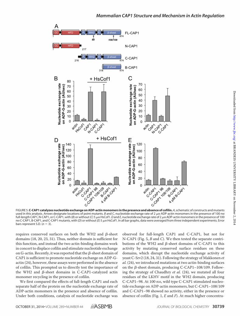
requires conserved surfaces on both the WH2 and �-sheetdomains (18, 20, 23, 31). Thus, neither domain is sufficient forthis function, and instead the two actin-binding domains workin concert to displace cofilin and stimulate nucleotide exchangeon G-actin. Recently, it was reported that the �-sheet domain ofCAP1 is sufficient to promote nucleotide exchange on ADP-G-actin (24), however, these assays were performed in the absenceof cofilin. This prompted us to directly test the importance ofthe WH2 and �-sheet domains in C-CAP1-catalyzed actinmonomer recycling in the presence of cofilin.
We first compared the effects of full-length CAP1 and eachseparate half of the protein on the nucleotide exchange rate ofADP-actin monomers in the presence and absence of cofilin.Under both conditions, catalysis of nucleotide exchange was
observed for full-length CAP1 and C-CAP1, but not forN-CAP1 (Fig. 5, B and C). We then tested the separate contri-butions of the WH2 and �-sheet domains of C-CAP1 to thisactivity by mutating conserved surface residues on thesedomains, which disrupt the nucleotide exchange activity ofyeast C-Srv2 (18, 24, 31). Following the strategy of Makkonen etal. (24), we introduced mutations at two actin-binding surfaceson the �-sheet domain, producing C-CAP1–108/109. Follow-ing the strategy of Chaudhry et al. (24), we mutated all fourresidues of the LKHV motif in the WH2 domain, producingC-CAP1–98. At 100 nM, wild type C-CAP1 stimulated nucleo-tide exchange on ADP-actin monomers, but C-CAP1–108/109and C-CAP1–98 showed no activity, either in the presence orabsence of cofilin (Fig. 1, E and F). At much higher concentra-
FIGURE 5. C-CAP1 catalyzes nucleotide exchange on ADP-actin monomers in the presence and absence of cofilin. A, schematic of constructs and mutantsused in this analysis. Arrows designate locations of point mutants. B and C, nucleotide exchange rate of 2 �M ADP-actin monomers in the presence of 100 nM
full-length CAP1, N-CAP1, or C-CAP1, with (B) or without (C) 5 �M HsCof1. D and E, nucleotide exchange rate of 2 �M ADP-actin monomers in the presence of 100nM C-CAP1, B-CAP1, and C-CAP1 mutants, with (D) or without (E) 5 �M HsCof1. In all bar graphs, data were averaged from three independent experiments. Errorbars represent S.D. (n � 3).
Mammalian CAP1 Structure and Mechanism in Actin Regulation
OCTOBER 31, 2014 • VOLUME 289 • NUMBER 44 JOURNAL OF BIOLOGICAL CHEMISTRY 30739
at BR
AN
DE
IS UN
IVE
RSIT
Y L
IBR
AR
Y on N
ovember 2, 2014
http://ww
w.jbc.org/
Dow
nloaded from

tions (4 �M), the �-sheet alone showed nucleotide exchangeactivity in the absence of cofilin (Fig. 5E), but its effects wereminimal in the presence of cofilin (Fig. 5D). These results showconclusively that both the WH2 and �-sheet domains ofC-CAP1 are required to stimulate recycling of cofilin-boundADP-actin monomers.
Cross-species Compatibility of Srv2/CAP and CofilinActivities—Finally, based on the results above, we askedwhether mouse CAP1 could function with yeast cofilin, andconversely whether yeast Srv2 could function with mammaliancofilin in promoting actin turnover. In F-actin disassemblyassays, mouse CAP1 enhanced the effects of yeast cofilin (Fig.6A, yCof1), and conversely, yeast Srv2 strongly enhanced theeffects of human cofilin (Fig. 6B, HsCof1). Further, the magni-tude of the enhancement was similar for cross-species reactionscompared with same-species reactions. Similarly, in nucleotideexchange assays, mouse CAP1 strongly catalyzed recycling ofactin monomers bound by yCof1, and yeast Srv2 strongly cata-lyzed recycling of actin monomers bound by HsCof1 (Fig. 6C).These results strengthen the view that the Srv2/CAP mecha-nisms in promoting actin turnover are conserved, and furthersuggest that the underlying molecular interactions amongSrv2/CAP, actin, and cofilin are also conserved.
DISCUSSION
Srv2/CAP and cofilin are two of the most highly conservedcomponents of the eukaryotic actin cytoskeleton, and haveclear homologs in a diverse range of plants, animals, and fungi.A wealth of genetic studies in different systems has establishedthe physiological importance of Srv2/CAP in regulating actindynamics and organization (reviewed in Ref. 16). However, themechanisms underlying these cellular functions remain onlypartially understood. Biochemical studies have shown thatSrv2/CAP homologs have two separate activities, one inenhancing cofilin-mediated severing of filaments, and one indisplacing cofilin from ADP-actin monomers and catalyzingnucleotide exchange on actin (17–21). For yeast Srv2, these twofunctions were assigned to separate halves of the protein thatfunction in a largely autonomous manner (22). The N-termi-nal-half mediates enhanced severing, which depends on its abil-ity to form large hexameric structures (20), while the C-termi-nal-half mediates actin monomer recycling, which depends onactin-binding surfaces located in both its WH2 and �-sheetdomains (18, 23). One question that has remained open is howthe hexameric Srv2/CAP structures enhance cofilin-mediatedsevering of filaments. A second important question is whetherCAP homologs from other species adopt related structuresand/or promote F-actin severing and G-actin recycling by sim-ilar mechanisms.
Our data reveal that the N-terminal-half of mouse CAP1(N-CAP1) forms a hexameric structure with six symmetricalprotrusions, highly similar to yeast N-Srv2. N-CAP1 was suffi-cient to enhance cofilin-mediated severing by 8-fold, and thisactivity was abolished by mutations at conserved surfaces onthe HFD domain. These results suggest that both the structureand activity of the N-terminal-half of Srv2/CAP are well con-served across distant species. To better understand the mech-anism by which N-CAP1 enhances cofilin-mediated severing,
we investigated its interactions with F-actin, and found thatN-CAP1 hexamers bind autonomously to F-actin, independentof cofilin (Kd � 7.6 �M at pH 7.5; Kd � 1.6 �M at pH 8.0). Underthese conditions, we also observed that N-CAP1 bindingalters actin filament structure, leading to an increase in fila-ment crossover distance and opposite to cofilin’s twisting
FIGURE 6. Testing cross-species compatibility of Srv2/CAP and cofilinactivities. A and B, comparative effects of full-length CAP1, full-length Srv2,N-CAP1, and N-Srv2 on the disassembly of 2 �M F-actin (10% pyrene-labeled)in the presence of 100 nM yeast cofilin (yCof1) (A) or 250 nM HsCof1 (B). Reac-tions also contain 100 nM CapZ and 3 �M vitamin D-binding protein. C, com-parative effects of full-length CAP1 or Srv2 (200 nM each) on the nucleotideexchange rate of ADP-actin monomers (2 �M) with 5 �M HsCof1 or yeastcofilin. Error bars represent S.D. (n � 3).
Mammalian CAP1 Structure and Mechanism in Actin Regulation
30740 JOURNAL OF BIOLOGICAL CHEMISTRY VOLUME 289 • NUMBER 44 • OCTOBER 31, 2014
at BR
AN
DE
IS UN
IVE
RSIT
Y L
IBR
AR
Y on N
ovember 2, 2014
http://ww
w.jbc.org/
Dow
nloaded from

effects. These observations raise the possibility that N-CAP1enhances severing by introducing local discontinuities in fil-ament topology to accelerate cofilin-mediated fragmenta-tion (34). Our ultrastructural analysis also showed that mostN-CAP1 particles contact actin filaments with only one ortwo of their HFD protrusions. Thus, hexamerization mayserve primarily to create a multivalent structure where avid-ity effects increase the likelihood of binding between HFDdomains and actin filaments.
Our data also shed important light on the mechanism bywhich mammalian CAP1 stimulates recycling of cofilin andactin monomers. Previously it was shown that the ability ofyeast Srv2 to catalyze recycling of ADP-actin monomers in thepresence of cofilin depends on both its WH2 and �-sheetdomains. However, a more recent study has suggested thatmammalian CAP1 may use a different mechanism (24). Specif-ically, it was shown that CAP1 �-sheet domain alone is suffi-cient to promote nucleotide exchange on ADP-actin mono-mers, and that this activity is abolished by mutations atconserved actin-binding residues on this domain (referred toherein as CAP1–108/109). It is critical to note however, thatthese assays were performed in the absence of cofilin, whichis abundant in cells and has a strong inhibitory effect on nucle-otide exchange (35, 36). For this reason, we directly comparedthe activities of C-CAP1 and its �-sheet domain alone(B-CAP1) on nucleotide exchange both in the presence andabsence of cofilin. In the absence of cofilin, C-CAP1 andB-CAP1 each stimulated nucleotide exchange, although highconcentrations of B-CAP1 were required for this activity. In thepresence of cofilin, C-CAP1 effectively catalyzed nucleotideexchange, whereas B-CAP1 did not, demonstrating that theWH2 domain is crucial for this function when cofilin is boundto ADP-actin monomers. Further, C-CAP1–98 (which mutatesa key actin-binding surface on the WH2 domain) almost com-pletely abolished the activity. These results demonstrate thatthe WH2 domain of CAP1 is critical for actin monomer recy-cling specifically in the presence of cofilin, similar to what hasbeen observed for yeast Srv2 (23). Thus, the mechanisms usedby yeast and mammalian CAP homologs for catalyzing actinmonomer recycling appear to be highly conserved.
We also note that in the above-mentioned study on C-CAP1(24) a point mutant in the WH2 domain targeting the con-served Lys in the LKHV motif disrupted binding to ATP-actinmonomers, but failed to disrupt C-CAP1 stimulation of nucle-otide exchange on ADP-actin monomers. However, earlierstudies on yeast Srv2 showed that mutating the central pair ofLys residues in its LKKV motif had little effect on functioneither in vitro or in vivo (18, 23). Instead, mutating both of theflanking hydrophobic residues (Leu and Val) was required todisrupt WH2 function in vitro and in vivo (23). Similarly here,we were able to abolish WH2 function in CAP1 by targeting allfour residues in its LKHV motif. Thus, future studies address-ing WH2 function in other CAP homologs should use a similarmutational strategy. Additionally, these results show that whilethe central charged residues are important for binding ATP-actin monomers, the flanking hydrophobic residues are criticalfor the nucleotide exchange function (ATP for ADP), andtherefore may be important for binding ADP-G-actin.
Overall, our results demonstrate that the structure, activities,and mechanisms of Srv2/CAP in stimulating actin turnover areremarkably well conserved between species as evolutionarilydistant as yeast and mice. This is supported further by ourobservation of cross-species compatibility in the activities ofyeast and mammalian Srv2/CAP and cofilin. A broad implica-tion of our findings is that many of the diverse cellular andphysiological functions reported for CAP in different systemsmay stem from the same underlying activities and mechanismsin stimulating actin dynamics. It is our hope that the tools gen-erated here by dissection of CAP1 will inspire and facilitateadditional genetic testing of CAP function and mechanism indifferent organisms.
Acknowledgments—We thank Dr. Chen Xu for assistance with theMorgani electron microscope. The three-dimensional reconstructionof N-CAP1 was accomplished with grant support from the RussianScientific Foundation (Project 14-14-00234) (to O. S.).
REFERENCES1. Blanchoin, L., and Pollard, T. D. (1999) Mechanism of interaction of Acan-
thamoeba actophorin (ADF/Cofilin) with actin filaments. J. Biol. Chem.274, 15538 –15546
2. Mabuchi, I. (1983) An actin-depolymerizing protein (depactin) from star-fish oocytes: properties and interaction with actin. J. Cell Biol. 97,1612–1621
3. Maciver, S. K., Zot, H. G., and Pollard, T. D. (1991) Characterization ofactin filament severing by actophorin from Acanthamoeba castellanii.J. Cell Biol. 115, 1611–1620
4. Nishida, E., Maekawa, S., and Sakai, H. (1984) Characterization of theaction of porcine brain profilin on actin polymerization. J. Biochem. 95,399 – 404
5. Hotulainen, P., Paunola, E., Vartiainen, M. K., and Lappalainen, P. (2005)Actin-depolymerizing factor and cofilin-1 play overlapping roles in pro-moting rapid F-actin depolymerization in mammalian nonmuscle cells.Mol. Biol. Cell 16, 649 – 664
6. Lappalainen, P., and Drubin, D. G. (1997) Cofilin promotes rapid actinfilament turnover in vivo. Nature 388, 78 – 82
7. Bamburg, J. R., McGough, A., and Ono, S. (1999) Putting a new twist onactin: ADF/cofilins modulate actin dynamics. Trends Cell Biol. 9, 364 –370
8. Elam, W. A., Kang, H., and De la Cruz, E. M. (2013) Biophysics of actinfilament severing by cofilin. FEBS Lett. 587, 1215–1219
9. Ono, S. (2007) Mechanism of depolymerization and severing of actin fil-aments and its significance in cytoskeletal dynamics. Int. Rev. Cytol. 258,1– 82
10. Galkin, V. E., Orlova, A., Kudryashov, D. S., Solodukhin, A., Reisler, E.,Schröder, G. F., and Egelman, E. H. (2011) Remodeling of actin filamentsby ADF/cofilin proteins. Proc. Natl. Acad. Sci. U. S.A. 108, 20568 –20572
11. McGough, A., Pope, B., Chiu, W., and Weeds, A. (1997) Cofilin changesthe twist of F-actin: implications for actin filament dynamics and cellularfunction. J. Cell Biol. 138, 771–781
12. McCullough, B. R., Blanchoin, L., Martiel, J. L., and De la Cruz, E. M.(2008) Cofilin increases the bending flexibility of actin filaments: implica-tions for severing and cell mechanics. J. Mol. Biol. 381, 550 –558
13. Suarez, C., Roland, J., Boujemaa-Paterski, R., Kang, H., McCullough, B. R.,Reymann, A. C., Guérin, C., Martiel, J. L., De la Cruz, E. M., and Blanchoin,L. (2011) Cofilin tunes the nucleotide state of actin filaments and severs atbare and decorated segment boundaries. Curr. Biol. 21, 862– 868
14. Andrianantoandro, E., and Pollard, T. D. (2006) Mechanism of actin fila-ment turnover by severing and nucleation at different concentrations ofADF/cofilin. Mol. Cell 24, 13–23
15. Brieher, W. (2013) Mechanisms of actin disassembly. Mol. Biol. Cell 24,2299 –2302
16. Ono, S. (2013) The role of cyclase-associated protein in regulating actin
Mammalian CAP1 Structure and Mechanism in Actin Regulation
OCTOBER 31, 2014 • VOLUME 289 • NUMBER 44 JOURNAL OF BIOLOGICAL CHEMISTRY 30741
at BR
AN
DE
IS UN
IVE
RSIT
Y L
IBR
AR
Y on N
ovember 2, 2014
http://ww
w.jbc.org/
Dow
nloaded from

filament dynamics - more than a monomer-sequestration factor. J. CellSci. 126, 3249 –3258
17. Balcer, H. I., Goodman, A. L., Rodal, A. A., Smith, E., Kugler, J., Heuser,J. E., and Goode, B. L. (2003) Coordinated regulation of actin filamentturnover by a high-molecular-weight Srv2/CAP complex, cofilin, profilin,and Aip1. Curr. Biol. 13, 2159 –2169
18. Mattila, P. K., Quintero-Monzon, O., Kugler, J., Moseley, J. B., Almo, S. C.,Lappalainen, P., and Goode, B. L. (2004) A high-affinity interaction withADP-actin monomers underlies the mechanism and in vivo function ofSrv2/cyclase-associated protein. Mol. Biol. Cell 15, 5158 –5171
19. Moriyama, K., and Yahara, I. (2002) The actin-severing activity of cofilin isexerted by the interplay of three distinct sites on cofilin and essential forcell viability. Biochem. J. 365, 147–155
20. Chaudhry, F., Breitsprecher, D., Little, K., Sharov, G., Sokolova, O., andGoode, B. L. (2013) Srv2/cyclase-associated protein forms hexamericshurikens that directly catalyze actin filament severing by cofilin. Mol.Biol. Cell 24, 31– 41
21. Normoyle, K. P., and Brieher, W. M. (2012) Cyclase-associated protein(CAP) acts directly on F-actin to accelerate cofilin-mediated actin sever-ing across the range of physiological pH. J. Biol. Chem. 287, 35722–35732
22. Chaudhry, F., Jansen, S., Little, K., Suarez, C., Boujemaa-Paterski, R., Blan-choin, L., and Goode, B. L. (2014) Autonomous and in trans functions forthe two halves of Srv2/CAP in promoting actin turnover. Cytoskeleton 71,351–360
23. Chaudhry, F., Little, K., Talarico, L., Quintero-Monzon, O., and Goode,B. L. (2010) A central role for the WH2 domain of Srv2/CAP in rechargingactin monomers to drive actin turnover in vitro and in vivo. Cytoskeleton67, 120 –133
24. Makkonen, M., Bertling, E., Chebotareva, N. A., Baum, J., and Lappa-lainen, P. (2013) Mammalian and malaria parasite cyclase-associated pro-teins catalyze nucleotide exchange on G-actin through a conserved mech-anism. J. Biol. Chem. 288, 984 –994
25. Graziano, B. R., Jonasson, E. M., Pullen, J. G., Gould, C. J., and Goode, B. L.(2013) Ligand-induced activation of a formin-NPF pair leads to collabor-ative actin nucleation. J. Cell Biol. 201, 595– 611
26. Ludtke, S. J., Baldwin, P. R., and Chiu, W. (1999) EMAN: semiautomatedsoftware for high-resolution single-particle reconstructions. J. Struct. Biol.128, 82–97
27. van Heel, M., Harauz, G., Orlova, E. V., Schmidt, R., and Schatz, M. (1996)A new generation of the IMAGIC image processing system. J. Struct. Biol.116, 17–24
28. Van Heel, M. (1987) Angular reconstitution: a posteriori assignment ofprojection directions for 3D reconstruction. Ultramicroscopy 21, 111–123
29. Grigorieff, N. (2007) FREALIGN: high-resolution refinement of singleparticle structures. J. Struct. Biol. 157, 117–125
30. Kuhn, J. R., and Pollard, T. D. (2005) Real-time measurements of actinfilament polymerization by total internal reflection fluorescence micros-copy. Biophys. J. 88, 1387–1402
31. Quintero-Monzon, O., Jonasson, E. M., Bertling, E., Talarico, L.,Chaudhry, F., Sihvo, M., Lappalainen, P., and Goode, B. L. (2009) Recon-stitution and dissection of the 600-kDa Srv2/CAP complex: roles for olig-omerization and cofilin-actin binding in driving actin turnover. J. Biol.Chem. 284, 10923–10934
32. Mintzer, K. A., and Field, J. (1994) Interactions between adenylyl cyclase,CAP and RAS from Saccharomyces cerevisiae. Cell. Signal. 6, 681– 694
33. Shima, F., Okada, T., Kido, M., Sen, H., Tanaka, Y., Tamada, M., Hu, C. D.,Yamawaki-Kataoka, Y., Kariya, K., and Kataoka, T. (2000) Association ofyeast adenylyl cyclase with cyclase-associated protein CAP forms a secondRas-binding site which mediates its Ras-dependent activation. Mol. Cell.Biol. 20, 26 –33
34. McCullough, B. R., Grintsevich, E. E., Chen, C. K., Kang, H., Hutchison,A. L., Henn, A., Cao, W., Suarez, C., Martiel, J. L., Blanchoin, L., Reisler, E.,and De La Cruz, E. M. (2011) Cofilin-linked changes in actin filamentflexibility promote severing. Biophys. J. 101, 151–159
35. Blanchoin, L., and Pollard, T. D. (1998) Interaction of actin monomerswith Acanthamoeba actophorin (ADF/cofilin) and profilin. J. Biol. Chem.273, 25106 –25111
36. Nishida, E. (1985) Opposite effects of cofilin and profilin from porcinebrain on rate of exchange of actin-bound adenosine 5�-triphosphate. Bio-chemistry 24, 1160 –1164
Mammalian CAP1 Structure and Mechanism in Actin Regulation
30742 JOURNAL OF BIOLOGICAL CHEMISTRY VOLUME 289 • NUMBER 44 • OCTOBER 31, 2014
at BR
AN
DE
IS UN
IVE
RSIT
Y L
IBR
AR
Y on N
ovember 2, 2014
http://ww
w.jbc.org/
Dow
nloaded from