cell isolation optimizing system - worthington biochem
TRANSCRIPT

Worthington Biochemical CorporationTel: 800.445.9603 • 732.942.1660 • Fax: 800.368.3108 • 732.942.9270
www.worthington-biochem.com • www.tissuedissociation.com
Cell Isolation Optimizing System
Kit contains an assortment of the enzymes most frequenty used for tissue dissociation and primary cell isolation applications and a Cell Isolation Guide which provides information on theory, techniques and lists hundreds of references according to tissue and species.
Store at 2 - 8°C

Worthington Biochemical Corporation • Lakewood, New Jersey 08701732.942.1660 • 800.445.9603 • Fax: 732.942.9270
www.worthington-biochem.com
CELL ISOLATION OPTIMIZING SYSTEM
1
Introduction .................................................1
Cell Isolation Theory
Tissue Types .......................................2
Dissociating Enzymes .......................2
Cell Isolation Techniques (using the kit)
Methods and Materials ...................5
Working with Enzymes
Media Table
Equilibration of Media
Tituration
Cell Harvesting
Trypsinization
Optimization Techniques ................7
General Guidelines
Optimization Strategy
Cell Quantitation
Measure of Viability
General References .................................10
Tissue Tables ..........................................11-39
Collagenase Sampling Program .............40
Related Worthington Products ................41
IntroductionTissue dissociation/primary cell isolation and cell harvesting are principal applications for enzymes in tissue culture research and cell biology studies. Despite the widespread use of enzymes for these applications over the years, their mechanisms of action in dissociation and harvesting are not well understood. As a result, the choice of one technique over another is often arbitrary and based more on past experience than on an understanding of why the method works and what modifications could lead to even better results.
The goal of a cell isolation procedure is to maximize the yield of functionally viable, dissociated cells, and there are nine primary parameters which affect the outcome of any particular procedure:
1. Type of tissue2. Species of origin3. Age of the animal4. Dissociation medium used5. Enzyme(s) used6. Impurities in any crude enzyme preparation used7. Concentration(s) of enzyme(s) used8. Temperature9. Incubation times
The first three items generally are not a matter of choice. To achieve suitable results the other variable conditions are best defined empirically.
• • • •
Researchers searching the scientific literature for information on the ideal enzymes and optimal conditions for tissue dissociation are often confronted with conflicting data. Much of the variation stems from the complex and dynamic nature of the extracellular matrix and from the historical use of relatively crude, undefined enzyme preparations for cell isolation applications. The extracellular matrix is composed of a wide variety of proteins, glycoproteins, lipids and glycolipids, all of which can differ in abundance from species to species, tissue to tissue and with developmental age. Furthermore, commonly used crude enzyme preparations such as Pronase, NF 1:250 and collagenase contain several proteases in variable concentrations, as well as a variety of polysaccharidases, nucleases and lipases.
Contents
This guide summarizes our knowledge of how these enzymes accomplish the “routine” operations of tissue dissociation and cell harvesting, describes standard lab procedures; offers a logical experimental approach for establishing a cell isolation protocol, and lists many tissue-specific references.
Note: We have not limited the references listed to only those papers using Worthington enzymes. Generally speaking, the enzymes supplied in this kit can be used interchangeably for most preparations cited.
Cell Isolation Optimizing System
Enzyme Code* Qty/Vial Page # Collagenase Type 1 CLS1 500 mg dw ...........3Collagenase Type 2 CLS2 500 mg dw ...........3Collagenase Type 3 CLS3 500 mg dw ...........3Collagenase Type 4 CLS4 500 mg dw ...........3Trypsin TRL 500 mg dw ...........4Hyaluronidase HSE 50,000 Units ..........4Elastase ESL 100 mg P ............4Papain PAPL 100 mg P ............4Deoxyribonuclease I DP 25 mg dw ............5Neutral Protease (Dispase®) NPRO 10 mg dw ............5Trypsin Inhibitor SIC 100 mg dw ...........5
dw = dry weight P = protein * The code which appears in the table for each of the enzymes corresponds to the codes found in our regular price list.
Kit Contents

Worthington Biochemical Corporation • Lakewood, New Jersey 08701732.942.1660 • 800.445.9603 • Fax: 732.942.9270
www.worthington-biochem.com
CELL ISOLATION OPTIMIZING SYSTEM
2
Cell Isolation Theory
Tissue Types
This section summarizes the general characteristics of extracellular matrices associated with various types of tissue. Coupled with the descriptions of individual enzymes offered in the next section, this information will aid in choosing the enzyme(s) best suited for a particular tissue.
Epithelial Tissue
In the adult, epithelium forms tissues such as the epidermis, the glandular appendages of skin, the outer layer of the cornea, the lining of the alimentary and reproductive tracts, peritoneal and serous cavities, and blood and lymph vessels (where it is usually referred to as “endothelium”). Structures derived from outpouchings from the primitive gut, including portions of the liver, pancreas, pituitary, gastric and intestinal glands are also composed of epithelial tissue.
Epithelial cells are typically packed so closely together that there is very little intercellular material between them. An extremely tight bond exists between adjacent cells making dissociation of epithelium a difficult process.
On the lateral surfaces of adjacent epithelial cells there are four distinct types of intercellular bonds: the zonula occludens, zonula adherens, macula adherens and nexus. The former three are often closely associated to form a junctional complex. In the zonula occludens, or “tight junction”, there are multiple sites of actual fusion of the adjacent unit membranes interspersed by short regions of unit membrane separation of approximately 100-150 Å. In a zonula adherens, or “intermediate junction”, a fine network of cytoplasmic filaments radiates from the cell membrane into the cytoplasm. The space between unit membranes of adjacent cells is approximately 150-200 Å and is composed of an intercellular amorphous substance of unknown composition. In the macula adherens, or “desmosome”, there is a somewhat similar array of intracellular filaments. The adjacent unit membrane space is approximately 150-200 Å and consists of an extracellular protein and glycoprotein ground substance, often with an electron-dense bar visible within it. The integrity of the desmosome requires calcium, and it is broken down by EDTA and calcium-free media. The enzymes collagenase, trypsin and hyaluronidase can also dissociate the desmosome. The nexus, or “gap junction”, covers most of the epithelial cell surface.
In these areas, the unit membranes appear tightly attached and are separated by only 20Å. The intercellular material consists of an amorphous, darkly-staining substance.
On the basal surface of the epithelium where it overlays connective tissue, there is an extracellular bonding layer or sheet called the basal lamina. The lamina is composed of a network of fine, collagen-like reticular fibers embedded in an amorphous matrix of high and low molecular weight glycoproteins.
Connective Tissue
Connective tissue develops from mesenchymal cells and forms the dermis of skin, the capsules and stroma of several organs, the sheaths of neural and muscular cells and bundles, mucous and serous membranes, cartilage, bone, tendons, ligaments and adipose tissue.
Connective tissue is composed of cells and extracellular fibers embedded in an amorphous ground substance and is classified as loose or dense, depending upon the relative abundance of the fibers. The cells, which may be either fixed or wandering, include fibroblasts, adipocytes, histiocytes, lymphocytes, monocytes, eosinophils, neutrophils, macrophages, mast cells, and mesenchymal cells.
There are three types of fibers: collagenous, reticular, and elastic, although there is evidence that the former two may simply be different morphological forms of the same basic protein. The proportion of cells, fibers and ground substance varies greatly in different tissues and changes markedly during the course of development.
Collagen fibers are present in varying concentrations in virtually all connective tissues. Measuring 1-10 µm in thickness, they are unbranched and often wavy, and contain repeating transverse bands at regular intervals. Biochemically, native collagen is a major fibrous component of animal extracellular connective tissue; skin, tendon, blood vessels, bone, etc. In brief, collagen consists of fibrils composed of laterally aggregated polarized tropocollagen molecules (M.W. 300,000). Each rod-like tropocollagen unit consists of three helical polypeptide α-chains wound around a single axis. The strands have repetitive glycine residues at every third position and an abundance of proline and hydroxyproline. The amino acid sequence is characteristic of the tissue of origin. Tropocollagen units combine uniformly in a lateral arrangement reflecting charged and uncharged amino acids along the molecule, thus creating an axially repeating periodicity. Fibroblasts and possibly other mesenchymal cells synthesize the tropocollagen subunits and release them into the extracellular matrix where they undergo enzymatic processing and aggregation into native
collagen fibers. Interchain cross-linking of hydroxyprolyl residues stabilizes the collagen complex and makes it more insoluble and resistant to hydrolytic attack by most proteases. The abundance of collagen fibers and the degree of cross-linking tend to increase with advancing age, making cell isolation more difficult.
Reticular fibers form a delicate branching network in loose connective tissue. They exhibit a regular, repeating subunit structure similar to collagen and may be a morphological variant of the typical collagen fibers described above. Reticular fibers tend to be more prevalent in tissues of younger animals.
Elastic fibers are less abundant than the collagen varieties. They are similar to reticular fibers in that they form branching networks in connective tissues. Individual fibers are usually less than 1 µm thick and exhibit no transverse periodicity. The fibers contain longitudinally-arranged bundles of microfibrils embedded in an amorphous substance called elastin. Like collagen, elastin contains high concentrations of glycine and proline, but in contrast has a high content of valine and two unusual amino acids, desmosine and isodesmosine. Fibroblasts and possibly other mesenchymal cells synthesize the elastin precursor, tropoelastin, and release it into the extracellular matrix where enzymes convert the lysine residues into the desmosines. Polymerization of elastin occurs during interchain cross-linking of the latter. In this state, elastin is very stable and also highly resistant to hydrolytic attack by most proteases.
The viscous extracellular ground substance in which connective tissue cells and fibers are embedded is a complex mixture of various glycoproteins, the most common being hyaluronic acid, chondroitin sulfate A, B, and C and keratin sulfate. Each of these glycoproteins is an unbranching polymer of two different alternating monosaccharides attached to a protein moiety. Hyaluronic acid, for example, contains acetyl glucosamine and glucuronate monomers and about 2% protein, while the chondroitin sulfates contain acetyl galactosamine and glucuronate or iduronate monomers and more than 15% protein. The relative abundance of these glycoproteins varies with the origin of the connective tissue.
Dissociating EnzymesWhile many enzyme systems have been investigated by researchers performing primary cell isolations, the enzymes discussed here have been found satisfactory for a wide variety of tissues from many different species of various ages.

Worthington Biochemical Corporation • Lakewood, New Jersey 08701732.942.1660 • 800.445.9603 • Fax: 732.942.9270
www.worthington-biochem.com
CELL ISOLATION OPTIMIZING SYSTEM
3
Collagenase
Bacterial collagenase is a crude complex containing a collagenase more accurately referred to as clostridiopeptidase A which is a protease with a specificity for the X-Gly bond in the sequence Pro-X-Gly-Pro, where X is most frequently a neutral amino acid. Such sequences are often found in collagen, but only rarely in other proteins. While many proteases can hydrolyze single-stranded, denatured collagen polypeptides, clostridiopeptidase A is unique among proteases in its ability to attack and degrade the triple-helical native collagen fibrils commonly found in connective tissue.
True collagenase may cleave simultaneously across all three chains or attack at a single strand. Mammalian collagenases split collagen in its native triple-helical conformation at a specific site yielding fragments, TC A and TC B, representing 3/4 and 1/4 lengths of the tropocollagen molecule. After fragmentation the pieces tend to uncoil into random polypeptides and are more susceptable to attack by other proteases.
Bacterial collagenases are usually extracted from host invasive strains. These enzymes differ from mammalian collagenases in that they attack many sites along the helix. Collagenases from Clostridium histolyticum, first prepared by Mandl, et al., have been most thoroughly studied. Commercially available collagenase has been limited primarily to that from Cl. histolyticum; although, other sources have recently become available. Clostridial collagenase also degrades the helical regions in native collagen preferentially at the X-Gly bond in the sequence Pro-X-Gly-Pro where X is most frequently a neutral amino acid. This bond in synthetic peptide substrates may also be split.
Purified clostridiopeptidase A alone is usually inefficient in dissociating tissues due to incomplete hydrolysis of all collagenous polypeptides and its limited activity against the high concentrations of non-collagen proteins and other macromolecules found in the extracellular matrix. The collagenase most commonly used for tissue dissociation is a crude preparation containing clostridiopeptidase A in addition to a number of other proteases, polysaccharidases and lipases. Crude collagenase is well suited for tissue dissociation since it contains the enzyme required to attack native collagen and reticular fibers in addition to the enzymes which hydrolyze the other proteins, polysaccharides and lipids in the extracelluar matrix of connective and epithelial tissues.
The first commercially available collagenase was offered by Worthington in 1959. At that time we offered one type of crude enzyme which we tested only for collagenase activity. Eventually, with the cooperation of many in the research community, four basic profiles were identified:
Type 1 containing average amounts of assayed activities (collagenase, caseinase, clostripain, and tryptic activities). It is generally recommended for epithelial, liver, lung, fat, and adrenal tissue cell preparations.
Type 2 containing greater proteolytic activities, especally clostripain activities. It is generally used for heart, bone, muscle, thyroid and cartilage.
Type 3 selected because of low proteolytic activity. It is usually used for mammary cells.
Type 4 selected because of low tryptic activity. It is commonly used for islets and other applications where receptor integrity is crucial.
Also available, Animal Origin Free collagenase is derived from cultures grown in medium completely devoid of animal based components and designed for bioprocessing applications where introduction of potential animal derived pathogens must be prevented. Levels of secondary proteases are similar to Types 1 and 2 for code CLSAFA and other grades are pending development.
Correlations between type and effectiveness with different tissues have been good, but not perfect, due in part to variable parameters of use. Nevertheless most researchers consider the tissue-typing of crude collagenase lots to be a valuable service. A detailed description of the Worthington collagenase assay can be found in the Worthington Enzyme Manual or at: www.worthington-biochem.com.
If you find one of the types of collagenases suitable for your cell isolation procedure, you may want to try Worthington’s Collagenase Sampling Program. This cost-free program lets researchers pre-sample different lots of collagenase and evaluate them in their specific applications to achieve the best combination of cell yield and viability. (See page 40 of this guide for further information.)
COLLAGENASE
Code Qty/Vial Description Activity CLS1 500 mg dw The usual balance of ≥125 U/mg dw enymatic activities.
CLS2 500 mg dw Especially prepared to ≥125 U/mg dw contain higher clostri- pain activity.
CLS3 500 mg dw Usually low in all ≥100 U/mg dw proteolytic activities, but with normal collagenase activity.
CLS4 500 mg dw Selected to contain ≥160 U/mg dw low tryptic activity, collagenase generally high, clostripain level low to normal.
Animal Origin Free (AOF) grades of collagenase also now available, please inquire.
Activity definition: [1 Unit liberates 1µ mole of L-leucine equivalents from collagen in 5 hours at 37°C, pH 7.5]

Worthington Biochemical Corporation • Lakewood, New Jersey 08701732.942.1660 • 800.445.9603 • Fax: 732.942.9270
www.worthington-biochem.com
CELL ISOLATION OPTIMIZING SYSTEM
4
Trypsin
Trypsin is a pancreatic serine protease with a specificity for peptide bonds involving the carboxyl group of the basic amino acids, arginine and lysine. Trypsin is one of the most highly specific proteases known, although it also exhibits some esterase and amidase activity.
Purified trypsin alone is usually ineffective for tissue dissociation since it shows little selectivity for extracellular proteins. Combinations of purified trypsin and other enzymes such as elastase and/or collagenase have proven effective for dissociation.
“Trypsin” is also the name commercial suppliers have given to pancreatin, a crude mixture of proteases, polysaccharidases, nucleases and lipases extracted from porcine pancreas. NF 1:250, a commonly used “trypsin” preparation, has the potency to bring about the proteolytic digestion of 250 times its weight of casein under assay conditions specified by the National Formulary. It is important to realize that this assay procedure is not specific for trypsin, although pancreatin does contain this enzyme. Nomenclature notwithstanding, crude “trypsins” like NF 1:250 and 1:300 are widely used for dissociating tissues, perhaps because the tryptic and contaminating proteolytic and polysaccharidase activities do bring about a preferential attack of the extracellular matrix. It appears, however, that crude trypsin and crude collagenase dissociate tissues by different mechanisms, and difficulties are often encountered when using NF 1:250 preparations -- the most common being incomplete solubility, lot-to-lot variability, cell toxicity, and cell surface protein/receptor damage.
In tissue culture laboratories, researchers use purified trypsin to release cells into suspension from monolayers growing on the interior surfaces of culture vessels. Most cells originating from normal tissues and not highly adapted to artificial culture conditions grow in monolayers, i.e., a layer of cells one cell thick adhering to the interior surface of the culture vessel. Because such cells are more like cells in normal tissues, many tissue culture researchers are studying cells that grow in monolayer culture.
Monolayer cultures are commonly grown in glass or polystyrene roller bottles, culture flasks, or Petri dishes. Plastic vessels used in tissue culture work are specially treated to ensure good adherence of cells to the vessel walls. For a detailed discussion of cell harvesting, see page 6 of this guide.
Some of the most frequently used grades of purified trypsin for cell isolation procedures are the Worthington product Codes: TRL, TRLS, and TRTVMF. These products are suitable for cell harvesting as well as tissue dissociation.
Elastase
Pancreatic elastase is a serine protease with a specificity for peptide bonds adjacent to neutral amino acids. It also exhibits esterase and amidase activity. While elastase will hydrolyze a wide variety of protein substrates, it is unique among proteases in its ability to hydrolyze native elastin, a substrate not attacked by trypsin, chymotrypsin or pepsin. It is produced in the pancreas as an inactive zymogen, proelastase, and activated in the duodenum by trypsin. Elastase is also found in blood components and bacteria.
Because elastin is found in highest concentrations in the elastic fibers of connective tissues, elastase is frequently used to dissociate tissues which contain extensive intercellular fiber networks. For this purpose, it is usually used with other enzymes such as collagenase, trypsin, and chymotrypsin. Elastase is the enzyme of choice for the isolation of Type II cells from the lung.
Code Qty/Vial Description Activity TRL 500 mg dw Supplied as a ≥180 U/mg P chromatographically purified, diafiltered and lyophilized powder.
Activity definition: [1 Unit liberates 1µ mole of p-toluene-sulfonyl-L-arginine methyl ester (TAME) per minute at 25°C, pH 8.2, in the presence of 0.01 M calcium ion. 1 TAME unit=57.5 BAEE units=19.2 USP/NF units.]
TRYPSIN
Code Qty/Vial Description Activity ESL 100 mg P A lyophilized powder ≥3 U/mg P prepared from 2X crystallized suspension.
Activity definition: [1 Unit converts 1µ mole of N-succinyl-trialynyl-p-nitroanilide per minute at 25°C, pH 8.0.]
ELASTASE
Code Qty/Vial Description Activity HSE 50 KU A partially purified, ≥300 U/mgdw dialyzed, lyophilized powder.
Activity definition: A partially purified, dialyzed, lyophilized powder.
HYALURONIDASE
Code Qty/Vial Description Activity PAPL 100 mg P A lyophilized powder activates* to prepared from 2X ≥15 U/mg P crystallized suspension containing sodium acetate.
Activity definition: [1 Unit hydrolyzes 1µ mole of BAEE per minute at 25° C, pH 6.2 after activation.]
*It is recommended that the enzyme be fully activated prior to use in a solution containing 1.1 mM EDTA, 0.067 mM mercaptoethanol and 5.5 mM cysteine-HCI for 30 minutes.
PAPAIN
Hyaluronidase
Hyaluronidase is a polysaccharidase with a specificity for endo-N-acetylhexosaminic bonds between 2-acetoamido-2-deoxy-beta-D-glucose and D-glucuronate. These bonds are common in hyaluronic acid and chondroitin sulfate A and C. Because these substances are found in high concentrations in the ground substance of virtually all connective tissues, hyaluronidase is often used for the dissociation of tissues, usually in combination with a crude protease such as collagenase.
Papain
Papain is a sulfhydryl protease from Carica papaya latex. Papain has wide specificity and it will degrade most protein substrates more extensively than the pancreatic proteases. It also exhibits esterase activity.
With some tissues papain has proven less damaging and more effective than other proteases. Huettner and Baughman (J. Neuroscience, 6, 3044 (1986)) describe a method using papain to obtain high yields of viable, morphologically intact cortical neurons from postnatal rats which is the basis of the Worthington Papain Dissociation System.

Worthington Biochemical Corporation • Lakewood, New Jersey 08701732.942.1660 • 800.445.9603 • Fax: 732.942.9270
www.worthington-biochem.com
CELL ISOLATION OPTIMIZING SYSTEM
5
Deoxyribonuclease I
Often as a result of cell damage, deoxyribonucleic acid leaks into the dissociation medium increasing viscosity and causing handling and recovery problems. Purified deoxyribonuclease (DNase) is sometimes included in cell isolation procedures to digest the nucleic acids without damaging the intact cells.
Code Qty/Vial Description Activity SIC 100 mg dw An acetone powder 1 mg inhibits prepared from soy- ≥0.75 mg trypsin bean extract. Code: TRL
Activity definition: [0.75 mg TRL ≈ 135 TAME units ≈ 7760 BAEE units ≈ 2590 USP/NF units]
TRYPSIN INHIbITOR (SOYbEAN)
Code Qty/Vial Description Activity DP 25 mg dw Partially purified. ≥2000 U/mg dw A lyophilized powder.*
Activity definition: [1 Unit causes an increase in absorbance at 260 nm of 0.001 per minute per ml at 25°C when acting upon highly polymerized DNA at pH 5.0 (Kunitz Unit).]
*DNase is sensitive to shear denaturation –– Mix gently.
DEOxYRIbONUCLEASE I
Code Qty/Vial Description Activity NPRO 10 mg dw Chromatigraphically ≥4 U/mg dw purified lyophilized powder.
Activity definition: [1 Unit releases Folin-positive amino acids equivalent to 1µmole tyrosine per minute from casein at 37°C, pH 7.8]
NEUTRAL PROTEASE (DISPASE®)
Neutral Protease (Dispase®)
Neutral Protease (Dispase®) is a bacterial enzyme produced by Bacillus polymyxa that hydrolyses N-terminal peptide bonds of non-polar amino acid residues and is classified as an amino-endopeptidase. Its mild proteolytic action makes the enzyme especially useful for the isolation of primary and secondary (subcultivation) cell culture since it maintains cell membrane integrity.
Neutral Protease (Dispase®) is also frequently used as a secondary enzyme in conjunction with collagenase and/or other proteases in many primary cell isolation and tissue dissociation applications. Neutral Protease (Dispase®) dissociates fibroblast-like cells more efficiently than epithelial-like cells so it has also been used for differential isolation and culture applications. Other advantages are its non-mammalian (bacterial) source and its ability to be inhibited by EDTA.
Trypsin Inhibitor (Soybean)
The trypsin inhibitor from soybean inactivates trypsin on an equimolar basis; however it exhibits no effects on the esterolytic, proteolytic or elastolytic activities of porcine elastase. Cell isolation procedures occasionally call for a trypsin inhibitor, usually the inhibitor from soybean (Worthington Code: SIC).
Cell Isolation TechniquesMethods and Materials
Working With Enzymes
• All of the enzymes Worthington offers for tissue dissociation applications are available as lyophilized powders for convenience, versatility, and stability. As such they may be stored at 2 – 8°C, and they can be shipped without special handling. While lyophilization makes shipping and storing the enzymes easier, special care is required when opening any of the vials.
• All enzymes, upon reconstitution, can be sterile filtered through a 0.22µm pore size membrane.
• Generally most of the enzymes used in cell isolation procedures (except trypsin) can be directly dissolved in a balanced salt solution or buffer of choice. Stock solutions of trypsin should be made initially by reconstituting the enzyme in 0.001N HCl. This solution can be diluted into the digestion medium or buffer immediately prior to use.
• Lyophilized proteins tend to be very hydroscopic so they should not be opened in humid areas. Be sure that any vial has been brought to room temperature before opening. Ideally, the vials should be taken from the refrigerator at least a half hour before opening, and they should be left in a dessicator. Before opening any of the vials, be
sure it is not at all cool to the touch. All of the cell isolation enzymes cited in this section can be repeatedly warmed to room temperature and then returned to the refrigerator as long as these precautions are followed.
• Once diluted with media or buffer, proteolytic enzymes may undergo autolysis. Dissolve enzymes immediately before use.
• Special care must be taken with the deoxyribonuclease. This product is very prone to shear denaturation. Mix gently.
• Reconstituted enzymes should not be stored at 2–8°C. If necessary they can be aliquoted and frozen at –20°C. Avoid repeated freeze-thaw cycles.
Basic Primary Cell Isolation Protocol(Refer to references for application specific parameters)
• For non-perfusion, mince or cut the isolated piece of tissue into 2-4 millimeter pieces with sterile scissors or scalpel.
• Add the tissue pieces to the appropriate buffer or balanced salt solution on ice and wash 2-3 times.
• Add appropriate amount of enzyme(s) and incubate at optimum temperature (usually 37°C) for appropriate time, mixing intermittently.
• Gently disperse the cells by pipeting (trituration).
• Filter the cell suspension through fine mesh.
• Allow the cells to settle and decant excess liquid containing enzymes. Wash and repeat 2-3 times.
• Resuspend cells in appropriate medium or buffer.
• Quantitate cell yield and viability.
• Seed cells for culture, if required.
Perfusion procedures require special equipment and techniques for recirculating the buffers, media and enzymes. Please refer to referenced texts for additional information and guidance.

Worthington Biochemical Corporation • Lakewood, New Jersey 08701732.942.1660 • 800.445.9603 • Fax: 732.942.9270
www.worthington-biochem.com
CELL ISOLATION OPTIMIZING SYSTEM
6
Equilibration with 95%O2:5%CO2
In many cell isolation procedures it is important to the survival of the tissue during dissociation that the incubation medium be both well oxygenated and buffered at physiological pH. Both requirements are satisfied when the medium is equilibrated with 95%O2:5%CO2. Several balanced salt solutions contain the pH sensitive indicator dye, phenol red. When it is red or purple in color, the medium is too alkaline. This sometimes occurs when the tissue is placed in the dissociation enzyme solution. Reequilibration with O2:CO2 is usually necessary prior to incubation.
Gas should not be bubbled directly into any solution containing protein. This can result in frothing and denaturation of the protein with loss of biological activity. Gas can be sterilized by passage through a 0.22 micron membrane filter or through a sterile fiber plug such as the cotton plug in a sterile Pasteur or volumetric pipette. While mixing the solution, pass O2:CO2 continuously through the space above the liquid until color indicates pH 7.2-7.4. The balanced salt solution is often pre-gassed but should be equilibrated with sterile O2:CO2 each time the bottle is opened.
Buffered balanced salt solutions will usually maintain constant pH regardless of the degree of oxygenation/carbonation and as a result can be easier to work with. Certain cell types may be sensitive to particular buffer salts. The reference tables can be useful in selecting an appropriate balanced salt solution, buffer, or dissociation media for a specific application.
Trituration(Cell dispersion through mild pumping action)
This can be a crucial procedure. It serves to break up the tissue fragments following incubation in the dissociation mix. If done too vigorously, cells will be destroyed lowering viability; too weakly and tissue fragments will be left intact lowering yield. Gentle trituration, using a 10ml pipette, constitutes filling and emptying the barrel at a rate of about 3.0ml per sec. You can best determine a suitable rate for your tissue through trial and error. Avoid bubbling the cell suspension.
Enzymatic Cell Harvesting
Most non-malignant cells growing in vitro move about and divide until they form a monolayer one cell thick completely covering the surfaces of the culture vessel. Movement and proliferation normally cease when confluence is reached. Harvesting cells for study, processing or subculture requires dissociation and detachment of the monolayer.
A compilation of standard balanced salt solutions follows for reference. A review of the reference section can be helpful in selecting an appropriate dissociation solution.
Ringerc Tyrodede Geyf Earleg Puckh Hanksl Dulbecco (PBS)jk
NaCl 9.00 8.00 7.00 6.80 8.00 8.00 8.00
KCl 0.42 0.20 0.37 0.40 0.40 0.40 0.20
CaCl2 0.25 0.20 0.17 0.20 0.012 0.14 0.40
MgCl2-6H2O - 0.10 0.21 - - 0.10 0.10
MgSO4-7H2O - - 0.07 0.10 0.154 0.10 -
Na2HPO4-12H2O - 3.00 - 0.39 0.12 2.31
NaH2PO4-H2O - 0.05 - 0.125 - - -
KH2PO4 - - 0.03 0.15 0.06 0.20
NaHCO3 - 1.00 2.27 2.20 - 0.35 -
Glucose - 1.00 1.00 1.00 1.10 1.00 -
Phenol Red - - - 0.05 0.005 0.02 -
Atmosphere air air 95% air/5% CO2 95% air/5% CO2 air air air
Standard Solution Table Composition of Selected Balanced Salt Solutionsa,b
a Amounts are given as grams per liter of solutionb In some instances the values given represent calculations from data presented by the authors to account for the use of hydrated or anhydrous saltsc S.Ringer, J. Physiol. (London) 18, 425 (1895)d M.V. Tyrode, Arch. Int. Pharmacodyn. Ther., 20, 2025 (1910)e R.C. Parker, “Methods of Tissue Culture”, 3rd ed., p. 57, Harper, New York, 1961f G.O. Gey and M.K. Hey, Am J. Cancer, 27, 55 (1936)g W.R. Earle, J. Natl. Cancer Inst, 4, 165 (1943)h T.T. Puck, S.J. Cieciura, and A. Robinson, J. Exp. Med. 108, 945 (1958)i J.H. Hanks and R.E. Wallace, Proc. Soc. Exp. Biol. Med., 71, 196 (1949)j PBS, phosphate-buffered salinek R. Dulbecco and M. Vogt, J. Exp. Med., 99, 167 (1954)

Worthington Biochemical Corporation • Lakewood, New Jersey 08701732.942.1660 • 800.445.9603 • Fax: 732.942.9270
www.worthington-biochem.com
CELL ISOLATION OPTIMIZING SYSTEM
7
Optimization TechniquesGeneral Guidelines
Although optimization of a cell isolation procedure for a particular cell type is dependent upon the adequate recovery of cells having various required characteristics, some guidelines can be established. The information in this guide regarding cell isolation and the enzymes used, when combined with logic and suitable experimental design, should lead to the development of a satisfactory cell isolation method. (See Freshney, 1987 for a detailed discussion)
The complex relationship between cell yield and viability can be represented by the simplified illustrations shown below. In general there is an area of optimized recovery balanced between yield and viability; working near the middle of this range will reduce variability in the results of the cell isolation procedure. Understanding this relationship and how it can vary with a particular cell type and application, can make the optimization process easier.
For troubleshooting purposes various possible results, along with suggested corrective actions are listed below. Keep in mind that there are no clear lines between the quadrants but rather converging zones with variable areas of overlap.
Working
Ce
ll V
iab
ility
Cell Yield
Range
Ce
ll V
iab
ility
Cell Yield
Low YieldHigh Viability
High YieldHigh Viability
High YieldLow Viability
Low YieldLow Viability
plasma in which human fibroblasts were growing prior to subculturing. Techniques using trypsin similar to those used today were introduced by Scherer, Syverton and Gey in 1953 to harvest the then newly cultivated HeLa cell strain for subculturing and biochemical analysis. These workers tested both recrystallized trypsin and NF 1:250 trypsin for cell harvesting and found that the purified trypsin was more potent and less toxic to cells. Nevertheless the NF 1:250 preparation was employed for routine harvesting simply because it was less expensive.
Relatively crude pancreatic preparations like NF 1:250 trypsin are still used today for cell harvesting in spite of the fact that they exhibit considerable lot-to-lot variability and contain extraneous substances and other enzymatic activities. Impurities in crude trypsin can cause unnecessary damage to cells and a reduction of cloning efficiency. Use of higher purity crystalline trypsin can eliminate many of these difficulties.
None of the contaminants present in the NF 1:250 materials appears to be essential for cell harvesting activity since purified trypsin is very effective for monolayer dissociation, and since crude NF 1:250 trypsin plus soybean trypsin inhibitor is ineffective.
McKeehan and Ham report markedly improved viability and multiplication potential to single cells in low serum medium when harvesting with crystalline trypsin at reduced temperatures, i.e., at 4°C.
Cell Release Procedure
In order to transfer or pass cells in monolayer culture from one culture vessel to another it is necessary to release cells from the monolayer into suspension so that they can be easily handled by pipetting and diluting.
Releasing cells from the monolayer is almost always accomplished with purified trypsin by a procedure known as trypsinization. A usual trypsinization procedure follows.
Trypsinization Procedure
1. Remove culture medium from cells.
2. Add sterile trypsin solution (in BSS-balanced salt solution, normally calcium-free Hanks).
3. Allow trypsin solution to act on monolayer for several minutes at room temperature or 37°C. (or longer at 2-8°C.)
4. Remove trypsin solution gently, so as not to disturb cells.
5. Add BSS or media (often with serum or trypsin inhibitor to inactivate residual trypsin) and agitate vessel to disrupt monolayer and suspend cells.
Limited treatment of the cell layer with the enzyme trypsin is the method most frequently applied.
It was formerly thought that trypsin preparations simply hydrolyzed a proteinaceous adhesive bonding substance responsible for the tenacious attachment of cells to their substratum with the resultant detachment of the cells from the culture vessel. It is now felt that the mechanism of action of trypsin in cell harvesting is more complex. This section summarizes recent information on this subject.
Cell Adhesion and Harvesting
During interphase, fibroblast-like cells in culture are spread out on the substratum in a characteristic, spindle-shaped configuration. There are differences of opinion as to whether the actual areas of cell adhesion are distributed over most of the undersurface of the cell or are localized in relatively narrow patches near the cell margins, principally in the vicinity of ruffling activity. In either case, these areas of adhesion appear to be composed of clusters of attachment points, each about 1 µm in diameter. The individual attachment points are apparently the distal portions of a cell cytoskeleton structure bound to the substratum.
Within minutes after subjecting cultured cells to cold temperatures, chelating agents or trypsin solutions, they change shape drastically by rounding up and blebbing. Electron micrographs show many long retraction fibers with a diameter of 0.25 – 0.5µm running from the surface of the rounded cell body to enlarged, terminal bulb attachment points previously located on the flattened cell’s undersurface.
The cells remain attached to the substratum until the fibers are broken, either mechanically by tapping or shaking the culture vessel, or chemically by the continued action of chelators and/or trypsin. (Cold temperatures alone are sufficient for rounding up but not for detachment. These conditions also greatly diminish the entry of trypsin into the cell.) Soon after cell detachment from the surface of the culture vessel, and subculture into new vessels containing trypsin-free medium, cytoplasm flows into the broken retraction fibers and refills them. Within an hour the rounded cells begin to take on their characteristic shape.
Trypsin for Cell Harvesting
In 1916, Rous and Jones used “the trypsin powders of Merck, Brubler and Kahlbaum” to digest the plasma clots in which living cells were growing in order to obtain a cell suspension for subculturing. Vogelaar and Erlichman in 1934 were the next researchers to utilize the digestive enzymes in a crude trypsin preparation to liquify the coagulated

Worthington Biochemical Corporation • Lakewood, New Jersey 08701732.942.1660 • 800.445.9603 • Fax: 732.942.9270
www.worthington-biochem.com
CELL ISOLATION OPTIMIZING SYSTEM
8
2. Adding by activity can result in a possible two-fold difference in the amount of weight added to a dissociation; however, normalizes the potency used based upon the primary activity for each lot.
Both methods ignore the relative contaminant activity levels. Upon establishing a basic method, consider pre-sampling different lots of enzyme(s) to evaluate these factors and to select a lot of enzyme which has minimal effect upon the critical parameters of a specific application.
Important: For accurate evaluation of a particular procedure’s performance, cell yield and viability should be quantitated and compared. After optimizing basic dissociation and isolation conditions, the specific application parameters such as metabolic function(s) or receptor binding capability should also be evaluated. Based upon these results the method may be judged suitable for use or re-optimized for higher retention of native cellular charactaristics.
Cell QuantitationIt is important to quantitate the results of each dissociation step in order to effectively evaluate each procedure. The use of a cell counting chamber (hemocytometer) for yield quantitation and the use of trypan blue for viability quantitation are recommended. The use of a hemocytometer for cell yield quantitation is outlined; however, newcomers to this procedure can refer to more detailed discussions (see Freshney, Culture of Animal Cells, page 335).
Required Supplies:Improved Neubauer Hemocytometer
Cell Compatible Media or BSS
Pasteur Pipet or Micropipettor
Microscope (10X)
Counter
Optimization Strategy
Review the reference tables starting on page 11 for the particular tissue and cell type of interest, and then apply this information to the practical application of tissue dissociation. An example of a basic optimization strategy follows:
Based upon the enzyme(s) cited, working concentrations and the buffer or media system used, set up proposed preliminary dissociation conditions similar to the closest available reference(s) listed in the tables.
Note: If a majority of the most similar referenced procedures cite the use of more than one enzyme, optimize the concentration of the primary enzyme (the one at the highest relative concentration) before adding the secondary enzyme(s). For example, if the two most similar references cite collagenase 0.1% with DNase 0.01% and collagenase 0.075% with hyaluronidase 0.025%, optimize the collagenase concentration empirically before evaluating the effects of either the hyaluronidase or the deoxyribonuclease. After optimizing the primary enzyme’s concentration and incubation conditions evaluate any secondary enzyme(s).
Initially vary the concentration of the primary enzyme approximately 1. 50% relative to the referenced procedure(s). The above example of collagenase concentrations 0.1% and 0.075% suggests an evaluation of enzyme concentrations between 0.025% and 0.15%. The concentration increments should be evenly distributed to cover this entire range. As a result incremental concentrations of 0.025%, 0.05%,0.075%, 0.10%, 0.125% and 0.15% would be indicated. To simplify the initial screening the middle of the range can be selected and, after evaluation of yield and viability results, a decision can be made regarding the need for further studies. In this case initial collagenase concentrations evaluated may be 0.05%, 0.075%, 0.10% and 0.125%.
Note: Historically, most tissue dissociation and cell isolation protocols have cited the enzyme concentration used in terms of weight per unit volume (w/v). More recently, however, some researchers have begun to use the enzymes on an activity basis, that is, units per milliliter (u/ml). Use either method but consider the advantages and disadvantages of each:
The traditional weight per unit volume method most likely resulted 1. from the use of cruder, partially purified mixtures of enzymes and is used independently of any specific or contaminating activities which may be present. With some of these crude preparations the lot-to-lot variation can be significant resulting in up to a two-fold difference in the amount of enzymatic activity added on a weight basis.
High Yield/Low Viability: Good dissociation, cellular damage. Enzyme overly digestive and/or at too high a working concentration. Reduce concentration and/or incubation time and monitor yield and viability response. Try diluting the proteolytic action by adding bovine serum albumin (BSA) (0.1 = 0.5% w/v) or soybean trypsin inhibitor (0.01 - 0.1% w/v) to the dissociation. Try using less proteolytic enzyme although yield may be affected and should be monitored.
High Yield/High Viability: The place to be! Consider evaluating the effect of dissociation parameters to learn their limitations for future reference.
A scale showing the relative digestive power of the enzymes commonly used follows for reference. Refer to this scale when troubleshooting a dissociation and planning isolation strategy.
TrypsinPapain
ElastaseHyaluronidase
Deoxyribonuclease INeutral Protease (Dispase®)
Collagenase Type 3Collagenase Type 4
Collagenase Type 1Collagenase Type 2
Strongly Digestive
Weakly Digestive
Enzyme Digestion Scale
Low Yield/Low Viability: Over/under dissociation, cellular damage. Change to less digestive type enzyme and/or decrease working concentration. (e.g. from trypsin to collagenase/ from Type 2 collagenase to Type 1).
Low Yield/High Viability: Under dissociation. Increase enzyme concentration and/or incubation time and monitor both yield and viability response. If yield remains poor, evaluate a more digestive type enzyme and/or the addition of secondary enzyme(s).

Worthington Biochemical Corporation • Lakewood, New Jersey 08701732.942.1660 • 800.445.9603 • Fax: 732.942.9270
www.worthington-biochem.com
CELL ISOLATION OPTIMIZING SYSTEM
9
Measure of Viability
One of the simplest methods to approximate cell viability is the dye exclusion technique. This method utilizes an indicator dye to demonstrate cell membrane damage. Cells which absorb the dye become stained and are considered non-viable. Dyes such as trypan blue, erythrosin, and nigrosin are commonly used with trypan blue being the most common in preliminary cell isolation procedures.
This procedure can be performed along with the cell counting procedure but cell density may require adjustment in order to obtain approximately 106 cells per milliliter.
Procedure
Mix 1 drop of trypan blue with one drop of the cell suspension and 1. allow 1 - 2 minutes for absorption
Prepare hemocytometer and load chambers as described in “Cell 2. Quantitation”.
Count both the total number of cells and the number of stained (dark) 3. cells.
Calculation
Percent Viability = Total Cells Counted - Stained Cells x 100 Total Cells Counted
Example
Total Cells / 1 mm2 = 182Stained Cells = 24
% Viability = 182 - 24 = 158 x 100
182 182
= 86.8% Viability
Note: Dye exclusion viability procedures tend to give high estimates of cell viability when compared to cell attachment or metabolic assays, but for optimization of cell isolation procedures trypan blue does provide a rapid estimate of dissociation performance in conjunction with yield quantitation.
7. For accuracy count at least 100-500 cells. Depending upon yield and density more or fewer areas may be counted.
8. Repeat the count for the second chamber. If no second chamber exists, the slide should be cleaned and the process repeated.
Calculation:
C = Ñ x 104
where C = cells per milliliter Ñ = average of cells counted 104 = volume conversion factor for 1 mm2
Total Yield = C x V where V = total volume of cells (ml)
Example:
Count1 = 182 cells/mm2
Count2 = 175 cells/mm2
Volume of Cells = 55 ml Average cells counted = Count1 + Count2 2
= 182 + 175 2 = 178.5
C = 178.5 x 104 = 1,785,000 cells/ml
Total yield = C x V = 1,785,000 x 55 = 98,175,000 cells
Note: For best results the cell density should be at least 105 cells per milliliter. Common errors occur by improper mixing of the cell suspension prior to sampling and/or by allowing the cells to settle in the pipet prior to loading the hemocytometer counting chamber. Avoid the counting of multiple cell aggregates; the presence of aggregates indicates incomplete dissociation which may require further optimization of the isolation parameters. A single cell suspension provides the best results.
Procedure:
Carefully clean the counting chamber surface and the coverslip of 1. the hemocytometer with 70% isopropanol and allow to air dry. Be careful not to scratch these surfaces.
Wet the sides of the coverslip with reagent grade water and align 2. the coverslip over the counting chamber.
Take a well mixed 20-50 µl aliquot of the dissociated cell suspension 3. using either a Pasteur pipet or a micropipettor only drawing the cells into the tip. Immediately transfer the cell suspension to the counting chamber by placing the tip of the pipet at the edge of the chamber and allowing the chamber to fill completely via capillary action. Do not over- or underfill the chamber.
Repeat this procedure using another aliquot sample for the second 4. chamber on the opposite side of the hemocytometer.
Place the hemocytometer on the microscope stage and, using 5. the 10X objective, focus on the counting chamber grid lines. Adjust the contrast as needed to clearly see both the grid and the dispersed cells.
Adjust the field area by slowly moving the slide to obtain a central 6. grid bounded by three lines on all sides (see figure below). Count the total number of cells present in this 1 mm2 area including those cells which are on the top and left borders and excluding those on the right and bottom borders.
1 mm2 workingcount
1 mm}

Worthington Biochemical Corporation • Lakewood, New Jersey 08701732.942.1660 • 800.445.9603 • Fax: 732.942.9270
www.worthington-biochem.com
CELL ISOLATION OPTIMIZING SYSTEM
10
Tissue TablesEach of the following tables lists references related to a specific type of tissue. For your convenience the listings are arranged according to species. We have attempted to find references for a large variety of species of various ages, and from those to select the most recent papers. Worthington also publishes the Tissue Dissociation Guide which includes additional information. The Tissue Dissociation Guide is available on our websites, www.worthingtonbiochem.com or www.tissuedissociation.com. To obtain a printed copy, please contact Customer Service at 800.445.9603 from anywhere within the United States or Canada or via e-mail at: [email protected].
Index to TablesAdipose/Fat ....................... 11 Adrenal .............................. 12 Bone .................................. 14 Brain.............................14-15 Cartilage .......................15-16Colon ................................. 16Endothelial ....................16-18 Epithelial .......................18-20Eye .................................... 20Heart ................................. 20Intestine ............................ 21Kidney ..........................21-22 Liver .............................23-25 Lung .............................25-26Lymph Nodes ..................... 26
Mammary .....................26-27 Misc. ............................27-28Muscle ..........................28-30 Neural...........................31-32 Pancreas ......................33-34 Parotid ............................... 34Pituitary ........................34-35Prostate ............................. 35Reproductive ................35-37 Scales ............................... 37 Skin ................................... 37Spleen ............................... 37 Stem.................................. 38 Thymus ............................. 38 Thyroid/Parathyroid............ 39Tonsil ................................. 39 Tumor ................................ 39
Huettner, J.E. and Baughman, R.W.: Primary Culture of Identified Neurons From the Visual Cortex of Postnatal Rats, Journal of Neuroscience 6, 3044-3060 (1986).
Jakoby, W.B., and Pastan, I.H.: Methods in Enzymology Vol. LVIII p. 121, Academic Press (1988).
Ludowieg, J., Vennesland, B., and Dorfman, A.: The Mechanism of Action of Hyaluronidase, J. Biol. Chem., 236, 333 (1961).
Mahler, H.R. and Cordes, E.H.: Biological Chemistry, Harper and Row, New York, NY (1965).Mandl, I., ed.: Collagenase, Gordon and Breach, New York (1972).
Mandl, I., Keller, S., and Manahan, J.: Multiplicity of Clostridium histolyticum Collagenases, Biochemistry, 3, 1737 (1964).
Mandl, I., MacLennan, J.D., Howes, E.L., DeBellis, R.H., and Sohler, A.: Isolation and Characterization of Proteinase and Collagenase from Cl. histolyticum, J. Clin. Invest., 32 ,1323 (1953).
McKeehan, W.L., McKeehan, K.A., Hammond, S.L., and Ham, R.G.: In Vitro, 13, 399 (1977).
Rous, P. and Jones, F.S.: J. Experimental Medicine, 23, 549 (1916).
Scherer, W.F., Syverton, J.T. and Gey, G.O.: J. Experimental Medicine, 97, 695 (1953).
Seglen, P.O.: Experimental Cell Research 82, 391 (1973).
Speicher, D.W., and McCarl, R.L.: Pancreatic Enzyme Requirements for the Dissociation of Rat Hearts for Culture, In Vitro 10, 30 (1974).
Vogelaar, J.P.M. and Erlichman, E.: American Journal of Cancer, 22, 66 (1934).
Worthington Enzyme Manual, Worthington Biochemical Corp., Lakewood, NJ, 1993
Worthington Tissue Dissociation Guide, Worthington Biochemical Corp., Lakewood, NJ, 2012
General ReferencesBloom, W. and Fawcett, D.W.: A Textbook of Histology, 10th ed., W.B. Saunders
Co., Philadelphia, PA (1975).
Bonney, R.J., Becker, J.E., Walker, P.R., and Potter,V.R.: Primary Monolayer Cultures of Adult Rat Liver Parenchymal Cells Suitable for Study of the Regulation of Enzyme Synthesis, In Vitro 9, 399-413 (1974).
Bonney, R.J., Walker, P.R., and Potter, V.R.: Isoenzyme Patterns in Parenchymal and non-Parenchymal Cells Isolated from Regenerating and Regenerated Rat Liver, Biochem. Journal 136, 947-954 (1973).
Brown, W.E., and Wold, F.: Alkyl Isocyanates as Active-Site-Specific Reagents for Serine Proteases. Reaction Properties, Biochemistry, 12, 828 (1973).
DeRobertis, E.D.P., Saez, F.A. and DeRobertis, E.M.F.: Cell Biology, 6th ed., W.B. Saunders Co., Philadelphia, PA (1975).
Glick, M., Burns, A., and Reddy, W.: Dispersion and Isolation of Beating Cells from Adult Rat Heart, Analytical Biochem. 61, 32-42 (1974).
Freshney, R. Ian: Culture of Epithelial Cells, Wiley-Liss, Inc., New York (1992).
Freshney, R. Ian: Culture of Animal Cells, 5th ed. Alan R. Liss, Inc., New York (2010).
Harris, E.D., Jr., and Krane, S.M.: Collagenases, New Eng. J. Med., 291, 557, 605, 652 (1974).
Hilfer, R.S.: Collagenase Treatment of Chick Heart and Thyroid, Tissue Culture Methods & Applications (Kruse,P., Patterson, M. eds.), 16 (1973).
Hoffman, P., Meyer, K., and Linker, A.: Transglycosylation During the Mixed Digestion of Hyaluronic Acid and Chrondroitin Sulfate by Testicular Hyaluronidase, J. Biol. Chem., 219, 653 (1956).
Note: The following abbreviations appear throughout the Cell Isolation Optimizing Insert:
BALB ..............................Bagg Albine (obtained from H.J Bagg in 1923)BSA ................................................................... Bovine Serum Albumin BSS ....................................................................Balanced Salt SolutionCF.....................................................................................Calcium FreeCLSPA................................................ Worthington Purified CollagenaseCMF ...............................................................Calcium Magnesium FreeDMEM ............................................ Dulbecco’s Modified Eagle MediumEBSS ...................................................... Earle’s Balanced Salt SolutionFBS ........................................................................ Fetal Bovine Serum HBSS ...................................................... Hank’s Balanced Salt Solution HIS ............................................................Hepatocyte Isolation SystemL-15 .................................................................Liebowitz L-15 Medium MEM .......................................................... Minimum Essential MediumNCIS ..................Worthington Neonatal Cardiomyocyte Isolation System PBS ............................................................. Phosphate Buffered Saline PDS ........................................ Worthington Papain Dissociation System RPMI ............................... Roswell Park Memorial Institute (Moore, et al, Tissue Culture Association Manual, 3, 503-508, 1976) SD .............................................................................. Sprague-Dawley SW .................................................................................Swiss Webster

Worthington Biochemical Corporation • Lakewood, New Jersey 08701732.942.1660 • 800.445.9603 • Fax: 732.942.9270
www.worthington-biochem.com
CELL ISOLATION OPTIMIZING SYSTEM
11
[CF=Calcium Free • CMF=Calcium Magnesium Free • BSS=Balanced Salt Solution]
Adipose/Fat Adipose/FatSpecies Cell(s) Enzyme(s) Medium ReferenceBovine Bovine Adipocytes Collagenase Type 1: 40 u/ml Krebs-Ringer bicarbonate Yang YT, Baldwin RL: Preparation and metabolism of isolated cells from bovine adipose tissue, J Dairy Sci
56, 350-65, 1973 (10340)Canine Canine Renal adipose derived cells Collagenase Type 1: 0.3% DMEM Basu, J., Genheimer, C., Sangha, N., Quinlan, S., Guthrie, K., Kelley, R., Ilagan, R., Jain, D., Bertram, T.
and Ludlow, J.: Organ Specific Regenerative Markers in Peri-Organ Adipose: Kidney., Lipids Health Dis Vol. 10, 171, 2011 (10665)
Canine, 20-25 kg Adipose stem cells Collagenase: see reference Media-199 Fischer, L., McIlhenny, S., Tulenko, T., Golesorkhi, N., Zhang, P., Larson, R., Lombardi, J., Shapiro, I. and DiMuzio, P.: Endothelial Differentiation of Adipose-Derived Stem Cells: Effects of Endothelial Cell Growth Supplement and Shear Force., J Surg Res 152, 157, 2009 (10599)
Equine Equine Adipose derived stem cells Collagenase Type 1: 0.1% PBS Vidal, M., Robinson, S., Lopez, M., Paulsen, D., Borkhsenious, O., Johnson, J., Moore, R. and Gimble, J.: Comparison of Chondrogenic Potential in Equine Mesenchymal Stromal Cells Derived from Adipose Tissue and Bone Marrow., Vet Surg Vol. 37, 713, 2008 (10561)
Fish Fish, Atlantic salmon Preadipocytes Collagenase Type 1: 0.1% HBSS Todorcevic, M., Vegusdal, A., Gjoen, T., Sundvold, H., Torstensen, B., Kjaer, M. and Ruyter, B.: Changes in Fatty Acids Metabolism During Differentiation of Atlantic Salmon Preadipocytes; Effects of n-3 and n-9 Fatty Acids., Biochim Biophys Acta 1781, 326, 2008 (10597)
Gerbil Gerbil of unknown age (also rat, hamster, rabbit, lamb, guinea-pig)
Brown fat Collagenase Type 1: 0.10% Bicarbonate buffer Nedergaard, J. and Lindberg, O.: The Brown Fat Cell, Int Rev Cytol 74, 187, 1982 (544)
Guinea-Pig Guinea-pig, adult (also rat, hamster, gerbil, rabbit, lamb)
Brown fat Collagenase Type 1: 0.10% Bicarbonate buffer Nedergaard, J. and Lindberg, O.: The Brown Fat Cell, Int Rev Cytol 74, 187, 1982 (544)
Hamster Hamster, adult (also rat, gerbil, rabbit, lamb, guinea-pig)
Brown fat Collagenase Type 1: 0.10% Bicarbonate buffer Nedergaard, J. and Lindberg, O.: The Brown Fat Cell, Int Rev Cytol 74, 187, 1982 (544)
Human Human, male 40-60 years Adipose derived stem cells Collagenase: 0.25%Deoxyribonuclease I: 0.002%
PBS Blasi, A., Martino, C., Balducci, L., Saldarelli, M., Soleti, A., Navone, S., Canzi, L., Cristini, S., Invernici, G., Parati, E. and Alessandri, G.: Dermal Fibroblasts Display Similar Phenotypic and Differentiation Capacity to Fat-Derived Mesenchymal Stem Cells, but Differ in Anti-Inflammatory and Angiogenic Potential, Vasc Cell Vol. 3, 5, 2011 (10486)
Mouse Mouse Stem and progenitor Collagenase Type 2: 0.2% HBSS Han, J., Koh, Y., Moon, H., Ryoo, H., Cho, C., Kim, I. and Koh, G.: Adipose Tissue is an Extramedullary Reservoir for Functional Hematopoietic Stem and Progenitor Cells., Blood 115, 957, 2010 (10494)
Mouse, 3 week Adipocytes Collagenase Type 1: 0.2% HBSS De Matteis, R., Zingaretti, M., Murano, I., Vitali, A., Frontini, A., Giannulis, I., Barbatelli, G., Marcucci, F., Bordicchia, M., Sarzani, R., Raviola, E. and Cinti, S.: In Vivo Physiological Transdifferentiation of Adult Adipose Cells., Stem Cells 27, 2761, 2009 (10552)
Porcine Porcine, female, <1 year Adipose mesenchymal stem Collagenase Type 1: 0.1% DMEM Williams, K., Picou, A., Kish, S., Giraldo, A., Godke, R. and Bondioli, K: Isolation and Characterization of Porcine Adipose Tissue-Derived Adult Stem Cells., Cells Tissues Organs 188, 251, 2008 (10370)
Rat Rat, Lewis, male Renal adipose derived cells Collagenase Type 1: 0.3% DMEM Basu, J., Genheimer, C., Sangha, N., Quinlan, S., Guthrie, K., Kelley, R., Ilagan, R., Jain, D., Bertram, T. and Ludlow, J.: Organ Specific Regenerative Markers in Peri-Organ Adipose: Kidney., Lipids Health Dis Vol. 10, 171, 2011 (10665)
Rat, SD, neonatal Brown adipocytes Collagenase Type 4: 0.1%Neutral Protease: 0.1%Trypsin: 0.05%
PBS Liu, Z., Wang, H., Zhang, Y., Zhou, J., Lin, Q., Wang, Y., Duan, C., Wu, K. and Wang, C.: Efficient Isolation of Cardiac Stem Cells from Brown Adipose., J Biomed Biotechnol Vol. 2010, 104296, 2010 (10598)
Rat, Wistar, 4 week Adipocytes Collagenase: 0.2% Ham’s F12 Aoki, S., Toda, S., Sakemi, T., and Sugihara, H.: Coculture of Endothelial Cells and Mature Adipocytes Actively Promotes Immature Preadipocyte Development In Vitro, Cell Struct Funct 28, 55, 2003 (9791)
Rat, SD Adipocytes Collagenase Type 1: 0.2% KRHB Mora, S., Yang, C., Ryder, J., Boeglin, D and Pessin, J: The MEF2A and MEF2D Isoforms are Differentially Regulated in Muscle and Adipose Tissue during States of Insulin Deficiency, Endocrinology 142, 1999, 2001 (9796)
Rat, SD, male, 4-7 weeks Brown adipocytes Deoxyribonuclease I: 0.5% DMEM Omatsu-Kanbe, M., and Matsuura, H.: Inhibition of Store-operated Ca2+ Entry by Extracellular ATP in Rat Brown Adipocytes, J Physiol 521 (3), 601, 1999 (1307)
Rat Adipocytes Collagenase Type 2: 0.2% DMEM /F-12 Serrero, G: Primary Culture in Defined Medium of Adipocyte Precursors,Cell & Tissue Culture: Laboratory Procedures Vol. 1,Doyle, A., Griffiths, J., and Newell, D., John Wiley and Sons, Ltd., 11B:6.1, 1995 (1285)
Rat, SD, male, 130-160 g Adipose Epididymal fat pads
Collagenase: 0.3% Kreb’s-Ringer bicarbonate buffer modified Charron, M.J. and Kahn, B.B.: Divergent Molecular Mechanisms for Insulin-Resistant Glucose Transport in Muscle and Adipose Cells In Vivo, J Biol Chem 265, 7994, 1990 (571)
Rat, 3 day Preadipocytes Collagenase Type 3: 0.10% Parker Medium 199 Gaben-Cogneville, A., Poussin, B., Chamblier, M., Forgue-Fafitte, M., and Rosselin, G.: Development of Insulin and Epidermal Growth Factor Receptors During the Differentiation of Rat Preadipocytes in Primary Culture, Biochim Biophys Acta 968, 231, 1988 (336)
Rat, SD, various weights and ages Brown adipocytes Interscapular & cervical depots
Collagenase: 0.2% Soybean Trypsin Inhibitor: 0.3%
Krebs Ringer bicarbonate buffer Woodward, Julie A. and Saggerson, E.: Effect of Adenosine Deaminase, N6-Phenylisopropyladenosine and Hypothroidism on the Responsiveness of Rat Brown Adipocytes to Noradrenaline, Biochem J 238, 395, 1986 (311)
Rat, CD, male, 150-200 g Adipocytes Epididymal fat pads
Collagenase: 0.1% Krebs Ringer bicarbonate buffer Pessin, J.E., Gitomer, W., Oka, Y., Oppenheimer, C.L., and Czech, M.P.: ß-Adrenergic Regulation of Insulin and Epidermal Growth Factor Receptors in Rat Adipocytes, J Biol Chem 258, 7386, 1983 (558)

Worthington Biochemical Corporation • Lakewood, New Jersey 08701732.942.1660 • 800.445.9603 • Fax: 732.942.9270
www.worthington-biochem.com
CELL ISOLATION OPTIMIZING SYSTEM
12
[CF=Calcium Free • CMF=Calcium Magnesium Free • BSS=Balanced Salt Solution]
Adrenal AdrenalSpecies Cell(s) Enzyme(s) Medium ReferenceBovine Bovine Chromaffin cells Collagenase: 0.1%
Deoxyribonuclease I: 30 u/mlDMEM Hahm, S., Chen, Y., Vinson, C. and Eiden, L.: A Calcium-Initiated Signaling Pathway Propagated
Through Calcineurin and cAMP Response Element-Binding Protein Activates Proenkephalin Gene Transcription after Depolarization., Mol Pharmacol 64, 1503, 2003 (10565)
Bovine, 6 month Chromaffin Collagenase Type 1: 0.125% Locke’s solution Moustafa T, Girod S, Tortosa F, Li R, Sol JC, Rodriguez F, Bastide R, Lazorthes Y, Sallerin B: Viability and functionality of bovine chromaffin cells encapsulated into alginate-PLL microcapsules with a liquefied inner core, Cell Transplant 15, 121-33, 2006 (10341)
Bovine Chromaffin Collagenase: 0.2% Locke’s solution Ortega, J., Sagen, J., and Pappas, G.: Short-term Immunosuppression Enhances Long-term Survival of Bovine Chromaffin Cell Xenografts in Rat CNS, Cell Transplant 1, 33, 1992 (359)
Bovine Chromaffin Deoxyribonuclease I: 30 u/mg HEPES Zhu, J., Li, W., Toews, M., and Hexum, T.: Neuropeptide Y Inhibits Forskolin-Stimulated Adenylate Cyclase in Bovine Adrenal Chromaffin Cells via a Pertussis Toxin-Sensitive Process, J Pharmacol Exp Ther 263 (3), 1479, 1992 (1232)
Bovine (also rat)
Heart Adrenal chromaffin Paraneurons
Trypsin: 0.06% 25mM HEPES buffered Locke’s solution, CMF
Trifaro, J., Tang, R., and Novas, M.: Monolayer Co-Culture of Rat Heart Cells and Bovine Adrenal Chromaffin Paraneurons, In Vitro Cell Dev Biol 26, 335, 1990 (438)
Bovine Chromaffin Collagenase Type 1: 0.25 % DMEM Dahmer, M., Hart, P., and Perlman, R.: Studies on the Effect of Insulin-Like Growth Factor-I on Catecholamine Secretion from Chromaffin Cells, J Neurochem 54 (3), 931, 1990 (1231)
Bovine Chromaffin Collagenase: 0.05% Locke’s solution, CMF Aunis, D., Rotllan, P., and Miras-Portugal, M.: Incorporation of Adenosine into Nucleotides of Chromaffin Cells in Culture, Neurochem Int 7, 89, 1985 (644)
Bovine Chromaffin Collagenase: 0.15% Kreb’s, CMF Almazan, G., Aunis, D., Garcia, A., Montiel, C., Nicolas, G., and Sanchez-Garcia, P.: Effects Of CLS on the Release of Noradrenaline From Chromaffin Cells, Br J Biomed Sci 81, 599, 1984 (343)
Bovine Chromaffin Collagenase: 0.1% (see reference) Pollard, H., Pazoles, C., Creutz, C., Scott, J., Zinder, O., and Hotchkiss, A.: An Osmotic Mechanism For Exocytosis From Dissociated Chromaffin Cells, J Biol Chem 259, 1114, 1984 (559)
Bovine Chromaffin Collagenase Type 1: 0.05% CF Kreb’s Cena, V., Garcia, A., Montiel, C., and Sanchez-Garcia, P.: Uptake of [3H]-nicotine and [3H]-noradrenaline by Cultured Chromaffin Cells, Br J Pharmacol 81, 119, 1984 (342)
Bovine Chromaffin Collagenase Type 1: 0.025% HBSS, modified Waymire, J., Bennett, W., Boehme, R., Hankins, L., Gilmer-Waymire, K., and Haycock, J.: Bovine Adrenal Chromaffin Cells: High-Yield Purification and Viability in Suspension Culture, J Neurosci Methods 7, 329, 1983 (608)
Bovine Medulla Hyaluronidase: 0.2% Saline w/BSA 0.5% Knight, D. and Baker, P.: Stimulus-Secretion Coupling in Isolated Bovine Adrenal Medullary Cells, Q J Exp Physiol 68, 123, 1983 (715)
Bovine Medulla Collagenase: 0.2% Krebs-Ringer bicarbonate buffer, CMF Greenberg, A. and Zinder, O.: alpha- and beta-Receptor Control of Catecholamine Secretion from Isolated Adrenal Medulla Cells, Cell Tissue Res 226, 655, 1982 (356)
Bovine Chromaffin Deoxyribonuclease I: 15 µg/ml Medium A (see reference)
Wilson, S.P., and Viveros, O.H.: Primary Culture of Adrenal Medullary Chromaffin Cells in a Chemically Defined Medium, Exp Cell Res 133, 159, 1981 (392)
Bovine Medulla Protease: 0.2% Saline Baker, P., and Knight., D: Calcium Control of Exocytosis and Endocytosis in Bovine Adrenal Medullary Cells, Phil Trans R Soc Lond 296, 83, 1981 (1158)
Bovine Chromaffin Collagenase: 0.05% Locke’s solution, CMF Trifaro, J.M., and Lee, R.W.: Morphological Characteristics and Stimulus-Secretion Coupling in Bovine Adrenal Chromaffin Cell Cultures, Neuroscience 5, 1533, 1980 (647)
Bovine, adult (also rat, Hanover-Wistar, young; guinea-pig, newborn)
Chromaffin Collagenase: 0.5% HBSS Unsicker, K., Rieffert, B., and Ziegler, W.: Effects of Cell Culture Conditions, Nerve Growth Factor, Dexamethasone, and Cyclic AMP on Adrenal Chromaffin Cells In Vitro, Adv Biochem Psychopharmacol 255, 51, 1980 (713)
Bovine Chromaffin Collagenase: 0.25% F-12 medium Kumakura, K., Karoum, F., Guidotti, A., and Costa, E.: Modulation of Nicotinic Receptors by Opiate Receptor Agonists in Cultured Adrenal Chromaffin Cells, Nature 283, 489, 1980 (714)
Guinea-Pig Guinea-pig, 500-700 g Chromaffin Medulla
Collagenase: BSS (see reference)
Role, L.W., Leeman, S.E., and Perlman, R.L.: Somatostain and Substance P Inhibit Catecholamine Secretion from Isolated Cells of Guinea-pig Adrenal Medulla, Neurochem Int 6, 1813, 1981 (643)
Guinea-pig (also rat, Hanover-Wistar, young; newborn; cattle)
Chromaffin Collagenase: 0.5% HBSS Unsicker, K., Rieffert, B., and Ziegler, W.: Effects of Cell Culture Conditions, Nerve Growth Factor, Dexamethasone, and Cyclic AMP on Adrenal Chromaffin Cells In Vitro, Adv Biochem Psychopharmacol 255, 51, 1980 (713)
Guinea-pig Adrenal Chromaffin
Collagenase: 0.05%-0.20% Kreb’s-Ringer bicarb glucose buffer, CF Hochman, J., and Perlman, R.L.: Catecholamine Secretion by Isolated Adrenal Cells, Biochim Biophys Acta 421, 168, 1976 (320)
Hamster Hamster (Mesocricetus auratus) 100-150 g Adrenal Chromaffin
Hyaluronidase: 0.20% Kreb’s Ringer bicarbonate buffer Liang, B.T., and Perlman, R.L.: Catecholamine Secretion by Hamster Adrenal Cells, J Neurochem 32, 927, 1979 (606)
Human Human Chromaffin cells Collagenase: 0.2% Locke’s solution Jeon, Y., Baek, W., Chung, S., Shin, N., Kim, H., and Lee, S.: Cultured Human Chromaffin Cells Grafted in Spinal Subarachnoid Space Relieves Allodynia in a Pain Rat Model., Korean J Anesthesiol Vol. 60, 357, 2011 (10566)
Human Adrenocortical Collagenase Type 1: 0.2%Deoxyribonuclease I: 0.01%
Kreb’s Ringer Caroccia, B., Fassina, A., Seccia, T., Recarti, C., Petrelli, L., Belloni, A., Pelizzo, M. and Rossi, G.: Isolation of Human Adrenocortical Aldosterone-Producing Cells by a Novel Immunomagnetic Beads Method., Endocrinology 151, 1375, 2010 (10680)
Mouse Mouse, embryonic Chromafin cells Papain: 20-25 u/ml DMEM Tian Jin-Hua, Wu Zheng-Xing, Unzicker Michael, Lu Li, Cai Qian, Li Cuiling, Schirra Claudia, Matti Ulf, Stevens David, Deng Chuxia, Rettig Jens, Sheng Zu-Hang: The role of Snapin in neurosecretion: snapin knock-out mice exhibit impaired calcium-dependent exocytosis of large dense-core vesicles in chromaffin cells, J Neurosci 25, 10546-55, 2005 (10118)

Worthington Biochemical Corporation • Lakewood, New Jersey 08701732.942.1660 • 800.445.9603 • Fax: 732.942.9270
www.worthington-biochem.com
CELL ISOLATION OPTIMIZING SYSTEM
13
[CF=Calcium Free • CMF=Calcium Magnesium Free • BSS=Balanced Salt Solution]
Rat Chromaffin Trypsin: 0.10% Ham’s F-12 w/HEPES Englert, D.F.: An Optical Study of Isolated Rat Adrenal Chromaffin Cells, Exp Cell Res 125, 369, 1980 (389)
Rat, Hanover-Wistar, 2nd postnatal week (also guinea-pig, cattle)
Chromaffin Collagenase: 0.5% HBSS Unsicker, K., Rieffert, B., and Ziegler, W.: Effects of Cell Culture Conditions, Nerve Growth Factor, Dexamethasone, and Cyclic AMP on Adrenal Chromaffin Cells, Adv Biochem Psychopharmacol 25, 51, 1980 (711)
Rat , SD, female, 200 g Glomerulosa Deoxyribonuclease I: 0.05% Kreb’s Braley, L., Williams, G., and Bradwin, G.: The Effect of Unit Gravity Sedimentation on Adrenal Steroidogenesis by Isolated Rat Glomerulosa and Fasciculata Cells, Endocrinology 106 (1), 50, 1980 (769)
Rat Foreskin Collagenase: 0.5% Dulbecco’s MEM w/10% calf serum Folkman, J., Haudenschild, C. C., and Zetter, B. R.: Long-term Culture of Capillary Endothlial Cells, Proc Natl Acad Sci U S A 76, 5217, 1979 (653)
Rat, Wistar-Hanover, 7-12 day Medullary Trypsin: 0.125% HBSS Unsicker, K., Krisch, B., Otten, U., and Thoenen, H.: Nerve Growth Factor-Induced Fiber Outgrowth From Isolated Rat Adrenal Chromaffin Cells: Impairment by Glucocorticoids , Proc Natl Acad Sci U S A 75 (7), 3498, 1978 (988)
Rat, SD, male Cortical Trypsin: 0.25% Kreb’s Ringer bicarbonate buffer Barofsky, A., Feinstein, M., and Halkerston, I.: Enzymatic and Mechanical Requirements for the Dissociation of Cortical Cells From Rat Adrenal Glands, Exp Cell Res 79, 263, 1973 (1010)
Rat, Holtzman, male, 180-250 g Adrenal Collagenase Type 1: 0.5% Kreb’s Ringer bicarbonate buffer Kloppenborg, P., Island, D., Liddle, G., Michelakis, A., and Nicholson, W.: A Method of Preparing Adrenal Cell Suspensions and Its Applicability to the In Vitro Study of Adrenal Metabolism, Endocrinology 82, 1053, 1968 (383)
Adrenal (Con’t) Adrenal (Con’t)
Species Cell(s) Enzyme(s) Medium ReferenceOvine Ovine, fetal Adrenocortical Collagenase Type 1: 0.4% DMEM/Ham’s F12 Valego, N., Su, Y., Carey, L., Young, S., Tatter, S., Wang, J. and Rose, J.: Hypothalamic-Pituitary
Disconnection in Fetal Sheep Blocks the Peripartum Increases in Adrenal Responsiveness and Adrenal ACTH Receptor Expression., Am J Physiol Regul Integr Comp Physiol 289, R410, 2005 (10563)
Ovine, adult Chromaffin cells Collagenase Type 2: 0.2%Deoxyribonuclease I: 100 u/ml
Locke’s solution Keating, D., Rychkov, G., Adams, M., Holgert, H., McMillen, I.C. and Roberts, M.: Opioid Receptor Stimulation Suppresses the Adrenal Medulla Hypoxic Response in Sheep by Actions on Ca(2+) and K(+) Channels., J Physiol 555, 489, 2004 (10567)
Ovine, 3 year Anterior pituitary Trypsin: 2.5% Deoxyribonuclease I: 0.004%
DMEM Canny B J, O’Farrell K A, Clarke I J, Tilbrook A J: The influence of sex and gonadectomy on the hypothalamo-pituitary-adrenal axis of the sheep, J Endocrinol 162, 215-25, 1999 (10324)
Rat Rat, SD Chromaffin cells Collagenase Type 1: 0.26%Deoxyribonuclease I: 0.015%Hyaluronidase: 0.015%
HBSS Gilabert, J: Necessary Conditions to Maintain Rat Adrenal Chromaffin Cells in Primary Culture, Cell Biology of the Chromaffin Cell, Borges, R. and Gandia, L., Instituto Teofilo Hernando, 2004 (10564)
Rat Chromaffin Collagenase Type 1: 0.26% Deoxyribonuclease I: 0.015% Hyaluronidase: 0.015%
HBSS Gilabert, J, Montalvo, G, and Artalejo A.: Rat Chromaffin cells primary cultures: Standardization and quality assessment for single-cell assays, Nat Protoc , 294, 2006 (10349)
Rat, SD, male Zona fasciculata/reticularis Collagenase: 0.4% Krebs-HEPES Bruder Eric D, Ball Dennis L, Goodfriend Theodore L, Raff Hershel: An oxidized metabolite of linoleic acid stimulates corticosterone production by rat adrenal cells, Am J Physiol Regul Integr Comp Physiol 284, R1631-5, 2003 (10134)
Rat, Wistar, newborn Chromaffin cells Collagenase Type 1: 0.025% Deoxyribonuclease I: 0.0015%
DMEM Zhang L, Castell A, Avila E, Drucker-Colín R, Escobar A: Immunocytochemical, ultrastructural and neurochemical evidences on synaptogenesis and dopamine release of rat chromaffin cells co-cultured with striatal neurons, J Neuropathol Exp Neurol 59, 170-4, 2000 (10247)
Rat, SD, male ZG ZFR
Collagenase Type 1: 0.2% Kreb’s Sayed, S., Whitehouse, B., and Jones, P.: Phosphoserine/Threonine Phosphatases in the Rat Adrenal Cortex: A Role in the Control of Steroidogenesis, J Endocrinol 154, 449, 1997 (1072)
Rat, Fischer, male, 10-16 weeks Adrenocortical Deoxyribonuclease I: 0.005% BSS Roskelley, C.D. and Auersperg, N.: Density Separation of Rat Adrenocortical Cells: Morphology, Steroidogenesis, and P-450scc Expression in Primary Culture, In Vitro Cell Dev Biol 26, 493, 1990 (425)
Rat, male, 120-160 g, Rat, SD, male, 400-450 g
Leydig Adrenal
Collagenase Type 2: 0.03% (adrenal) Krebs Ringer bicarbonate buffer Ng, T. and Liu, W.: Toxic Effect of Heavy Metals on Cells Isolated from the Rat Adrenal and Testis, In Vitro Cell Dev Biol 26, 24, 1990 (435)
Rat, SD, 2-4 day old (also bovine)
Heart Adrenal chromaffin Paraneurons
Trypsin: 0.06% 25mM HEPES buffered Locke’s solution, CMF
Trifaro, J., Tang, R., and Novas, M.: Monolayer Co-Culture of Rat Heart Cells and Bovine Adrenal Chromaffin Paraneurons, In Vitro Cell Dev Biol 26, 335, 1990 (438)
Rat, Long-Evans, female, 150-200 g Glomerulosa Collagenase: 0.2% MEM-d-Val Payet, N., Deziel, Y., and Lehoux, J.-G.: Vasopressin: A Potent Growth Factor in Adrenal Glomerulosa Cells in Culture, J Steroid Biochem 20, 449, 1984 (621)
Rat, Fischer, male, 1-10 months Adrenocortical Deoxyribonuclease I: 0.005% BSS Leonard, R.K., Auersperg, N., and Parkes, C.O.: Ascorbic Acid Accumulation by Cultured Rat Adrenocortical Cells, In Vitro 19, 46, 1983 (527)
Rat, SD, male, 400-450 g Decapular Capsular Glomerulosa
Deoxyribonuclease I: 0.01% Medium 199 Li, C.H., Ng, T.B., and Cheng, C.H.K.: Melanotropins: Aldosterone- and Corticosterone-Stimulating Activity in Isolated Rat Adrenal Cells, Int J Pept Protein Res 19, 361, 1982 (543)
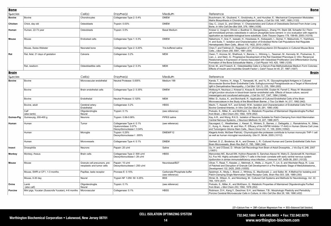
Worthington Biochemical Corporation • Lakewood, New Jersey 08701732.942.1660 • 800.445.9603 • Fax: 732.942.9270
www.worthington-biochem.com
CELL ISOLATION OPTIMIZING SYSTEM
14
[CF=Calcium Free • CMF=Calcium Magnesium Free • BSS=Balanced Salt Solution]
bone boneSpecies Cell(s) Enzyme(s) Medium ReferenceBovine Bovine Chondrocytes Collagenase Type 2: 0.4% DMEM Buschmann, M., Gluzband, Y., Grodzinsky, A., and Hunziker, E.: Mechanical Compression Modulates
Matrix Biosynthesis in Chondrocyte/Agarose Culture, J Cell Sci 108, 1497, 1995 (1133)Chicken Chick, day old Osteoblasts Trypsin: 0.03% DMEM Gay, C., Lloyd, Q., and Gilman, V.: Characteristics and Culture of Osteoblasts Derived From Avian Long
Bone, In Vitro Cell Dev Biol 30A, 379, 1994 (1036)Human Human, 22-73 year Osteoblasts Trypsin: 0.5% Basal Medium Kneser U, Voogd A, Ohnolz J, Buettner O, Stangenberg L, Zhang YH, Stark GB, Schaefer DJ: Fibrin
gel-immobilized primary osteoblasts in calcium phosphate bone cement: in vivo evaluation with regard to application as injectable biological bone substitute, Cells Tissues Organs 179, 158-69, 2005 (10316)
Mouse Mouse Endosteal cells Collagenase Type 1: 0.3% DMEM Nakamura, Y., Arai, F., Iwasaki, H., Hosokawa, K., Kobayashi, I., Gomei, Y., Matsumoto, Y., Yoshihara, H. and Suda, T.: Isolation and Characterization of Endosteal Niche Cell Populations that Regulate Hematopoietic Stem Cells., Blood 116, 1422, 2010 (10621)
Mouse, Swiss-Webster Neonatal bone Collagenase Type 2: 0.20% Tris-buffered saline Chen,T. and Feldman,D.: Regulation of 1,25-Dihydroxyvitamin D3 Receptors in Cultured Mouse Bone Cells, J Biol Chem 256, 5561, 1981 (554)
Rat Rat, fetal, 21 days of gestation Calvaria Collagenase: 0.2% MEM Owen, T., Aronow, M., Shalhoub, V., Barone, L., Wilming, L., Tassinari, M., Kennedy, M., Pockwinse, S., Lian, J., and Stein, G.: Progressive Development of the Rat Oseoblast Phenotype In Vitro: Reciprocal Relationships in Expression of Genes Associated with Osteoblast Proliferation and Differentiation During Formation of the Bone Extracellular Matrix, J Cell Physiol 143, 420, 1990 (1235)
Rat, newborn Osteoblastlike cells Collagenase Type 2: 0.3% MEM Ernst, M., and Froesch, E.: Osteoblastlike Cells in a Serum-Free Methylcellulose Medium Form Colonies: Effects of Insulin and Insulinlike Growth Factor I, Calcif Tissue Int 40, 27, 1987 (1007)
brain brainSpecies Cell(s) Enzyme(s) Medium ReferenceBovine Bovine Microvascular endothelial Neutral Protease: 0.005% Medium 199 Kanda, T., Yoshino, H., Ariga, T., Yamawaki, M., and Yu, R.: Glycosphingolipid Antigens in Cultured
Microvasular Bovine Brain Endothelial Cells: Sulfoglucuronosyl Paragloboside as a Target of Monoclonal IgM in Demyelinative Neuropathy, J Cell Biol 126 (1), 235, 1994 (950)
Bovine Brain endothelial cells Collagenase Type 2: 0.35% DMEM Wolburg H, Neuhaus J, Kniesel U, Krauss B, Schmid EM, Ocalan M, Farrell C, Risau W: Modulation of tight junction structure in blood-brain barrier endothelial cells. Effects of tissue culture, second messengers and cocultured astrocytes, J Cell Sci 107, 1347, 1994 (10048)
Bovine Endothelial Neutral Protease: 0.125% MEM Miller, D., Audus, K., and Borchardt, R.: Application of Cultured Endothelial Cells of the Brain Microvasculature in the Study of the Blood-Brain Barrier, J Tiss Cul Meth 14, 217, 1992 (942)
Bovine, adult Cerebral artery Endothelial
Collagenase: 0.2% HBSS Machi, T., Kassell, N.F., and Scheld, W.M.: Isolation and Characterization of Endothelial Cells From Bovine Cerebral Arteries, In Vitro Cell Dev Biol 26, 291, 1990 (436)
Calf (also lamb)
Oligodendroglia Neural
Trypsin: 0.1% (see reference) Poduslo, S., Miller, K., and McKhann, G.: Metabolic Properties of Maintained Oligodendroglia Purified from Brain, J Biol Chem 253, 1592, 1978 (552)
Guinea-Pig Guinea-pig, 200-400 g Neurons Trypsin: 0.06-0.08% PIPES saline Kay, A.R., and Wong, R.K.S.: Isolation of Neurons Suitable for Patch-Clamping from Adult Mammalian Central Nervous Systems, J Neurosci Methods 16, 227, 1986 (607)
Human Human Tumor Collagenase Type 4: 0.1%Hyaluronidase: 0.07%Deoxyribonuclease I: 0.04%
(see reference) Sauvageot, C., Weatherbee, J., Kesari, S., Winters, S., Barnes, J., Dellagatta, J., Ramakrishna, N., Stiles, C., Kung, A., Kieran, M. and Wen, P.: Efficacy of the HSP90 Inhibitor 17-AAG in Human Glioma Cell Lines and Tumorigenic Glioma Stem Cells., Neuro Oncol Vol. 11, 109, 2009 (10592)
Human Microglia Trypsin: 0.25% Deoxyribonuclease I: .005%
DMEM/F12 Klegeris Andis, McGeer PatrickL: Chymotrypsin-like proteases contribute to human monocytic THP-1 cell as well as human microglial neurotoxicity, Glia 51, 56-64, 2005 (10112)
Human Microvessels Collagenase Type 4: 0.1% DMEM Gerhart, D. Z., Broderius, M. A., and Drewes, L. R.: Cutlured Human and Canine Endothelial Cells from Brain Microvessels, Brain Res Bull 21, 785, 1988 (344)
Insect Drosophilia Neurons Papain: 20 u/ml Saline Gu, H. and O’Dowd, D.: Whole Cell Recordings from Brain of Adult Drosophila., J Vis Exp 6, 248, 2007 (10651)
Monkey Monkey, rhesus Brain cells Collagenase Type 2: 500 u/ml Deoxyribonuclease I: 28 u/ml
HBSS Marcondes MC, Burudi EM, Huitron-Resendiz S, Sanchez-Alavez M, Watry D, Zandonatti M, Henriksen SJ, Fox HS: Highly activated CD8(+) T cells in the brain correlate with early central nervous system dysfunction in simian immunodeficiency virus infection, J Immunol 167, 5429-38, 2001 (10125)
Mouse Mouse Granule cell precursors, pre-neoplastic and tumor cells
Papain: 10 u/mlDeoxyribonuclease I: 250 u/ml
Neurobasal/B27 Oliver, T., Read, T., Kessler, J., Mehmeti, A., Wells, J., Huynh, T., Lin, S. and Wechsler-Reya, R.: Loss of Patched and Disruption of Granule Cell Development in a Pre-Neoplastic Stage of Medulloblastoma., Development 132, 2425, 2005 (10555)
Mouse, SWR or CF1, 1-3 months Papillae, taste receptor Pronase E: 0.15% Carbonate-Phosphate buffer (see reference)
Spielman, A., Mody, I., Brand, J., Whitney, G., MacDonald, J., and Salter, M.: A Method for Isolating and Patch-Clamping Single Mammalian Taste Receptor Cells, Brain Res 503, 326, 1989 (350)
Mouse, 0-30 day Neural Trypsin NF 1:250: 50 0.25% BSS Shrier, B., Wilson, S., and Nirenberg, M.: Cultured Cell Systems and Methods for Neurobiology, Vol. 32, 765, 1974 (637)
Ovine Lamb (also calf)
Oligodendroglia Neural
Trypsin: 0.1% (see reference) Poduslo, S., Miller, K., and McKhann, G.: Metabolic Properties of Maintained Oligodendroglia Purified from Brain, J Biol Chem 253, 1592, 1978 (552)
Porcine Mini pigs, Yucatan (Susscrofa Yucatan), 4-6 months Microvascular Collagenase: 0.1% HBSS Robinson, D.H., Kang,Y., Deschner, S.H., and Nielsen, T.B.: Morphologic Plasticity and Periodicity : Porcine Cerebral Microvascular Cells in Culture, In Vitro Cell Dev Biol 26, 169, 1990 (432)

Worthington Biochemical Corporation • Lakewood, New Jersey 08701732.942.1660 • 800.445.9603 • Fax: 732.942.9270
www.worthington-biochem.com
CELL ISOLATION OPTIMIZING SYSTEM
15
[CF=Calcium Free • CMF=Calcium Magnesium Free • BSS=Balanced Salt Solution]
brain (Con’t) brain (Con’t)
Species Cell(s) Enzyme(s) Medium ReferenceRat Rat, Fisher, 7-21 month Hippocampal neurons Papain: 0.2% Hibernate A Chen, N., Newcomb, J., Garbuzova-Davis, S., Davis Sanberg, C., Sanberg, P. and Willing, A.: Human
Umbilical Cord Blood Cells Have Trophic Effects on Young and Aging Hippocampal Neurons in Vitro., Aging Dis 1, 173, 2010 (10663)
Rat, SD, 7 day Cerebellar granule neurons PDS kit: per instructions PBS Tanaka, S., Shaikh, I., Chiocca, E. and Saeki, Y.: The Gs-Linked Receptor GPR3 Inhibits the Proliferation of Cerebellar Granule Cells During Postnatal Development., PLoS ONE 4, e5922, 2009 (10487)
Rat, neonatal Astrocytes Papain: 20 u/ml EBSS Shigetomi, E. and Khakh, B.: Measuring Near Plasma Membrane and Global Intracellular Calcium Dynamics in Astrocytes., J Vis Exp 26, 1142, 2009 (10656)
Rat, neonatal Hippocampal neurons Papain: 10 u/ml EBSS Richler Esther, Chaumont Severine, Shigetomi Eiji, Sagasti Alvaro, Khakh Baljit S: Tracking transmitter-gated P2X cation channel activation in vitro and in vivo, Nat Methods 5, 87-93, 2008 (10319)
Rat, E18 Hippocampal neurons Papain: 0.2% Hibernate Jekabsons MB, Nicholls DG: Bioenergetic analysis of cerebellar granule neurons undergoing apoptosis by potassium/serum deprivation, Cell Death Differ 13, 1595-610, 2006 (10129)
Rat, Wistar, 1-3 day Hippocampal neurons Trypsin: 0.05% DMEM Velasco Myrian, Garcia Esperanza, Onetti Carlos G: Glucose deprivation activates diversity of potassium channels in cultured rat hippocampal neurons, Cell Mol Neurobiol 26, 307-19, 2006 (10321)
Rat, E19 Hippocampal neuron Papain: 10 u/ml MEM Khakh Baljit S, Fisher James A, Nashmi Raad, Bowser David N, Lester Henry A: An angstrom scale interaction between plasma membrane ATP-gated P2X2 and alpha4beta2 nicotinic channels measured with fluorescence resonance energy transfer and total internal reflection fluorescence microscopy, J Neurosci 25, 6911-20, 2005 (10307)
Rat, Wistar, 14 day Visual cortical PDS kit: EBSS Mizoguchi Y, Kanematsu T, Hirata M, Nabekura J: A rapid increase in the total number of cell surface functional GABAA receptors induced by brain-derived neurotrophic factor in rat visual cortex, J Biol Chem 278, 44097, 2003 (10022)
Rat, Wistar, male Cerebral endothelial Collagenase Type 3: 0.2% MEM Floris S, Van den Born J, van der Pol SM, Dijkstra CD, De Vries HE: Heparan sulfate proteoglycans modulate monocyte migration across cerebral endothelium, J Neuropathol Exp Neurol 62, 780, 2003 (10041)
Shellfish Helisoma trivolvis Neurons Buccal ganglia
Trypsin: 0.2% Saline, sterile Hadley, R.D., Bodnar, D.A., and Kater, S.B.: Formation of Electrical Synapses Between Isolated, Cultured Helisoma Neurons Requires Mutual Neurite Elongation, J Neurosci 5, 3145, 1985 (615)
Cartilage CartilageSpecies Cell(s) Enzyme(s) Medium ReferenceBovine Bovine, 18-36 month Chondrocytes Collagenase Type 1: 0.1% HBSS White, R. and Gibson, J.: The Effect of Oxygen Tension on Calcium Homeostasis in Bovine Articular
Chondrocytes., J Orthop Surg Res Vol. 5, 27, 2010 (10610)Bovine, 6-8 week Chondrocytes Collagenase Type 2: 0.2% DMEM Hwang, Y., Sangaj, N. and Varghese, S.: Interconnected Macroporous Poly(ethylene glycol) Cryogels as a
Cell Scaffold for Cartilage Tissue Engineering., Tissue Eng Part A Vol. 16,, , 3033-41, 2010 (10631)Steers, 1-2 years Chondrocytes Trypsin: 0.20% HBSS Mackintosh, D., and Mason, R.: Pharmacological Actions of 17 Beta-oestradiol on Articular Cartilage
Chondrocytes and Chondrosarcoma Chondrocytes in the Absence of Oestrogen Receptors, Biochim Biophys Acta 964, 295, 1988 (334)
Calf, 1-14 days Chondrocytes Collagenase Type 2: 0.20% PBS Klagsbrun, M.: Large Scale Preparation of Chondrocytes, Vol. 58,, , 560, 1979 (1263)
Chicken Chick embryos, HH stage Mesenchymal Trypsin: 0.1% DMEM (see reference)
Wong, M., and Tuan, R.: Nuserum, A Synthetic Serum Replacement, Supports Chondrogenesis of Embryonic Chick Limb Bud Mesenchymal Cells in Micromass Culture, In Vitro Cell Dev Biol 29A, 917, 1993 (965)
Chicken, broiler strain, 8-10 weeks Matrix vesicles Epiphyseal growth plate
Trypsin: 0.1% Tris-buffered saline Genge, B., Wu, L. and Wuthier, R.: Differential Fractionation of Matrix Vesicle Proteins: Further Characterization of the Acidic Phospholipid-dependent Ca2+-Binding Proteins, J Biol Chem 265, 4703, 1990 (569)
Chicken, broiler strain, 8-10 week Matrix vesicles Trypsin: 0.1% (see reference) Genge, B.R., Wu, L.N.Y., and Wuthier, R.E.: Identification of Phospholipid-dependent Calcium-binding Proteins as Constituents of Matrix Vesicles, J Biol Chem 264, 10917, 1989 (564)
Human Human Synoviocytes Collagenase Type 1: 0.4% DMEM Kim Wan-Uk, Kwok Seung-Ki, Hong Kyung-Hee, Yoo Seung-Ah, Kong Jin-Sun, Choe Jongseon, Cho Chul-Soo: Soluble Fas ligand inhibits angiogenesis in rheumatoid arthritis, Arthritis Res Ther 9, R42, 2007 (10173)
Human, 13-62 years Articular chondrocytes Trypsin: 0.2% BSS Srivastava, V.M.L., Malemud, C.J., Hough, A.J., Bland, J.H., and Sokoloff, L.: Preliminary Experience with Cell Culture of Human Articular Chondrocytes, Arthritis Rheum 17, 165, 1974 (726)
Human, 26-84 years Chondrocytes Collagenase: GBSS Manning, W.K., and Bonner, W.M.: Isolation and Culture of Chondrocytes From Human Adult Articular Cartilage, Arthritis Rheum 10, 235, 1967 (727)
Mouse Mouse, 1 day Chondrocytes Collagenase: 0.2% DMEM Terpstra, L, Prud’homme, J, Arabian, A, Takeda, S, Karsenty, G, Dedhar, S, and St-Arnaud, R.: Reduced Chondrocyte Proliferation and Chondrodysplasia in Mice Lacking the Integrin-linked Kinase in Chondrocytes, J Cell Biol 162, 139, 2003 (9756)
Ovine Sheep, 2 month Chondrocytes Collagenase Type 2: 0.3% Ham’s F-12 Kojima Koji, Bonassar Lawrence J, Roy Amit K, Mizuno Hirokazu, Cortiella Joaquin, Vacanti Charles A: A composite tissue-engineered trachea using sheep nasal chondrocyte and epithelial cells, FASEB J 17, 823-8, 2003 (10216)
Porcine Porcine, 1 year Chondrocytes Collagenase Type 1: 0.2% DEMEM Chowdhury, T., Schulz, R., Rai, S., Thuemmler, C., Wuestneck, N., Bader, A and Homandberg, G: Biomechanical Modulation of Collagen Fragment-Induced Anabolic and Catabolic Activities in Chondrocyte/Agarose Constructs., Arthritis Res Ther 12, R82, 2010 (10611)

Worthington Biochemical Corporation • Lakewood, New Jersey 08701732.942.1660 • 800.445.9603 • Fax: 732.942.9270
www.worthington-biochem.com
CELL ISOLATION OPTIMIZING SYSTEM
16
[CF=Calcium Free • CMF=Calcium Magnesium Free • BSS=Balanced Salt Solution]
Cartilage (Con’t) Cartilage (Con’t)
Species Cell(s) Enzyme(s) Medium ReferencePorcine Porcine, 2-4 month Chondrons Neutral Protease: 0.3%
Collagenase: 0.2% PBS Graff RD, Lazarowski ER, Banes AJ, Lee GM: ATP release by mechanically loaded porcine chondrons in
pellet culture, Arthritis Rheum 43, 1571-9, 2000 (10253)Rabbit Rabbit, New Zealand, white, 4-6 wk & 22-25 wk Chondrocytes Protease XIV: 5 mg/g of tissue Ham’s F-12 Plaas, A., Sandy, J., and Kimura, J.: Biosynthesis of Cartilage Proteoglycan & Link Protein Articular
Chondrocytes, J Biol Chem 263, 7560, 1988 (562)Rabbit, white, male, 8 weeks Chondrocytes Trypsin: 0.2% Gey’s BSS Benya, P.D., Padilla, S.R., and Nimni, M.E.: The Progeny of Rabbit Articular Chondrocytes Synthesize
Collagen Types I and III and Type I Trimer, but Not Type II, Biochem 16, 865, 1977 (312)Rabbit, New Zealand white or Dutch, 1 week (also human, newborn)
Chondrocytes Trypsin: 0.1% Saline G, CMF Schindler, F.H., Ose, M.A., and Solursh, M.: The Synthesis of Cartilage Collagen by Rabbit and Human Chondrocytes in Primary Cell Culture, In Vitro 12, 44, 1976 (495)
Rabbit, New Zealand white, immature, 2.25 - 3.3 Kg Articular chondrocytes Hyaline
Trypsin: 0.2% Gey’s BSS Green, J.R., and William, T.: Articular Cartilage Repair. Behavior of Rabbit Chondrocytes During Tissue Culture and Subsequent Allografting, Clin Orthop Relat Res , 237, 1976 (710)
Rabbit, New Zealand white, male, 250-350 g Epiphyseal Articular cartilage
Trypsin: 0.25% Eagle’s basal medium Bentley, G., and Greer , R.: Homotransplantation of Isolated Epiphyseal and Articular Cartilage Chondrocytes into Joint Surfaces of Rabbits, Nature 230, 385, 1971 (641)
Rat Rat, SD, young, 100-120 g Chondrocytes Trypsin: 0.2% Ham’s F-12 medium Shimomura, Y., Yoneda, T., and Suzuki, F.: Osteogenesis by Chondrocytes from Growth Cartilage of Rat Rib, Calcif Tissue Res 19, 179, 1975 (351)
Colon ColonSpecies Cell(s) Enzyme(s) Medium ReferenceHuman Human Cancer stem cells
Enzyme(s)Collagenase: Type 3: 200 u/mlDeoxyribonuclease 1: 100 u/ml
RPMI-1640 Dalerba P., Dylla S.J., Park I., Liu R., Wang X., Cho R. W., Hoey T., Gurney A., Huang E.H., Simeone D.M., Shelton A. A., Parmiani G., Castelli C., Clarke M.F: Phenotypic characterization of human colorectal cancer stem cells, Proc Natl Acad Sci U S A 104, 10158-63, 2007
Mouse Mouse Dendridic Enzyme(s) Collagenase: 300 u/mlDeoxyribonuclease 1: .002% u/ml
RPMI-1640 Abe K., Nguyen K., Fine S.D., Mo Ji-Hun, Shen C., Shenouda S., Corr M., Jung S., Lee J., Eckmann L., Raz E.: Conventional dendritic cells regulate the outcome of colonic inflammation independently of T cells., Proc Natl Acad Sci U S A 104, 17022-7, 2007
Endothelial EndothelialSpecies Cell(s) Enzyme(s) Medium ReferenceBovine Bovine, adult Cerebral artery
EndothelialCollagenase: 0.2% HBSS Machi, T., Kassell, N.F., and Scheld, W.M.: Isolation and Characterization of Endothelial Cells From
Bovine Cerebral Arteries, In Vitro Cell Dev Biol 26, 291, 1990 (436)Bovine Endothelial
AorticTrypsin: 0.05% Krebs Ringer solution DeNucci, G., Gryglewski, R.J., Warner, T.D., and Vane, J.R.: Receptor-Mediated Release of Endothelium-
Derived Relaxing Factor and Prostacyclin From Bovine Aortic Endothelial Cells Is Coupled, Proc Natl Acad Sci U S A 85, 2334, 1988 (659)
Bovine Endothelial, pulmonary artery Collagenase: 0.1% CMF-Dulbecco’s PBS Martin, T.: Formation of Diacylglycerol by a Phospholipase D-phosphatidate Phosphatase Pathway Specific for Phosphatidylcholine in Endothelial Cells, Biochim Biophys Acta 962, 282, 1988 (333)
Bovine Endothelial Aorta
Collagenase Type 2: 0.1% PBS Carson, M.P. and Haudenschild, C.C.: Microvascular Endothelium and Pericytes: High Yield, Low Passage Cultures, In Vitro Cell Dev Biol 22, 344, 1986 (417)
Bovine Endothelial Brain arteries
Collagenase Type 2: 0.2% Dulbecco’s PBS Goetz, I.., Warren, J., Estrada, C., Roberts, E., and Krause, D.: Long-Term Cultivation of Arterial and Capillary Endothelium From Adult Bovine Brain, In Vitro Cell Dev Biol 21, 172, 1985 (413)
Bovine Endothelial Corneal
Trypsin: 0.2% PBS: DMEM Scott, D., Murray, J., and Barnes, M.: Investigation of the Attachment of Bovine Corneal Endothelial Cells, Exp Cell Res 144, 472, 1983 (393)
Bovine, 2-3 weeks Endothelial Pulmonary artery
Collagenase Type 1: 0.2% RPMI 1640 w/1% Fetal Bovine Serum Lee, S., Douglas, W., Deneke, S., and Fanburg, B.: Ultrastructural Changes in Bovine Pulmonary Artery Endothelial Cells Exposed to 80% O2 In Vitro, In Vitro 19, 714, 1983 (531)
Bovine Endothelial, corneal Trypsin: 0.05% 0.01M Phosphate buffer with 0.02% EDTA, 0.9% NaCl (see reference)
Robinson, J. and Gospodarowicz, D.: Glycosaminoglycans Synthesized by Cultured Bovine Corneal Endothelial Cells, J Cell Physiol 117, 368, 1983 (594)
Bovine Endothelial Subclavian vein
Collagenase Type 1: 0.10% PBS Olander, J., Marasa, J., Kimes, R., Johnston, G., and Feder, J.: An assay measuring the stimulation of several types of bovine endothelial cells by growth factor(s) derived from cultured human tumor cells, In Vitro 18, 99, 1982 (525)
Bovine Aortic Pulmonary artery
Collagenase Type 2: 0.10% PBS Makarski, J. S.: Stimulation of cyclic AMP production by vasoactive agents in cultured bovine aortic and pulmonary artery endothelial cells, In Vitro 17, 450, 1981 (513)
Calf, fetal, 4-9 months Endothelial Collagenase Type 1: 0.25% PBS Rosen, E., Mueller, S., Noveral, J., and Levine, E.: Proliferative Characteristics of Clonal Endothelial Cell Strains, J Cell Physiol 107, 123, 1981 (880)
Bovine, adult Aorta Collagenase Type 1: 125 u/ml Dulbecco’s PBS with calcium and magnesium
Cotta-Pereira, G., Sage, H., Bornstein, P., Ross, R., and Schwartz, S.: Studies of Morphologically Atypical (“Sprouting”) Cultures of Bovine Aortic Endothelial Cells. Growth Characteristics and Connective Tissue Protein Synthesis, J Cell Physiol 102, 183, 1980 (592)
Bovine, young Pulmonary artery Collagenase: 0.1% Medium 199 Ryan, U., Mortara, M., and Whitaker, C.: Methods for Microcarrier Culture of Bovine Pulmonary Artery Endothelial Cells Avoiding the Use of Enzymes, Tissue Cell 12, 619, 1980 (670)
Bovine Foreskin Collagenase: 0.5% Dulbecco’s MEM w/10% calf serum Folkman, J., Haudenschild, C. C., and Zetter, B. R.: Long-term Culture of Capillary Endothlial Cells, Proc Natl Acad Sci U S A 76, 5217, 1979 (653)
Bovine Saphenous Vein Aorta
Collagenase: 0.01% PBS Eskin, S., Sybers, H., Trevino, L., Lie, J., and Chimoskey, J.: Comparison of Tissue-Cultured Bovine Endothelial Cells from Aorta and Saphenous Vein, In Vitro 14, 903, 1978 (500)
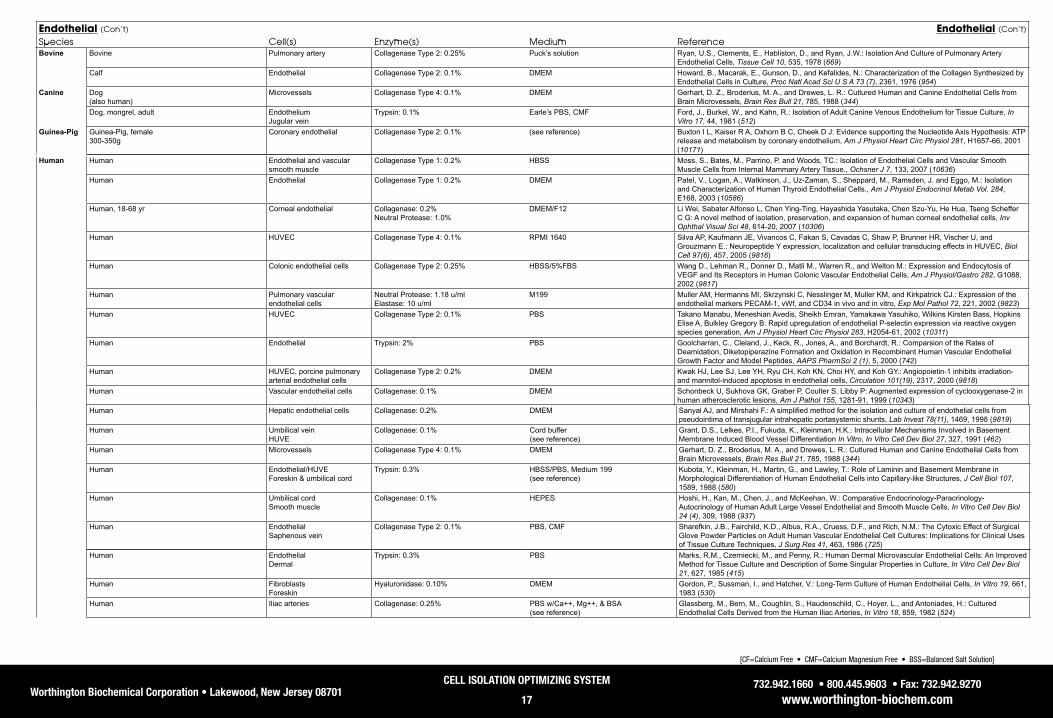
Worthington Biochemical Corporation • Lakewood, New Jersey 08701732.942.1660 • 800.445.9603 • Fax: 732.942.9270
www.worthington-biochem.com
CELL ISOLATION OPTIMIZING SYSTEM
17
[CF=Calcium Free • CMF=Calcium Magnesium Free • BSS=Balanced Salt Solution]
Human Hepatic endothelial cells Collagenase: 0.2% DMEM Sanyal AJ, and Mirshahi F.: A simplified method for the isolation and culture of endothelial cells from pseudointima of transjugular intrahepatic portasystemic shunts, Lab Invest 78(11), 1469, 1998 (9819)
Human Umbilical vein HUVE
Collagenase: 0.1% Cord buffer (see reference)
Grant, D.S., Lelkes, P.I., Fukuda, K., Kleinman, H.K.: Intracellular Mechanisms Involved in Basement Membrane Induced Blood Vessel Differentiation In Vitro, In Vitro Cell Dev Biol 27, 327, 1991 (462)
Human Microvessels Collagenase Type 4: 0.1% DMEM Gerhart, D. Z., Broderius, M. A., and Drewes, L. R.: Cutlured Human and Canine Endothelial Cells from Brain Microvessels, Brain Res Bull 21, 785, 1988 (344)
Human Endothelial/HUVE Foreskin & umbilical cord
Trypsin: 0.3% HBSS/PBS, Medium 199 (see reference)
Kubota, Y., Kleinman, H., Martin, G., and Lawley, T.: Role of Laminin and Basement Membrane in Morphological Differentiation of Human Endothelial Cells into Capillary-like Structures, J Cell Biol 107, 1589, 1988 (580)
Human Umbilical cord Smooth muscle
Collagenase: 0.1% HEPES Hoshi, H., Kan, M., Chen, J., and McKeehan, W.: Comparative Endocrinology-Paracrinology-Autocrinology of Human Adult Large Vessel Endothelial and Smooth Muscle Cells, In Vitro Cell Dev Biol 24 (4), 309, 1988 (937)
Human Endothelial Saphenous vein
Collagenase Type 2: 0.1% PBS, CMF Sharefkin, J.B., Fairchild, K.D., Albus, R.A., Cruess, D.F., and Rich, N.M.: The Cytoxic Effect of Surgical Glove Powder Particles on Adult Human Vascular Endothelial Cell Cultures: Implications for Clinical Uses of Tissue Culture Techniques, J Surg Res 41, 463, 1986 (725)
Endothelial (Con’t) Endothelial (Con’t)
Species Cell(s) Enzyme(s) Medium ReferenceBovine Bovine Pulmonary artery Collagenase Type 2: 0.25% Puck’s solution Ryan, U.S., Clements, E., Habliston, D., and Ryan, J.W.: Isolation And Culture of Pulmonary Artery
Endothelial Cells, Tissue Cell 10, 535, 1978 (669)Calf Endothelial Collagenase Type 2: 0.1% DMEM Howard, B., Macarak, E., Gunson, D., and Kefalides, N.: Characterization of the Collagen Synthesized by
Endothelial Cells in Culture, Proc Natl Acad Sci U S A 73 (7), 2361, 1976 (954)Canine Dog
(also human)Microvessels Collagenase Type 4: 0.1% DMEM Gerhart, D. Z., Broderius, M. A., and Drewes, L. R.: Cutlured Human and Canine Endothelial Cells from
Brain Microvessels, Brain Res Bull 21, 785, 1988 (344)Dog, mongrel, adult Endothelium
Jugular veinTrypsin: 0.1% Earle’s PBS, CMF Ford, J., Burkel, W., and Kahn, R.: Isolation of Adult Canine Venous Endothelium for Tissue Culture, In
Vitro 17, 44, 1981 (512)Guinea-Pig Guinea-Pig, female
300-350gCoronary endothelial Collagenase Type 2: 0.1% (see reference) Buxton I L, Kaiser R A, Oxhorn B C, Cheek D J: Evidence supporting the Nucleotide Axis Hypothesis: ATP
release and metabolism by coronary endothelium, Am J Physiol Heart Circ Physiol 281, H1657-66, 2001 (10171)
Human Human Endothelial and vascular smooth muscle
Collagenase Type 1: 0.2% HBSS Moss, S., Bates, M., Parrino, P. and Woods, TC.: Isolation of Endothelial Cells and Vascular Smooth Muscle Cells from Internal Mammary Artery Tissue., Ochsner J 7, 133, 2007 (10636)
Human Endothelial Collagenase Type 1: 0.2% DMEM Patel, V., Logan, A., Watkinson, J., Uz-Zaman, S., Sheppard, M., Ramsden, J. and Eggo, M.: Isolation and Characterization of Human Thyroid Endothelial Cells., Am J Physiol Endocrinol Metab Vol. 284, E168, 2003 (10586)
Human, 18-68 yr Corneal endothelial Collagenase: 0.2% Neutral Protease: 1.0%
DMEM/F12 Li Wei, Sabater Alfonso L, Chen Ying-Ting, Hayashida Yasutaka, Chen Szu-Yu, He Hua, Tseng Scheffer C G: A novel method of isolation, preservation, and expansion of human corneal endothelial cells, Inv Ophthal Visual Sci 48, 614-20, 2007 (10306)
Human HUVEC Collagenase Type 4: 0.1% RPMI 1640 Silva AP, Kaufmann JE, Vivancos C, Fakan S, Cavadas C, Shaw P, Brunner HR, Vischer U, and Grouzmann E.: Neuropeptide Y expression, localization and cellular transducing effects in HUVEC, Biol Cell 97(6), 457, 2005 (9816)
Human Colonic endothelial cells Collagenase Type 2: 0.25% HBSS/5%FBS Wang D., Lehman R., Donner D., Matli M., Warren R., and Welton M.: Expression and Endocytosis of VEGF and Its Receptors in Human Colonic Vascular Endothelial Cells, Am J Physiol/Gastro 282, G1088, 2002 (9817)
Human Pulmonary vascular endothelial cells
Neutral Protease: 1.18 u/ml Elastase: 10 u/ml
M199 Muller AM, Hermanns MI, Skrzynski C, Nesslinger M, Muller KM, and Kirkpatrick CJ.: Expression of the endothelial markers PECAM-1, vWf, and CD34 in vivo and in vitro, Exp Mol Pathol 72, 221, 2002 (9823)
Human HUVEC Collagenase Type 2: 0.1% PBS Takano Manabu, Meneshian Avedis, Sheikh Emran, Yamakawa Yasuhiko, Wilkins Kirsten Bass, Hopkins Elise A, Bulkley Gregory B: Rapid upregulation of endothelial P-selectin expression via reactive oxygen species generation, Am J Physiol Heart Circ Physiol 283, H2054-61, 2002 (10311)
Human Endothelial Trypsin: 2% PBS Goolcharran, C., Cleland, J., Keck, R., Jones, A., and Borchardt, R.: Comparsion of the Rates of Deamidation, Diketopiperazine Formation and Oxidation in Recombinant Human Vascular Endothelial Growth Factor and Model Peptides, AAPS PharmSci 2 (1), 5, 2000 (742)
Human HUVEC, porcine pulmonary arterial endothelial cells
Collagenase Type 2: 0.2% DMEM Kwak HJ, Lee SJ, Lee YH, Ryu CH, Koh KN, Choi HY, and Koh GY.: Angiopoietin-1 inhibits irradiation- and mannitol-induced apoptosis in endothelial cells, Circulation 101(19), 2317, 2000 (9818)
Human Vascular endothelial cells Collagenase: 0.1% DMEM Schonbeck U, Sukhova GK, Graber P, Coulter S, Libby P: Augmented expression of cyclooxygenase-2 in human atherosclerotic lesions, Am J Pathol 155, 1281-91, 1999 (10343)
Human Endothelial Dermal
Trypsin: 0.3% PBS Marks, R.M., Czerniecki, M., and Penny, R.: Human Dermal Microvascular Endothelial Cells: An Improved Method for Tissue Culture and Description of Some Singular Properties in Culture, In Vitro Cell Dev Biol 21, 627, 1985 (415)
Human Fibroblasts Foreskin
Hyaluronidase: 0.10% DMEM Gordon, P., Sussman, I., and Hatcher, V.: Long-Term Culture of Human Endothelial Cells, In Vitro 19, 661, 1983 (530)
Human Iliac arteries Collagenase: 0.25% PBS w/Ca++, Mg++, & BSA (see reference)
Glassberg, M., Bern, M., Coughlin, S., Haudenschild, C., Hoyer, L., and Antoniades, H.: Cultured Endothelial Cells Derived from the Human Iliac Arteries, In Vitro 18, 859, 1982 (524)

Worthington Biochemical Corporation • Lakewood, New Jersey 08701732.942.1660 • 800.445.9603 • Fax: 732.942.9270
www.worthington-biochem.com
CELL ISOLATION OPTIMIZING SYSTEM
18
[CF=Calcium Free • CMF=Calcium Magnesium Free • BSS=Balanced Salt Solution]
Endothelial (Con’t) Endothelial (Con’t)
Species Cell(s) Enzyme(s) Medium ReferenceHuman Human (adult and child) Foreskin Collagenase: 0.5% Dulbecco’s MEM w/10% calf serum Folkman, J., Haudenschild, C. C., and Zetter, B. R.: Long-term Culture of Capillary Endothlial Cells, Proc
Natl Acad Sci U S A 76, 5217, 1979 (653)Human Umbilical vein Trypsin: 100 µg/ml Tris-HCl,0.2 M Jaffe, E.A., Minick, C.R., Adelman, B., Becker, C.G., and Nachman, R.: Synthesis of Basement
Membrane Collagen By Cultured Human Endothelial Cells, J Exp Med 144, 209, 1976 (602)Human Umbilical vein Collagenase Type 1: 125 u/ml Dulbecco’s PBS Gimbrone Jr., M.A.: Culture of Vascular Endothelium, Prog Hemost Thromb 3, 1, 1976 (709)
Human Umbilical cord Collagenase: 0.1% Dulbecco’s PBS Gimbrone, M.A., Cotran, R.S., and Folkman, J.: Human Vascular Endothelial Cells in Culture: Growth and DNA Synthesis, J Cell Biol 60, 673, 1974 (589)
Human Umbilical cord Collagenase: 0.2% Cord buffer (see reference)
Jaffe, E., Nachman, R., Becker, C., and Minick, C.: Culture of Human Endothelial Cells Derived from Umbilical Veins. Identification by Morphologic and Immunologic Criteria, J Clin Invest 52, 2745, 1973 (598)
Human Umbilical cord Trypsin NF 1:250: 0.125% Saline, normal Lewis, L.J., Haok, J.C., Maca, R.D., and Fry, G.L.: Replication of Human Endothelial Cells in Culture, Science 181, 452, 1973 (666)
Human Umbilical cord Trypsin NF 1:250: 0.25% CMF solution Fryer, D.G., Birnbaum, G., and Luttrell, C.N.: Human Endothelium in Cell Culture, J Atheroscler Res 6, 151, 1966 (547)
Mouse Mouse, 7-10 week Liver endothelial Collagenase: 0.03% DMEM Follenzi, A., Benten, D., Novikoff, P., Faulkner, L., Raut, S. and Gupta, S.: Transplanted Endothelial CellsRepopulate the Liver Endothelium and Correct the Phenotype of Hemophilia A Mice., J Clin Invest 118, 935, 2008 (10632)
Porcine Porcine Endothelial Aortic
Collagenase Type 2: 0.1% Dulbecco-Vogt MEM w/o serum Dickinson, E. and Slakey, L.: Plasma-derived Serum as a Selective Agent to Obtain Endothelial Cultures from Swine Aorta, In Vitro 18, 63, 1982 (523)
Porcine, 30-40 kg Endothelia Aortic
Collagenase Type 4: 0.025% Medium 199 Merrilees, M.J., and Scott, L.: Interaction of Aortic Endothelial and Smooth Muscle Cells in Culture, Atherosclerosis 39, 147, 1981 (307)
Porcine, 20-30 week Endothelial Aortas Veins
Collagenase: 0.1% Medium 199 w/BSS & HEPES or NaHCO3 Slater, D.N., and Sloan, J.M.: The Porcine Endothelial Cell in Tissue Culture, Atherosclerosis 21, 259, 1975 (305)
Porcine, 60-100 days Aorta Trypsin: 0.1% Phosphate buffer (see reference)
Coulson, W.F.: The Effect Of Proteolytic Enzymes on the Tensile Strength of Whole Aorta and Isolated Aortic Elastin, Biochim Biophys Acta 237, 378, 1971 (319)
Rabbit Rabbit, 2-3Kg Endothelial, aortic Elastase: 0.2% Hanks solution Haley, N., Shio, H., Fowler, S.: Characterization of lipid-laden aortic cells from cholesterol-fed rabbits. I. Resolution of aortic cell populations by metrizamide density gradient centrifugation, Lab Invest 37, 287, 1977 (624)
Rat Rat, SD, male, 250-300 g Smooth muscle, aorta Soybean Trypsin Inhibitor: 0.25% HBSS with 0.2 mM Ca++ Schwertschlag, U.S., and Whorton, A.R.: Platelet-Activating Factor-Induced Homologous and Heterologous Desensitization In Cultured Vascular Smooth Muscle Cells, J Biol Chem 263, 13791, 1988 (560)
Rat, SD, male, 350 - 450 g Lipocytes Kupffer Sinusoidal endothelial
Collagenase: 0.015% DMEM/Ham’s F-12 Friedman, S. and Roll, F.: Isolation and Culture of Hepatic Lipocytes,Kupffer Cells, and Sinusoidal Endothelial Cells by Density Gradient Centrifugation with Stractan, Anal Biochem 161, 207, 1987 (301)
Rat, Wistar, male, 3 mo Endothelial Kupffer Parenchymal
Pronase: 0.25% HBSS Nagelkenke, J., Barto, K., and VanBerkel, T.: In vivo and in vitro uptake and degradation of acetylated low density lipoprotein by rat liver endothelial, Kupffer, and parenchymal cells, J Biol Chem 258, 12221, 1983 (557)
Rat, Wistar-Kyoto, male, 100 - 200 g Endothelial Cerebral
Collagenase Type 2: 0.05% HBSS Diglio, C.A., Grammas, P., Filiberto Giacomelli, M.S., and Wiener, J.: Primary Culture of Rat Cerebral Microvascular Endothelial Cells, Lab Invest 46, 554, 1982 (626)
Rat, 300 G, and pig, 30-40 Kg Endothelial Thoracic aorta
Trypsin: 0.05% Medium 199 and 0.01M EDTA Merrilees, M.J., and Scott, L.: Interaction of aortic endothelial and smooth muscle cells in culture. Effect on glycosaminoglycan levels, Atherosclerosis 39, 147, 1981 (306)
Rat Foreskin Collagenase: 0.5% Dulbecco’s MEM w/10% calf serum Folkman, J., Haudenschild, C. C., and Zetter, B. R.: Long-term Culture of Capillary Endothlial Cells, Proc Natl Acad Sci U S A 76, 5217, 1979 (653)
Rat, Wistar, adult, 170 g Endothelial Trypsin: 0.5% BSS Phillips, P., Kumar, P., Kumar, S., and Waghe, M.: Isolation And Characterization of Endothelial Cells From Rat And Cow Brain White Matter, J Anat 129, 261, 1979 (708)
Epithelial EpithelialSpecies Cell(s) Enzyme(s) Medium ReferenceBovine Bovine, fetal Epithelial
TrachealNeutral Protease: 2% Dissociation medium, CMF Schumann, B.L., Cody, T.E., Miller, M.L., Leikauf, G.D.: Isolation, Characterization, and Long-Term
Culture of Fetal Bovine Tracheal Epithelial Cells, In Vitro Cell Dev Biol 24, 211, 1988 (422)Canine Dog Tracheal Pronase: 0.1% DMEM Virmani, A., Naziruddin, B., Desai, V., Lowry, J., Graves, D., and Sachdev, G.: Evidence for Secretion of
High Molecular Weight Mucins by Canine Tracheal Epithelial Cells in Primary Culture: Effects of Select Secretagogues on Mucin Secretion, In Vitro Cell Dev Biol 28A, 120, 1992 (1194)
Chicken Chick, 5 day old Intestinal mesenchymal and epithelial
Collagenase: 0.03% DMEM Simon-Assmann, P and Kedinger, M: Embryonic Gut-Dissagregated Culture,Cell & Tissue Culture: Laboratory Procedures Vol. 1,Doyle, A., Griffiths, J., and Newell, D., John Wiley and Sons, Ltd., 12A:3.1, 1995 (1271)
Fish Shark (Squalus acanthias) Rectal gland Collagenase: 0.2% Ringer’s solution Karnaky, Jr., K.J., Valentich, J.D., Currie, M.G., Oehlenschlager, W.F., and Kennedy, M.P.: Atriopeptin Stimulates Chloride Secretion in Cultured Shark Rectal Gland Cells, Am J Physiol 260, 1125, 1991 (287)
Shark (Squalus acanthias) Rectal gland Collagenase: 0.2% Ringer’s solution Valentich, J: , J Tiss Cul Meth 13, 149, 1991 (1265)

Worthington Biochemical Corporation • Lakewood, New Jersey 08701732.942.1660 • 800.445.9603 • Fax: 732.942.9270
www.worthington-biochem.com
CELL ISOLATION OPTIMIZING SYSTEM
19
[CF=Calcium Free • CMF=Calcium Magnesium Free • BSS=Balanced Salt Solution]
Epithelial (Con’t) Epithelial (Con’t)
Species Cell(s) Enzyme(s) Medium ReferenceGuinea-Pig Guinea-pig, Hartley albino,
500-600 gEndometrial Collagenase Type 1: 0.1% HBSS Chaminadas, G., Alkhalaf, M., Remy-Martin, J.P., Propper, A.Y., and Adessi, G.L.: Specific Effect of
Oestrone Sulphate on Protein Synthesis and Secretion by Cultured Epithelial Cells from Guinea-pig Endometrium, J Endocrinol 123, 233, 1989 (600)
Hamster Hamster, Syrian gold, male, 100-120 g Tracheal Pronase: 0.1% MEM with Hepes, CMF Niles,R., Kim, K.C., Hyman, B., Christensen, T., Wasano, K., Brody, J.: Characterization Of Extended Primary And Secondary Cultures Of Hamster Tracheal Epithelial Cells, In Vitro Cell Dev Biol 24, 457, 1988 (423)
Hamster, Syrian golden (strain CR:RGH) Tracheal Trypsin: 0.1% Ham’s F-12 McDowell, E., et al.: Differentiation of Tracheal Mucociliary Epithelium in Primary Cell Culture Recapitulates Normal Fetal Development and Regeneration Following Injury in Hamsters, Am J Pathol 129, 511, 1987 (283)
Hamster (strain 1516 EHS and Lakeview), 8-12 weeks
Tracheal Trypsin: 0.05% Medium 199 Lee, T., Wu, R., Brody, A., Barrett, J., and Nettesheim, P.: Growth and Differentiation of Hamster Tracheal Epithelial Cells in Culture, Exp Lung Res 6, 27, 1984 (406)
Hamster, Syrian gold, male, 6 weeks- 4 months Tracheal Trypsin: 0.25% PBS with EDTA Goldman, W.E., Baseman, J.B.: Selective Isolation and Culture of a Proliferating Epithelial Cell Population from the Hamster Trachea, In Vitro 16, 313, 1980 (506)
Human Human Colonic epithelial Collagenase: Neutral Protease: 0.3%Deoxyribonuclease I: 0.05%
RPMI 1640 Fukushima, K. and Fiocchi, C.: Paradoxical Decrease of Mitochondrial DNA Deletions in Epithelial Cells of Active Ulcerative Colitis Patients., Am J Physiol Gastrointest Liver Physiol Vol. 286, G804-13, 2004 (10355)
Human Human tracheal epithelium Protease Type XIV: 0.04% DMEM/F12 Widdicombe JH, Sachs LA, Morrow JL, and Finkbeiner WE.: Expansion of cultures of human tracheal epithelium with maintenance of differentiated structure and function, Biotechniques 39(2), 249, 2005 (9824)
Human Epithelial Collagenase: 2.0% DMEM/Ham’s F-12 Emerman, J. and Wilkinson, D.: Routine Culturing of Normal, Dysplastic and Malignant Human Mammary Epithelial Cells from Small Tissue Samples, In Vitro Cell Dev Biol 26, 1186, 1990 (429)
Human Epithelial Pronase: 0.1% PBS Gruenert, D.C., Basbaum, C.B., and Widdicombe, J.H.: Long-Term Culture of Normal and Cystic Fibrosis Epithelial Cells Grown Under Serum-Free Conditions, In Vitro Cell Dev Biol 26, 411, 1990 (440)
Human Epithelial Sweat gland
Collagenase Type 2: 0.2% (see reference) Wood, L. and Neufeld, E.: A Cystic Fibrosis Phenotype in Cells Cultured from Sweat Gland Secretory Coil. Altered Kinetics of 36Cl Efflux, J Biol Chem 265, 12796, 1990 (568)
Human, women, 27-49 years Epithelial Ovary
Trypsin: 0.125% HBSS, CMF Auersperg, N., Siemens, C.H., and Myrdal, S.E.: Human Ovarian Surface Epithelium In Primary Culture, In Vitro 20, 743, 1984 (535)
Human, infant and neonate Epithelial Prostate
Trypsin: 0.1% HBSS Lechner, J., Babcock, M., Marnell, M., Narayan, K., and Kaighn, M.: Normal Human Prostate Epithelial Cell Cultures, Methods Cell Biol 21, 195, 1980 (631)
Mouse Mouse Submandibular salivary Collagenase Type 2 or 3: 0.16% DMEM Durban, E: Submandibular Salivary Epithelial Cells,Cell & Tissue Culture: Laboratory Procedures Vol. 1,Doyle, A., Griffiths, J., and Newell, D., John Wiley and Sons, Ltd., 12B:2.1, 1995 (1272)
Mouse Esophageal Trypsin: 0.25% PBS, CMF Katayama, M., Kan, M.: Heparin-Binding (Fibroblast) Growth Factors Are Potential Autocrine Regulators of Esophageal Epithelial Cell Proliferation, In Vitro Cell Dev Biol 27, 533, 1991 (467)
Mouse, BALB/c, male, 3-5 months Epithelial Submandibular salivary gland
Collagenase Type 3: 0.16% , 1:1 v/v DMEM with 15 mM HEPES Durban, E.M.: Mouse Submandibular Salivary Epithelial Cell Growth and Differentiation in Long-Term Culture:Influence of the Extracellular Matrix, In Vitro Cell Dev Biol 26, 33, 1990 (437)
Mouse, BALB/cfC3H or BALB/c 8-12 day mid-pregnant
Epithelial Submandibular gland
Collagenase Type 3: 0.1% HBSS Yang, J., Flynn, D., Larson, L., and Hamamoto, S.: Growth in Primary Culture of Mouse Submandibular Epithelial Cells, In Vitro 18, 435, 1982 (520)
Mouse BALB/cfC3H Mammary tumors Epithelial
Collagenase: 1.0% HBSS Yang, J., Guzman, R., Richards, J., and Nandi, S.: Primary Cultures of Mouse Mammary Tumor Epithelial Cells Embedded in Collagen Gels, In Vitro 16, 502, 1980 (507)
Mouse, C3H, 6-8 weeks Epithelial Collagenase: 0.10% DMEM Lillehaug, J., Mondal, S., and Heidelberger, C.: Establishment of Epithelial Cell Lines from Mouse Regenerating Liver, In Vitro 15, 910, 1979 (504)
Porcine Porcine, 5-60 kg Retinal pigment epithelial cells Collagenase: 2% DMEM Wiencke, A., Kiilgaard, J., Nicolini, J., Bundgaard, M., Ropke, C., and La Cour, M.: Growth of Cultured Porcine Retinal Pigment Epithelial Cells, Acta Opthalmol Scand 81(2), 170, 2003 (9825)
Porcine Trachea Neutral Protease: 0.2% HBSS De Buysscher, E., Kennedy, J., and Mendicino, J.: Synthesis of Mucin Glycoproteins by Epithelial Cells Isolated from Swine Trachea by Specific Proteolysis, In Vitro 20, 433, 1984 (534)
Rabbit Rabbit, New Zealand white, adult Colon Neutral Protease: 0.3% PBS Vidrich, A., Racindranath, R., Farsi, K., and Targan, S.: A Method for the Rapid Establishment of Normal Adult Mammalian Colonic Epithelial Cell Cultures, In Vitro Cell Dev Biol 24 (3), 188, 1988 (918)
Rabbit, New Zealand white, male, 4-5lb. Gastric Parietal and chief
Collagenase Type 2: 0.08% Sodium phosphate buffer Chew, C.S., Brown,M.R.: Release of Intracellular Ca2+ and Elevation of Inositol Triphosphate by Secretagogues in Parietal and Chief Cells Isolated from Rabbit Gastric Mucosa, Biochim Biophys Acta 888, 116, 1986 (326)
Rabbit, New Zealand white estrous, female, 4-5 months
Mesothelial and surface epithelial Ovaries
Trypsin: 0.125%-0.5% Medium 199 Nicosia, S., Johnson, J., and Streibel, E.: Isolation and Ultrastructure of Rabbit Ovarian Mesothelium(Surface Epithelium), Int J Gynecol Pathol 3, 348, 1984 (542)
Rabbit, fetal Epithelial Gastric
Collagenase Type 3: 0.10% HBSS Logsdon, C.D., Bisbee, C.A., Rutten, M.J. and Machen, T.E.: Fetal Rabbit Gastric Epithelial Cells Cultured on Floating Collagen Gel, In Vitro 18, 233, 1981 (517)
Rat Rat, SD, male, 150 - 250 g Epithelial Stomach
Pronase: 0.15% Medium 199 Dial, E., Kao, Y., and Lichtenberger, L.: Effects of 16,16-Dimethyl Prostaglandin E2 On Glycoprotein And Lipid Synthesis of Gastric Epithelial Cells Grown in a Primary Culture, In Vitro Cell Dev Biol 27, 39, 1991 (464)
Rat, Wistar, neonatal Epithelial Trypsin: 0.1% HBSS Jassal, D., Han, R., Caniggia, I., Post, M., and Tanswell, A.: Growth of Distal Fetal Rat Lung Epithelial Cells in a Defined Serum-Free Medium, In Vitro Cell Dev Biol 27A, 625, 1991 (471)
Rodent, various (see reference) Epithelial Trypsin: 0.2% MEM, PBS Robinson, C., and Wu, R.: Culture of Conducting Airway Epithelial Cells in Serum-Free Medium, J Tiss Cul Meth 13, 95, 1991 (1239)
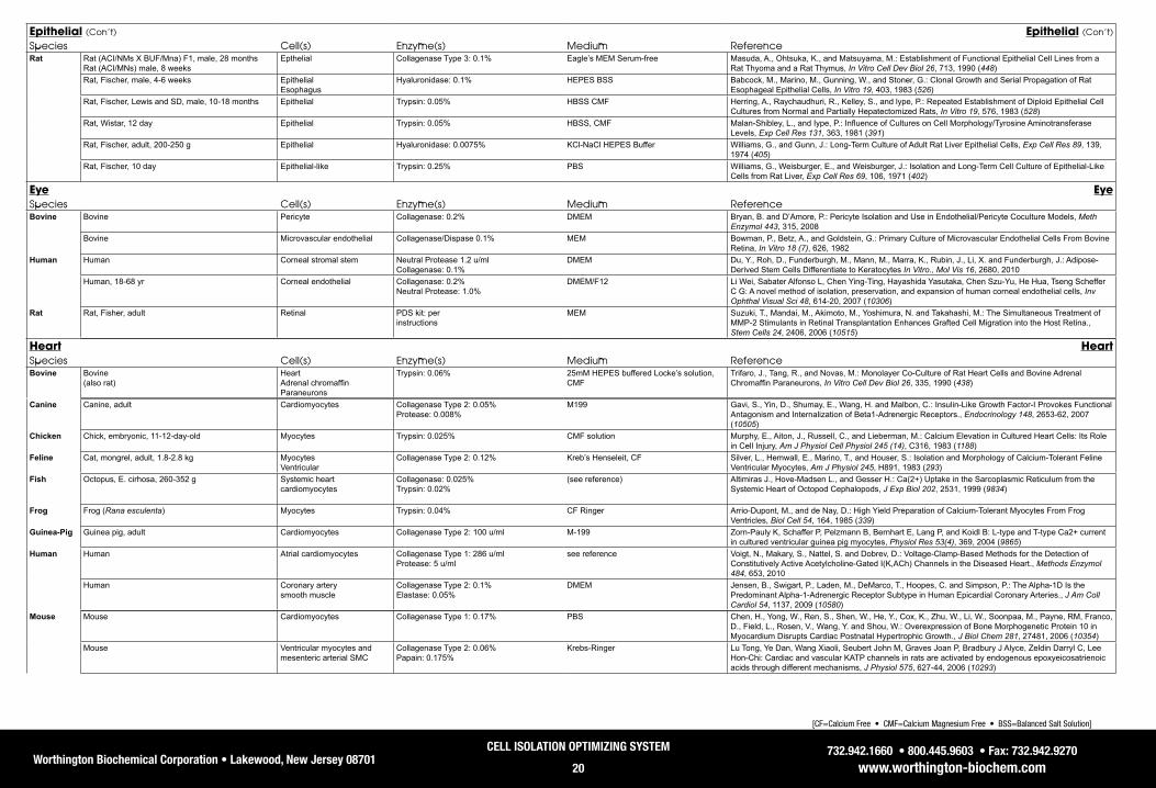
Worthington Biochemical Corporation • Lakewood, New Jersey 08701732.942.1660 • 800.445.9603 • Fax: 732.942.9270
www.worthington-biochem.com
CELL ISOLATION OPTIMIZING SYSTEM
20
[CF=Calcium Free • CMF=Calcium Magnesium Free • BSS=Balanced Salt Solution]
Epithelial (Con’t) Epithelial (Con’t)
Species Cell(s) Enzyme(s) Medium ReferenceRat Rat (ACI/NMs X BUF/Mna) F1, male, 28 months
Rat (ACI/MNs) male, 8 weeksEpthelial Collagenase Type 3: 0.1% Eagle’s MEM Serum-free Masuda, A., Ohtsuka, K., and Matsuyama, M.: Establishment of Functional Epithelial Cell Lines from a
Rat Thyoma and a Rat Thymus, In Vitro Cell Dev Biol 26, 713, 1990 (448)Rat, Fischer, male, 4-6 weeks Epithelial
EsophagusHyaluronidase: 0.1% HEPES BSS Babcock, M., Marino, M., Gunning, W., and Stoner, G.: Clonal Growth and Serial Propagation of Rat
Esophageal Epithelial Cells, In Vitro 19, 403, 1983 (526)Rat, Fischer, Lewis and SD, male, 10-18 months Epithelial Trypsin: 0.05% HBSS CMF Herring, A., Raychaudhuri, R., Kelley, S., and lype, P.: Repeated Establishment of Diploid Epithelial Cell
Cultures from Normal and Partially Hepatectomized Rats, In Vitro 19, 576, 1983 (528)Rat, Wistar, 12 day Epithelial Trypsin: 0.05% HBSS, CMF Malan-Shibley, L., and Iype, P.: Influence of Cultures on Cell Morphology/Tyrosine Aminotransferase
Levels, Exp Cell Res 131, 363, 1981 (391)Rat, Fischer, adult, 200-250 g Epithelial Hyaluronidase: 0.0075% KCl-NaCl HEPES Buffer Williams, G., and Gunn, J.: Long-Term Culture of Adult Rat Liver Epithelial Cells, Exp Cell Res 89, 139,
1974 (405)Rat, Fischer, 10 day Epithelial-like Trypsin: 0.25% PBS Williams, G., Weisburger, E., and Weisburger, J.: Isolation and Long-Term Cell Culture of Epithelial-Like
Cells from Rat Liver, Exp Cell Res 69, 106, 1971 (402)
Eye EyeSpecies Cell(s) Enzyme(s) Medium ReferenceBovine Bovine Pericyte Collagenase: 0.2% DMEM Bryan, B. and D’Amore, P.: Pericyte Isolation and Use in Endothelial/Pericyte Coculture Models, Meth
Enzymol 443, 315, 2008Bovine Microvascular endothelial Collagenase/Dispase 0.1% MEM Bowman, P., Betz, A., and Goldstein, G.: Primary Culture of Microvascular Endothelial Cells From Bovine
Retina, In Vitro 18 (7), 626, 1982Human Human Corneal stromal stem Neutral Protease 1.2 u/ml
Collagenase: 0.1%DMEM Du, Y., Roh, D., Funderburgh, M., Mann, M., Marra, K., Rubin, J., Li, X. and Funderburgh, J.: Adipose-
Derived Stem Cells Differentiate to Keratocytes In Vitro., Mol Vis 16, 2680, 2010Human, 18-68 yr Corneal endothelial Collagenase: 0.2%
Neutral Protease: 1.0% DMEM/F12 Li Wei, Sabater Alfonso L, Chen Ying-Ting, Hayashida Yasutaka, Chen Szu-Yu, He Hua, Tseng Scheffer
C G: A novel method of isolation, preservation, and expansion of human corneal endothelial cells, Inv Ophthal Visual Sci 48, 614-20, 2007 (10306)
Rat Rat, Fisher, adult Retinal PDS kit: perinstructions
MEM Suzuki, T., Mandai, M., Akimoto, M., Yoshimura, N. and Takahashi, M.: The Simultaneous Treatment of MMP-2 Stimulants in Retinal Transplantation Enhances Grafted Cell Migration into the Host Retina., Stem Cells 24, 2406, 2006 (10515)
Heart HeartSpecies Cell(s) Enzyme(s) Medium ReferenceBovine Bovine
(also rat)Heart Adrenal chromaffin Paraneurons
Trypsin: 0.06% 25mM HEPES buffered Locke’s solution, CMF
Trifaro, J., Tang, R., and Novas, M.: Monolayer Co-Culture of Rat Heart Cells and Bovine Adrenal Chromaffin Paraneurons, In Vitro Cell Dev Biol 26, 335, 1990 (438)
Canine Canine, adult Cardiomyocytes Collagenase Type 2: 0.05%Protease: 0.008%
M199 Gavi, S., Yin, D., Shumay, E., Wang, H. and Malbon, C.: Insulin-Like Growth Factor-I Provokes FunctionalAntagonism and Internalization of Beta1-Adrenergic Receptors., Endocrinology 148, 2653-62, 2007 (10505)
Chicken Chick, embryonic, 11-12-day-old Myocytes Trypsin: 0.025% CMF solution Murphy, E., Aiton, J., Russell, C., and Lieberman, M.: Calcium Elevation in Cultured Heart Cells: Its Role in Cell Injury, Am J Physiol Cell Physiol 245 (14), C316, 1983 (1188)
Feline Cat, mongrel, adult, 1.8-2.8 kg Myocytes Ventricular
Collagenase Type 2: 0.12% Kreb’s Henseleit, CF Silver, L., Hemwall, E., Marino, T., and Houser, S.: Isolation and Morphology of Calcium-Tolerant Feline Ventricular Myocytes, Am J Physiol 245, H891, 1983 (293)
Fish Octopus, E. cirhosa, 260-352 g Systemic heart cardiomyocytes
Collagenase: 0.025% Trypsin: 0.02%
(see reference) Altimiras J., Hove-Madsen L., and Gesser H.: Ca(2+) Uptake in the Sarcoplasmic Reticulum from the Systemic Heart of Octopod Cephalopods, J Exp Biol 202, 2531, 1999 (9834)
Frog Frog (Rana esculenta) Myocytes Trypsin: 0.04% CF Ringer Arrio-Dupont, M., and de Nay, D.: High Yield Preparation of Calcium-Tolerant Myocytes From Frog Ventricles, Biol Cell 54, 164, 1985 (339)
Guinea-Pig Guinea pig, adult Cardiomyocytes Collagenase Type 2: 100 u/ml M-199 Zorn-Pauly K, Schaffer P, Pelzmann B, Bernhart E, Lang P, and Koidl B: L-type and T-type Ca2+ current in cultured ventricular guinea pig myocytes, Physiol Res 53(4), 369, 2004 (9865)
Human Human Atrial cardiomyocytes Collagenase Type 1: 286 u/mlProtease: 5 u/ml
see reference Voigt, N., Makary, S., Nattel, S. and Dobrev, D.: Voltage-Clamp-Based Methods for the Detection of Constitutively Active Acetylcholine-Gated I(K,ACh) Channels in the Diseased Heart., Methods Enzymol 484, 653, 2010
Human Coronary arterysmooth muscle
Collagenase Type 2: 0.1%Elastase: 0.05%
DMEM Jensen, B., Swigart, P., Laden, M., DeMarco, T., Hoopes, C. and Simpson, P.: The Alpha-1D Is the Predominant Alpha-1-Adrenergic Receptor Subtype in Human Epicardial Coronary Arteries., J Am Coll Cardiol 54, 1137, 2009 (10580)
Mouse Mouse Cardiomyocytes Collagenase Type 1: 0.17% PBS Chen, H., Yong, W., Ren, S., Shen, W., He, Y., Cox, K., Zhu, W., Li, W., Soonpaa, M., Payne, RM, Franco, D., Field, L., Rosen, V., Wang, Y. and Shou, W.: Overexpression of Bone Morphogenetic Protein 10 in Myocardium Disrupts Cardiac Postnatal Hypertrophic Growth., J Biol Chem 281, 27481, 2006 (10354)
Mouse Ventricular myocytes and mesenteric arterial SMC
Collagenase Type 2: 0.06% Papain: 0.175%
Krebs-Ringer Lu Tong, Ye Dan, Wang Xiaoli, Seubert John M, Graves Joan P, Bradbury J Alyce, Zeldin Darryl C, Lee Hon-Chi: Cardiac and vascular KATP channels in rats are activated by endogenous epoxyeicosatrienoic acids through different mechanisms, J Physiol 575, 627-44, 2006 (10293)

Worthington Biochemical Corporation • Lakewood, New Jersey 08701732.942.1660 • 800.445.9603 • Fax: 732.942.9270
www.worthington-biochem.com
CELL ISOLATION OPTIMIZING SYSTEM
21
[CF=Calcium Free • CMF=Calcium Magnesium Free • BSS=Balanced Salt Solution]
Mouse Mouse, neonatal Cardiomyoctes Collagenase Type 1: 150 u/ml Trypsin: 0.01%
M199 Takahashi N, Wang X, Tanabe S, Uramoto H, Jishage K, Uchida S, Sasaki S, Okada Y.: ClC-3-independent sensitivity of apoptosis to Cl- channel blockers in mouse cardiomyocytes, Cell Physiol Biochem 15, 263, 2005 (10033)
Mouse, 6-8 week Vascular smooth muscle Papain: 10 u/ml Elastase: .005% Collagenase: 0.05% Deoxyribonuclease I: 1000 u/ml PDS kit: with modifications
EBSS Qian, Q., Hunter, L., Li, M., Marin-Padilla, M., Prakash, Y., Somlo, S., Harris, P., Torres, V., and Sieck, G.: Pkd2 Haploinsufficiency Alters Intracellular Calcium Regulation in Vascular Smooth Muscle Cells, Hum Mol Genet 12(15), 1875, 2003 (9846)
Heart (Con’t) Heart (Con’t)
Species Cell(s) Enzyme(s) Medium Reference
Rabbit Rabbit Smooth muscle cells Collagenase Type 2: 300 u/ml Elastase: 5 u/ml
F10 Ham Verheye, S., Martinet, W., Kockx, M., Knaapen, M., Salu, K., Timmermans, J., Ellis, J., Kilpatrick, D., DeMayer, G.: Selective Clearance of Macrophages in Atherosclerotic Plaques by Autophagy, J Am Coll Cardiol 49, 706, 2007 (10335)
Rat Rat, SD, male Ventricular myocytes Collagenase: 0.13% Hyaluronidase: 0.06%
DMEM Stagg Mark A, Coppen Steven R, Suzuki Ken, Varela-Carver Anabel, Lee Joon, Brand Nigel J, Fukushima Satsuki, Yacoub Magdi H, Terracciano Cesare M: Evaluation of frequency, type, and function of gap junctions between skeletal myoblasts overexpressing connexin43 and cardiomyocytes: relevance to cell transplantation, FASEB J 20, 744-6, 2006 (10275)
Rat, SD, 200-250 g Ventricular myocytes and mesenteric arterial SMC
Collagenase Type 2: 0.06% Papain: 0.175%
Krebs-Ringer Lu Tong, Ye Dan, Wang Xiaoli, Seubert John M, Graves Joan P, Bradbury J Alyce, Zeldin Darryl C, Lee Hon-Chi: Cardiac and vascular KATP channels in rats are activated by endogenous epoxyeicosatrienoic acids through different mechanisms, J Physiol 575, 627-44, 2006 (10293)
Rat, neonatal, adult, 17 day Ventricular cardiomyocytes Collagenase Type 2: 0.7-1% DMEM Lam ML, Bartoli M, and Claycomb WC.: The 21-day postnatal rat ventricular cardiac muscle cell in culture as an experimental model to study adult cardiomyocyte gene expression, Mol Cell Biochem 229, 51, 2002 (9842)
Intestine IntestineSpecies Cell(s) Enzyme(s) Medium ReferenceHuman Human Intestinal epithelial Collagenase Type 4: 72.5 u/ml HBSS Fahlgren, A., Hammarstrom, S., Danielsson, A. and Hammarstrom, M.: Increased Expression of
Antimicrobial Peptides and Lysozyme in Colonic Epithelial Cells of Patients with Ulcerative Colitis., Clin Exp Immunol 131, 90, 2003 (10358)
Human Lamina propria mononuclear cells
Collagenase: 0.02% HBSS Kanai Takanori, Totsuka Teruji, Uraushihara Koji, Makita Shin, Nakamura Tetsuya, Koganei Kazutaka, Fukushima Tsuneo, Akiba Hisaya, Yagita Hideo, Okumura Ko, Machida Utako, Iwai Hideyuki, Azuma Miyuki, Chen Lieping, Watanabe Mamoru: Blockade of B7-H1 suppresses the development of chronic intestinal inflammation, J Immunol 171, 4156-63, 2003 (10232)
Human Mucosal mononuclear cells Collagenase Type 3: 0.01% Deoxyribonuclease I: 0.01% Soybean Trypsin Inhibitor: 0.01%
RPMI Stallmach A, Schaffer F, Hoffmann S, Weber S, Muller-Molaian I, Schneider T, Kohne G, Ecker KW, Feifel G, Zeitz M: Increased state of activation of CD4 positive T cells and elevated interferon gamma production in pouchitis, Gut 43, 499-505, 1998 (10240)
Mouse Mouse, 7 week Interstitial cells Collagenase: 0.04% Trypsin: 0.02%
(see reference) Goto Kazunori, Matsuoka Satoshi, Noma Akinori: Two types of spontaneous depolarizations in the interstitial cells freshly prepared from the murine small intestine, J Physiol 559, 411-22, 2004 (10145)
Mouse, 0-15 day Interstitial cells of Cajal Collagenase Type 2: 0.13% Hanks Lee Young Mee, Kim Byung Joo, Kim Hyun Jin, Yang Dong Ki, Zhu Mei Hong, Lee Kyu Pil, So Insuk, Kim Ki Whan: TRPC5 as a candidate for the nonselective cation channel activated by muscarinic stimulation in murine stomach, Am J Physiol/Gastro 284, G604-16, 2003 (10233)
Mouse Lamina propria lymphocytes Collagenase Type 4: 300 u/ml PBS Wu Y, Wang X, Csencsits KL, Haddad A, Walters N, Pascual DW: M cell-targeted DNA vaccination, Proc Natl Acad Sci U S A 98, 9318-23, 2001 (10271)
Kidney KidneySpecies Cell(s) Enzyme(s) Medium ReferenceBovine Bovine Papillary duct Hyaluronidase: 0.2% Kreb’s buffer
HEPES buffered salineHusted, R., Hayashi, M., and Stokes, J.: Characteristics of Papillary Collecting Duct Cells in Primary Culture, Am J Physiol 255, F1160, 1988 (298)
Canine Dog, 1-5 months Proximal tubules Collagenase Type 1: 0.15% Krebs Ringer bicarbonate buffer States, B., Reynolds, R., Lee, J., and Segal, S.: Cystine Uptake By Cultured Cells Originating From Dog Proximal Tubule Segments, In Vitro Cell Dev Biol 26, 105, 1990 (426)
Dog, mongrel, 20 Kg Proximal tubular Deoxyribonuclease I: 0.0125% (see reference) Yau, C., Rao, L., and Silverman, M.: Sugar Uptake Into a Primary Culture of Dog Kidney Proximal Tubular Cells, Can J Physiol Pharmacol 63, 417, 1985 (707)
Fish Winter flounder (Pseudopleur-onectes americanus) 200 - 500 g
Renal tubule Trypsin: 0.2% CMF solution (see reference)
Dickman, K. and Renfro, J.: Primary Culture of Flounder Renal Tubule Cells:Transepithelial Transport, Am J Physiol 251, 424, 1986 (297)
Guinea-Pig Guinea-pig, male, 100 g Single cells Trypsin: 0.25% CF salt solution Phillips, H.: Dissociation of Single Cells from Lung or Kidney Tissue with Elastase, In Vitro 8, 101, 1972 (538)
Hamster Hamster (also rat, SD and Wistar, 150-225 g, rabbit, bovine)
Papillary duct Hyaluronidase: 0.2% Kreb’s buffer HEPES buffered saline
Husted, R., Hayashi, M., and Stokes, J.: Characteristics of Papillary Collecting Duct Cells in Primary Culture, Am J Physiol 255, F1160, 1988 (298)
Human Human Renal cortex Trypsin: 0.1% Tissue Culture Grade Water McAteer, J, Kempson, S., and Evan, A: Culture of Human Renal Cortex Epithelial Cells, J Tiss Cul Meth 13, 143, 1991 (1266)
Human Papillary duct Collagenase: 400 u/ml Eagle’s MEM-HEPES buffer w/L-glutamine Trifillis, A. and Kahng, M.: Characterization of an In Vitro System of Human Renal Papillary Collecting Duct Cells, In Vitro Cell Dev Biol 26, 441, 1990 (441)

Worthington Biochemical Corporation • Lakewood, New Jersey 08701732.942.1660 • 800.445.9603 • Fax: 732.942.9270
www.worthington-biochem.com
CELL ISOLATION OPTIMIZING SYSTEM
22
[CF=Calcium Free • CMF=Calcium Magnesium Free • BSS=Balanced Salt Solution]
Kidney (Con’t) Kidney (Con’t)
Species Cell(s) Enzyme(s) Medium ReferenceHuman Human Mesangial Trypsin: 0.25% DMEM/Ham’s F-12 Heieren, M., van der Woude, F., and Balfour Jr., H.: Cytomegalovirus Replicates Efficiently in Human
Kidney Mesangial Cells, Proc Natl Acad Sci U S A 85, 1642, 1988 (657)Monkey Monkey Kidney Trypsin: 0.01% - 0.00001% Eagle’s MEM Melnick, J., and Wallis, C.: Problems Related to the Use of Serum and Trypsin in the Growth of Monkey
Kidney Cells, Dev Biol 37, 77, 1976 (706)Mouse Mouse, adult Cortex, proximal tubule Collagenase: 0.15% DMEM Syal Ashu, Schiavi Susan, Chakravarty Sumana, Dwarakanath Vangipuram, Quigley Raymond, Baum
Michel: Fibroblast growth factor-23 increases mouse PGE2 production in vivo and in vitro, Am J Physiol/Renal 290, F450-5, 2006 (10281)
Mouse, naive SJL (H-2S) Proximal tubular epithelial Deoxyribonuclease I: 15 µg/ml RPMI 1640 Haverty, T., Kelly, C., Hines, W., Amenta, P., Watanabe, M., Harper, R., Kefalides, N., and Neilson, E.: Characterization of a Renal Tubular Epithelial Cell Line Which Secretes the Autologous Target Antigen of Autoimmune Experimental Interstitial Nephritis, J Cell Biol 107, 1359, 1988 (578)
Rabbit Rabbit, New Zealand white, male, 1.5 - 2.0 Kg Duct Soybean Trypsin Inhibitor: 0.025% Hank’s Solution with calcium and HEPES Naray-Fejes-Toth, A., and Fejes-Toth, G.: Immunoelection and Culture of Cortical Collecting Duct Cells, J Tiss Cul Meth 13, 179, 1991 (1267)
Rabbit, New Zealand white, female, 2-3 Kg Renal proximal tubules Deoxyribonuclease I: 70 u/ml Modified DME-F12 Rodeheaver, D., Aleo, M., and Schnellmann, R.: Differences in Enzymatic and Mechanical Isolated Rabbit Renal Proximal Tubules: Comparison in Long-Term Incubation, In Vitro Cell Dev Biol 26, 898, 1990 (454)
Rabbit (also rat, SD and Wistar, 150-225 g, hamster, bovine)
Papillary duct Hyaluronidase: 0.2% Kreb’s buffer HEPES buffered saline
Husted, R., Hayashi, M., and Stokes, J.: Characteristics of Papillary Collecting Duct Cells in Primary Culture, Am J Physiol 255, F1160, 1988 (298)
Rat Rat, Wistar, 300g Renal proximal tubules Collagenase Type 2: 0.2% DMEM/F12 Deng Aihua, Miracle Cynthia M, Lortie Mark, Satriano Joseph, Gabbai Francis B, Munger Karen A, Thomson Scott C, Blantz Roland C: Kidney oxygen consumption, carbonic anhydrase, and proton secretion, Am J Physiol/Renal 290, F1009-15, 2006 (10305)
Rat, male, 150-200 g Epithelial Proximal tubule
Protease: 0.1% HBSS/CMF Elliget, K., and Trump, B.: Primary Cultures of Normal Rat Kidney Proximal Tubule Epithelial Cells for Studies of Renal Cell Injury, In Vitro Cell Dev Biol 27, 739, 1991 (476)
Rat, adult Inner medullary collecting duct Papillae
Collagenase Type 2: 0.1% PBS Brion, L., Schwartz, J., Lachman, H., Zavilowitz, B., and Schwartz, G.: Development of H+ Secretion by Cultured Renal Inner Medullary Collecting Duct Cells, Am J Physiol 257, F486, 1989 (300)
Rat, Wistar, female, 100-150 g Glomerular mesangial Collagenase: 0.025% RPMI 1640 Wang, J., Kester, M., and Dunn, M.: The Effects of Endotoxin on Platelet-Activating Factor Synthesis in Cultured Rat Glomerular Mesangila Cells, Biochim Biophys Acta 969, 217, 1988 (338)
Rat, Wistar, male, 150 - 200 g Renal target Collagenase: 1.0% (also 0.7%) Eagle’s MEM Barlet-Bas, C., Khadouri, C., Marsey, S., and Doucet, A.: Sodium-Independent In Vitro Induction of Na+,K+-ATPase by Aldosterone in Renal Target Cells: Permissive Effect of Triiodothyronine, Proc Natl Acad Sci U S A 85, 1707, 1988 (658)
Rat,Wistar, 300-400 g Proximal tubules Collagenase: 0.15 % Krebs Henseleit solution Vinay, P., Gougoux, A., and Lemieux, G.: Isolation of a Pure Suspension of Rat Proximal Tubules, Am J Physiol 241, F403, 1981 (289)
Rat Fibroblasts Kidney
Trypsin: up to 0.25% Dulbecco-Vogt MEM Wallach, D., Anderson, W., and Pastan, I.: Activation of Adenylate Cyclase in Cultured Fibroblasts by Trypsin, J Biol Chem 253, 24, 1978 (553)
Liver LiverSpecies Cell(s) Enzyme(s) Medium ReferenceAvian Ducklings, 7 day Hepatocytes Collagenase: 0.05%
Hyaluronidase: 0.05% DMEM/ F12 Lee J., Culvenor J., Angus P., Smallwood R., Nicoll A., and Locarnini S.: Duck Hepatitis B Virus
Replication in Primary Bile Duct Epithelial Cells, J Virol 75(16), 7651, 2001 (9883)
Canine Dog, 13 years old (also rat, guinea-pig, rabbit, human)
Hepatocytes Collagenase: 90 u/ml CF EGTA perfusate Reese, J. and Byard, J.: Isolation And Culture of Adult Hepatocytes from Liver Biopsies, In Vitro 17, 935, 1981 (412)
Dog, adult Hepatocytes Trypsin: 0.1% HBSS, CMF Vickrey, H., Ramon, J., and McCann, D.: Continuous Culture of Normal Adult Mammalian Hepatocytes Exhibiting Parenchymal Functions, In Vitro 15, 120, 1979 (502)
Chicken Chicken, adult Hepatocytes Collagenase: 0.02% HEPES, CF Fraslin, J., Touquette, L., Douaire, M., Menezo, Y., Guillemot, J., and Mallard, J.: Isolation and Long Term Maintenance of Differentiated Adult Chicken Hepatocytes in Primary Culture, In Vitro Cell Dev Biol 28, 615, 1992 (486)
Chicken (also rat, SD, 150-250 g)
Hepatocytes Collagenase Type 4: 6000 units Medium A Roseman, S., Weigel, P., Schnaar, R., Kuhlenschmidt, M., Schmell, E., Lee, R., and Lee, Y.: Adhesion of Hepatocytes to Immobilized Sugars. A Threshold Phenomenon, J Cell Biol 254, 10830, 1979 (582)
Chicken, Leghorn, white, 10-15 day Hepatocytes Deoxyribonuclease I: 0.00125% PBS Tarlow, D., Watkins, P., Reed, R., Miller, R., Zwergel, E., and Lane, M.: Lipogenesis and the Synthesis and Secretion of Very Low Density Lipoprotein by Avian Liver Cells in Nonproliferating Monolayer Culture, J Cell Biol 73, 332, 1974 (590)
Chick embryos, 5 day Heart Liver
Trypsin: 3.0% Tyrode’s solution, CMF Steinberg, M.: “ECM”: Its Nature, Origin, And Function in Cell Aggregation, Exp Cell Res 30, 257, 1963 (396)
Chick embryonic Various tissues (heart, liver, skeletal, cardiac)
Trypsin: various grades CMF Tyrode’s solution Rinaldini, L.: An Improved Method for the Isolation and Quantitative Cultivation of Embryonic Cells, Exp Cell Res 16, 477, 1959 (394)
Fish Rainbow trout (Oncorhynchus mykiss), male & female, 120-600 g
Hepatocytes Collagenase Type 2: 0.045% Kreb’s-Ringer bicarbonate buffer, CF Blair, J.B., Miller, M.R., Pack, D., Barnes, R., Teh, S.J. and Hinton, D.E.: Isolated Trout Liver Cells:Establishing Short-Term Primary Cultures Exhibiting Cell-to-Cell Interactions, In Vitro Cell Dev Biol 26, 237, 1990 (434)
Trout, male, 100 - 200 g Hepatocytes Collagenase Type 2: 0.045% HBSS with 0.05M HEPES Lipsky, M., Sheridan, T., Bennett, R., and May, E.: Comparison of Trout Hepatocyte Culture on Different Substrates, In Vitro Cell Dev Biol 22, 360, 1986 (418)

Worthington Biochemical Corporation • Lakewood, New Jersey 08701732.942.1660 • 800.445.9603 • Fax: 732.942.9270
www.worthington-biochem.com
CELL ISOLATION OPTIMIZING SYSTEM
23
[CF=Calcium Free • CMF=Calcium Magnesium Free • BSS=Balanced Salt Solution]
Liver (Con’t) Liver (Con’t)
Species Cell(s) Enzyme(s) Medium ReferenceTrout 100g - 5kg Hepatocytes Hyaluronidase: 0.08% (see reference) Bailey, G., Taylor, M., and Selivonchick, D.: Aflatoxin B1 Metabolism and DNA Binding in Isolated
Hepatocytes From Rainbow Trout Salmo gairdner, Carcinogenesis 3, 511, 1982 (1264)Trout (Salmo gairdneri), male, 150-200 g Catfish (Ictalurus punctatus), male, 100 - 150 g
Hepatocytes Collagenase: 100u/ml HBSS Klaunig, J.: Establishment of Fish Hepatocyte Cultures For Use in In Vitro Carcinogenicity Studies, Natl Cancer Inst Monogr 65, 163, 1981 (703)
Guinea-Pig Guinea-pig, Hartley, male, 250-300 g Hepatocytes Collagenase Type 2: Kreb’s Ringer bicarbonate buffer Arinze, I. and Kawai, Y.: Adrenergic Regulation of Glycogenolysis in Isolated Guinea-Pig Hepatocytes: Evidence that B2-Receptors Mediate Catecholamine Stimulation of Glycogenolysis, Arch Biochem Biophys 225, 196, 1983 (304)
Guinea-pig, young Hepatocytes Collagenase: 90 u/ml CF EGTA perfusate Reese, J. and Byard, J.: Isolation And Culture of Adult Hepatocytes from Liver Biopsies, In Vitro 17, 935, 1981 (412)
Human Human Hepatocytes Collagenase: 0.05% EBSS Hughes, R., Mitry, R., Dhawan, A., Lehec, S., Girlanda, R., Rela, M., Heaton, N., and Muiesan, P.: Isolation of Hepatocytes from Livers from Non-Heart-Beating Donors for Cell Transplantation, Liver Transpl 12, 713, 2006 (10205)
Human Hepatocytes Collagenase: 0.05% HEPES buffer (see reference)
Gomez-Lechon, M., Lopez, P., Donato, T., Montoya, A., Larrauri, A., Gimenez, P., Trullenque, R., Fabra, R., and Castell, J.: Culture of Human Hepatocytes From Small Surgical Liver Biopsies: Biochemical Characterization And Comparison With in vivo, In Vitro Cell Dev Biol 26, 67, 1990 (445)
Human, male, 17-40 yrs. Hepatocytes Collagenase: 0.05% HEPES buffer Begue, J., Baffet, G., Campion, J., and Guillouzo, A.: Differential Response of Primary Cultures of Human and Rat Hepatocytes to Aflatoxin B1-Induced Cytotoxicity and Protection by the Hepatoprotective agent(+)-Cyanidanol-3, Biol Cell 63, 327, 1988 (341)
Human, 51-75 years old (also rat, guinea-pig, rabbit, dog)
Hepatocytes Collagenase: 90 u/ml CF EGTA perfusate Reese, J. and Byard, J.: Isolation And Culture of Adult Hepatocytes from Liver Biopsies, In Vitro 17, 935, 1981 (412)
Human Hepatocytes Trypsin: 0.1% HBSS, CMF Kaighn, M.: Human Liver Cells,Tissue Culture Methods / Applications,(Kruse, P., Patterson, M. eds), 54, 1973 (702)
Monkey Monkey (Macaca fascicularis), adult, 5-6 Kg Hepatocytes Trypsin: 160 u/ml HEPES buffer Ulrich, R., Aspar, D., Cramer, C., Kletzien, R., and Ginsberg, L.: Isolation and Culture of Hepatocytes from the Cynomolgus Monkey (Macaca fascicularis), In Vitro Cell Dev Biol 26, 815, 1990 (452)
Mouse Mouse Liver epithelial progenitor cells Collagenase Type 4: 0.1% Deoxyribonuclease I: 0.05%
DMEM Li Wen-Lin, Su Juan, Yao Yu-Cheng, Tao Xin-Rong, Yan Yong-Bi, Yu Hong-Yu, Wang Xin-Min, Li Jian-Xiu, Yang Yong-Ji, Lau Joseph T Y, Hu Yi-Ping: Isolation and characterization of bipotent liver progenitor cells from adult mouse, Stem Cells 24, 322-32, 2006 (10248)
Mouse, C3H, 6-8 weeks Epithelial Collagenase: 0.10% DMEM Lillehaug, J., Mondal, S., and Heidelberger, C.: Establishment of Epithelial Cell Lines from Mouse Regenerating Liver, In Vitro 15, 910, 1979 (504)
Mouse, 20-30 g Parenchymal and non-parenchymal
Hyaluronidase: 0.1% Hank’s w/ Insulin, CMF Crisp, D., and Pogson, C.: Glycolytic and Gluconeogenic Enzyme Activities in Parenchymal and Non-parenchymal Cells from Mouse Liver, Biochem J 126, 1009, 1972 (309)
Porcine Porcine, male, 2-3 week Hepatocytes Collagenase: 0.05% DMEM Raman Priya, Donkin Shawn S, Spurlock Michael E: Regulation of hepatic glucose metabolism by leptin in pig and rat primary hepatocyte cultures, Am J Physiol Regul Integr Comp Physiol 286, R206-16, 2004 (10268)
Rabbit Rabbit, New Zealand white Hepatocytes Collagenase: 90 u/ml CF EGTA perfusate Reese, J. and Byard, J.: Isolation And Culture of Adult Hepatocytes from Liver Biopsies, In Vitro 17, 935, 1981 (412)
Rat Rat, Fisher, E14 Hepatocytes Collagenase Type 1: 0.22% HBSS Isabel Zvibel, Miri Bronstein, Einav Hubel, Ella Bar-Lev, Zamir Halpern, Ran Oren: Isolation, characterization and culture of Thy1-positive cells from fetal rat livers, World J Gastroenterol 12, 3841-7, 2006 (10303)
Rat, 200-300 g Hepatocytes Collagenase Type 1: 0.067% HBSS Mula, N., Cubero, F., Codesal, J., de Andres, S., Escudero, C., Garcia-Barrutia, S., Millan, I., Arahuetes, R. and Maganto, P.: Survival of Allogeneic Hepatocytes Transplanted into the Thymus., Cells Tissues Organs 188, 270, 2008 (10371)
Rat, SD, 8 - 12 day Parenchymal hepatocytes Collagenase Type 4: 80 u/ml HBSS Davila, J., Reddy, C., Davis, P. and Acosta, D.: Toxicity Assessment of Papaverine Hydrochloride and Papaverine-Derived Metabolites in Primary Cultures of Rat Hepatocytes, In Vitro Cell Dev Biol 26, 515, 1990 (444)
Rat, SD, male, 250-350 g Hepatocytes Collagenase Type 2: 0.05% HBSS, CMF Liu, J., Kershaw, W., and Klaassen, C.: Rat Primary Hepatocyte Cultures are a Good Model for Examining Metallothionein-Induced Tolerance to Cadmium Toxicity, In Vitro Cell Dev Biol 26, 75, 1990 (450)
Rat, Wistar, male, 200 g Parenchymal Collagenase Type 1: HEPES, modified Kindberg, G., Gudmundsen, O., and Berg, T.: The Effect of Vandate on Receptor-mediated Endocytosis of Asialoorosomucoid in Rat Liver Parenchymal Cells, J Biol Chem 265, 8999, 1990 (572)
Rat, SD, male, 175-225 g Hepatocytes Collagenase: Kreb’s Ringer bicarbonate buffer Reddy, S., Amick, G., Cooper, R., and Damun, Z.: Insulin Stimulates the Activity of a Protamine in Isolated Rat Hepatocytes, J Biol Chem 265, 7748, 1990 (570)
Rat, SD, neonatal, 8 - 10 days Hepatocytes Collagenase Type 4: 0.05% Hanks’ BSS, CF Davila, J., Lenherr, A., and Acosta, D.: Protective Effect of Flavonoids on Drug-Induced Hepatotoxicity In Vitro, Toxicology 57, 267, 1989 (673)
Rat, Fischer 344, male, 14 months Hepatocytes Collagenase: 0.05% Ringer’s biocarbonate buffer Nagy, I., Ohno-Iwashita, Y., Ohta, M., Nagy, V., Kitani, K., Ando, S., and Imahori, K.: Effect of Perfringolysin O on the Lateral Diffusion Constant of Membrane Proteins of Hepatocytes as Revealed by Fluorescence Recovery After Photobleaching, Biochim Biophys Acta 939, 551, 1988 (327)
Rat, SD, male, 200-300 g Hepatocytes Collagenase: 0.05% HEPES Voss, A. and Sprecher, H.: Metabolism of 6,9,12-Octadecatetraenoic and 6,9,12,15-Octadecatetraenoic Acid, Biochim Biophys Acta 958, 153, 1988 (328)
Rat, Wistar, male,3 month, 200 g Parenchymal Endothelial Kupffer
Collagenase Type 1: 0.05% Krebs Henseleit Kuiper, J., Zijlstra, F., Kamps, J, and vanBerkel, T.: Identification of Prostaglandin D2 as the Major Eiconsanoid from Liver Endothelial and Kupffer Cells, Biochim Biophys Acta 959, 143, 1988 (329)

Worthington Biochemical Corporation • Lakewood, New Jersey 08701732.942.1660 • 800.445.9603 • Fax: 732.942.9270
www.worthington-biochem.com
CELL ISOLATION OPTIMIZING SYSTEM
24
[CF=Calcium Free • CMF=Calcium Magnesium Free • BSS=Balanced Salt Solution]
Liver (Con’t) Liver (Con’t)
Species Cell(s) Enzyme(s) Medium ReferenceRat Rat, Wistar, male, 200 - 300 g Parenchymal
KupfferCollagenase Type 2: 0.05% HBSS with CaCl2 Cai, H., He, Z., and Ding, Y.: Effects of Monocyte Macrophages Stimulation on Hepatic Lipoprotein
Receptors, Biochim Biophys Acta 958, 334, 1988 (331)Rat, Wistar, female, 17 days Hepatocytes Collagenase: 0.05% HBSS Cotariu, D., Barr-Nea, L., Papo, N., and Zaidman, J.: Induction of gamma-Glutamyl Transferase by
Dexamethasone in Cultured Rat Hepatocytes, Enzyme 40, 212, 1988 (386)Rat, SD, male, 350 - 450 g Lipocytes
Kupffer Sinusoidal endothelial
Collagenase: 0.015% DMEM/Ham’s F-12 Friedman, S. and Roll, F.: Isolation and Culture of Hepatic Lipocytes,Kupffer Cells, and Sinusoidal Endothelial Cells by Density Gradient Centrifugation with Stractan, Anal Biochem 161, 207, 1987 (301)
Rat, SD, male Hepatocytes Collagenase: 100 - 200 µg/g body weight Eagle’s Eagle’s w/HEPES HBSS
Oka, J. and Weigel, P.: Monensin Inhibits Ligand Dissociation Only Transiently and Partially and distinguishes two galactosyl receptor pathways in isolated rat hepatocytes, J Cell Physiol 133, 243, 1987 (595)
Rat, SD, female, 100-180 g Hepatocytes Collagenase: 0.5% Krebs Ringer bicarbonate buffer Schwarz, K., Lanier, S., Carter, E., Homcy, C., and Graham, R.: Rapid Reciprocal Changes in Adrenergic Receptors in Intact Isolated Hepatocytes During Primary Cell Culture, Mol Pharmacol 27, 200, 1985 (639)
Rat, SD, male, 270 - 320 g Hepatocytes Collagenase: 0.04% Bicarbonate buffer with calcium added Brass, E., Garrity, M., and Robertson, R.: Inhibition of Glucagon-Stimulated Hepatic Glycogenolysis by E-Series Prostaglandins, FEBS Lett 169, 293, 1984 (410)
Rat, Fischer, Lewis and SD, male 10-18 months Epithelial Trypsin: 0.05% HBSS CMF Herring, A., Raychaudhuri, R., Kelley, S., and Iybe, P.: Repeated Establishment of Diploid Epithelial Cell Cultures from Normal and Partially Hepatectomized Rats, In Vitro 19, 576, 1983 (528)
Rat, Wistar, male, 3 month old Endothelial Kupffer Parenchymal
Collagenase: 0.05% HBSS Nagelkenke, J., Barto, K., and Berkel, T.: In Vivo and in Vitro Uptake and Degradation of Acetylated Low Density Lipoprotein by Rat Liver Endothelial, Kupffer, and Parenchymal Cells, J Biol Chem 258 (20), 12221, 1983 (940)
Rat, SD, male/female, 250-300 g Hepatocytes Collagenase: 100 u/ml Krebs Henseleit bicarbonate buffer Studer, R. and Borle, A.: Differences between Male and Female Rats in the Regulation of Hepatic Glycogenolysis. The Relative Role of Calcium and cAMP in Phosphorylase Activation by Catecholamines, J Biol Chem 257, 7987, 1982 (556)
Rat, Wistar, 12 day Epithelial Trypsin: 0.05% HBSS, CMF Malan-Shibley, L., and Iype, P.: Influence of Cultures on Cell Morphology/Tyrosine Aminotransferase Levels, Exp Cell Res 131, 363, 1981 (391)
Rat, SD Hepatocytes Collagenase: 90 u/ml CF EGTA perfusate Reese, J. and Byard, J.: Isolation And Culture of Adult Hepatocytes from Liver Biopsies, In Vitro 17, 935, 1981 (412)
Rat, Wistar, female, fetus Hepatocytes Collagenase: 0.025% HEPES buffer Gugen-Guillouzo, C., Tichonicky, L., Szajnert, M., and Kruh, J.: Changes in Some Chromatin and Cytoplastic Enzymes of Perinatal Rat Hepatocytes, In Vitro 16, 1, 1980 (505)
Rat, SD, male, 250-300 g Parenchymal Collagenase Type 2: 0.05% Kreb’s Henseleit bicarbonate buffer Yamada, S., Otto, P., Kennedy, D., and Whayne, T.: The Effects of Dexamethasone on Metabolic Activity of Hepatocytes in Primary Monolayer Culture, In Vitro 16, 559, 1980 (508)
Rat, SD, 7-10 day Liver Collagenase: 0.05% HBSS modified (see reference) Acosta, D., Anuforo, D., and Smith, R: , J Tiss Cul Meth 6, 35, 1980 (1268)
Rat, Wistar, male, 200-250 g Hepatocytes Hyaluronidase: 460 u/ml Saline Poli, G., Gravela, E., Albano, E., and Dianzani, M.: Studies on Fatty Liver with Isolated Hepatocytes: II., The Action of Carbon Tetrachloride on Lipid Peroxidation, Protein and Triglyceride Synthesis and Secretion, Exp Mol Pathol 30, 116, 1979 (408)
Rat (also chicken)
Hepatocytes Collagenase Type 3 & 4: HEPES Weigel, P., Schnaar, R., Kuhlenschmidt, M., Schmell, E., Lee, R., Lee, Y., and Roseman, S.: Adhesion of Hepatocytes to Immobilized Sugars, J Biol Chem 254 (21), 10830, 1979 (1032)
Rat, SD, male, 200 g Hepatocytes Collagenase Type 1: 100 u/ml Buffers 1 & 2 (see reference)
Rubin, K., Kjellen, L., and Oslashbrink, B.: Intercellular Adhesion between Juvenile Liver Cells.. A Method to Measure the Formation of Stable Lateral Contacts Between Cells Attached to a Collagen Gel, Exp Cell Res 109, 413, 1977 (387)
Rat (WAG/RIJ), female, 24, 30, and 37 months Parenchymal Collagenase Type 1: 0.05% - 0.06% HEPES buffer VanBezodijen, C., Grell, T., and Knook, D.: Effect of Age on Protein Synthesis by Isolated Liver Parenchymal Cells, Mech Ageing Dev 6, 293, 1977 (630)
Rat, Fisher, adult, male Hepatocytes Collagenase Type 1: 100 u/ml HBSS (see reference)
Williams, G., Bermudez, E., and Scaramuzzino, D.: Rat Hepatocytes Primary Cell Cultures III. Improved Dissociation and Attachment Techniques and the Enchancement of Survival by Culture Medium, In Vitro 13 (12), 809, 1977 (826)
Rat, Fischer, male, adult, 170-265 g Hepatocytes Collagenase: 0.05%-0.10% Williams E Laishes, B., and Williams, G.: Conditions Affecting Primary Cell Cultures of Functional Adult Rat Hepatocytes. I.. The Effect of Insulin, In Vitro 12, 521, 1976 (496)
Rat, neonate, 3 day Hepatocytes Trypsin: 0.25% PBS, CMF Bausher, J., and Schaeffer, W.: A Diploid Rat Liver Cell Culture. 1. Characterization and Sensitivity to Aflatoxin B1, In Vitro 9, 286, 1974 (540)
Rat, SD, albino, male Parenchymal Hyaluronidase: 0.1% Hank’s solution, CMF Bonney, R., Becker, J., Walker, P., and Potter, V.: Primary Monolayer Cultures of Adult Rat Liver Parenchymal Cells Suitable for Study of the Regulation of Enzyme Synthesis, In Vitro 9, 399, 1974 (541)
Rat, embryos, 1-3 days Hepatocytes Hyaluronidase: 0.10% HBSS Gerschenson, L., Berliner, J., and Davidson, M.: The Isolation and Culture of Liver Cells, Vol. 32, 733, 1974 (635)
Rat, SD, adult, male, 200-250 g Parenchymal Hyaluronidase: 0.10% HBSS, CF Howard, R., Lee, J., and Pesch, L.: The fine structure, potassium content, and respiratory activity of isolated rat liver parenchymal cells prepared by improved enzymatic techniques, J Cell Biol 57, 642, 1973 (586)
Rat, SD, adult, male, 180-300 g Parenchymal Collagenase Type 1: 0.05% Hank’s solution, CF Bissell, D., Hammaker, L., and Meyer, U.: Parenchymal Cells from Adult Rat Liver in Nonproliferating Monolayer Culture. I. Functional Studies, J Cell Biol 59, 722, 1973 (588)
Rat, Wistar, 200-250 g Hepatocytes Hyaluronidase: 0.10% HBSS, CF Berg, T., Boman, D., and Seglen, P.O.: Induction of Tryptophan Oxygenase in Primary Rat Liver Cell Suspensions by Glucocorticoid Hormone, Exp Cell Res 72, 571, 1972 (404)

Worthington Biochemical Corporation • Lakewood, New Jersey 08701732.942.1660 • 800.445.9603 • Fax: 732.942.9270
www.worthington-biochem.com
CELL ISOLATION OPTIMIZING SYSTEM
25
[CF=Calcium Free • CMF=Calcium Magnesium Free • BSS=Balanced Salt Solution]
Liver (Con’t) Liver (Con’t)
Species Cell(s) Enzyme(s) Medium ReferenceRat Rat, SD, female, 130-160 g Hepatocytes Hyaluronidase: 0.08% HBSS, CF Johnson, M., Das, N., Butcher, F., and Fain, J.: The Regulation of Gluconeogenesis in Isolated Rat Liver
Cells by Glucagon, Insulin, Dibutyryl Cyclic Adenosine Monophosphate, and Fatty Acids, J Biol Chem 247, 3229, 1972 (550)
Rat, Wistar, male, 260-310 g Hepatocytes Collagenase: 0.01 - 0.08% HEPES Seglen, P.: Preparation of Rat Liver Cells, Exp Cell Res 74, 450, 1972 (840)
Rat, Fisher, 10 day Epithelial-like Trypsin: 0.25% PBS Williams, G., Weisburger, E., and Weisburger, J.: Isolation and Long-Term Cell Culture of Epithelial-Like Cells from Rat Liver, Exp Cell Res 69, 106, 1971 (402)
Rat, Wistar, male, 6-8 weeks, 80-160 g Hepatocytes Hyaluronidase: 1.0% HBSS, CMF Iype, P.: Cultures from Adult Rat Liver Cells. 1.Establishment of Monolayer Cell-Cultures from Normal Lver, J Cell Physiol 78, 281, 1971 (596)
Rat, 100-200 g Hepatocytes Hyaluronidase: 0.10% HBSS, CF Haung, Y., and Ebner, K.: Induction of Tyrosine Aminotransferase in Isolated Liver Cells, Biochim Biophys Acta 191, 161, 1969 (318)
Rat, SD, adult, 200-300 g Parenchymal Hyaluronidase: 0.10% Collagenase Type 1: 0.05%
HBSS, CF Berry, M., and Friend, D.: High Yield Preparation of Isolated Rat Liver Parenchymal Cells, J Cell Biol 43, 506, 1969 (583)
Lung LungSpecies Cell(s) Enzyme(s) Medium ReferenceBovine Bovine Pulmonary microvessel
endothelialCollagenase Type 2: 1000 u/ml PBS Del Vecchio, P., Siflinger-Birnboim, A., Belloni, P., Holleran, L., Lum, H., and Malik, A.: Culture and
Characterization of Pulmonary Microvascular Endothelial Cells, In Vitro Cell Dev Biol 28A, 711, 1992 (947)Guinea-Pig Guinea-pig, male, 100 g Single cells Trypsin: 0.25% CF salt solution Phillips, H.: Dissociation of Single Cells from Lung or Kidney Tissue with Elastase, In Vitro 8, 101, 1972
(538)Human Human fetuses, 80 day
(also swine fetuses, 70 day, adult Amer Dutch, 250 day)
Lung Collagenase: 0.01% HBSS Hinz, R., and Syverton, J.: Mammalian Cell Cultures for Study of Influenza Virus. I. Preparation of Monolayer Cultures with Collagenase, Proc Soc Exp Biol Med 101, 19, 1959 (662)
Mouse Mouse, 8-12 week Mononuclear cells Collagenase Type 1: 300 u/ml Deoxyribonuclease I: 50 u/ml
RPMI 1640 medium Woolard MD, Hodge LM, Jones HP, Schoeb TR, and Simecka JW: The upper and lower respiratory tracts differ in their requirement of IFN-gamma and IL-4 in controlling respiratory mycoplasma infection and disease, J Immunol 172, 6875, 2004 (10014)
Mouse Alveolar epithelial cells Neutral Protease: Deoxyribonuclease I: 0.01%
DMEM Paine R 3rd, Wilcoxen SE, Morris SB, Sartori C, Baleeiro CE, Matthay MA, and Christensen PJ: Transgenic overexpression of granulocyte macrophage-colony stimulating factor in the lung prevents hyperoxic lung injury, Am J Pathol 163, 2397, 2003 (10011)
Mouse, 6-8 week Lung and lymph node cells Collagenase Type 2: 0.1% Deoxyribonuclease I: 0.002%
RPMI 1640 Vermaelen KY, Carro-Muino I, Lambrecht BN, and Pauwels RA.: Specific migratory dendritic cells rapidly transport antigen from the airways to the thoracic lymph nodes, J Exp Med 193, 51, 2001 (10015)
Porcine Swine fetuses (70 day), adult Amer Dutch 250 day (also human fetuses, 80 day)
Lung Collagenase: 0.01% HBSS Hinz, R., and Syverton, J.: Mammalian Cell Cultures for Study of Influenza Virus. I. Preparation of Monolayer Cultures with Collagenase, Proc Soc Exp Biol Med 101, 19, 1959 (662)
Rabbit Rabbit, New Zealand white, adult,male Alveolar type II Trypsin: 0.0025% Joklik’s MEM Finkelstein, J., Maniscalco, W., and Shapiro, D.: Properties of Freshly Isolated Type II Alveolar Epithelial Cells, Biochim Biophys Acta 762, 398, 1983 (323)
Rabbit, New Zealand, white, male, 1.7 kg Lung Pronase: 0.2% Kreb’s serum substitute solution, CMF Gould, M., Clements, J., Jones, A., and Felts, J.: Dispersal of Rabbit Lung into Individual Viable Cells: A New Model for the Study of Lung Metabolism, Science 178, 1209, 1972 (665)
Rat Rat, SD, 250-300g Alveolar epithelial Elastase: 3-4.5 u/ml RPMI 1640 Chen J., Chen Z., Narasaraju T., Jin N., and Liu L.: Isolation of Highly Pure Alveolar Epithelial Type I and Type II Cells from Rat Lungs, Lab Invest 84, 727, 2004 (10006)
Rat, SD, 300-400 g Pulmonary endothelial cells Collagenase Type 2: 1000 u/ml DMEM/F-12 King J, Hamil T, Creighton J, Wu S, Bhat P, McDonald F, and Stevens T: Structural and functional characteristics of lung macro- and microvascular endothelial cell phenotypes, Microvasc Res 67, 139, 2004 (10010)
Rat, Wistar Epithelial Trypsin: 0.1% HBSS Jassal, D., Han, R., Caniggia, I., Post, M., and Tanswell, A.: Growth of Distal Fetal Rat Lung Epithelial Cells in a Defined Serum-Free Medium, In Vitro Cell Dev Biol 27A, 625, 1991 (471)
Rat, SD, 8 day Interstitial Trypsin: 1.125% HEPES buffer Berk, J., Franzblau, C., and Goldstein, R.: Recombinant Interleukin-1beta Inhibits Elastin Formation by a Neonatal Rat Lung Fibroblast Subtype, J Biol Chem 266, 3192, 1991 (574)
Rat, Wistar, female, virgin Alveolar epithelial type II Trypsin: 1% Eagle’s MEM Fraslon, C., Rolland, G., Bourbon, J., Rieutort, M., and Valenza, C.: Culture of Fetal Alveolar Epithelial Type II Cells in Serum-Free Medium, In Vitro Cell Dev Biol 27A, 843, 1991 (927)
Rat, Fischer 344, male Alveolar type II pneumocytes Elastase: 40 u/ml HEPES buffer (see reference)
Mangum, J., Everitt, J., Bonner, J., Moore, L., and Brody, A.: Co-Culture of Primary Pulmonary Cells to Model Alveolar Injury and Translocation of Proteins, In Vitro Cell Dev Biol 26, 1135, 1990 (428)
Rat, SD, male, 250-400 g Alveolar type II Elastase: 40 u/ml Phosphate-buffered medium (see reference)
Ma, J., LaCagnin, L., Bowman, L., and Miles, P.: Carbon Tetrachloride Inhibits Synthesis of Pulmonary Surfactant Disaturated Phosphatidylcholines and ATP Production in Alveolar Type II Cells, Biochim Biophys Acta 1003, 136, 1989 (314)
Rat, Wistar, adult, male and pregnant female (known gestation)
Alveolar type II Trypsin: 0.1% RPMI 1640 Batenburg, J., Otto-Verberne, C., Have-Opbroek, A., and Klazinga, W.: Isolation of Alveolar Type II Cells from Fetal Rat Lung by Differential Adherence in Monolayer Culture, Biochim Biophys Acta 960, 441, 1988 (332)
Rat, SD, male, 150 - 200 g Pneumocytes type II Trypsin: 0.30% BSS Brown, L. and Longmore, W.: Altered Phospholipid Secretion in Type II Pneumocytes Isolated from Streptozotocin-diabetic Rats, Biochim Biophys Acta 878, 258, 1986 (325)
Rat, SD, male, 180-200 g Alveolar type II Elastase: 4.3 u/ml HEPES (see reference)
Dobbs, L., Gonzalez R., and Williams, M.: An Improved Method for Isolating Type ll Cells in High Yield and Purity, Am Rev Respir Dis 134, 141, 1986 (700)

Worthington Biochemical Corporation • Lakewood, New Jersey 08701732.942.1660 • 800.445.9603 • Fax: 732.942.9270
www.worthington-biochem.com
CELL ISOLATION OPTIMIZING SYSTEM
26
[CF=Calcium Free • CMF=Calcium Magnesium Free • BSS=Balanced Salt Solution]
Lung (Con’t) Lung (Con’t)
Species Cell(s) Enzyme(s) Medium ReferenceRat Rat, SD, male, 150-400 g Alveolar type II Elastase: 4 u/ml Auto-Pow Eagle’s modified MEM Goodman, B., Fleischer, R., and Crandall, E.: Evidence for Active Na+ Transport by Cultured Monolayers
of Pulmonary Alveolar Epithelial Cells, Am J Physiol 245, C78, 1983 (292)Rat, SD, male/female, 180-250 g Alveolar type II Trypsin: 0.30% BSS Mason, R., Williams, M., Greenleaf, R., and Clements, J.: Isolation and Properties of Type II Alveolar Cells
from Rat Lung, Am Rev Respir Dis 115, 1015, 1977 (697)Rat, Wistar, pathogen free Alveolar type II Trypsin: 0.50% Earle’s MEM King, R.: Metabolic Fate of the Apoproteins of Pulmonary Surfactant, Am Rev Respir Dis 115, 73, 1977
(699)Rat, fetus, 19 days Alveolar pneumonocytes,
type IITrypsin: 0.1 % HBSS, CMF Douglas, W., and Teel, R.: An Organotypic in Vitro Model System for Studying Pulmonary Surfactant
Production by Type II Alveolar Pneumonocytes, Am Rev Respir Dis 113, 17, 1976 (698)Rat, adult Lung Collagenase: 0.1% Moscona saline, CMF Douglas, W., and Kaighn, M.: Clonal Isolation of Differentiated Rat Lung Cells, In Vitro 10, 230, 1974 (493)
Rat, SD, male, 100 g Alveolar type II Trypsin: 1.0% Joklik’s medium Kikkawa, Y., and Yoneda, K.: Type II Epithelial Cell of the Lung. I. Method of Isolation, Lab Invest 30, 76, 1974 (623)
Lymph nodes Lymph nodesSpecies Cell(s) Enzyme(s) Medium ReferenceMouse Mouse Follicular dentritic Collagenase Type 4: 0.25%
Deoxyribonuclease I: 0.5% HBSS Kapasi ZF, Qin D, Kerr WG, Kosco-Vilbois MH, Shultz LD, Tew JG, Szakal AK: Follicular dendritic cell
(FDC) precursors in primary lymphoid tissues, J Immunol 160, 1078-84, 1998 (10270)
Mouse Lung draining Collagenase Type 4: 0.1-0.125% HBSS Rayamajhi, M., Redente, E., Condon, T., Gonzalez-Juarrero, M., Riches, D. and Lenz, L.: Non-Surgical Intratracheal Instillation of Mice with Analysis of Lungs and Lung Draining Lymph Nodes by Flow Cytometry., J Vis Exp 51, 2702, 2011 (10661)
Mammary MammarySpecies Cell(s) Enzyme(s) Medium ReferenceBovine Bovine Epithelial Hyaluronidase: 0.005% HBSS Gibson, C., Vega, J., Baumrucker, C., Oakley, C., and Welsch, C.: Establishment And Characterization Of
Bovine Mammary Epithelial Cell Lines, In Vitro Cell Dev Biol 27, 585, 1991 (469)Bovine Epithelial Deoxyribonuclease I: 0.04% HBSS/Medium 199 Baumrucker, C., Deemer, K., Walsh, R., Riss, T., and Akers, R.: Primary Culture of Bovine Mammary Acini
on a Collagen Matrix, Tissue Cell 20 (4), 541, 1988 (874)Bovine, young, lactating, female Mammary Collagenase: 0.30 % HBSS or EBSS Anderson, C., and Larson, B.: Comparative Maintenance of Function in Dispersed Cell and Organ
Cultures, Exp Cell Res 61, 24, 1970 (399)Bovine, dairy, purebred, (also rat, Holtzmann, albino, white)
Secretory Mammary gland
Collagenase: 0.02 - 0.03% HBSS or EBSS Schingoethe, D., Hageman, E., and Larson, B.: Essential Amino Acids for Milk Protein Synthesis in the In Vitro Secretory Cell and Stimulation by Elevated Levels, Biochim Biophys Acta 148, 469, 1967 (316)
Goat Goat, lactating, 1 month Mammary gland Collagenase: 0.02 - 0.03% HBSS or EBSS Blanco, A., Rife, U., and Larson, B.: Lactate Dehydrogenase Isozymes during Dedifferentiation in Cultures of Mammary Secretory Cells, Nature 214, 1331, 1967 (640)
Guinea-Pig Guinea-pig, pregnant, 4-10 day Mammary gland Trypsin NF 1:250: 0.25% Dulbecco phosphate Turba, F., and Hilpert, N.: Secretion and Resorption of Proteins by Isolated Mammary Gland Cells. German, Biochem Z 334, 501, 1961 (1282)
Human Human Epithelial Collagenase: 2.0% DMEM/Ham’s F-12 Emerman, J. and Wilkinson, D.: Routine Culturing of Normal, Dysplastic and Malignant Human Mammary Epithelial Cells from Small Tissue Samples, In Vitro Cell Dev Biol 26, 1186, 1990 (429)
Human Tumor, breast Neuraminidase: 0.8 u/ml HBSS Leung, C., and Shiu, R.: Morphological and Proliferative Characteristics of Human Breast Tumor Cells Cultured on Plastic and in Collagen Matrix, In Vitro 18, 476, 1981 (521)
Human Epithelial Hyaluronidase: 100 u/ml DMEM/Ham’s F-12 Stampfer, M., Hallowes, R., and Hackett, A.: Growth of Normal Human Mammary Cells in Culture, In Vitro 16 (5), 415, 1980 (856)
Mouse Mouse, 12 week Mammary epithelial Collagenase: 0.3% Hyaluronidase: 100 u/ml Trypsin: 0.25% Neutral Protease: 0.5% Deoxyribonuclease I: 0.01%
(see reference) Taddei Ilaria, Deugnier Marie-Ange, Faraldo Marisa M, Petit Valerie, Bouvard Daniel, Medina Daniel, Fsssler Reinhard, Thiery Jean Paul, Glukhova Marina: Beta1 integrin deletion from the basal compartment of the mammary epithelium affects stem cells, Nat Cell Biol 10, 716-22, 2008 (10320)
Mouse BALB/cCrgl, mature Epithelial Pronase: 0.01% Hepes buffered medium 199 Bandyopadhyay, G., Imagawa, W., Wallace, D., and Nandi, S.: Proliferative effects of insulin and epidermal growth factor on mouse mammary epithelial cells in primary culture, J Biol Chem 263, 7567, 1988 (563)
Mouse, BALB/c, female, 6-8 week Epithelial Collagenase Type 3: 0.1% DMEM Ehmann, U., Peterson, W., and Misfeldt, D.: To Grow Mouse Mammary Epithelial Cells in Culture, J Cell Biol 98, 1026, 1984 (926)
Mouse C3H/HeN, female Epithelial Deoxyribonuclease I: 0.0001% Medium 199 Taketani, Y., and Oka, T.: EGF Stimulates Cell Proliferation and Inhibits Functional Differentiation of Mouse Mammalian Dipithelial Cells in Culture, Endocrinology 113, 871, 1983 (380)
Mouse, BALB/c/Crgl Me, female, pregnant Epithelial Pepsin: 0.1% and 0.05% HBSS with 0.2% EDTA, CMF Riser, M., Huff, B., and Medina, D.: Pepsin Can Be Used To Subculture Viable Mammary Epithelial Cells, In Vitro 19, 730, 1983 (532)
Mouse, female Epithelial Deoxyribonuclease I: 0.1% DMEM Jones, W., and Hallowes, R.: Isolation of the Epithelial Subcomponents of the Mouse Mammary Gland for Tissue-Level Culture Studies, J Tiss Cul Meth 8 (1), 17, 1983 (873)
Mouse NMuMG, female, 2 months Epithelial Collagenase Type 3: 0.1% DMEM Ehmann, U., and Misfeldt, D.: Mouse Mammary Cells in D-Valine Medium, In Vitro 18, 407, 1982 (519)

Worthington Biochemical Corporation • Lakewood, New Jersey 08701732.942.1660 • 800.445.9603 • Fax: 732.942.9270
www.worthington-biochem.com
CELL ISOLATION OPTIMIZING SYSTEM
27
[CF=Calcium Free • CMF=Calcium Magnesium Free • BSS=Balanced Salt Solution]
Mammary (Con’t) Mammary (Con’t)
Species Cell(s) Enzyme(s) Medium ReferenceMouse Mouse BALB/cCrgl, virgin, female, 4-8 month Epithelial
Mammary glandCollagenase Type 3: 0.1% HBSS White, M., Hu, A., Hamamoto, S. and Nandi: In vitro analysis of proliferating epithelial cell populations from
the mouse mammary gland: fibroblast-free growth and serial passage, In Vitro 14, 271, 1978 (498)Mouse BALB/c, pregnant, 60-80 days Mammary Hyaluronidase: 0.1% CMF medium Ceriani, R., Peterson, J., and Abraham, S.: Removal of Cell Surface Material by Enzymes Used to
Dissociate Mammary Gland Cells, In Vitro 14, 887, 1978 (499)Mouse, BALB/cCrgl, female also guinea-pig
Swiss 3T3 Collagenase Type 3: 0.2% PBS CMF Asch, B., and Medina, D.: Concanavalin A-Induced Agglutinability of Normal, Preneoplastic, and Neoplastic Mouse Mammary Cells, J Natl Cancer Inst 61 (6), 1423, 1978 (1012)
Mouse, lactating Parenchymal Collagenase Type 1: 0.3% Kreb’s Ringer bicarbonate buffer Kerkof, P., and Abraham, S.: Preparation of Adipose Cell-Free Suspensions of Mammary Gland Parenchymal Cells from Lactating Mice, Methods Enzymol 69, 693, 1976 (696)
Mouse, female, early pregnancy <10 days Epithelial Collagenase: 0.1% Eagle’s MEM Moore, D., and Lasfargues, E.: Method for the Continuous Cultivation of Mammary Epithelium, In Vitro 7, 21, 1971 (537)
Mouse, CBA, virgin, 9/10 wk Mammary Hyaluronidase: 0.1% BSS, CMF Prop, F., and Wiepjes, G.: Improved Method for Preparation of Single-cell Suspensions from Mammary Glands of Adult Virgin Mouse, Exp Cell Res 61, 451, 1970 (400)
Mouse, lactating, 14-18 day Parenchymal Collagenase: 0.33% Kreb’s buffer Pitelka, D., Kerkof, P., Gagne, H., Smith, S., and Abraham, S.: Characteristics of Cells Dissociated from Mouse Mammary Glands. I. Method of Separation and Morphology of Parenchymal Cells from Lactating Glands, Exp Cell Res 57, 43, 1969 (398)
Mouse, lactating, 14 day Mammary Trypsin NF 1:250: 0.25% HBSS Kopelovich, L., Abraham, S., McGrath, H., DeOme, K., Chaikoff, I.: Metabolic Characteristics of a Naturally Ocurring Preneoplastic Tissue. I. Glycolytic Enzyme Activators of Hyperplastic Alveolar Nodule Outgrowths and Adenocarcinomas of Mouse Mammary Gland, Cancer Res 26, 1534, 1966 (352)
Mouse, pregnant, 14-17 day Mammary Collagenase: 0.05% - 0.1% HBSS Daniel, C., and DeOme, K.: Growth of Mouse Mammary Glands In Vivo After Monolayer Culture, Science 149, 634, 1965 (663)
Mouse, adult, 1-10 days pregnant Epithelial Mammary
Collagenase: 0.02% Simm’s Lasfargues, E.: Cultivation and Behavior In Vitro of the Normal Mammary Epithelium of the Adult Mouse. II. Observations on the Secretory Activity, Exp Cell Res 13, 553, 1957 (390)
Rat Rat, female, 50 day Mammary epithelial Collagenase Type 3: 0.15% DMEM/F12 Maffini M., Soto A., Calabro J., Ucci A., and Sonnenschein C.: The Stroma as a Crucial Target in Rat Mammary Gland Carcinogenesis, J Cell Sci 117, 1495, 2004 (10001)
Rat, Lewis, female, 90 days Epithelial Mammary
Collagenase: 0.05% Medium 199 Lin, T., Hom, Y.K., Richards, J. and Nandi, S.: Effects of Antioxidants and Reduced Oxygen Tension on Rat Mammary Epithelial Cells in Culture, In Vitro Cell Dev Biol 27A, 191, 1991 (458)
Rat, SD, female, 55 day Epithelial Neutral Protease: 3 u/ml Medium 199 Ehmann, U., Osborn, R., Guzman, R., and Fajardo, L.: Cultured Proliferating Rat Mammary Epithelial Cells, In Vitro Cell Dev Biol 27, 749, 1991 (477)
Rat, 50-60 days Epithelial Neutral Protease: 0.2% EBSS Hahm, H.A., Ip, M.M.: Primary Culture of Normal Rat Mammary Epithelial Cells Within a Basement Matrix. 1. Regulation of Proliferation by Hormones and Growth Factors, In Vitro Cell Dev Biol 26, 791, 1990 (451)
Rat, Wistar, 13-18 day postpartum Acini Collagenase: 0.05% HBSS Katz, J., Wals, P. and Van de Velde, R: Lipogenesis by Acini from Mammary Gland of Lactating Rats, J Biol Chem 249, 7348, 1974 (551)
Rat, SD, 15-20 day postpartum Mammary Collagenase: 0.2% Kreb’s Ringer bicarbonate buffer Martin, R., and Baldwin, R.: Effects of Insulin on Isolated Rat Mammary Cell Metabolism: Glucose Utilization and Metabolite Patterns, Endocrinology 89, 1263, 1971 (384)
Miscellaneous MiscellaneousSpecies Cell(s) Enzyme(s) Medium ReferenceHuman Human Synoviocytes Collagenase: 0.2% DMEM/F12 Chen V, Croft D, Purkis P, Kramer IM: Co-culture of synovial fibroblasts and differentiated U937 cells is
sufficient for high interleukin-6 but not interleukin-1beta or tumour necrosis factor-alpha release., Br J Rheumatol Vol. 37, 148-56, 1998 (10360)
Human Carotid artery plaque macrophage
Collagenase Type 4: 450 u/ml Deoxyribonuclease I: 500 u/ml Soybean Trypsin Inhibitor: 0.1%
HBSS Patino Willmar D, Kang Ju-Gyeong, Matoba Satoaki, Mian Omar Y, Gochuico Bernadette R, Hwang Paul M: Atherosclerotic plaque macrophage transcriptional regulators are expressed in blood and modulated by tristetraprolin, Circ Res 98, 1282-9, 2006 (10336)
Human Periapical granuloma CLSPA: 0.25% RPMI-1640 Stern MH, Dreizen S, Mackler BF, Levy BM: Isolation and characterization of inflammatory cells from the human periapical granuloma, J Dent Res 61, 1408-12, 1982 (10292)
Insect Insect, Lepidoptera Lepitopteran Collagenase Type 3: 0.35%Hyaluronidase: 0.01%
Dulbecco PBS Goodwin, R and McCawley, P: Initiating Attached Cell Lines From the Lepidoptera (Insecta), Meth Cell Sci 3, 567, 1977 (10675)
Mouse Mouse Spleen, bone marrow endothelial
Collagenase Type 4: 0.3-1.0%Deoxyribonuclease I: 20 u/ml
PBS Shi, C., Jia, T., Mendez-Ferrer, S., Hohl, T., Serbina, N., Lipuma, L., Leiner, I., Li, M., Frenette, P. and Pamer, E.: Bone Marrow Mesenchymal Stem and Progenitor Cells Induce Monocyte Emigration in Response to Circulating Toll-Like Receptor Ligands., Immunity 34, 590, 2011 (10641)
Mouse, 25-30 g Tracheal inflammatory cells Collagenase Type 4: 0.1% Deoxyribonuclease I: 50 u/ml Soybean Trypsin Inhibitor: 0.1%
RPMI 1640 Minamoto Kanji, Pinsky DavidJ: Recipient iNOS but not eNOS deficiency reduces luminal narrowing in tracheal allografts, J Exp Med 196, 1321-33, 2002 (10299)
Mouse, 6-8 week Bone marrow mesenchymal stem
Collagenase Type 1: 0.25% RPMI 1640 Xu, S., De Becker, A., Van Camp, B., Vanderkerken, K. and Van Riet, I.: An Improved Harvest and In Vitro Expansion Protocol for Murine Bone Marrow-Derived Mesenchymal Stem Cells., J Biomed Biotechnol Vol. 2010, 105940, 2010 (10617)
Rabbit Rabbit, New Zealand, 8-10 week Tenocytes and tendon stem cells
Collagenase Type 1: 0.3%Neutral Protease: 0.4%
DMEM Zhang, J. and Wang, J.: Characterization of Differential Properties of Rabbit Tendon Stem Cells and Tenocytes., BMC Musculoskelet Disord 11, 10, 2010 (10639)
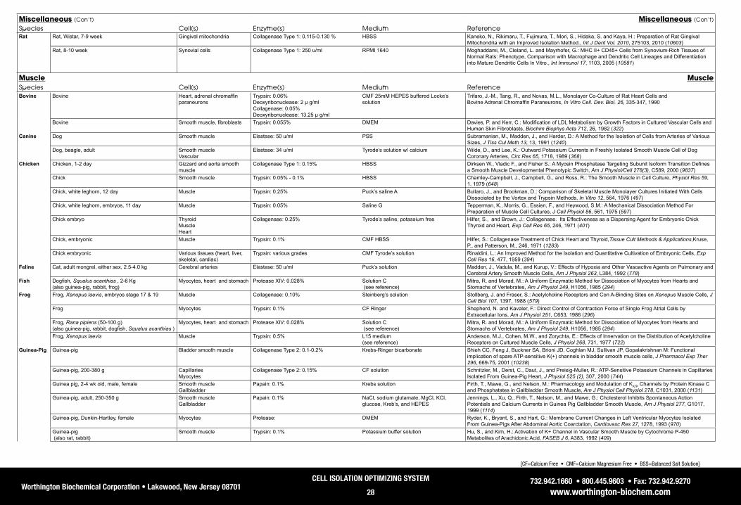
Worthington Biochemical Corporation • Lakewood, New Jersey 08701732.942.1660 • 800.445.9603 • Fax: 732.942.9270
www.worthington-biochem.com
CELL ISOLATION OPTIMIZING SYSTEM
28
[CF=Calcium Free • CMF=Calcium Magnesium Free • BSS=Balanced Salt Solution]
Miscellaneous (Con’t) Miscellaneous (Con’t)
Species Cell(s) Enzyme(s) Medium ReferenceRat Rat, Wistar, 7-9 week Gingival mitochondria Collagenase Type 1: 0.115-0.130 % HBSS Kaneko, N., Rikimaru, T., Fujimura, T., Mori, S., Hidaka, S. and Kaya, H.: Preparation of Rat Gingival
Mitochondria with an Improved Isolation Method., Int J Dent Vol. 2010, 275103, 2010 (10603)Rat, 8-10 week Synovial cells Collagenase Type 1: 250 u/ml RPMI 1640 Moghaddami, M., Cleland, L. and Mayrhofer, G.: MHC II+ CD45+ Cells from Synovium-Rich Tissues of
Normal Rats: Phenotype, Comparison with Macrophage and Dendritic Cell Lineages and Differentiation into Mature Dendritic Cells In Vitro., Int Immunol 17, 1103, 2005 (10581)
Muscle MuscleSpecies Cell(s) Enzyme(s) Medium ReferenceBovine Bovine Heart, adrenal chromaffin
paraneuronsTrypsin: 0.06%Deoxyribonuclease: 2 μ g/mlCollagenase: 0.05%Deoxyribonuclease: 13.25 μ g/ml
CMF 25mM HEPES buffered Locke’ssolution
Trifaro, J.-M., Tang, R., and Novas, M.L., Monolayer Co-Culture of Rat Heart Cells andBovine Adrenal Chromaffin Paraneurons, In Vitro Cell. Dev. Biol. 26, 335-347, 1990
Bovine Smooth muscle, fibroblasts Trypsin: 0.055% DMEM Davies, P. and Kerr, C.: Modification of LDL Metabolism by Growth Factors in Cultured Vascular Cells and Human Skin Fibroblasts, Biochim Biophys Acta 712, 26, 1982 (322)
Canine Dog Smooth muscle Elastase: 50 u/ml PSS Subramanian, M., Madden, J., and Harder, D.: A Method for the Isolation of Cells from Arteries of Various Sizes, J Tiss Cul Meth 13, 13, 1991 (1240)
Dog, beagle, adult Smooth muscle Vascular
Elastase: 34 u/ml Tyrode’s solution w/ calcium Wilde, D., and Lee, K.: Outward Potassium Currents in Freshly Isolated Smooth Muscle Cell of Dog Coronary Arteries, Circ Res 65, 1718, 1989 (368)
Chicken Chicken, 1-2 day Gizzard and aorta smooth muscle
Collagenase Type 1: 0.15% HBSS Dirksen W., Vladic F., and Fisher S.: A Myosin Phosphatase Targeting Subunit Isoform Transition Defines a Smooth Muscle Developmental Phenotypic Switch, Am J Physiol/Cell 278(3), C589, 2000 (9837)
Chick Smooth muscle Trypsin: 0.05% - 0.1% HBSS Chamley-Campbell, J., Campbell, G., and Ross, R.: The Smooth Muscle in Cell Culture, Physiol Res 59, 1, 1979 (648)
Chick, white leghorn, 12 day Muscle Trypsin: 0.25% Puck’s saline A Bullaro, J., and Brookman, D.: Comparison of Skeletal Muscle Monolayer Cultures Initiated With Cells Dissociated by the Vortex and Trypsin Methods, In Vitro 12, 564, 1976 (497)
Chick, white leghorn, embryos, 11 day Muscle Trypsin: 0.05% Saline G Tepperman, K., Morris, G., Essien, F., and Heywood, S.M.: A Mechanical Dissociation Method For Preparation of Muscle Cell Cultures, J Cell Physiol 86, 561, 1975 (597)
Chick embryo Thyroid Muscle Heart
Collagenase: 0.25% Tyrode’s saline, potassium free Hilfer, S., and Brown, J.: Collagenase. Its Effectiveness as a Dispersing Agent for Embryonic Chick Thyroid and Heart, Exp Cell Res 65, 246, 1971 (401)
Chick, embryonic Muscle Trypsin: 0.1% CMF HBSS Hilfer, S.: Collagenase Treatment of Chick Heart and Thyroid,Tissue Cult Methods & Applications,Kruse, P., and Patterson, M., 246, 1971 (1283)
Chick embryonic Various tissues (heart, liver, skeletal, cardiac)
Trypsin: various grades CMF Tyrode’s solution Rinaldini, L.: An Improved Method for the Isolation and Quantitative Cultivation of Embryonic Cells, Exp Cell Res 16, 477, 1959 (394)
Feline Cat, adult mongrel, either sex, 2.5-4.0 kg Cerebral arteries Elastase: 50 u/ml Puck’s solution Madden, J., Vadula, M., and Kurup, V.: Effects of Hypoxia and Other Vasoactive Agents on Pulmonary and Cerebral Artery Smooth Muscle Cells, Am J Physiol 263, L384, 1992 (778)
Fish Dogfish, Squalus acanthias , 2-6 Kg (also guinea-pig, rabbit, frog)
Myocytes, heart and stomach Protease XIV: 0.028% Solution C (see reference)
Mitra, R. and Morad, M.: A Uniform Enzymatic Method for Dissociation of Myocytes from Hearts and Stomachs of Vertebrates, Am J Physiol 249, H1056, 1985 (294)
Frog Frog, Xenopus laevis, embryos stage 17 & 19 Muscle Collagenase: 0.10% Steinberg’s solution Stollberg, J. and Fraser, S.: Acetylcholine Receptors and Con A-Binding Sites on Xenopus Muscle Cells, J Cell Biol 107, 1397, 1988 (579)
Frog Myocytes Trypsin: 0.1% CF Ringer Shepherd, N. and Kavaler, F.: Direct Control of Contraction Force of Single Frog Atrial Cells by Extracellular Ions, Am J Physiol 251, C653, 1986 (296)
Frog, Rana pipiens (50-100 g) (also guinea-pig, rabbit, dogfish, Squalus acanthias )
Myocytes, heart and stomach Protease XIV: 0.028% Solution C (see reference)
Mitra, R. and Morad, M.: A Uniform Enzymatic Method for Dissociation of Myocytes from Hearts and Stomachs of Vertebrates, Am J Physiol 249, H1056, 1985 (294)
Frog, Xenopus laevis Muscle Trypsin: 0.5% L15 medium (see reference)
Anderson, M.J., Cohen, M.W., and Zorychta, E.: Effects of Innervation on the Distribution of Acetylcholine Receptors on Cultured Muscle Cells, J Physiol 268, 731, 1977 (722)
Guinea-Pig Guinea-pig Bladder smooth muscle Collagenase Type 2: 0.1-0.2% Krebs-Ringer bicarbonate Shieh CC, Feng J, Buckner SA, Brioni JD, Coghlan MJ, Sullivan JP, Gopalakrishnan M: Functional implication of spare ATP-sensitive K(+) channels in bladder smooth muscle cells, J Pharmacol Exp Ther 296, 669-75, 2001 (10238)
Guinea-pig, 200-380 g Capillaries Myocytes
Collagenase Type 2: 0.15% CF solution Schnitzler, M., Derst, C., Daut, J., and Preisig-Muller, R.: ATP-Sensitive Potassium Channels in Capillaries Isolated From Guinea-Pig Heart, J Physiol 525 (2), 307, 2000 (744)
Guinea pig, 2-4 wk old, male, female Smooth muscle Gallbladder
Papain: 0.1% Krebs solution Firth, T., Mawe, G., and Nelson, M.: Pharmacology and Modulation of KATP Channels by Protein Kinase C and Phosphatates in Gallbladder Smooth Muscle, Am J Physiol Cell Physiol 278, C1031, 2000 (1131)
Guinea-pig, adult, 250-350 g Smooth muscle Gallbladder
Papain: 0.1% NaCl, sodium glutamate, MgCl, KCl, glucose, Kreb’s, and HEPES
Jennings, L., Xu, Q., Firth, T., Nelson, M., and Mawe, G.: Cholesterol Inhibits Spontaneous Action Potentials and Calcium Currents in Guinea Pig Gallbladder Smooth Muscle, Am J Physiol 277, G1017, 1999 (1114)
Guinea-pig, Dunkin-Hartley, female Myocytes Protease: DMEM Ryder, K., Bryant, S., and Hart, G.: Membrane Current Changes in Left Ventricular Myocytes Isolated From Guinea-Pigs After Abdominal Aortic Coarctation, Cardiovasc Res 27, 1278, 1993 (970)
Guinea-pig (also rat, rabbit)
Smooth muscle Trypsin: 0.1% Potassium buffer solution Hu, S., and Kim, H.: Activation of K+ Channel in Vascular Smooth Muscle by Cytochrome P-450 Metabolites of Arachidonic Acid, FASEB J 6, A383, 1992 (409)

Worthington Biochemical Corporation • Lakewood, New Jersey 08701732.942.1660 • 800.445.9603 • Fax: 732.942.9270
www.worthington-biochem.com
CELL ISOLATION OPTIMIZING SYSTEM
29
[CF=Calcium Free • CMF=Calcium Magnesium Free • BSS=Balanced Salt Solution]
Muscle (Con’t) Muscle (Con’t)
Species Cell(s) Enzyme(s) Medium ReferenceGuinea-Pig Guinea-pig, 200-400 g
(also rabbit, frog, dogfish)Myocytes, heart and stomach Protease XIV: 0.028% Solution C
(see reference)Mitra, R. and Morad, M.: A Uniform Enzymatic Method for Dissociation of Myocytes from Hearts and Stomachs of Vertebrates, Am J Physiol 249, H1056, 1985 (294)
Guinea-pig, prepubertal Smooth muscle Aortic
Trypsin: 0.05% Dulbecco-Vogt modification of Eagle’s Ross, R.: The Smooth Muscle Cell . II. Growth of Smooth Muscle in Culture and Formation of Elastic Fibers, J Cell Biol 50, 172, 1971 (584)
Hamster Hamster, male, 60-70 day Satellite Trypsin: 0.25% DMEM Nakamura, T., Iwata, Y., Sampaolesi, M., Hanada, H., Saito, N., Artman, M., Coetzee, W., and Shigekawa, M.: Stretch-Activated Cation Channels in Skeletal Muscle Myotubes From Sarcoglycan-Deficient Hamsters, Am J Physiol Cell Physiol 281, C690, 2001 (747)
Human Human, male Myogenic Collagenase Type 4: 0.1%Neutral Protease: 2.4 u/ml
HBSS Stadler, G., Chen, J., Wagner, K., Robin, J., Shay, J., Emerson, C. and Wright, W.: Establishment of Clonal Myogenic Cell Lines from Severely Affected Dystrophic Muscles - CDK4 Maintains the Myogenic Population., Skelet Muscle 1, 12, 2011 (10667)
Human Muscle derived multiprogenitor cells
Collagenase Type 2: 0.05% DMEM Nesti, L., Jackson, W., Shanti, R., Koehler, S., Aragon, A., Bailey, J., Sracic, M., Freedman, B., Giuliani, J. and Tuan, R.: Differentiation Potential of Multipotent Progenitor Cells Derived from War-Traumatized Muscle Tissue., J Bone Joint Surg Am Vol. 90, 2390, 2008 (10490)
Human Endothelial and vascular smooth muscle
Collagenase Type 1: 0.2% HBSS Moss, S., Bates, M., Parrino, P. and Woods, TC.: Isolation of Endothelial Cells and Vascular Smooth Muscle Cells from Internal Mammary Artery Tissue., Ochsner J 7, 133, 2007 (10636)
Human Urinary tract smooth muscle Collagenase Type 4: 100 u/ml DMEM Kimuli, M., Eardley, I. and Southgate, J.: In Vitro Assessment of Decellularized Porcine Dermis as a Matrix for Urinary Tract Reconstruction., BJU Int Vol. 94, 859, 2004 (10570)
Human, female Smooth muscle Myometrial
Deoxyribonuclease I: 0.015% and 0.007% HBSS Richardson, M., Taylor, D., Casey, M., MacDonald, P., and Stull, J.: Biochemical Markers of Contraction in Human Myometrial Smooth Muscle Cells in Culture, In Vitro Cell Dev Biol 23, 21, 1987 (420)
Human, female Smooth muscle Myometrial
Deoxyribonuclease I: 0.12% HBSS Casey, M., MacDonald, P., Mitchell, M., and Snyder, J.: Maintenance and Characterization of Human Myometrial Smooth Muscle Cells in Monolayer Culture, In Vitro 20, 396, 1984 (533)
Human, fetal (also bovine)
Smooth muscle, fibroblasts Trypsin: 0.055% DMEM Davies, P. and Kerr, C.: Modification of LDL Metabolism by Growth Factors in Cultured Vascular Cells and Human Skin Fibroblasts, Biochim Biophys Acta 712, 26, 1982 (322)
Human Smooth muscle Trypsin: 0.25% DMEM Eskin, S., Sybers, H., Lester, J., Navarro, L., Gotto, A., and DeBakey, M.: Human Smooth Muscle Cells Cultured From Atherosclerotic Plaques and Uninvolved Vessel Wall, In Vitro 17 (8), 713, 1981 (864)
Human (also rat, guinea-pig, chick, monkey)
Smooth muscle Trypsin: 0.05% - 0.1% HBSS Chamley-Campbell, J., Campbell, G., and Ross, R.: The Smooth Muscle in Cell Culture, Physiol Res 59, 1, 1979 (648)
Lizard Lizard (Anolis carolinensis) Myoblasts, tail Collagenase: 0.2% GM III (see reference)
Cox, P., and Simpson, Jr., S.: A Microphotometric Study of Myogenic Lizard Cells Grown in Vitro, Dev Biol 23, 433, 1970 (369)
Monkey Monkey (Macaca nemestrina) Smooth muscle Trypsin: 0.05% Dulbecco-Vogt Chait, A., Ross, R., Albers, J., and Bierman, E.: Platelet-Derived Growth Factor Stimulates Activity of LDL Receptors, Proc Natl Acad Sci U S A 77, 4084, 1980 (654)
Rhesus monkey, 1 year (also human, rabbits)
Smooth muscle, saphenous vein
Elastase: 0.05% BSS Chamley, J., Campbell, G., McConnell, J., and Groschel-Stewart, U.: Comparison of Vascular Smooth Muscle Cells from Adult Human, Monkey and Rabbit in Primary Culture and in Subculture, Cell Tissue Res 177, 503, 1977 (354)
Mouse Mouse, 1-25 month Myocytes, endothelial Neutral Protease: 1.2 u/ml Collagenase Type 4: 0.2%
PBS Ieronimakis Nicholas, Balasundaram Gayathri, Reyes Morayma: Direct isolation, culture and transplant of mouse skeletal muscle derived endothelial cells with angiogenic potential, PLoS ONE 3, e0001753, 2008 (10313)
Mouse, 15-20 day Intersitial cells of Cajal Collagenase Type 2: 0.13% M199 Li CX, Liu BH, Tong WD, Zhang LY, and Jiang YP: Dissociation, culture and morphologic changes of interstitial cells of Cajal in vitro, World J Gastroenterol 11, 2838, 2005 (10007)
Ovine Sheep, adult and neonatal Tracheal smooth muscle cells Papain: 0.2% Deoxyribonuclease I: 0.1%
MOPS-PSS Driska S., Laudadio R., Wolfson M., and Shaffer T.: A Method for Isolating Adult and Neonatal Airway Smooth Muscle Cells and Measuring Shortening Velocity, J Appl Physiol 86(1), 427, 1999 (9841)
Porcine Porcine Smooth muscle, aorta Collagenase: 0.3% DMEM Xiong, Yimin, Xu, Shangzhe, and Slakey, Linda L: Modulation of Response to Adenosine in Vascular Smooth Muscle Cells Cultured in Defined Medium, In Vitro Cell Dev Biol 27, 355, 1991 (463)
Porcine Smooth muscle Aorta
Trypsin: 0.05% EDTA 0.02% Breton, M., Berrou, E., Deudon, E. and Picard, J.: Changes in Proteoglycans of Cultured Pig Aortic Smooth Muscle Cells During Subculture, In Vitro Cell Dev Biol 26, 157, 1990 (431)
Porcine Smooth muscle Aortic medial tissue
Collagenase: 0.30% DMEM Fehr, T., Dickinson, E., Goldman, S. and Slakey, L.: Cyclic AMP Efflux is Regulated by Occupancy of The Adenosine Receptor in Pig Aortic Smooth Muscle Cells, J Biol Chem 265, 10974, 1990 (566)
Quail Quail, embryo, 10 day Myoblasts Collagenase Type 2: 0.1% Puck’s solution Konigsberg, I.: Skeletal Myoblasts in Culture, Methods Enzymol LVIII, 511, 1979 (638)
Rabbit Rabbit, New Zealand White Aortic smooth muscle Collagenase Type 2: 300 u/ml Elastase: 5 u/ml
F10 Ham’s Croons Valerie, Martinet Wim, Herman Arnold G, Timmermans Jean-Pierre, De Meyer Guido R Y: Selective clearance of macrophages in atherosclerotic plaques by the protein synthesis inhibitor cycloheximide, J Pharmacol Exp Ther 320, 986-93, 2007 (10348)
Rabbit (also rat, guinea-pig)
Smooth muscle Trypsin: 0.1% Potassium buffer solution Hu, S., and Kim, H.: Activation of K+ Channel in Vascular Smooth Muscle by Cytochrome P-450 Metabolites of Arachidonic Acid, FASEB J 6, A383, 1992 (409)
Rabbit, adult, 1-2 kg Smooth muscle Elastase: 0.17 - 0.25% Saline Benham, C., Bolton, T., Byrne, N., and Large, W.: Action of Extremely Applied Adenosine Triphosphate O Single Smooth Muscle Cells Dispersed From Rabbit Ear Artery, J Physiol 387, 473, 1987 (863)
Rabbit, New Zealand white, 1500 g Smooth muscle, aortic Trypsin: 0.038% MEM Knodle, S., Anderson, S., and Papaioannou, S.: Large Scale Preparation of Rabbit Aortic Smooth Muscle Cells For Use in Calcium Uptake Studies, In Vitro Cell Dev Biol 22, 23, 1986 (416)
Rabbit, adult, 1-2 kg Smooth muscle, ear artery Trypsin: 0.1% CF solution (see reference)
Benham, C.D., Bolton, T.B.: Spontaneous Transient Outward Currents in Single Visceral and Vascular Smooth Muscle Cells of the Rabbit, J Physiol 381, 385, 1986 (720)

Worthington Biochemical Corporation • Lakewood, New Jersey 08701732.942.1660 • 800.445.9603 • Fax: 732.942.9270
www.worthington-biochem.com
CELL ISOLATION OPTIMIZING SYSTEM
30
[CF=Calcium Free • CMF=Calcium Magnesium Free • BSS=Balanced Salt Solution]
Muscle (Con’t) Muscle (Con’t)
Species Cell(s) Enzyme(s) Medium ReferenceRabbit Rabbit, 0.5-1 kg
(also guinea-pig, frog, dogfish)Myocytes, heart and stomach Protease XIV: 0.028% Solution C
(see reference)Mitra, R. and Morad, M.: A Uniform Enzymatic Method for Dissociation of Myocytes from Hearts and Stomachs of Vertebrates, Am J Physiol 249, H1056, 1985 (294)
Rabbit, white New Zealand, adult, male, 2 kg Smooth muscle, aorta Trypsin: 0.1% Krebs Ringer HEPES solution Ives, H., Schultz, G., Galardy, R., and Jamieson, J.: Preparation of Functional Smooth Muscle Cells from the Rabbit Aorta, J Exp Med 148, 1400, 1978 (603)
Rabbits, New Zealand white albino, 5-6 months (also human, Rhesus monkey, rabbit)
Smooth muscle, saphenous vein
Elastase: 0.05% BSS Chamley, J., Campbell, G., McConnell, J., and Groschel-Stewart, U.: Comparison of Vascular Smooth Muscle Cells from Adult Human, Monkey and Rabbit in Primary Culture and in Subculture, Cell Tissue Res 177, 503, 1977 (354)
Rabbit, chinchilla, 5-8 month, virgin, female, 2-3 kg Smooth muscle, aorta Hyaluronidase: 800 u/ml HBSS Peters, T., Muller, M., and deDuve, C.: Lysosomes of the Arterial Wall. I. Isolation and Subcellular Fractionation of Cells from Normal Rabbit Aorta, J Exp Med 136, 1117, 1972 (601)
Rabbit, New England, albino Thoracic aorta Elastase: 0.008% Kreb’s Ringer Day, A., Phil, D., and Newman, H.: Synthesis of Phospholipid by Foam Cells Isolated from Rabbit Atherosclerotic Lesions, Circ Res XIX, 122, 1966 (777)
Rat Rat, fetal 17-18 day Myotubes Trypsin: 0.05% (see reference) Das, M., Rumsey, J., Bhargava, N., Stancescu, M. and Hickman, J.: Skeletal Muscle Tissue Engineering: A Maturation Model Promoting Long-Term Survival of Myotubes, Structural Development of the Excitation-Contraction Coupling Apparatus and Neonatal Myosin Heavy Chain Expression., Biomaterials 30, 5392, 2009 (10654)
Rat, Lewis, neonatal Myoblasts Collagenase Type 2: 1.0%Neutral Protease: 2.4 u/ml
Ham’s F-10 Kim, J., Hadlock, T., Cheney, M., Varvares, M. and Marler, J.: Muscle Tissue Engineering for Partial Glossectomy Defects., Arch Facial Plast Surg 5, 403, 2003 (10637)
Rat, SD, adult and neonatal w/in 1 day of birth Myooids Neutral Protease: 4 u/ml Ham’s F-12 Dennis, R., Kosnik II, P., Gilbert, M., and Faulkner, J.: Excitability and Contractility of Skeletal Muscle Engineered from Primary Cultures and Cell Lines, Am J Physiol Cell Physiol 280, C288, 2001 (1111)
Rat, SD, 3 month Smooth muscle cells Collagenase Type 2: 0.2% Elastase: 0.04% Soybean Trypsin Inhibitor: 0.1%
M-199 Su E., Stevenson S., Rollence M., Marshall-Neff J., and Liau G.: A Genetically Modified Adenoviral Vector Exhibits Enhanced Gene Transfer of Human Smooth Muscle Cells, J Vasc Res 38(5), 471, 2001 (9860)
Rat, SD, 250 g Arterial smooth muscle Papain: 0.03% Collagenase: 0.1%
(see reference) Jaggar JH: Intravascular pressure regulates local and global Ca(2+) signaling in cerebral artery smooth muscle cells, Am J Physiol Cell Physiol 281, C439-48, 2001 (10325)
Rat, Wistar, adult, male Smooth & skeletal muscle Cardiac myocytes
Protease: 0.01% PSS Wellman, G., Barrett-Jolley, R., Koppel, H. Everitt, D., and Quayle, J.: Inhibition of Vascular KATP Channels by U-37883A: A Comparison with Cardiac and Skeletal Muscle, Br J Pharmacol 128, 909, 1999 (1065)
Rat, SD, male, 250-350 g Vascular smooth muscle Collagenase Type 2: 0.1% Elastase: 0.0125%
DMEM Hrometz, S., Edelmann, S., McCune, D., Olges, J., Hadley, R., Perez ,D., and Piascik, M.: Expression of Multiple Alpha1-Adrenoceptors on Vascular Smooth Muscle: Correlation with the Regulation of Contraction, J Pharmacol Exp Ther 290(1), 452, 1999 (9867)
Rat, SD, male, 150-175 g Smooth muscle, endothelial Trypsin: 0.04% MEM Redmond, E., Cahill, P., and Sitzmann, J.: Perfused Transcapillary Smooth Muscle and Endothelial Cell Co-Culture-A Novel In Vitro Model, In Vitro Cell Dev Biol Anim 31, 601, 1995 (1234)
Rat, Wistar Kyoto, 10-15 weeks Smooth muscle, mesenteric artery
Trypsin: 0.05% MEM McGuire, P., Walker-Caprioglio, H., Little, S., and McGuffee, L.: Isolation and Culture of Rat Superior Mesenteric Artery Smooth Muscle Cells, In Vitro Cell Dev Biol 29, 135, 1993 (491)
Rat (also rabbit, guinea-pig)
Smooth muscle Trypsin: 0.1% Potassium buffer solution Hu, S., and Kim, H.: Activation of K+ Channel in Vascular Smooth Muscle by Cytochrome P-450 Metabolites of Arachidonic Acid, FASEB J 6, A383, 1992 (409)
Rat, SD, 19 days Smooth muscle, myometrial Trypsin: 150 µg/ml HBSS or PSS, CMF Loch-Caruso, R., Pahl, M., and Juberg, D.: Rat Myometrial Smooth Muscle Cells Show High Levels of Gap Junctional Communication Under a Variety of Culture Conditions, In Vitro Cell Dev Biol 28, 97, 1992 (489)
Rat, SHRs and WKY, male, 10 - 14 weeks Smooth muscle, tail arteries Papain: 0.1% HEPES buffer (see reference)
Bolzon, B. and Cheung, D.: Isolation and Characterization of Single Vascular Smooth Muscle Cells From Spontaneously Hypertensive Rats, Hypertension 14, 137, 1989 (694)
Rat, 1-3 day Smooth muscle, aortic Elastase: 0.0125% DMEM Barone, L., Wolfe, L., Faris, B., and Franzblau, C.: Elastin mRNA Levels and Insoluble Elastin Accumulation in Neonatal Rat, Biochem 27, 3175, 1988 (313)
Rat Muscle, mesenteric arteries Trypsin: 0.05% HEPES KG solution (see reference)
Bean, B., Sturek, M., Puga, A., and Hermsmeyer, K.: Calcium Channels in Muscle Cells Isolated From Rat Mesenteric Arteries: Modulation by Dihydropyridine Drugs, Circ Res 59, 229, 1986 (364)
Rat, SHR, WKY. either sex, 12-19 day, 3 month, and retired breeders
Endothelial, aortic Elastase: 0.05% Waymouth’s culture medium Gordon, D., Mohai, L., and Schwartz, S.: Induction of Polyploidy in Cultures of Neonatal Rat Aortic Smooth Muscle Cells, Circ Res 59, 633, 1986 (866)
Rat, male, 150-250 g Endothelial, aortic Trypsin: RPMI 1640 Cole, O., Fan, T., and Lewis, G.: Isolation, Characterization, Growth and Culture of Endothelial Cells From the Rat Aorta, Cell Biol Int Rep 10 (6), 399, 1986 (884)
Rat, Wistar, female, 10 weeks Myocytes Collagenase: 0.1% HBSS Boulanger-Saunier, C., Kattenburg, D., and Stoclet, J.: Cyclic AMP-dependent Phosphorylation of a 16kDa Protein in a Plasma Membrane-enriched Fraction of Rat Aortic Myocytes, FEBS Lett 193, 283, 1985 (411)
Rat, SD, male, 200 - 250 g Smooth muscle, thoracic aorta
Trypsin: 0.0375% Eagle’s MEM with calcium Brock, T., Alexander, R., Ekstein, L., Atkinson, W., and Gimbrone, M.: Angiotensin Increases Cytosolic Free Calcium in Cultured Vascular Smooth Muscle Cells, Hypertension 7, 105, 1985 (693)
Rat, SD, 225-250g Mesenteric artery smooth muscle cells
Elastase: .0125% Soybean Trypsin Inhibitor: 0.025% Collagenase Type 1: 0.1%
HBSS Gunther S, Alexander RW, Atkinson WJ, and Gimbrone MA Jr.: Functional angiotensin II receptors in cultured vascular smooth muscle cells, J Cell Biol 92, 289, 1982 (10058)
Rat, 3-4 day Myocardial Trypsin NF 1:250: 0.125% HBSS CMF Kasten, F.: Rat Myocardial Cells In Vitro: Mitosis and Differentiated Properties, In Vitro 8, 128, 1972 (539)
Rat, 200 g Muscle Trypsin: 0.05% Kreb’s Henseleit bicarbonate buffer Kono, T.: Roles of Collagenases and Other Proteolytic Enzymes in the Dispersal of Animal Tissues, Biochim Biophys Acta 178, 397, 1969 (317)
Rat, Wistar, 3-10 day Heart Trypsin NF 1:250: 250: 0.1% Saline A (see reference)
Harary, I., and Farley, B.: In Vitro Studies on Single Beating Rat Heart Cells, Exp Cell Res 29, 451, 1963 (395)
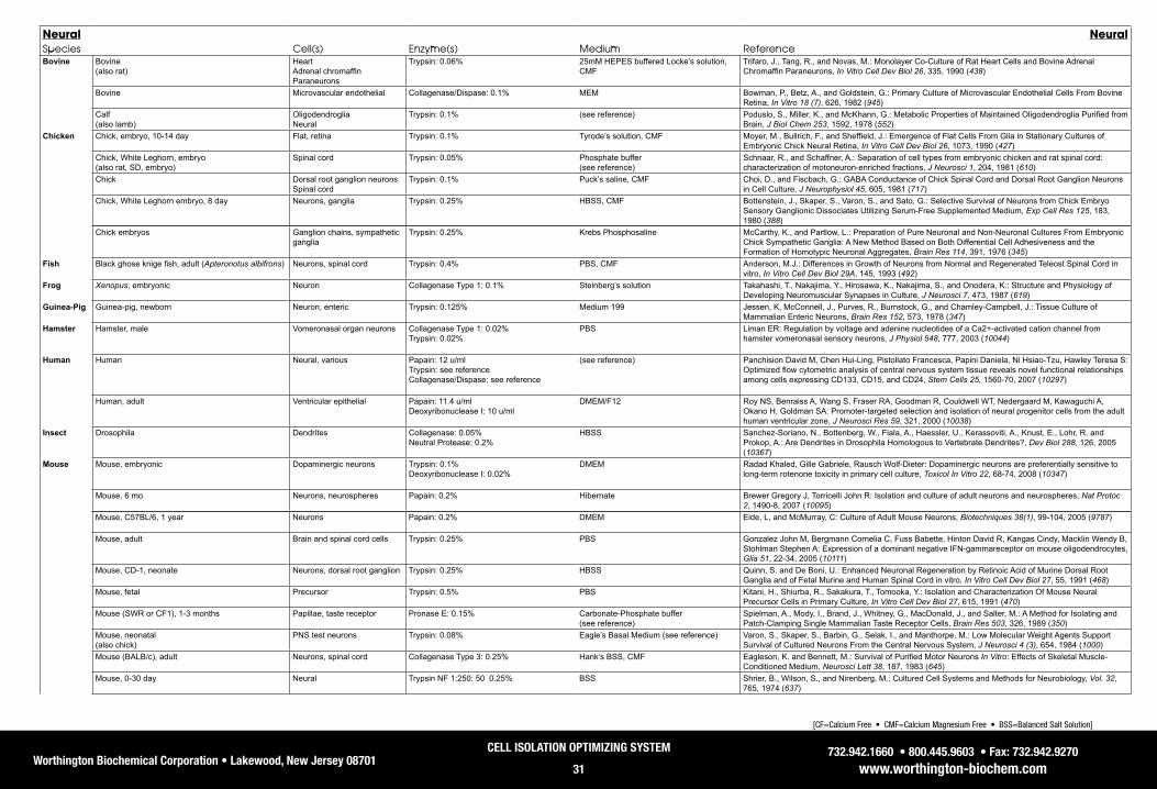
Worthington Biochemical Corporation • Lakewood, New Jersey 08701732.942.1660 • 800.445.9603 • Fax: 732.942.9270
www.worthington-biochem.com
CELL ISOLATION OPTIMIZING SYSTEM
31
[CF=Calcium Free • CMF=Calcium Magnesium Free • BSS=Balanced Salt Solution]
Neural NeuralSpecies Cell(s) Enzyme(s) Medium ReferenceBovine Bovine
(also rat)Heart Adrenal chromaffin Paraneurons
Trypsin: 0.06% 25mM HEPES buffered Locke’s solution, CMF
Trifaro, J., Tang, R., and Novas, M.: Monolayer Co-Culture of Rat Heart Cells and Bovine Adrenal Chromaffin Paraneurons, In Vitro Cell Dev Biol 26, 335, 1990 (438)
Bovine Microvascular endothelial Collagenase/Dispase: 0.1% MEM Bowman, P., Betz, A., and Goldstein, G.: Primary Culture of Microvascular Endothelial Cells From Bovine Retina, In Vitro 18 (7), 626, 1982 (945)
Calf (also lamb)
Oligodendroglia Neural
Trypsin: 0.1% (see reference) Poduslo, S., Miller, K., and McKhann, G.: Metabolic Properties of Maintained Oligodendroglia Purified from Brain, J Biol Chem 253, 1592, 1978 (552)
Chicken Chick, embryo, 10-14 day Flat, retina Trypsin: 0.1% Tyrode’s solution, CMF Moyer, M., Bullrich, F., and Sheffield, J.: Emergence of Flat Cells From Glia in Stationary Cultures of Embryonic Chick Neural Retina, In Vitro Cell Dev Biol 26, 1073, 1990 (427)
Chick, White Leghorn, embryo (also rat, SD, embryo)
Spinal cord Trypsin: 0.05% Phosphate buffer (see reference)
Schnaar, R., and Schaffner, A.: Separation of cell types from embryonic chicken and rat spinal cord: characterization of motoneuron-enriched fractions, J Neurosci 1, 204, 1981 (610)
Chick Dorsal root ganglion neurons Spinal cord
Trypsin: 0.1% Puck’s saline, CMF Choi, D., and Fiscbach, G.: GABA Conductance of Chick Spinal Cord and Dorsal Root Ganglion Neurons in Cell Culture, J Neurophysiol 45, 605, 1981 (717)
Chick, White Leghorn embryo, 8 day Neurons, ganglia Trypsin: 0.25% HBSS, CMF Bottenstein, J., Skaper, S., Varon, S., and Sato, G.: Selective Survival of Neurons from Chick Embryo Sensory Ganglionic Dissociates Utilizing Serum-Free Supplemented Medium, Exp Cell Res 125, 183, 1980 (388)
Chick embryos Ganglion chains, sympathetic ganglia
Trypsin: 0.25% Krebs Phosphosaline McCarthy, K., and Partlow, L.: Preparation of Pure Neuronal and Non-Neuronal Cultures From Embryonic Chick Sympathetic Ganglia: A New Method Based on Both Differential Cell Adhesiveness and the Formation of Homotypic Neuronal Aggregates, Brain Res 114, 391, 1976 (345)
Fish Black ghose knige fish, adult (Apteronotus albifrons) Neurons, spinal cord Trypsin: 0.4% PBS, CMF Anderson, M.J.: Differences in Growth of Neurons from Normal and Regenerated Teleost Spinal Cord in vitro, In Vitro Cell Dev Biol 29A, 145, 1993 (492)
Frog Xenopus, embryonic Neuron Collagenase Type 1: 0.1% Steinberg’s solution Takahashi, T., Nakajima, Y., Hirosawa, K., Nakajima, S., and Onodera, K.: Structure and Physiology of Developing Neuromuscular Synapses in Culture, J Neurosci 7, 473, 1987 (619)
Guinea-Pig Guinea-pig, newborn Neuron, enteric Trypsin: 0.125% Medium 199 Jessen, K, McConnell, J., Purves, R., Burnstock, G., and Chamley-Campbell, J.: Tissue Culture of Mammalian Enteric Neurons, Brain Res 152, 573, 1978 (347)
Hamster Hamster, male Vomeronasal organ neurons Collagenase Type 1: 0.02% Trypsin: 0.02%
PBS Liman ER: Regulation by voltage and adenine nucleotides of a Ca2+-activated cation channel from hamster vomeronasal sensory neurons, J Physiol 548, 777, 2003 (10044)
Human Human Neural, various Papain: 12 u/ml Trypsin: see reference Collagenase/Dispase: see reference
(see reference) Panchision David M, Chen Hui-Ling, Pistollato Francesca, Papini Daniela, Ni Hsiao-Tzu, Hawley Teresa S: Optimized flow cytometric analysis of central nervous system tissue reveals novel functional relationships among cells expressing CD133, CD15, and CD24, Stem Cells 25, 1560-70, 2007 (10297)
Human, adult Ventricular epithelial Papain: 11.4 u/ml Deoxyribonuclease I: 10 u/ml
DMEM/F12 Roy NS, Benraiss A, Wang S, Fraser RA, Goodman R, Couldwell WT, Nedergaard M, Kawaguchi A, Okano H, Goldman SA: Promoter-targeted selection and isolation of neural progenitor cells from the adult human ventricular zone, J Neurosci Res 59, 321, 2000 (10038)
Insect Drosophila Dendrites Collagenase: 0.05%Neutral Protease: 0.2%
HBSS Sanchez-Soriano, N., Bottenberg, W., Fiala, A., Haessler, U., Kerassoviti, A., Knust, E., Lohr, R. and Prokop, A.: Are Dendrites in Drosophila Homologous to Vertebrate Dendrites?, Dev Biol 288, 126, 2005 (10367)
Mouse Mouse, embryonic Dopaminergic neurons Trypsin: 0.1% Deoxyribonuclease I: 0.02%
DMEM Radad Khaled, Gille Gabriele, Rausch Wolf-Dieter: Dopaminergic neurons are preferentially sensitive to long-term rotenone toxicity in primary cell culture, Toxicol In Vitro 22, 68-74, 2008 (10347)
Mouse, 6 mo Neurons, neurospheres Papain: 0.2% Hibernate Brewer Gregory J, Torricelli John R: Isolation and culture of adult neurons and neurospheres, Nat Protoc 2, 1490-8, 2007 (10095)
Mouse, C57BL/6, 1 year Neurons Papain: 0.2% DMEM Eide, L, and McMurray, C: Culture of Adult Mouse Neurons, Biotechniques 38(1), 99-104, 2005 (9787)
Mouse, adult Brain and spinal cord cells Trypsin: 0.25% PBS Gonzalez John M, Bergmann Cornelia C, Fuss Babette, Hinton David R, Kangas Cindy, Macklin Wendy B, Stohlman Stephen A: Expression of a dominant negative IFN-gammareceptor on mouse oligodendrocytes, Glia 51, 22-34, 2005 (10111)
Mouse, CD-1, neonate Neurons, dorsal root ganglion Trypsin: 0.25% HBSS Quinn, S. and De Boni, U.: Enhanced Neuronal Regeneration by Retinoic Acid of Murine Dorsal Root Ganglia and of Fetal Murine and Human Spinal Cord in vitro, In Vitro Cell Dev Biol 27, 55, 1991 (468)
Mouse, fetal Precursor Trypsin: 0.5% PBS Kitani, H., Shiurba, R., Sakakura, T., Tomooka, Y.: Isolation and Characterization Of Mouse Neural Precursor Cells in Primary Culture, In Vitro Cell Dev Biol 27, 615, 1991 (470)
Mouse (SWR or CF1), 1-3 months Papillae, taste receptor Pronase E: 0.15% Carbonate-Phosphate buffer (see reference)
Spielman, A., Mody, I., Brand, J., Whitney, G., MacDonald, J., and Salter, M.: A Method for Isolating and Patch-Clamping Single Mammalian Taste Receptor Cells, Brain Res 503, 326, 1989 (350)
Mouse, neonatal (also chick)
PNS test neurons Trypsin: 0.08% Eagle’s Basal Medium (see reference) Varon, S., Skaper, S., Barbin, G., Selak, I., and Manthorpe, M.: Low Molecular Weight Agents Support Survival of Cultured Neurons From the Central Nervous System, J Neurosci 4 (3), 654, 1984 (1000)
Mouse (BALB/c), adult Neurons, spinal cord Collagenase Type 3: 0.25% Hank’s BSS, CMF Eagleson, K. and Bennett, M.: Survival of Purified Motor Neurons In Vitro: Effects of Skeletal Muscle-Conditioned Medium, Neurosci Lett 38, 187, 1983 (645)
Mouse, 0-30 day Neural Trypsin NF 1:250: 50 0.25% BSS Shrier, B., Wilson, S., and Nirenberg, M.: Cultured Cell Systems and Methods for Neurobiology, Vol. 32, 765, 1974 (637)

Worthington Biochemical Corporation • Lakewood, New Jersey 08701732.942.1660 • 800.445.9603 • Fax: 732.942.9270
www.worthington-biochem.com
CELL ISOLATION OPTIMIZING SYSTEM
32
[CF=Calcium Free • CMF=Calcium Magnesium Free • BSS=Balanced Salt Solution]
Neural (Con’t) Neural (Con’t)
Species Cell(s) Enzyme(s) Medium ReferencePorcine Porcine, adult, 60-100 kg Superior cervical ganglia Papain: 2 u/ml
Collagenase: 0.12% Neutral Protease: 0.48%
HBSS Si ML, Lee TJ.: Presynaptic alpha7-nicotinic acetylcholine receptors mediate nicotine-induced nitric oxidergic neurogenic vasodilation in porcine basilar arteries, J Pharmacol Exp Ther 298, 122, 2001 (10052)
Quail Quail Neural crest Trypsin: 0.05% MEM, HBSS Sieber-Blum, M., and Cohen, A.: Clonal Analysis of Quail Neural Crest Cells: They are Pluripotent and Differentiate in Vitro in the Absence of Noncrest Cells, Dev Biol 80, 96, 1980 (371)
Rat Rat, embryonic Hypothalamic neurons and glias
Papain: 0.5 u/ml DMEM Yokosuka Makoto, Ohtani-Kaneko Ritsuko, Yamashita Kayoko, Muraoka Daisuke, Kuroda Yoichiro, Watanabe Chiho: Estrogen and environmental estrogenic chemicals exert developmental effects on rat hypothalamic neurons and glias, Toxicol In Vitro 22, 1-9, 2008 (10346)
Rat, SD, embryos, 19-21 days gestation Schwann, dorsal root ganglia Trypsin: 0.25% HBSS, CMF Mithen, F., Reiker, M., and Birchem, R.: Effects of Ethanol on Rat Schwann Cell Proliferation and Myelination in Culture, In Vitro Cell Dev Biol 26, 129, 1990 (430)
Rat (also bovine)
Heart Adrenal chromaffin Paraneurons
Trypsin: 0.06% 25mM HEPES buffered Locke’s solution, CMF
Trifaro, J., Tang, R., and Novas, M.: Monolayer Co-Culture of Rat Heart Cells and Bovine Adrenal Chromaffin Paraneurons, In Vitro Cell Dev Biol 26, 335, 1990 (438)
Rat, postnatal Septal neurons Papain: 0.05% PBS, CMF Hatanaka,H., Tsukui, H., Nihonmatsu, I.: Septal Cholinergic Neurons From Postnatal Rat Can Survive In The Dissociate Culture Conditions In The Presence Of Nerve Growth Factor, Neurosci Lett 79, 85, 1987 (646)
Rat, Long Evans, 1-15 days Neurons, visual cortex Papain: 20 u/ml BSS (see reference) Huettner, J., and Baughman, R.: Primary Culture of Identified Neurons From the Visual Cortex of Postnatal Rats, J Neurosci 6, 3044, 1986 (617)
Rat, embryo, 15 day Dorsal horn neurons Spinal
Trypsin: 0.025% Ham’s F-12 Jahr, C. and Jessell, T.: Synaptic Transmission between Dorsal Root Ganglion and Dorsal Horn Neurons in Culture: Antagonism of Monosynaptic Excitatory Postsynaptic Potentials and Glutamate Excitation by Kynurenate, J Neurosci 5, 2281, 1985 (614)
Rat, fetus, 18-20 day Hippocampal neurons Trypsin: 0.25% HBSS, CMF Bartlett, W. and Banker, G.: An Electron Microscopic Study of the Development of Axons and Dendrites by Hippocampal Neurons in Culture. I. Cells Which Develop Without Intercellular Contacts, J Neurosci 4, 1944, 1984 (613)
Rat, postnatal Ganglion, retina Papain: 12.5 u/ml HBSS w/5 mM HEPES Leifer, D., Lipton, S., Barnstable, C., and Masland, R.: Monoclonal Antibody to Thy-1 Enhances Regeneration of Processes by Rat Retinal Ganglion Cells in Culture, Science 224, 303, 1984 (667)
Rat, SD, pups Retina Trypsin: 0.25% Ham’s F-12 Sarthy PV, Curtis BM, and Catterall WA.: Retrograde Labeling, Enrichment, and Characerization of Retinal Ganglion Cells from the Neonatal Rat, J Neurosci 3 (12), 2532, 1983 (1199)
Rat, W/FU, 5-8 day Neurons and glial Trypsin: 0.25% MEM (see reference)
Raff, M., Fields, K., Hakomori, S., Mirsky, R., Pruss, R., and Winter, J.: Cell-Type-Specific Markers for Distinguishing and Studying Neurons And the Major Classes of Glial Cells in Culture, Brain Res 174, 283, 1979 (348)
Rat, fetus Neurons, sympathetic Trypsin: 0.25% L-15 or HBSS, CMF Wakshull, E., Johnson, M., Burton, H.: Postnatal Rat Sympathetic Neurons In Culture. 1. A Comparison With Embryonic Neurons, J Neurophysiol 42, 1410, 1979 (716)
Rat, Wistar/Furth, newborn (also bovine)
Schwann Trypsin: 0.25% DMEM Brockes, J., Fields, K., and Raff, M.: Studies on Cultured Rat Schwann Cells. I. Establishment of Purified Populations From Cultures of Peripheral Nerve, Brain Res 165, 105, 1979 (991)
Rat, embryo Neurons, cortical Trypsin: 0.027% MEM Dichter, M.: Rat Cortical Neurons in Cell Culture: Culture Methods, Cell Morphology, Electrophysiology, and Synapse Formation, Brain Res 149, 279, 1978 (346)
Rat, newborn Neurons, sympathetic Collagenase Type 1: 0.01% Hank’s solution, CF Reichardt, L., Patterson, P.: Neurotransmitter Synthesis and Uptake by Isolated Sympathetic Neurones in Microcultures, Nature 270, 147, 1977 (642)
Rat, neonatal Neurons, superior cervical ganglia
Trypsin: 0.1% Basal L-15 medium Mains, R., and Patterson, P.: Primary Cultures of Dissociated Sympathetic Neurons I. Establishment of Long-Term Growth in Culture and Studies of Differentiated Properties, J Cell Biol 59, 329, 1973 (587)
Ovine Lamb (also calf)
Oligodendroglia Neural
Trypsin: 0.1% (see reference) Poduslo, S., Miller, K., and McKhann, G.: Metabolic Properties of Maintained Oligodendroglia Purified from Brain, J Biol Chem 253, 1592, 1978 (552)
Salamander Salamander (A.tigrinum) Photoreceptors, retina Papain: 0.05% (see reference) Bader, C., MacLeish, P., and Schwartz, E.: Responses to Light of Solitary Rod Photoreceptors Isolated From Tiger Salamander Retina, Proc Natl Acad Sci U S A 75, 3507, 1978 (652)
Shellfish Snails (Helisoma trivolis), albino, adult Buccal ganglia; SLT muscle Trypsin: 0.2% DMEM Zoran, M., Doyle, R. and Haydon, P.: Target Contact Regulates the Calcium Responsiveness of the Secretory Machinery During Synaptogenesis, Neuron 6, 145, 1991 (691)
Pond snail (Helisoma) albino, adult Somata, buccal ganglia Trypsin: 0.2% Antibiotic saline, Leibowitz 50% Haydon, P.: The Formation of Chemical Synapses Between Cell-Cultured Neuronal Somata, J Neurosci 8, 1032, 1988 (620)
Helisoma trivovlis Buccal ganglia Trypsin: 0.2% L-15 medium Cohan, C., Haydon, P., and Kater, S.: Single Channel Activity Differs in Growing and Nongrowing Growth Cones of Isolated Identified Neurons of Helisoma, J Neurosci Res 13, 285, 1985 (609)
Turtle Turtle (Pseudemys scripta elegans) Retinal Papain: 0.1% (13.5 u/mg) Kreb’s Ringer Lam, D.: Biosynthesis of Acetylcholine in Turtle Photoreceptors, Proc Natl Acad Sci U S A 69, 1987, 1972 (649)

Worthington Biochemical Corporation • Lakewood, New Jersey 08701732.942.1660 • 800.445.9603 • Fax: 732.942.9270
www.worthington-biochem.com
CELL ISOLATION OPTIMIZING SYSTEM
33
[CF=Calcium Free • CMF=Calcium Magnesium Free • BSS=Balanced Salt Solution]
Pancreas PancreasSpecies Cell(s) Enzyme(s) Medium ReferenceBovine Bovine (Bos taurus) Ductal Neutral Protease: 0.05% EBSS Sato, T., Sato, M., Hudson, E., and Jones, R.: Characterization of Bovine Pancreatic Ductal Cells Isolated
by a Perfusion-Digestion Technique, In Vitro 19, 651, 1983 (529)Canine Canine Buccal ganglia; SLT muscle Trypsin: 0.2% DMEM Zoran, M., Doyle, R. and Haydon, P.: Target Contact Regulates the Calcium Responsiveness of the
Secretory Machinery During Synaptogenesis, Neuron 6, 145, 1991 (691)Fish Fish, Osphronemus gourami, 3-month-old, 0.5 mg Islets Collagenase: 0.12 - 0.46 u/ml RPMI 1640 Schrezenmeir, J., Laue, C., Sternheim,E., Wolbert, K., Darquy, S., Chicheportiche, D., Kirchgessner,
J., and Reach, G.: Long-Term Function of Single-Cell Preparations of Piscine Principal Islets in Hollow Fibers, Transplant Proc 24 (6), 2941, 1992 (1221)
Guinea-Pig Guinea-pig, Hartley, albino, male, 350-400 g Acinar Soybean Trypsin Inhibitor: 0.01% Kreb’s Ringer Gardner, J., Conlon, T., Klaeveman, H., Adams, T., and Ondetti, M.: Action of Cholecystokinin and Cholinergic Agents on Calcium Transport in Isolated Pancreatic Acinar Cells, J Clin Invest 56, 366, 1975 (599)
Guinea-pig Exocrine Hyaluronidase: 0.15% - 0.2% Kreb’s Ringer Amsterdam, J., and Jamieson, J.: Structural and Functional Characterization of Isolated Pancreatic Exocrine Cells, Proc Natl Acad Sci U S A 69 (10), 3028, 1972 (1151)
Hamster Hamster Islets Collagenase Type 4: 1.3% - 2.0% HBSS Feldman, J., and Chapman, B.: Preparation of Islets of Langerhans from Rabbits and Hamsters by the Collagenase Digestion Technique, Acta Diabetol 12, 208, 1975 (686)
Human Human Islets Collagenase Type 4: 0.8% HBSS Izumi, R., Konishi, K., Ueno, K., Shimizu, K., Hirosawa, H., Takahashi, N., and Miyazaki, I.: Isolation of Human Pancreatic Islets from Cryopreserved Pancreas, Transplant Proc XVII, 383, 1985 (689)
Human Islets Collagenase (1 or 4): 0.60% HBSS Gray, D., McShane, P., Grant, A., and Morris, P.: A Method for Isolation of Islets of Langerhans from the Human Pancreas, Diabetes 33, 1055, 1984 (690)
Human, infant, age 1 day-1 year Islets Collagenase: 170-210 u/ml HBSS Sutherland, D., Matas, A., Steffes, M., and Najarian, J.: Infant Human Pancreas: A Potential Source of Islet Tissue for Transplantation, Diabetes 25 (12), 1123, 1976 (810)
Monkey Monkey, 3-5 Kg Islets Hyaluronidase: 0.05% HBSS Scharp, D., Murphy, J., Newton, W., Ballinger, W., and Lacy, P.: Application of an Improved Isolation Technique for Islet Transplantation in Primates and Rats, Transplant Proc 7, 739, 1975 (688)
Mouse Mouse Pancreatic islet Collagenase Type 4: 0.2% HBSS Haefliger Jacques-Antoine, Tawadros Thomas, Meylan Laure, Gurun SabineLe, Roehrich Marc-Estienne, Martin David, Thorens Bernard, Waeber Gerard: The scaffold protein IB1/JIP-1 is a critical mediator of cytokine-induced apoptosis in pancreatic beta cells, J Cell Sci 116, 1463-9, 2003 (10151)
Mouse Islets Collagenase Type 4: 0.2% HBSS Wu Yulian, Han Bing, Luo Hongyu, Roduit Raphael, Salcedo TheodoraW, Moore PaulA, Zhang Jun, Wu Jiangping: DcR3/TR6 effectively prevents islet primary nonfunction after transplantation, Diabetes 52, 2279-86, 2003 (10288)
Mouse, BALB/c, 6-8 week-old, either sex Acinar Collagenase: 0.1% Waymouth’s MB Kurup, S., and Bhonde, R.: Analysis and Optimization of Nutritional Set-up for Murine Pancreatic Acinar Cells, JOP 3 (1), 8, 2002 (1070)
Mouse Acinar Collagenase: 100 u/ml HEPES Jauch, P., Peterson, O., and Lauger, P.: Electrogenic Properties of the Na-Alanine Cotransporter in Acinar Cells, J Membr Biol 94, 99, 1986 (605)
Mouse (C57BL/6J-ob/ob), 9-12 wks, male Islets Hyaluronidase: 0.5% Kreb’s Ringer bicarbonate buffer Dalpe-Scott, M., Heick, H., and Begin-Heick, N.: Secretion in the Obese (ob/ob) Mouse. The Effect of Oxytetracycline on Insulin Release, Diabetes 32, 932, 1983 (687)
Porcine Porcine, juvenile Islets Collagenase: see reference Univ of Wisconsin solution Van der Burg Michael P M, Graham John M: Iodixanol Density Gradient Preparation in University of Wisconsin solution for porcine islet purification, ScientificWorldJournal 3, 1154-9, 2003 (10286)
Porcine, 3 month, 15-20 kg Acinar Collagenase Type 3: 200 u/ml RPMI 1640 Zhao, X., Han, J., and Tang, C.: Primary Culture of Porcine Pancreatic Acinar Cells, JOP 2 (2), 78, 2001 (768)
Porcine Acinar Collagenase: 100 u/ml Saline Iwatsuki, N., and Peterson, O.: Action of Tetraethylammonium on Calcium-Activated Potassim Channels in Pig Pancreatic Acinar Cells Studied by Patch-Clamp Single-Channel and Whole-Cell Current Recording, J Membr Biol 86, 139, 1985 (604)
Rabbit Rabbit, New Zealand white, Male/Female, 2-3 Kg Acinar Hyaluronidase: 0.2% Kreb’s Ringer bicarbonate buffer Renckens, B., Schrijen, J., Swarts, H., DePont, J., and Bonting, S.: Role of Calcium in Exocrine Pancreatic Secretion. IV. Calcium Movements in Isolated Acinar Cells of Rabbit Pancreas, Biochim Biophys Acta 544, 338, 1978 (321)
Rabbit, also hamster Islets Collagenase Type 4: 1.3% - 2.0% HBSS Feldman, J., and Chapman, B.: Preparation of Islets of Langerhans from Rabbits and Hamsters by the Collagenase Digestion Technique, Acta Diabetol 12, 208, 1975 (686)
Rat Rat, Wistar, male, 250-400g Pancreatic islets Collagenase: 0.75% RPMI 1640 Tian XH, Xue WJ, Ding XM, Pang XL, Teng Y, Tian PX, and Feng XS: Small intestinal submucosa improves islet survival and function during in vitro culture, World J Gastroenterol 11, 7378, 2005 (10008)
Rat, 250-350 g Pancreatic islet Collagenase Type 4: 0.2% HBSS Haefliger Jacques-Antoine, Tawadros Thomas, Meylan Laure, Gurun SabineLe, Roehrich Marc-Estienne, Martin David, Thorens Bernard, Waeber Gerard: The scaffold protein IB1/JIP-1 is a critical mediator of cytokine-induced apoptosis in pancreatic beta cells, J Cell Sci 116, 1463-9, 2003 (10151)
Rat Pancreatic acini CLSPA: 30 u/ml Collagenase Type 4: 30 u/ml Soybean Trypsin Inhibitor: 0.01%
M199 Blinman TA, Gukovsky I, Mouria M, Zaninovic V, Livingston E, Pandol SJ, Gukovskaya AS: Activation of pancreatic acinar cells on isolation from tissue: cytokine upregulation via p38 MAP kinase, Am J Physiol Cell Physiol 279, C1993-2003, 2000 (10133)
Rat, SD, male, 40 - 100 g Acinar Hyaluronidase: 462 u/ml Ham’s F12 Hirschi, K., Kenny, S., Justice, J., Brannon, P.: Effects of Secretin And Caerulein On Enzymes Of Cultured Pancreatic Acinar Cells, In Vitro Cell Dev Biol 27, 660, 1991 (472)
Rat, SD, male, 100 - 150 g Parotid acinar Trypsin: 0.001% F12 medium Yeh, C., Mertz, P., Oliver, C., Baum, B., and Kousvelari, E.: Cellular Characteristics of Long-Term Cultured Rat Parotid Acinar Cells, In Vitro Cell Dev Biol 27, 707, 1991 (473)

Worthington Biochemical Corporation • Lakewood, New Jersey 08701732.942.1660 • 800.445.9603 • Fax: 732.942.9270
www.worthington-biochem.com
CELL ISOLATION OPTIMIZING SYSTEM
34
[CF=Calcium Free • CMF=Calcium Magnesium Free • BSS=Balanced Salt Solution]
Pancreas (Con’t) Pancreas (Con’t)
Species Cell(s) Enzyme(s) Medium ReferenceRat Rat, Wistar, male Parotid acinar Trypsin: 0.02% Solution B
(see reference)Foskett, J., Roifman, C. and Wong, D.: Activation of Calcium Oscillations by Thapsigargin in Parotid Acinar Cells, J Biol Chem 266, 2778, 1991 (573)
Rat, SD, both sexes, 6-20 wks old Interlobular ducts Papain: 25 u/ml DMEM/Ham’s F-12 Githens, S., Schexnayder, J., Desai, K., and Patke, C.: Rat Pancreatic Interlobular Duct Epithelium: Isolation and Culture in Collagen Gel, In Vitro Cell Dev Biol 25 (8), 679, 1989 (790)
Rat, SD, male, 150 - 200 g Acinar Soybean Trypsin Inhibitor: 0.01% HEPES Menozzi, D., Sato, S., Jensen, R., and Gardner, J.: Cyclic GMP Does Not Inhibit Protein Kinase C-Mediated Enzyme Secretion in Rat Pancreatic Acini, J Biol Chem 264, 995, 1989 (565)
Rat, Wistar, male, 250 - 350 g Acinar, parotid Hyaluronidase: 0.015% Earle’s MEM Melvin, J., Kawaguchi, M., Baum, B., and Turner, R.: A Muscarinic Agonist-Stimulated Chloride Efflux Pathway is Associated With Fluid Secretion in Rat Parotid Acinar Cells, Biochem Biophys Res Commun 145, 754, 1987 (308)
Rat, SD, male, 50 - 125 g Acinar Exorbital lacrimal, parotid, pancreas
Trypsin: 0.01% HBSS, CMF Oliver, C., Waters, J., Tolbert, C., and Kleinman, H.: Growth of Exocrine Acinar Cells on a Reconstituted Basement Membrane Gel, In Vitro Cell Dev Biol 23, 465, 1987 (421)
Rat, SD, male, 42 - 48 day, 175 - 200 g Acinar, submandibular gland Hyaluronidase: 0.1 % HBSS, CF Quissell, D., Redman, R., and Mark, M.: Short-Term Primary Culture of Acinar-Intercalated Duct Complexes From Rat Submandibular Glands, In Vitro Cell Dev Biol 22, 469, 1986 (419)
Rat, SD, 125 - 350 g Duct Trypsin: 0.01% HBSS Githens, S., Holmquist, D., Whelan, J., and Ruby, J.: Ducts of the Rat Pancreas in Agarose Matrix Culture, In Vitro 16, 797, 1980 (510)
Rat, SD, male, 250 - 350 g (also mouse, white, Swiss, male, 20 - 24 g)
Acinar Hyaluronidase: 0.18% Kreb’s Henseleit bicarbonate buffer Williams, J., Korc, M., and Dormer, R.: Action of Secretagogues on a New Preparation of Functionally Intact, Isolated Pancreatic Acini, Am J Physiol 235, 517, 1978 (288)
Rat, Wistar, male, 200 - 300 g Islets Collagenase Type 4: 0.5% HBSS Shibata, A., Ludvigsen, C., Naber, S., McKaneil M., and Lacy, P.: Standardization of a Digestion-Filtration Method for Isolation of Islets, Diabetes 25, 667, 1976 (677)
Rat, neonate Islets Trypsin: 0.05% Puck’s saline buffered w/ EDTA 0.02% Leonard, R., Lazarow, A., and Hegre, O.: Pancreatic Islet Transplantation in the Rat, Diabetes 22, 413, 1973 (684)
Rat, Lewis, inbred Islets Collagenase: 0.5% HBSS Ballinger, W., and Lacy, P.: Transplantation of Intact Pancreatic Islets in Rats, Surgery 72 (2), 175, 1972 (1119)
Rat, Wistar, albino, male, 400 - 500 g Islets Collagenase Type 4: 1.0% - 1.2% HBSS Lacy, P., and Kostianovsky, M.: Method for the Isolation of Intact Islets of Langerhans from the Rat Pancreas, Diabetes 16, 35, 1967 (685)
Rat Ascites hepatoma Trypsin: 0.1% Phosphate buffer (see reference) Essner, E.: Experiments on an Ascites Hepatoma. I. Enzymatic Digestion and Alkaline Degradation of the Cementing Substance and Separation of Cells, in Tumor Islands, Exp Cell Res 7, 430, 1954 (403)
Parotid ParotidSpecies Cell(s) Enzyme(s) Medium ReferenceRat Rat, Wistar, male, 130 g Acinar Collagenase: 75 u/ml
Hyaluronidase: 153 u/mlRPMI 1640 Looms, D., Dissing, S., Tritsaris, K., Pedersen, A. and Nauntofte, B.: Adrenoceptor-Activated Nitric Oxide
Synthesis in Salivary Acinar Cells., Adv Dent Res Vol. 14, 62, 2000 (10629)Rat, SD, male, 100-150 g Parotid acinar Trypsin: 0.001% F12 medium Yeh, C., Mertz, P., Oliver, C., Baum, B., and Kousvelari, E.: Cellular Characteristics of Long-Term Cultured
Rat Parotid Acinar Cells, In Vitro Cell Dev Biol 27, 707, 1991 (473)Rat, Wistar, male Parotid acinar Trypsin: 0.02% Solution B
(see reference)Foskett, J., Roifman, C. and Wong, D.: Activation of Calcium Oscillations by Thapsigargin in Parotid Acinar Cells, J Biol Chem 266, 2778, 1991 (573)
Rat Parotid Hyaluronidase: 0.025% HBSS with 20mM HEPES Takuma, T. and Ichida, T.: Amylase Secretion From Saponin-Permeabilized Parotid Cells Evoked by Cyclic AMP, J Invest Dermatol 103, 95, 1988 (676)
Rat, Wistar, male, 250 - 350 g Acinar, parotid Hyaluronidase: 0.015% Earle’s MEM Melvin, J., Kawaguchi, M., Baum, B., and Turner, R.: A Muscarinic Agonist-Stimulated Chloride Efflux Pathway is Associated With Fluid Secretion in Rat Parotid Acinar Cells, Biochem Biophys Res Commun 145, 754, 1987 (308)
Rat, SD, male, 50 - 125 g Acinar Exorbital lacrimal, parotid, pancreas
Trypsin: 0.01% HBSS, CMF Oliver, C., Waters, J., Tolbert, C., and Kleinman, H.: Growth of Exocrine Acinar Cells on a Reconstituted Basement Membrane Gel, In Vitro Cell Dev Biol 23, 465, 1987 (421)
Pituitary PituitarySpecies Cell(s) Enzyme(s) Medium ReferenceBovine Bovine Pituitary Collagenase: 0.1% EBSS, CMF Mason, W. and Ingram, C.: Techniques for Studying the Role of Electrical Activity in Control of Secretion
by Normal Anterior Pituitary Cells, Vol. 124, 207, 1986 (632)Bovine Follicular, anterior pituitary
and pars tuberalisDeoxyribonuclease I: 200 µg/ml HBSS, CMF Ferrara, N., Goldsmith, P., Fujii, D., and Weiner, R.: Culture and Characterization of Follicular Cells of the
Bovine Anterior Pituitary and Pars Tuberalis, Vol. 124, 245, 1986 (633)Calf, male, 1-6 week old Pitituary Hyaluronidase: 0.1% DMEM Ridgway, E., Klibanski, A., Marorana, M., Milbury, P., Kieffer, J., and Chin, W.: The Effect of Somatostatin
on the Release of Thyrotropin and its Subunits from Bovine Anterior Pitituary Cells in Vitro, Endocrinology 112 (6), 1937, 1983 (1026)
Mouse Mouse, male Pituitary Collagenase: 0.4% Hyaluronidase: 0.1% Trypsin: 0.3%
DMEM/Han’s F12 Steveson Tami C, Ciccotosto Giuseppe D, Ma Xin-Ming, Mueller Gregory P, Mains Richard E, Eipper Betty A: Menkes protein contributes to the function of peptidylglycine alpha-amidating monooxygenase, Endocrinology 144, 188-200, 2003 (10241)
Ovine Ovine, adult Somatotropes Collagenase Type 1: 0.3% Hyaluronidase: Collagenase Type 1:
Medium 199 Xu Ruwei, Wang Qinling, Yan Ming, Hernandez Maria, Gong Changhong, Boon WahChin, Murata Yoko, Ueta Yoichi, Chen Chen: Orexin-A augments voltage-gated Ca2+ currents and synergistically increases growth hormone (GH) secretion with GH-releasing hormone in primary cultured ovine somatotropes, Endocrinology 143, 4609-19, 2002 (10246)

Worthington Biochemical Corporation • Lakewood, New Jersey 08701732.942.1660 • 800.445.9603 • Fax: 732.942.9270
www.worthington-biochem.com
CELL ISOLATION OPTIMIZING SYSTEM
35
[CF=Calcium Free • CMF=Calcium Magnesium Free • BSS=Balanced Salt Solution]
Pituitary (Con’t) Pituitary (Con’t)
Species Cell(s) Enzyme(s) Medium ReferenceRat Rat, SD, female, 200 - 250 g Anterior pituitary gland Trypsin: 0.1% EBSS, CMF D’Emden, M. and Wark, J.: Culture Requirements for Optimal Expression of 1,25-Dihydroxyvitamin D3-
Enhanced Thyrotropin Secretion, In Vitro Cell Dev Biol 27, 197, 1991 (459)
Prostate ProstateSpecies Cell(s) Enzyme(s) Medium ReferenceHuman Human, 52-56 yr Prostatic stromal cells Collagenase Type 1: 0.2% DMEM/F-12 Le Hanh, Arnold Julia T, McFann Kimberly K, Blackman Marc R: DHT and testosterone, but not DHEA or
E2, differentially modulate IGF-I, IGFBP-2, and IGFBP-3 in human prostatic stromal cells, Am J Physiol/Endo 290, E952-60, 2006 (10126)
Human Prostate stromal cells Collagenase Type 1: 0.1% RPMI 1640 Nakashiro Koh-Ichi, Hara Shingo, Shinohara Yuji, Oyasu Miho, Kawamata Hitoshi, Shintani Satoru, Hamakawa Hiroyuki, Oyasu Ryoichi: Phenotypic switch from paracrine to autocrine role of hepatocyte growth factor in an androgen-independent human prostatic carcinoma cell line, CWR22R, Am J Pathol 165, 533-40, 2004 (10163)
Human, fetal Prostatic fibroblasts Collagenase Type 1: 0.125% DMEM/F12 Levine AC, Liu XH, Greenberg PD, Eliashvili M, Schiff JD, Aaronson SA, Holland JF, Kirschenbaum A: Androgens induce the expression of vascular endothelial growth factor in human fetal prostatic fibroblasts, Endocrinology 139, 4672-8, 1998 (10124)
Mouse Mouse, 6-8 week Prostate epithelial/stem Collagenase Type 2: 0.5%Trypsin: 0.05%
HBSS Burger, P., Gupta, R., Xiong, X., Ontiveros, C., Salm, S., Moscatelli, D. and Wilson, E.: High Aldehyde Dehydrogenase Activity: A Novel Functional Marker of Murine Prostate Stem/Progenitor Cells., Stem Cells 27, 2220-8, 2009 (10488)
Mouse, 2 week Prostatic epithelial Collagenase Type 3: 170 u/ml DMEM Imamov Otabek, Morani Andrea, Shim Gil-Jin, Omoto Yoko, Thulin-Andersson Christina, Warner Margaret, Gustafsson Jan-Ake: Estrogen receptor beta regulates epithelial cellular differentiation in the mouse ventral prostate, Proc Natl Acad Sci U S A 101, 9375-80, 2004 (10224)
Mouse, male Prostatic stem Collagenase Type 1: 170 u/ml DMEM Dubey P, Wu H, Reiter RE, Witte ON: Alternative pathways to prostate carcinoma activate prostate stem cell antigen expression, Cancer Res 61, 3256-61, 2001 (10229)
Reproductive ReproductiveSpecies Cell(s) Enzyme(s) Medium ReferenceBovine Bovine Corpus leuteal cells Collagenase Type 4: 420 u/ml M-199 Levy N, Gordin M, Mamluk R, Yanagisawa M, Smith M F, Hampton J H, Meidan R: Distinct cellular
localization and regulation of endothelin-1 and endothelin-converting enzyme-1 expression in the bovine corpus luteum: implications for luteolysis, Endocrinology 142, 5254-60, 2001 (10169)
Bovine (also porcine, human)
Interna & corpus luteum Endometrium Ovarian Uterine
Pronase: 0.1% Moscona’s BSS Marcus, G., Connor, L., Domingo, M., Tsang, B., Downey, B., and Ainsworth, L.: Enzymatic Dissociation of Ovarian and Uterine Tissues, Endocr Res 10, 151, 1984 (372)
Chicken Chicken, Gallus Domesticus,20-30 week Primary follicles Trypsin: 0.15% Collagenase Type 1: 0.125%
Dulbecco’s phosphate buffered saline Du Meihong, Han Haitang, Jiang Bin, Zhao Chen, Qian Changsong, Shen Haiyan, Xu Yan, Li Zandong: An efficient isolation method for domestic hen (Gallus domesticus) ovarian primary follicles, J Reprod Dev 52, 569-76, 2006 (10301)
Frog Xenopus laevis, female Oocytes Collagenase Type 1: 0.2% (see reference) Pannaccione Anna, Castaldo Pasqualina, Ficker Eckhard, Annunziato Lucio, Taglialatela Maurizio: Histidines 578 and 587 in the S5-S6 linker of the human Ether-a-gogo Related Gene-1 K+ channels confer sensitivity to reactive oxygen species, J Biol Chem 277, 8912-9, 2002 (10166)
Xenopus laevis, mature female Oocytes Collagenase: 0.2% CF Medium Moriarty, T., Gillo, B., Carty, D., Premont, R., Landau, E., Iyengar, R.: Beta gamma Subunits of GTP-Binding Proteins Inhibit Muscarinic Receptor Stimulation of Phospholipase C, Proc Natl Acad Sci U S A 85, 8865, 1988 (661)
Hamster Hamster, Chinese Ovary Trypsin: 5% Dialyzed fetal calf serum, 10% and 0.5M Methotrexate
Wallis, R., and Drickamer, K.: Molecular Determinants of Oligomer Formation and Complement Fixation in Mannose-Binding Proteins, J Biol Chem 274 (6), 3580, 1999 (1125)
Human Human, female Decidual Collagenase: 0.25%Deoxyribonuclease I: 6.25 u/ml
DMEM/F12 Lockwood, C., Arcuri, F., Toti, P., Felice, C., Krikun, G., Guller, S., Buchwalder, L. and Schatz, F.: Tumor Necrosis Factor-Alpha and Interleukin-1 Beta Regulate Interleukin-8 Expression in Third Trimester Decidual Cells: Implications for the Genesis of Chorioamnionitis., Am J Pathol 169, 1294-302, 2006 (10353)
Human Sertoli cells Trypsin: 2.5% Collagenase Type 1: 2% Hyaluronidase: 1%
DMEM/F-12 Teng Yan, Xue Wu-jun, Ding Xiao-ming, Feng Xin-shun, Xiang He-li, Jiang Ya-zhuo, Tian Pu-xun: Isolation and culture of adult Sertoli cells and their effects on the function of co-cultured allogeneic islets in vitro, Chin Med J (Engl) 118, 1857-62, 2005 (10322)
Human, female, 34-51 yr Endometrial epithelial and stromal cells
Collagenase Type 3: 0.03% Deoxyribonuclease I: 0.004%
DMEM/F-12 Chan Rachel W S, Schwab Kjiana E, Gargett Caroline E: Clonogenicity of human endometrial epithelial and stromal cells, Biol Reprod 70, 1738-50, 2004 (10137)
Human, 20-40 yr Endometrium epithelial and stromal cells
Collagenase Type 1: 0.2% HBSS Arnold, J., Kaufman, D., Seppala, M., and Lessey, B.: Endometrial stromal cells regulate epithelial cell growth in vitro: a new co-culture model, Hum Reprod 16, 836, 2001 (9820)
Human Chorionic villi Trypsin: see reference Collagenase Type 3: 100 u/ml
HBSS Yusuf RZ, Naeem R: Cytogenetic studies of spontaneous miscarriages: a seven year study to compare significance of primary vs. secondary culture methods for assessment of fetal karyotype yield and maternal cell contamination, Early Pregnancy 5, 121-31, 2001 (10290)
Human Endometrial endothelial cells Collagenase Type 1: 0.2% McCoys medium Nikitenko LL, MacKenzie IZ,Rees MC,Bicknell R: Adrenomedullin is an autocrine regulator of endothelial growth in human endometrium, Mol Hum Reprod 6, 811, 2000 (10029)

Worthington Biochemical Corporation • Lakewood, New Jersey 08701732.942.1660 • 800.445.9603 • Fax: 732.942.9270
www.worthington-biochem.com
CELL ISOLATION OPTIMIZING SYSTEM
36
[CF=Calcium Free • CMF=Calcium Magnesium Free • BSS=Balanced Salt Solution]
Reproductive (Con’t) Reproductive (Con’t)
Species Cell(s) Enzyme(s) Medium ReferenceHuman Human Trophoblasts, placental Trypsin: 0.25% EBSS, CMF Branchaud, C.L., Goodyer, C.G., Guyda, H.J. and Lefebvre, Y.: A Serum-Free System for Culturing Human
Placental Trophoblasts, In Vitro Cell Dev Biol 26, 865, 1990 (453)Human Trophoblasts, placental Trypsin: 0.25% PBS Jie, Z., Fey, S., Hager, H., Hollsberg, P., Ebbesen, P., and Larsen, P.: Markers For Human Placental
Trophoblasts in Two-Dimensional Gel Electrophoresis, In Vitro Cell Dev Biol 26, 937, 1990 (455)Human Chorionic, placental Deoxyribonuclease I: 0.003% HBSS Egan, D., Grzegorczyk, V., Tricarico, K., Rueter, A.H., Olleman, W., and Marcotte, P.: Human Placental
Chorionic Renin: Production, Purification and Characterization, Biochim Biophys Acta 965, 68, 1988 (335)Human (also porcine, bovine)
Interna & corpus luteum Endometrium Ovarian Uterine
Pronase: 0.1% Moscona’s BSS Marcus, G., Connor, L., Domingo, M., Tsang, B., Downey, B., and Ainsworth, L.: Enzymatic Dissociation of Ovarian and Uterine Tissues, Endocr Res 10, 151, 1984 (372)
Human, female, 27-49 years Epithelial Ovary
Trypsin: 0.125% HBSS, CMF Auersperg, N., Siemens, C.H., and Myrdal, S.E.: Human Ovarian Surface Epithelium In Primary Culture, In Vitro 20, 743, 1984 (535)
Human, female, 27 years Smooth muscle, uterine Trypsin: 0.05% EBSS Rifas, L., Fant, J., Makman, M., and Seifter, S.: The Characterization of Human Uterine Smooth Muscle Cells in Culture, Cell Tissue Res 196, 385, 1979 (355)
Mouse Mouse, 6 day Sertoli Collagenase Type 2: 500 u/ml Hyaluronidase: 0.1% Deoxyribonuclease I: .0005%
DMEM Nalbandian Angele, Dettin Luis, Dym Martin, Ravindranath Neelakanta: Expression of vascular endothelial growth factor receptors during male germ cell differentiation in the mouse, Biol Reprod 69, 985-94, 2003 (10162)
Mouse, adult, neonatal and fetal Testicular cells Collagenase Type 1: 0.1% DMEM/F-12 O’Shaughnessy PJ, Fleming LM, Jackson G, Hochgeschwender U, Reed P, Baker PJ: Adrenocorticotropic hormone directly stimulates testosterone production by the fetal and neonatal mouse testis, Endocrinology 144, 3279-84, 2003 (10165)
Mouse, female,6-8 weeks Uterine Trypsin: 0.2% HBSS Ghosh, D., Danielson, K., Alston, J., Heyner, S.: Functional Differentiation of Mouse Uterine Epithelial Cells Grown On Collagen Gels Or Reconstituted Basement Membranes, In Vitro Cell Dev Biol 27, 713, 1991 (474)
Mouse, CF1, female Cumulus, one-cell embryos Hyaluronidase: 0.1% PBS, CMF Spindle, A.: In vitro Development of One-Cell Embryos from Outbred Mice: Influence of Culture Medium Composition, In Vitro Cell Dev Biol 26, 151, 1990 (424)
Mouse BALB/cCRGL, male, 2-3 months Epithelial, prostate gland Hyaluronidase: 0.1% Medium 199 Turner, T., Bern, H., Young,P., and Cunha, G.: Serum-Free Culture of Enriched Mouse Anterior and Ventral Prostatic Epithelial Cells in Collagen Gel, In Vitro Cell Dev Biol 26, 722, 1990 (449)
Mouse C57B/T, fetus, 16-17 days old Prostate Trypsin: 1.0% HBSS/ DMEM Thompson, T.C., Southgate, J., Kitchener, G., and Land, H.: Multistage Carcinogenesis Induced by ras and myc Oncogenes in a Reconstituted Organ, Cell 56, 917, 1989 (360)
Mouse, male, 10-13 wk Leydig Testis
Deoxyribonuclease I: 0.001% Medium 199 w/ BSA Stalvcey, J. and Payne, A.: Luteinizing Hormone Receptors and Testosterone Production in Whole Testes and Purified Leydig Cells from the Mouse: Differences among Inbred Strains, Endocrinology 112, 1696, 1983 (376)
Mouse BALB/cCrgl, female, 50 - 60 days Epithelial, vagina Collagenase Type 3: 0.1% HBSS Iguchi, T., Uchima, F.A., Ostrander, P., and Bern, H.: Growth of Normal Mouse Vaginal Epithelial Cells in and on Collagen Gels, Proc Natl Acad Sci U S A 80, 3743, 1983 (655)
Ovine Sheep Epithelial Collagenase: 125 - 190 u/ml DMEM Salamonsen, L., Sum O, W., Doughton, B., and Findlay, J.: The Effects of Estrogen and Progesterone In Vivo on Protein Synthesis and Secretion by Cultured Epithelial Cells from Sheep Endometrium, Endocrinology 117 (5), 2148, 1985 (1193)
Porcine Porcine (also bovine, human) Interna & corpus luteum Endometrium Ovarian Uterine
Pronase: 0.1% Moscona’s BSS Marcus, G., Connor, L., Domingo, M., Tsang, B., Downey, B., and Ainsworth, L.: Enzymatic Dissociation of Ovarian and Uterine Tissues, Endocr Res 10, 151, 1984 (372)
Porcine, 3-4 week Leydig Testis
Trypsin: 0.0003% Lebovitz L-15 Medium Mather, J., Saez, J., and Haour, F.: Regulaton of Gonadotropin Receptors and Steroidogenesis in Cultured Porcine Leydig Cells, Endocrinology 110, 933, 1982 (374)
Rabbit Rabbit, New Zealand white, adult, female (nonpregnant), 3-4 kg
Myocytes, uterine Deoxyribonuclease I: 200 µg/ml HBSS-HEPES buffer Phillippe, M., Saunders, T., and Bangalore, S.: Alpha-1,Alpha-2,and Beta Adrenergic Signal Transduction in Cultured Uterine Myocytes, In Vitro Cell Dev Biol 26, 369, 1990 (439)
Rabbit, New Zealand, mature, female, 4-5 Kg Ovarian surface epithelial and peritoneal mesothelial
Collagenase Type 1: 300 IU/ml (280 IU/mg) HBSS, CMF Piquette, G., and Timms, B.: Isolation and Characterization of Rabbit Ovarian Surface Epithelium, Granulosa Cells, and Peritoneal Mesothelium in Primary Culture, In Vitro Cell Dev Biol 26, 471, 1990 (443)
Rabbit, New Zealand white estrous, female, 4-5 months
Mesothelial and surface epithelial Ovaries
Trypsin: 0.125%-0.5% Medium 199 Nicosia, S., Johnson, J., and Streibel, E.: Isolation and Ultrastructure of Rabbit Ovarian Mesothelium(Surface Epithelium), Int J Gynecol Pathol 3, 348, 1984 (542)
Rat Rat, SD, 350-450 g Leydig cells Collagenase: .05-0.1% Medium 199 Sharma RS, Pal PC, Rajalakshmi M.: Isolation and Culture of Leydig Cells from Adult Rats, Indian J Clinical Biochem 21, 27, 2006 (10026)
Rat, SD, adult, male, 8 - 10 week Seminiferous tubules Trypsin: 0.05% Krebs-Ringer bicarbonate buffer (see reference)
Abou-Haila, A., and Tulsiani, D.: Acid Glycohydrolases in Rat Spermatocytes, Spermatids and Spermatozoa: Enzyme Activities, Biosynthesis and Immunolocalization, Biol Proced Online 3 (1), 35, 2001 (1074)
Rat, SD, male, 90 days Sertoli Hyaluronidase: 0.1% DMEM Onoda, M. and Djakiew, D.: Pachytene Spermatocyte Protein(s) Stimulate Sertoli Cells Grown in Bicameral Chambers: Dose-Dependent Secretion of Ceruloplasmin, Sulfated Glycoprotein-1, Sulfated Glycoprotein-2, and Transferrin, In Vitro Cell Dev Biol 27, 215, 1991 (460)
Rat, SD, male Leydig Trypsin: 0.02% DMEM Abayasekara, D., Kurlak, L., Band, A., Sullivan, M., and Cooke, B.: Effect of Cell Purity, Cell Concentration, and Incubation Conditions on Rat Testis Leydig Cell Steroidogenesis, In Vitro Cell Dev Biol 27, 253, 1991 (461)

Worthington Biochemical Corporation • Lakewood, New Jersey 08701732.942.1660 • 800.445.9603 • Fax: 732.942.9270
www.worthington-biochem.com
CELL ISOLATION OPTIMIZING SYSTEM
37
[CF=Calcium Free • CMF=Calcium Magnesium Free • BSS=Balanced Salt Solution]
Reproductive (Con’t) Reproductive (Con’t)
Species Cell(s) Enzyme(s) Medium ReferenceRat Rat, neonatal Uterine Trypsin: 1% HBSS Branham, W., Lyn-Cook, B., Andrews, A., McDaniel, M., Sheehan, D.: Growth of Neonatal Rat Uterine
Luminal Epithelium on Extracellular Matrix, In Vitro Cell Dev Biol 27, 442, 1991 (465)Rat, SD, male, 120-160 g Leydig
AdrenalCollagenase Type 2: 0.03% (adrenal) Krebs Ringer bicarbonate buffer Ng, T. and Liu, W.: Toxic Effect of Heavy Metals on Cells Isolated from the Rat Adrenal and Testis, In Vitro
Cell Dev Biol 26, 24, 1990 (435)Rat, Wistar, adult Uterine Trypsin: 0.5% PBS Pampfer, S., Vanderheyden, I., Michiels, B., and DeHertogh, R.: Co-Culture of Two-Cell Rat Embryos on
Cell Monolayers, In Vitro Cell Dev Biol 26, 944, 1990 (456)Rat, immature Luteal, ovaries Collagenase: 0.3% McCoy’s Rajan, V. and Menon, K.: Differential Uptake and Metabolism of Free and Esterified Cholesterol from High-
density Lipoproteins in the Ovary, Biochim Biophys Acta 959, 206, 1988 (330)Rat, male, 20 day Sertoli Trypsin: 0.15% (see reference) Skinner, M., Fetterolf, P., and Anthony, C.: Purification of a Paracrine Factor, P-Mod-S, Produced by
Testicular Peritubular Cells That Modulates Sertoli Cell Function, J Biol Chem 263, 2884, 1988 (561)Rat, SD, female, 21 day Luteal, ovaries Deoxyribonuclease I: 0.0004% McCoy’s Rajan, V. and Menon, K.: Involvement of Microtubules in Lipoprotein Degradation and Utilization for
Steroidogenesis in Cultured Rat Luteal Cells, Endocrinology 117, 2408, 1985 (382)Rat, SD, male, 10 day Sertoli Trypsin: 0.025% DMEM Hadley, M., Byers, S., Suarez-Quian, C., Kleinman, H., and Dym, M.: Extracellular Matrix Regulates
Seratoli Cell Differentiation, Testicular Cord Formation and Germ Cell Development In Vitro, J Cell Biol 101, 1511, 1985 (575)
Rat, SD, adult,male, 50-70 days Testicular Deoxyribonuclease I: 10 µg/ml HEPES Hsueh, A., Bambino, T., Zhuang, L., Welsh, T., and Ling, N.: Mechanism of the Direct Action of Gonadotropin Releasing Hormone and Its Antagonist on Androgen Biosynthesis by Cultured Rat Testicular Cells, Endocrinology 112, 1653, 1983 (375)
Rat, SD, female, pseudopregnant, 21 day Luteal, ovaries Hyaluronidase: 0.1% EBSS Rajendran, K., Hwang, J., and Menon, K.: Binding, Degradation an Utilization of Plasma High Density and Low Density Lipoproteins for Progesterone Production In Cultured Rat Luteal Cells, Endocrinology 112, 1746, 1983 (377)
Rat, SD, 4-6 day, male Sertolli, seminiforous tubules Collagenase: 0.03% Serum-free medium Rich, K., Bardin, C., Gunsalus, G., and Mather, J.: Age-Dependent Pattern of Androgen-Binding Protein Secretion from Rat Sertoli Cells in Primary Culture, Endocrinology 113, 2284, 1983 (379)
Rat, SD, female, 26 days Luteal Deoxyribonuclease I: 0.0004% Medium 199 Azhar, S. and Reaven, E.: Effect of Antimicrotubule Agents on Microtubules and Steroidogenesis in Luteal Cells, Am J Physiol 243, E380, 1982 (290)
Rat, SD, male Leydig Collagenase: 0.1% Krebs Ringer bicarbonate buffer Ramachandran, J., and Sairam, M.R.: The Effects Of Interstitial Cell-Stimulating Hormone, Its Subunits,and Recombinants on Isolated Rat Leydig Cells, Arch Biochem Biophys 167, 294, 1975 (303)
Scales ScalesSpecies Cell(s) Enzyme(s) Medium ReferenceFish Goldfish (Carassius Auratus L.) Pigment, xanthopores Deoxyribonuclease I: 0.005% Medium 199 w/BSA Lo, S., Grabowski, S., Lynch, T., Kern, D., Taylor, J.T., and Chen, T.: Isolation of xanthophores from the
goldfish, In Vitro 18, 356, 1982 (518)
Skin SkinSpecies Cell(s) Enzyme(s) Medium ReferenceFrog Frog Epidermal Trypsin: 0.18% Barth’s solution, CMF Nishikawa, A., Shimizu-Nishikawa, K., and Miler, L.: Isolation, characterization, and in vitro culture of larval
and adult epidermal cells of the frog Xenopus laevis, In Vitro Cell Dev Biol 26, 1128, 1990 (1287)
Human Human, 18-50 years Sweat duct Collagenase: 0.03% MEM Bell, C. and Quinton, P.: Effects of Media Buffer Systems on Growth and Electrophysiologic Characteristics of Cultured Sweat Duct Cells, In Vitro Cell Dev Biol 27, 47, 1991 (466)
Human Fibroblasts Trypsin: 0.25% CMF solution Limat, A., Hunziker, T., Boillat, C., Noser, F., and Wiesmann, U.: Postmitotic Human Dermal Fibroblasts Preserve Intact Feeder Properties for Epithelial Cell Growth After Long-Term Cryopreservation, In Vitro Cell Dev Biol 26, 709, 1990 (447)
Human Melanocytes, skin/ foreskin Trypsin: 0.25% PBS Peacocke, M., Yaar, M., Mansur, C., Chao, M., and Gilchrest, B.: Induction of Nerve Growth Factor Receptors on Cultured Human Melanocytes, Proc Natl Acad Sci U S A 85, 5282, 1988 (660)
Human Keratinocytes Trypsin: 0.25% DMEM Dover, R. and Potten, C.: Cell Cycle Kinetics of Cultured Human Epidermal Keratinocytes, J Invest Dermatol 80, 423, 1983 (683)
Human, fetal Smooth muscle, fibroblasts Trypsin: 0.055% DMEM Davies, P. and Kerr, C.: Modification of LDL Metabolism by Growth Factors in Cultured Vascular Cells and Human Skin Fibroblasts, Biochim Biophys Acta 712, 26, 1982 (322)
Human, 18-30 years, male, female Fibroblasts Trypsin: 0.1% HBSS McCoy, B., Galdun, J., and Cohen, I.: Effects of Density and Cellular Aging On Collagen Synthesis and Growth Kinetics in Keloid and Normal Skin Fibroblasts, In Vitro 18 (1), 79, 1982 (1129)
Spleen SpleenSpecies Cell(s) Enzyme(s) Medium ReferenceMouse Mouse Spleen, bone marrow
endothelialCollagenase Type 4: 0.3-1.0%Deoxyribonuclease I: 20 u/ml
PBS Shi, C., Jia, T., Mendez-Ferrer, S., Hohl, T., Serbina, N., Lipuma, L., Leiner, I., Li, M., Frenette, P. and Pamer, E.: Bone Marrow Mesenchymal Stem and Progenitor Cells Induce Monocyte Emigration in Response to Circulating Toll-Like Receptor Ligands., Immunity 34, 590, 2011 (10641)
Mouse Dendritic Collagenase: 300 u/mlDeoxyribonuclease I: 0.002%
RPMI 1640 Abe, K., Nguyen, K., Fine, S., Mo, J., Shen, C., Shenouda, S., Corr, M., Jung, S., Lee, J., Eckmann, L. and Raz, E.: Conventional Dendritic Cells Regulate the Outcome of Colonic Inflammation Independently of T Cells., Proc Natl Acad Sci U S A 104, 17022, 2007 (10356)
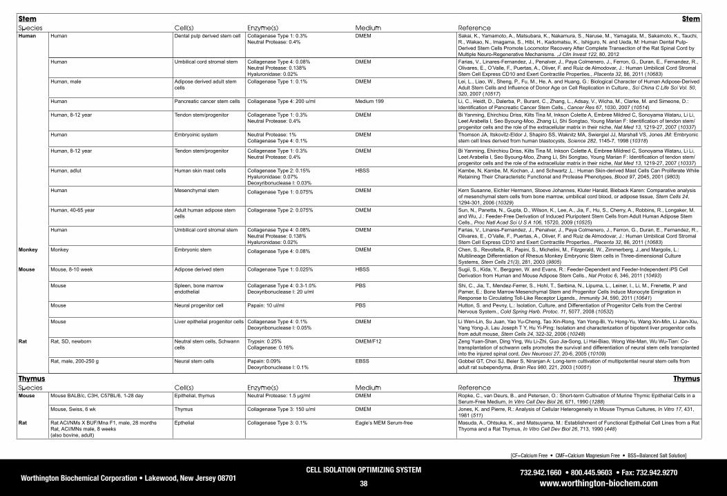
Worthington Biochemical Corporation • Lakewood, New Jersey 08701732.942.1660 • 800.445.9603 • Fax: 732.942.9270
www.worthington-biochem.com
CELL ISOLATION OPTIMIZING SYSTEM
38
[CF=Calcium Free • CMF=Calcium Magnesium Free • BSS=Balanced Salt Solution]
Stem StemSpecies Cell(s) Enzyme(s) Medium ReferenceHuman Human Dental pulp derived stem cell Collagenase Type 1: 0.3%
Neutral Protease: 0.4% DMEM Sakai, K., Yamamoto, A., Matsubara, K., Nakamura, S., Naruse, M., Yamagata, M., Sakamoto, K., Tauchi,
R., Wakao, N., Imagama, S., Hibi, H., Kadomatsu, K., Ishiguro, N. and Ueda, M: Human Dental Pulp-Derived Stem Cells Promote Locomotor Recovery After Complete Transection of the Rat Spinal Cord by Multiple Neuro-Regenerative Mechanisms. ,J Clin Invest 122, 80, 2012
Human Umbilical cord stromal stem Collagenase Type 4: 0.08%Neutral Protease: 0.138%Hyaluronidase: 0.02%
DMEM Farias, V., Linares-Fernandez, J., Penalver, J., Paya Colmenero, J., Ferron, G., Duran, E., Fernandez, R., Olivares, E., O’Valle, F., Puertas, A., Oliver, F. and Ruiz de Almodovar, J.: Human Umbilical Cord Stromal Stem Cell Express CD10 and Exert Contractile Properties., Placenta 32, 86, 2011 (10683)
Human, male Adipose derived adult stem cells
Collagenase Type 1: 0.1% DMEM Lei, L., Liao, W., Sheng, P., Fu, M., He, A. and Huang, G.: Biological Character of Human Adipose-Derived Adult Stem Cells and Influence of Donor Age on Cell Replication in Culture., Sci China C Life Sci Vol. 50, 320, 2007 (10517)
Human Pancreatic cancer stem cells Collagenase Type 4: 200 u/ml Medium 199 Li, C., Heidt, D., Dalerba, P., Burant, C., Zhang, L., Adsay, V., Wicha, M., Clarke, M. and Simeone, D.: Identification of Pancreatic Cancer Stem Cells., Cancer Res 67, 1030, 2007 (10514)
Human, 8-12 year Tendon stem/progenitor Collagenase Type 1: 0.3% Neutral Protease: 0.4%
DMEM Bi Yanming, Ehirchiou Driss, Kilts Tina M, Inkson Colette A, Embree Mildred C, Sonoyama Wataru, Li Li, Leet Arabella I, Seo Byoung-Moo, Zhang Li, Shi Songtao, Young Marian F: Identification of tendon stem/progenitor cells and the role of the extracellular matrix in their niche, Nat Med 13, 1219-27, 2007 (10337)
Human Embryoinic system Neutral Protease: 1% Collagenase Type 4: 0.1%
DMEM Thomson JA, Itskovitz-Eldor J, Shapiro SS, Waknitz MA, Swiergiel JJ, Marshall VS, Jones JM: Embryonic stem cell lines derived from human blastocysts, Science 282, 1145-7, 1998 (10318)
Human, 8-12 year Tendon stem/progenitor Collagenase Type 1: 0.3% Neutral Protease: 0.4%
DMEM Bi Yanming, Ehirchiou Driss, Kilts Tina M, Inkson Colette A, Embree Mildred C, Sonoyama Wataru, Li Li, Leet Arabella I, Seo Byoung-Moo, Zhang Li, Shi Songtao, Young Marian F: Identification of tendon stem/progenitor cells and the role of the extracellular matrix in their niche, Nat Med 13, 1219-27, 2007 (10337)
Human, adlut Human skin mast cells Collagenase Type 2: 0.15% Hyaluronidase: 0.07% Deoxyribonuclease I: 0.03%
HBSS Kambe, N, Kambe, M, Kochan, J, and Schwartz ,L.: Human Skin-derived Mast Cells Can Proliferate While Retaining Their Characteristic Functional and Protease Phenotypes, Blood 97, 2045, 2001 (9803)
Human Mesenchymal stem Collagenase Type 1: 0.075% DMEM Kern Susanne, Eichler Hermann, Stoeve Johannes, Kluter Harald, Bieback Karen: Comparative analysis of mesenchymal stem cells from bone marrow, umbilical cord blood, or adipose tissue, Stem Cells 24, 1294-301, 2006 (10329)
Human, 40-65 year Adult human adipose stem cells
Collagenase Type 2: 0.075% DMEM Sun, N., Panetta, N., Gupta, D., Wilson, K., Lee, A., Jia, F., Hu, S., Cherry, A., Robbins, R., Longaker, M. and Wu, J.: Feeder-Free Derivation of Induced Pluripotent Stem Cells from Adult Human Adipose Stem Cells., Proc Natl Acad Sci U S A 106, 15720, 2009 (10525)
Human Umbilical cord stromal stem Collagenase Type 4: 0.08%Neutral Protease: 0.138%Hyaluronidase: 0.02%
DMEM Farias, V., Linares-Fernandez, J., Penalver, J., Paya Colmenero, J., Ferron, G., Duran, E., Fernandez, R., Olivares, E., O’Valle, F., Puertas, A., Oliver, F. and Ruiz de Almodovar, J.: Human Umbilical Cord Stromal Stem Cell Express CD10 and Exert Contractile Properties., Placenta 32, 86, 2011 (10683)
Monkey Monkey Embryonic stem Collagenase Type 4: 0.08% DMEM Chen, S., Revoltella, R., Papini, S., Michelini, M., Fitzgerald, W., Zimmerberg, J.,and Margolis, L.: Multilineage Differentiation of Rhesus Monkey Embryonic Stem cells in Three-dimensional Culture Systems, Stem Cells 21(3), 281, 2003 (9805)
Mouse Mouse, 8-10 week Adipose derived stem Collagenase Type 1: 0.025% HBSS Sugii, S., Kida, Y., Berggren, W. and Evans, R.: Feeder-Dependent and Feeder-Independent iPS Cell Derivation from Human and Mouse Adipose Stem Cells., Nat Protoc 6, 346, 2011 (10493)
Mouse Spleen, bone marrow endothelial
Collagenase Type 4: 0.3-1.0%Deoxyribonuclease I: 20 u/ml
PBS Shi, C., Jia, T., Mendez-Ferrer, S., Hohl, T., Serbina, N., Lipuma, L., Leiner, I., Li, M., Frenette, P. and Pamer, E.: Bone Marrow Mesenchymal Stem and Progenitor Cells Induce Monocyte Emigration in Response to Circulating Toll-Like Receptor Ligands., Immunity 34, 590, 2011 (10641)
Mouse Neural progenitor cell Papain: 10 ul/ml PBS Hutton, S. and Pevny, L.: Isolation, Culture, and Differentiation of Progenitor Cells from the Central Nervous System., Cold Spring Harb. Protoc. 11, 5077, 2008 (10532)
Mouse Liver epithelial progenitor cells Collagenase Type 4: 0.1% Deoxyribonuclease I: 0.05%
DMEM Li Wen-Lin, Su Juan, Yao Yu-Cheng, Tao Xin-Rong, Yan Yong-Bi, Yu Hong-Yu, Wang Xin-Min, Li Jian-Xiu, Yang Yong-Ji, Lau Joseph T Y, Hu Yi-Ping: Isolation and characterization of bipotent liver progenitor cells from adult mouse, Stem Cells 24, 322-32, 2006 (10248)
Rat Rat, SD, newborn Neutral stem cells, Schwann cells
Trypsin: 0.25% Collagenase: 0.16%
DMEM/F12 Zeng Yuan-Shan, Ding Ying, Wu Li-Zhi, Guo Jia-Song, Li Hai-Biao, Wong Wai-Man, Wu Wu-Tian: Co-transplantation of schwann cells promotes the survival and differentiation of neural stem cells transplanted into the injured spinal cord, Dev Neurosci 27, 20-6, 2005 (10109)
Rat, male, 200-250 g Neural stem cells Papain: 0.09% Deoxyribonuclease I: 0.1%
EBSS Gobbel GT, Choi SJ, Beier S, Niranjan A: Long-term cultivation of multipotential neural stem cells from adult rat subependyma, Brain Res 980, 221, 2003 (10051)
Thymus ThymusSpecies Cell(s) Enzyme(s) Medium ReferenceMouse Mouse BALB/c, C3H, C57BL/6, 1-28 day Epithelial, thymus Neutral Protease: 1.5 µg/ml DMEM Ropke, C., van Deurs, B., and Petersen, O.: Short-term Cultivation of Murine Thymic Epithelial Cells in a
Serum-Free Medium, In Vitro Cell Dev Biol 26, 671, 1990 (1288)Mouse, Swiss, 6 wk Thymus Collagenase Type 3: 150 u/ml DMEM Jones, K. and Pierre, R.: Analysis of Cellular Heterogeneity in Mouse Thymus Cultures, In Vitro 17, 431,
1981 (511)Rat Rat ACI/NMs X BUF/Mna F1, male, 28 months
Rat, ACI/MNs male, 8 weeks (also bovine, adult)
Epthelial Collagenase Type 3: 0.1% Eagle’s MEM Serum-free Masuda, A., Ohtsuka, K., and Matsuyama, M.: Establishment of Functional Epithelial Cell Lines from a Rat Thyoma and a Rat Thymus, In Vitro Cell Dev Biol 26, 713, 1990 (448)

Worthington Biochemical Corporation • Lakewood, New Jersey 08701732.942.1660 • 800.445.9603 • Fax: 732.942.9270
www.worthington-biochem.com
CELL ISOLATION OPTIMIZING SYSTEM
39
[CF=Calcium Free • CMF=Calcium Magnesium Free • BSS=Balanced Salt Solution]
Human Human Epithelial Collagenase: 2.0% DMEM/Ham’s F-12 Emerman, J. and Wilkinson, D.: Routine Culturing of Normal, Dysplastic and Malignant Human Mammary Epithelial Cells from Small Tissue Samples, In Vitro Cell Dev Biol 26, 1186, 1990 (429)
Human Tumor Neutral Protease: 0.24% DMEM/Ham’s F-12 Boyd, J., Rinehart Jr., C., Walton, L., Siegal, G. and Kaufman, D.: Ultrastructural Characterization of Two New Human Endometrial Carcinoma Cell Lines and Normal Human Endometrial Epithelial Cells Cultured on Extracellular Matrix, In Vitro Cell Dev Biol 26, 701, 1990 (446)
Tumor TumorSpecies Cell(s) Enzyme(s) Medium ReferenceHamster Hamster, 6 week old Buccal pouch Neutral Protease: 0.24% CMF HBSS Min, B., Kim, K., Cherrick, H., and Park, N.: Three Cell Lines from Hamster Buccal Pouch Tumors
Induced by Topical 7,12-Dimethylbenz(a)Anthracene, Alone or in Conjunction with Herpes Simplex Virus Inoculation, In Vitro Cell Dev Biol 27A, 128, 1991 (457)
Human Epthelial and tumor Colon
Collagenase: 300 u/ml PBS medium 199 or medium F 12 Friedman, E., Higgins, P., Lipkin, M., Shinya, H., and Gelb, A.: Tissue Culture of Human Epithelial Cells from Benign Colonic Tumors, In Vitro 17, 632, 1981 (514)
Human Tumor, breast Neuraminidase: 0.8 u/ml HBSS Leung, C., and Shiu, R.: Morphological and Proliferative Characteristics of Human Breast Tumor Cells Cultured on Plastic and in Collagen Matrix, In Vitro 18, 476, 1981 (521)
Human Melanoma Metastatic tumors
Collagenase Type 3: 0.10% DMEM Creasey, A., Smith, H., Hackett, A., Fukuyama, K., Epstein, W., and Madin, S.: Biological Properties of Human Melanoma Cells in Culture, In Vitro 15, 342, 1979 (503)
Human Mammary tumors, hard Collagenase: 0.10% RPMI-1640 w/ 5% Fetal Calf Serum Lasfargues, E.: ,Tissue Culture Methods/Applications,Kruse, P., and Patterson, M., Academic Press, 45, 1973 (1293)
Mouse Mouse BALB/cfC3H Mammary tumors Epithelial
Collagenase: 1.0% HBSS Yang, J., Guzman, R., Richards, J., and Nandi, S.: Primary Cultures of Mouse Mammary Tumor Epithelial Cells Embedded in Collagen Gels, In Vitro 16, 502, 1980 (507)
Rat Rat ACI/NMs X BUF/Mna F1, male, 28 months, rat, ACI/MNs male, 8 weeks
Epthelial Collagenase Type 3: 0.1% Eagle’s MEM Serum-free Masuda, A., Ohtsuka, K., and Matsuyama, M.: Establishment of Functional Epithelial Cell Lines from a Rat Thyoma and a Rat Thymus, In Vitro Cell Dev Biol 26, 713, 1990 (448)
Rat Yolk sac tumor Trypsin: 0.01% DMEM Brennan, M., Oldberg, A., Hayman, E., and Ruoslahti, E.: Effect of a Proteoglycan Produced by Rat Tumor Cells on Their Adhesion to Fibronectin-Collagen Substrata, Cancer Res 43, 4302, 1983 (353)
Rat Ascites hepatoma Trypsin: 0.1% Phosphate buffer (see reference) Essner, E.: Experiments on an Ascites Hepatoma. I. Enzymatic Digestion and Alkaline Degradation of the Cementing Substance and Separation of Cells, in Tumor Islands, Exp Cell Res 7, 430, 1954 (403)
Thyroid/Parathyroid Thyroid/ParathyroidSpecies Cell(s) Enzyme(s) Medium ReferenceBovine Bovine, adult Parathyroid Deoxyribonuclease I: 0.005%, HEPES Ham’s F10 Nygren, P., Gylfe, E., Larsson, R., Johansson, H., Juhlin, C., Klareskoq, L., Akerstrom, G., and Rastad,
J.: Modulation of the Ca2+-Sensing Function of Parathyroid Cells In Vitro and in Hyperparathyroidism, Biochim Biophys Acta 968, 253, 1988 (337)
Bovine Parathyroid glands Deoxyribonuclease I: 0.0075% HEPES buffer Wallace, J., and Scarpa, A.: Regulation of Parathyroid Hormone Secretion in Vitro by Divalent Cations, J Biol Chem 257, 10613, 1982 (555)
Bovine Parathyroid Deoxyribonuclease I: 0.004% Eagle’s #2 medium without bicarbonate Brown, E., Hurwitz, S., and Aurbach, G.: Preparation of Viable Isolated Bovine Parathyroid Cells, Endocrinology 99, 1582, 1976 (385)
Bovine (also porcine)
Thyroid Trypsin: 0.004% EBSS Tong, W.: The Isolation and Culture of Thyroid Cells, Meth Enzymol 32, 745, 1974 (636)
Chicken Chick embryo Thyroid Muscle Heart
Collagenase: 0.25% Tyrode’s saline, potassium free Hilfer, S., and Brown, J.: Collagenase. Its Effectiveness as a Dispersing Agent for Embryonic Chick Thyroid and Heart, Exp Cell Res 65, 246, 1971 (401)
Chicken, Rhode Island Red, embryo Thyroid follicular Collagenase: 0.2% Tyrode’s solution, CMF Spooner, B.: The Expression of Differentiation by Chick Embryo Throid in Cell Culture. I. Functional and Fine Structural Stability in Mass and Clonal Culture, J Cell Physiol 75, 33, 1970 (682)
Ovine Sheep Thyroid Collagenase: 0.2% Puck’s Saline F Kerkof, P.: Preparation of Primary Cultures of Ovine Thyroid Gland Cells, J Tiss Cul Meth 7, 23, 1982 (1289)
Porcine Porcine (also bovine)
Thyroid Trypsin: 0.004% EBSS Tong, W.: The Isolation and Culture of Thyroid Cells, Meth Enzymol 32, 745, 1974 (636)
Tonsil TonsilSpecies Cell(s) Enzyme(s) Medium ReferenceHuman Human Tonsillar mononuclear cells Collagenase Type 1: 210 u/ml
Deoxyribonuclease I: 90 u/ml RPMI Grammer AC, McFarland RD, Heaney J, Darnell BF, Lipsky PE: Expression, regulation, and function of B
cell-expressed CD154 in germinal centers, J Immunol 163, 4150-9, 1999 (10230)

Worthington Biochemical Corporation • Lakewood, New Jersey 08701732.942.1660 • 800.445.9603 • Fax: 732.942.9270
www.worthington-biochem.com
CELL ISOLATION OPTIMIZING SYSTEM
40
Worthington Collagenase Sampling Program
The lot-to-lot variation which is typical of partially purified enzyme preparations such as Worthington collagenase Types 1-4 and Animal Origin Free CLSAF types makes it important to pre-test a particular lot of enzyme you are planning to use in your experiment. Many years ago we found that the most practical approach for the researcher is to presample several different lots of collagenase at a time and select the best of the group. As the world’s leading manufacturer of collagenase, Worthington is able to offer the greatest number of different lots at any given time and recommend specific lots for an application.
There is no charge for participating in the collagenase sampling program. Under the program, individual researchers are provided with 100 mg samples of up to three different lots of collagenase for evaluation in their own assay systems. A period of 60 days is allowed for your evaluation of these samples. A minimum of 3 grams of each lot will be placed on HOLD, reserved in your name. When you determine which lot performs best for you, simply specify the lot desired when ordering. The only requirement, once a suitable lot of collagenase is found, is that you purchase a minimum of 3 grams of the material.
To become part of this program, or to discuss any of the Worthington products, just call our Technical Service group toll-free at 800.445.9603 from anywhere in the United States or Canada or e-mail [email protected]
International customers should check our International Distributor listing for a distributor. If you do not have a Worthington Distributor for your country, please contact International Sales or Technical Service.
Related Worthington Products
The enzymes included in this kit are all samples of standard Worthington products; however, some of the package sizes used in the kit are non-standard. The following pages list the standard package sizes for each of the products along with brief descriptions and catalog numbers. Worthington also offers multiple grades of most of these same enzymes. Refer to the Catalog and Price List for a complete listing. To obtain a copy call Customer Service at:
800.445.9603.Please note that bulk purchases are also possible with each of the products. If your experiments require substantial quantities of one or more of these enzymes, we recommend that you contact our Customer Service department to discuss bulk pricing.
Ordering information along with our discount structure appears inside the back cover of this guide. If you have any questions concerning our products, please do not hesitate to call Technical Service toll-free at 800.445.9603 from anywhere in the United States or Canada or at 732.942.1660 from elsewhere. Twenty-four hour FAX service is available at 732.942.9270

Worthington Biochemical Corporation • Lakewood, New Jersey 08701732.942.1660 • 800.445.9603 • Fax: 732.942.9270
www.worthington-biochem.com
CELL ISOLATION OPTIMIZING SYSTEM
41
Cell Isolation Product List
Cell Isolation Optimizing System CIT
A complete method development kit containing an assortment of enzymes most frequently used in tissue dissociation and cell isolation procedures. Includes instructions, references and strategies for the handling, use and optimization of enzymatic cell isolation methods to achieve maximum yield of viable cells. Kit includes 500 mg of each of four types of collagenase, 500 mg trypsin, 50 ku hyaluronidase, 100 mg elastase, 100 mg papain, 25 mg DNase I, 10 mg neutral protease (Dispase) and 100 mg trypsin inhibitor. Store at 2-8°C.
LK003200 1 bx
Chymotrypsin, Alpha, 1X CDAG1X crystallized as zymogen and activated. Dialyzed against 1mM HCl and lyophilized. Store at 2-8°C.
≥35 units per mg protein
LS001333 LS001334 LS001332
1 gm 10 gm
Bulk Chymotrypsin, Alpha, 3X CDI3X crystallized alpha chymotrypsin which is an activation product of a 3X crystallized zymogen. Dialyzed against 1mM HCl and lyophilized. Store at 2-8°C.
≥45 units per mg protein
LS001448 LS001450 LS001451 LS001453
250 mg 1 gm
10 gm Bulk
Chymotrypsin, Alpha, Purified CDSChromatographically prepared by the procedure of Yapel, et al., J. Amer. Chem. Soc., 88, 2573 (1966). A lyophilized powder. Store at 2-8°C.
≥45 units per mg protein
LS001475 LS001479 LS001477
100 mg 1 gm Bulk
Chymotrypsin, Alpha, TLCK Treated CDTLCK3X crystallized and treated with 1-chloro-3-tosylamido-7-amino-2-heptanone (TLCK) to inhibit trypsin activity [Shaw, et al., Biochemistry, 4, 2219 (1965)]. Dialyzed against 1 mM HCl to remove autolysis products and low molecular weight contaminants. Supplied as a dialyzed, lyophilized powder. Store at 2-8°C.
≥45 units per mg protein
LS001430 LS001432 LS001434 LS001438
25 mg 100 mg
1 gm Bulk
Collagen CLType I collagen prepared by the method of Einbinder and Schubert, J. Biol. Chem., 188, 335 (1951). Supplied as a shredded, lyophilized, insoluble preparation. Store at 2-8°C.
LS001654 LS001652 LS001656 LS001658
1 gm 5 gm
10 gm Bulk
Collagen, Soluble CLCSType I collagen supplied as a 6mg/ml liquid preparation in 75mM sodiuim citrate, pH 3.6 - 4.0, containing 0.01% merthiolate as a preservative. Store at 2-8°C REQUIRES SPECIAL SHIPPING: ICE PACK. Note: Contains thimerisol as a preservative; proper disposal required.
Less than 20 minutes gel time
LS001663 Bulk
Product Catalog # Size Code

Worthington Biochemical Corporation • Lakewood, New Jersey 08701732.942.1660 • 800.445.9603 • Fax: 732.942.9270
www.worthington-biochem.com
CELL ISOLATION OPTIMIZING SYSTEM
42
Cell Isolation Product List
Collagenase, Type 1, Filtered CLSS-1
Collagenase, Type 1 (Code: CLS-1), which is filtered through a 0.22 micron membrane and lyophilized in vials to contain > 50 milligrams or 1 gram per vial. Store at 2-8°C.
≥125 units per mg dry weight
LS004214 LS004216 LS004217
50 mg 5x50 mg
1 gm
Collagenase, Animal Origin-Free CLSAFA
Collagenase derived from cultures grown in animal-free medium. Suitable for applications needing to avoid introduction of anumal derived pathogens into bioprocessing procedures.Store at 2-8°C.
≥150 units per mg dry weight
LS004152 LS004154 LS004156 LS004158
100 mg 1 gm 5 gm Bulk
Collagenase, Type 1 CLS-1
The original balance of enzymatic activities. Each lot assayed for collagenase, caseinase, clostripain and tryptic activities. Suggested for epithelial, liver, lung and adrenal primary cell isolations. A dialyzed, lyophilized powder. Store at 2-8°C.
≥125 units per mg dry weight
LS004194 LS004196 LS004197 LS004200
100 mg 1 gm 5 gm Bulk
Collagenase, Type 2 CLS-2
Prepared to contain higher clostripain activity. Suggested for bone, heart, liver, thyroid and salivary primary cell isolation. Supplied as a dialyzed, lyophilized powder. Store at 2-8°C.
≥125 units per mg dry weight
LS004174 LS004176 LS004177 LS004179
100 mg 1 gm 5 gm Bulk
Collagenase, Type 3 CLS-3
Lower in secondary proteolytic contaminant activities but wIth typical collagenase activity. Suggested for mammary primary cell isolation. A dialyzed, lyophilized powder. Store at 2-8°C.
≥100 units per mg dry weight
LS004180 LS004182 LS004183 LS004185
100 mg 1 gm 5 gm Bulk
Collagenase, Type 4 CLS-4
Prepared to contain lower tryptic activity levels to limit damage to membrane proteins and receptors but with normal to above normal collagenase activity. Suggested for pancreatic islet primary isolation. A dialyzed, lyophilized powder. Store at 2-8°C.
≥160 units per mg dry weight
LS004186 LS004188 LS004189 LS004191
100 mg 1 gm 5 gm Bulk
Collagenase, Purified CLSPA
Chromatographically purified. ≤50 caseinase units per milligram. Supplied as a lyophilized powder. Store at 2-8°C.
≥500 units per mg dry weight
LS005275 LS005273 LS005277
4 ku 10 ku Bulk
CLS-1
Product Catalog # Size Code

Worthington Biochemical Corporation • Lakewood, New Jersey 08701732.942.1660 • 800.445.9603 • Fax: 732.942.9270
www.worthington-biochem.com
CELL ISOLATION OPTIMIZING SYSTEM
43
Cell Isolation Product List
Collagenase, Type 3, Filtered CLSS-3
Collagenase, Type 3 (Code: CLS-3), which is filtered through a 0.22 micron membrane and lyophilized in vials to contain ≥ 50 milligrams per vial. Store at 2-8°C.
≥100 units per mg dry weight
LS004206 LS004208
50 mg 5x50 mg
Collagenase, Type 4, Filtered CLSS-4
Collagenase, Type 4 (Code: CLS-4), which is filtered through a 0.22 micron membrane and lyophilized in vials to contain > 50 milligrams or 1 gram per vial. Store at 2-8°C.
≥160 units per mg dry weight
LS004210 LS004212 LS004209
50 mg 5x50 mg
1 gmDeoxyribonuclease I, Ribonuclease & Protease Free, Solution DPRFS
Molecular Biology Grade. Chromatographically purified to remove RNase and protease. Supplied as a solution at approximately 2 Kunitz units per microliter (approximately 1 mg/ml) containing 50% glycerol and 1mM calcium chloride. Store at 2-8°C or -20°C.
≥2,000 Kunitz units per ml
LS006342 LS006344 LS006348
100 un 500 un
Bulk
Deoxyribonuclease I, Ribonuclease & Protease Free DPRF
Molecular Biology Grade. Chromatographically purified to remove RNase and protease. Lyophilized in vials. Each 10,000 unit vial contains 2 mg glycine, 2µmoles calcium, and ≥10,000 units of DNase I. Each 2,500 unit vial contains 0.5mg glycine, 0.5µmoles calcium, and ≥2,500 units of DNase I. Dissolving the entire 10,000 unit vial in 5 ml, or the entire 2,500 unit vial in 1.25 ml, provides the equivalent of a 1 mg/ml solution. (ku = 1000un) Store at 2-8°C. PROTECT FROM MOISTURE
≥2,000 units per mg dry weight
LS006331 LS006333 LS006334
2500 un 10 ku Bulk
Deoxyribonuclease I DPFF
Chromatographically purified. A lyophilized powder containing glycine as a stabilizer. Contains ≤ 0.0005% RNase. Store at 2-8°C. PROTECT FROM MOISTURE. RNase & Protease Free
≥2,000 Kunitz units per mg
dry weight
LS006330 LS006328 LS006332
25 ku 125 ku
Bulk
Deoxyribonuclease I D
Chromatographically purified. A lyophilized powder with glycine as a stabilizer. Store at 2-8°C. PROTECT FROM MOISTURE
≥2,000 Kunitz units per mg
dry weight
LS002004 LS002006 LS002007 LS002009
5 mg 20 mg
100 mg Bulk
Collagenase, Type 2, Filtered CLSS-2
Collagenase, Type 2 (Code: CLS-2), which is filtered through a 0.22 micron membrane and lyophilized in vials to contain > 50 milligrams or 1 gram per vial. Store at 2-8°C.
≥125 units per mg dry weight
LS004202 LS004204 LS004205
50 mg 5x50 mg
1 gm
Product Catalog # Size Code

Worthington Biochemical Corporation • Lakewood, New Jersey 08701732.942.1660 • 800.445.9603 • Fax: 732.942.9270
www.worthington-biochem.com
CELL ISOLATION OPTIMIZING SYSTEM
44
Cell Isolation Product List
Deoxyribonuclease I, Filtered DCLS
Filtered through a 0.22 micron membrane and lyophilized in vials. Material is not tested for pyrogenicity. Store at 2-8°C. PROTECT FROM MOISTURE
≥2,000 units per mg dry weight
LS002058 LS002060
11 mg 25 mg
Deoxyribonuclease I, Standard Vial DSV
Lyophilized in vials for assay standardization. Labeled to show established activity. Not suitable for assays at neutral pH. Store at 2-8°C. PROTECT FROM MOISTURE
~2,000 Kunitz units per vial
LS002173 LS002172
2 ku 5x2 ku
Deoxyribonuclease I DPPartially purified. A lyophilized powder. Store at 2-8°C. PROTECT FROM MOISTURE
≥2,000 Kunitz units per mg dry weight
LS002138 LS002139 LS002140 LS002141
25 mg 100 mg
1 gm Bulk
Deoxyribonuclease I DPBPartially purified. Supplied as lyophilized powder. Store at 2-8°C. PROTECT FROM MOISTURE
≥1,250 Kunitz units per mg dry weight
LS002145 LS002147 LS002149
100 mg 1 gm Bulk
Deoxyribonuclease I, Recombinant DR1Recombinant protein produced in Pichia pastoris. Free of animal derived components, RNases and proteases. Chromatographically purified. A lyophilized powder containing glycine as a stabilizer. Store at 2-8°C. PROTECT FROM MOISTURE
≥5,000 units per mg protein
LS006361 LS006362 LS006360
10 ku 50 ku Bulk
Deoxyribonuclease I, Recombinant Solution DR1SRecombinant protein produced in Pichia pastoris. Free of animal derived components, RNases and proteases. Chromatographically purified. A liquid preparation in 5mM Calcium Acetate, 4mg/ml glycine, pH 5.0 and 50% glycerol. Supplied with 10x reaction buffer. Store at -20°C. REQUIRES SPECIAL SHIPPING: ICE PACK
≥2 units per microliter LS006353 LS006355 LS006357
2 ku 5 x 2 ku
Bulk
Product Catalog # Size Code

Worthington Biochemical Corporation • Lakewood, New Jersey 08701732.942.1660 • 800.445.9603 • Fax: 732.942.9270
www.worthington-biochem.com
CELL ISOLATION OPTIMIZING SYSTEM
45
Cell Isolation Product List
Elastase, Suspension ES2X crystallized. Supplied as an aqueous suspension. This preparation must be diluted to dissolve the enzyme. The diluted enzyme should be 0.22 micron filtered before use. Suitable for the isolation of Type II lung cells. Store at 2-8°C. DO NOT FREEZE REQUIRES SPECIAL SHIPPING: ICE PACK.
≥3 units per mg protein
LS002274 LS002279 LS002280 LS002276
25 mg 100 mg
1 gm Bulk
Elastase, Purified ESFFChromatographically purified. A lyophilized powder. Store at 2-8°C. REQUIRES SPECIAL SHIPPING: ICE PACK
≥8 units per mg protein
LS006363 LS006365 LS006367
5 mg 20 mg
Bulk
Elastase, Lyophilized ESL2X Crystallized, (Code: ESL), supplied as a dialyzed, lyophilized powder. The enzyme should be 0.22 micron filtered after reconstitution and prior to use. Suitable for the isolation of Type II lung cells. Store at 2-8°C.
≥3 units per mg protein
LS002290 LS002292 LS002294 LS002298
25 mg 100 mg
1 gm Bulk
Hepatocyte Isolation System HISThe package contains sufficient materials for five separate adult rat liver perfusions including five single use CLSH enzyme vials, Five single use DNase vials, 10X CMF-Hank’s Balanced Salt Solution, L-15 Media Powder, 0.15M MOPS buffer. 7.5% Sodium Bicarbonate Solution and optimized protocol. Store at 2-8°C.
LK002060 bx
HIS-Collagenase/Elastase CLSHWorthington collagenase (Code: CLS-1) and elastase (Code: ESL), filtered through 0.22µm pore size membrane, and lyophilized. Before use, reconstitute with the L-15/MOPS solution and swirl gently to dissolve contents as directed in the following procedure. Store unreconstituted vials at 2–8°C.
LK002066 LK002067
1 VI 5 VI
HIS Kit, DNase Vial D2A component of the Hepatocyte Isolation kit containing 1,000 Units DNase I each, 5 VialsWorthington DNase I (Code: D), filtered through 0.22µm pore size membrane, and lyophilized. Before use, reconstitute with L-15/MOPS solution and swirl gently to dissolve contents as directed in the procedure. Store unreconstituted vials at 2–8°C.
≥1,000 units per vial
LK003170 LK003172
1 vi 5 vi
Hank’s Balanced Salt Solution (HBSS-CMF) 10X Solution HBSS1010X CMF-HBSS Concentrate, 1 bottle, 500mlSterile calcium- and magnesium-free Hank’s Balanced Salt Solution (CMF-HBSS). The solution is used for washing and perfusing the liver prior to the addition of the dissociating enzyme solution.
LK002064 1 ea
Product Catalog # Size Code

Worthington Biochemical Corporation • Lakewood, New Jersey 08701732.942.1660 • 800.445.9603 • Fax: 732.942.9270
www.worthington-biochem.com
CELL ISOLATION OPTIMIZING SYSTEM
46
Cell Isolation Product List
L-15 Media Powder L15NKLeibovitz L-15 media powder, a component of the HIS kit. Reconstitute entire contents of pouch, QS to 1 liter with cell culture grade water, and 0.22 micron filter. Suitable for cell isolation and culture applications. Store at 2-8°C.
LK003250 1 ea
0.15M, MOPS Buffer, HIS MOPS0.15M MOPS, pH 7.5, 0.22u filtered. Buffer concentrate used to buffer the constituted Leibovitz L-15 media in Hepatocyte Isolation System. Store at 2-8°C.
LK002070 1 ea
Sodium Bicarbonate 7.5% NAH7.5% Sodium Bicarbonate (NaHCO3),1 bottle, 100ml 7.5% Sodium bicarbonate concentrate, used to buffer the diluted CMF-HBSS. Store at 2-8°C.
LK002069 1 ea
Hyaluronidase HSEA partially purified, dialyzed, lyophilized powder. Store at -20°C.
≥300 USP/NF units per mg dry weight
LS002594 LS002592 LS002591
50 ku 300 ku
Bulk Hyaluronidase, Purified HSEPChromatographically purified. A dialyzed, lyophilized powder. Store at -20°C.
≥3,000 USP/NF units per mg dry weight
LS005477 LS005475 LS005474 LS005479
5 ku 15 ku 30 ku Bulk
Lysozyme LYA lyophilized powder containing sodium chloride and acetate. Store at 2-8°C.
≥5,000 units per mg dry weight
LS002880 LS002881 LS002883
1 gm 10 gm
Bulk Lysozyme, Purified, Salt Free LYSFA dialyzed, lyophilized powder. Store at 2-8°C.
≥8,000 units per mg dry weight
LS002931 LS002933 LS002934
1 gm 5 gm Bulk
Neonatal Cardiomyocyte Isolation System NCISKit for performing five separate tissue dissociations each containing up to twelve hearts. Contains single use vials of purified collagenase and trypsin, CMF-HBSS, Leibovitz L-15 media and Falcon cell strainers along with a detailed protocol. The kit is use-tested by Worthington to assure performance. Store at 2-8°C.
LK003300 LK003303
1 ki 3 ki
Product Catalog # Size Code

Worthington Biochemical Corporation • Lakewood, New Jersey 08701732.942.1660 • 800.445.9603 • Fax: 732.942.9270
www.worthington-biochem.com
CELL ISOLATION OPTIMIZING SYSTEM
47
Cell Isolation Product List
Collagenase Vial, NCIS CLSPANKA component of the NCIS kit. This material is 0.22 micron membrane filtered and lyophilized in autoclaved vials. A vial reconstituted with 5 ml of HBSS or equivalent yields a solution of 300 units/ml of collagenase, Code: CLSPA. Suitable for cell isolation and culture applications. Store at 2-8°C.
≥500 units per mg dry weight
LK003240 LK003245
1 vi 5 vi
Trypsin Vial, NCIS TRLSNKA component of the NCIS kit. This material is 0.22 micron membrane filtered and lyophilized in autoclaved vials. A vial reconstituted with 2 ml of HBSS yields a solution of 500µg/ml of trypsin, Code: TRLS. Suitable for cell isolation and culture applications. Store at 2-8°C.
≥180 units per vial
LK003220 LK003225
1 vi 5 vi
Inhibitor Vial, NCIS SICNKA component of the NCIS kit. This material is 0.22 micron membrane filtered and lyophilized in autoclaved vials. A vial reconstituted with 1 ml of HBSS or equivalent yields a solution of 2 mg/ml of trypsin inhibitor, Code: SIC. Suitable for cell isolation and culture applications. Store at 2-8°C.
1 mg inhibits at least 0.75 mg
trypsin, Code: TRL
LK003230 LK003235
1 vi 5 vi
HBSS Solution HBSSSterile calcium and magnesium free Hank’s balanced salt solution (CMFHBSS), pH 7.4, as supplied in the NCIS kit; 1 x 500 ml. Store at 2-8°C.
LK003210 1 ea
L-15 Media Powder L15NKLeibovitz L-15 media powder, a component of the NCIS kit. Reconstitute entire contents of pouch, QS to 1 liter with cell culture grade water, and 0.22 micron filter. Suitable for cell isolation and culture applications. Store at 2-8°C.
LK003250 1 ea
Cell Strainers (Falcon) CELSTRNKCell strainers (Falcon), components of the NCIS kit. Suitable for removal of tissue debris in cell isolation applications. Store at room temperature.
LK003265 5 ea
Neutral Protease (Dispase®), Purified NPROChromatographically purified. A lyophilized powder. Store at 2-8°C.
≥4 units per mg dry weight
LS02100 LS02104 LS02108
10 mg 50 mg
Bulk Neutral Protease, Partially Purified NPRO2Partially purified. A lyophilized powder. Store at 2-8°C.
≥0.1 units per mg dry weight
LS02109 LS02111 LS02112
1 gm 5 gm Bulk
Product Catalog # Size Code

Worthington Biochemical Corporation • Lakewood, New Jersey 08701732.942.1660 • 800.445.9603 • Fax: 732.942.9270
www.worthington-biochem.com
CELL ISOLATION OPTIMIZING SYSTEM
48
Cell Isolation Product List
Ovalbumin OAMajor protein of egg white, with a molecular weight of 43,000. A lyophilized powder. Store at 2-8°C.
LS003049 LS003048 LS003050
1 gm 5 gm Bulk
Ovalbumin, Purified OACChromatographically purified. Major protein of egg white, with a molecular weight of 43,000. A dialyzed, lyophilized powder. Store at 2-8°C.
LS003056 LS003054 LS003052
100 mg 1 gm Bulk
Papain, Suspension PAPSupplied as a 2X crystalline suspension in 50mM sodium acetate, pH 4.5. To insure full activity, the enzyme should be incubated in a solution containing 1.1mM EDTA, 0.067mM mercaptoethanol and 5.5mM cysteine-HCl for 30 minutes. It is recommended that the enzyme be 0.22 micron filtered after dissolution and prior to use. Store at 2-8°C. REQUIRES SPECIAL SHIPPING: ICE PACK
Activates to at least 20 units per mg
protein
LS003124 LS003126 LS003127 LS003128
25 mg 100 mg
1 gm Bulk
Papain, Lyophilized PAPLSupplied as a lyophilized powder prepared from a 2X crystalline suspension, Code: PAP. To insure full activity, the enzyme should be incubated in a solution containing 1.1mM EDTA, 0.067mM mercaptoethanol and 5.5mM cysteine-HCl for 30 minutes. It is recommended that the enzyme be 0.22 micron filtered after dissolution and prior to use. Store at 2-8°C.
Activates to at least 15 units per mg
protein
LS003118 LS003119 LS003120 LS003122
25 mg 100 mg
1 gm Bulk
PDS Kit, Papain Vial PAP2A component of the Papain Dissociation System, for use in the tissue dissociation method of Huettner, J.E., and Baughman, R.W.: J. Neuroscience, 6, 3044 (1986). Contains papain, L-cysteine, and EDTA. This material is 0.22 micron membrane filtered and lyophilized in autoclaved vials. A vial reconstituted with 5 ml of EBSS or equivalent yields a solution at 20 units of papain per ml in 1mM L-cysteine with 0.5mM EDTA. Store at 2-8°C.
≥100 unitsper vial
LK003176 LK003178
1 vi 5 vi
Papain Dissociation System PDSSet of five single use vials of papain and five single use vials of DNase, 100 ml of Earle’s balanced salt solution (EBSS), and an inhibitor vial for use in the tissue dissociation method of Huettner, J.E., and Baughman, R.W.: J. Neuroscience, 6, 3044 (1986). Use tested by Worthington using new-born rat pup spinal cord. The package contains sufficient materials for dissociation of five separate tissue aliquots of up to 0.3-0.4 cm3 each. Store at 2-8°C.
LK003150 LK003153
1 bx 3 bx
Product Catalog # Size Code

Worthington Biochemical Corporation • Lakewood, New Jersey 08701732.942.1660 • 800.445.9603 • Fax: 732.942.9270
www.worthington-biochem.com
CELL ISOLATION OPTIMIZING SYSTEM
49
Cell Isolation Product List
Papain Dissociation System, Without EBSS PDS2Complete kit as described for product Code: PDS, but without the Earle’s Balanced Salt Solution (EBSS). Store at 2-8°C.
LK003160 LK003163
1 bx 3 bx
PDS Kit, DNase Vial D2A component of the Papain Dissociation System. This material is 0.22 micron membrane filtered and lyophilized in autoclaved vials. A vial reconstituted with 0.5 ml of EBSS or equivalent yields a solution of 2000 units/ml of deoxyribonuclease (1 mg/ml). Store at 2-8°C.
≥1,000 units per vial LK003170 LK003172
1 vi 5 vi
PDS Kit, Inhibitor Vial OI-BSAOvomucoid protease inhibitor and bovine serum albumin which is 0.22 micron filtered and lyophilized in autoclaved vials to contain 10 mg/ml each upon reconstitution with 32 ml of EBSS. Store at 2-8°C.
≥300 mg TRL inhibited per vial
LK003182 1 vi
PDS Kit, EBSS Vial EBSSEarle’s balanced salt solution (EBSS) as supplied in the Papain Dissociation System. Store at 2-8°C.
LK003188 1 vi
Trypsin 2X Lyo TRLSupplied as a chromatographically purified, diafiltered and lyophilized powder. Store at 2-8°C. PROTECT FROM MOISTURE
≥180 units per mg protein
LS003702 LS003703 LS003704 LS003706
100 mg 1 gm Bulk Bulk
Trypsin TRL3Supplied as a chromatographically purified, diafiltered and lyophilized powder. Store at 2-8°C. PROTECT FROM MOISTURE
≥180 units per mg protein
LS003708 LS003707 LS003709
100 mg 1 gm Bulk
Proteinase K PROKA lyophilized powder. Purified to remove DNase and RNase. Store at 2-8°C.
≥20 units per mg dry weight
LS004220 LS004222 LS004224 LS004226
25 mg 100 mg
1 gm Bulk
Proteinase K, Solution, 20mg/ml PROKSA concentrated, ready to use liquid formulation. Proteinase K prepared at 20 mg/ml in 10 mM Tris-HCI, 1 mM calcium acetate,pH 7.5 containing 50% glycerol. DNase and RNase free.Store at -20°C
≥400 units per milliliter
LS004240 LS004242 LS004244
5 ml 25 ml Bulk
Product Catalog # Size Code

Worthington Biochemical Corporation • Lakewood, New Jersey 08701732.942.1660 • 800.445.9603 • Fax: 732.942.9270
www.worthington-biochem.com
CELL ISOLATION OPTIMIZING SYSTEM
50
Cell Isolation Product List
Trypsin, 2X, Sterile, Irradiated TRLVMF2X crystallized (Code: TRL), lyophilized, irradiated and tested for the absence of mycoplasma and extraneous virus according to 9 CFR113.53c. Each vial is filled to contain ≥100 mg. Store at 2-8°C.
≥180 units per mg protein (at least
10,350 BAEE/3450 USP/NF units per
mg protein)
LS004454 LS004452 LS004458
100 mg 5x100 mg
Bulk
Trypsin, Purified, Sequencing Grade II TRSEQIIBovine trypsin that has been treated with L-(tosylamido-2-phenyl) ethyl chloromethyl ketone (TPCK) to inhibit contaminating chymotryptic activity and extensively purified to remove autolysis products. Supplied as a lyophilized powder. Store at -20°C. PROTECT FROM MOISTURE. REQUIRES SPECIAL SHIPPING: ICE PACK
≥150 units per mg protein (at least
8,625 BAEE/2875 USP/NF units per
mg protein)
LS02115 LS02117 LS02118 LS02119
4x25 µg 4x100 µg
Bulk 1 mg
Trypsin, Modified, Sequencing Grade TRSEQZTrypsin, treated with L-(tosylamido-2-phenyl) ethyl chloromethyl ketone to inhibit contaminating chymotryptic activity, chemically modified to promote stability and further purified to remove autolysis fragments, resulting in a highly stable trypsin product resistant to autolysis while retaining specificity. Store at -20°C PROTECT FROM MOISTURE. REQUIRES SPECIAL SHIPPING: ICE PACK.
≥150 units per mg protein (at least
8,625 BAEE/2875 USP/NF units per
mg protein)
LS02120 LS02122 LS02124
4x25 µg 4x100 µg
Bulk
Trypsin, 0.22µ Filtered TRLSTrypsin chromatographically purified, diafiltered, (Code TRL3) filtered thorugh a 0.22 micron pore size membrane and lyophilized in sterile vials. This product is not tested for pyrogenicity. Store at 2-8°C.
≥180 units per mg protein (at least
10,350 BAEE/3,450 USP/NF u/mgP)
LS003736 LS003734 LS003738
50 mg 5x50 mg
Bulk
Trypsin, TPCK Treated TRTPCKA chromatographically purified, diafiltered, lyophilized powder that has been treated with L-(tosylamido-2-phenyl) ethyl chloromethyl ketone (TPCK) to inhibit contaminating chymotryptic activity [Kostka, V., and Carpenter, F.: JBC, 239, 1799 (1964)]. Store at 2-8°C. PROTECT FROM MOISTURE
≥180 units per mg protein (at least
10,350 BAEE/3,450 USP/NF u/mgP)
LS003740 LS003741 LS003744 LS003742
100 mg 500 mg
1 gm Bulk
Product Catalog # Size Code

Worthington Biochemical Corporation • Lakewood, New Jersey 08701732.942.1660 • 800.445.9603 • Fax: 732.942.9270
www.worthington-biochem.com
CELL ISOLATION OPTIMIZING SYSTEM
51
Cell Isolation Product List
Trypsin, TPCK-Treated, Irradiated TRTVMFChromatographically purified trypsin treated with L-(tosylamido-2-phenyl) ethyl chloromethyl ketone (TPCK) to inhibit contaminating chymotryptic activity according to Kostka, V., and Carpenter, F.H.: JBC, 239, 1799 (1964), Code: TRTPCK, lyophilized, irradiated and tested for the absence of mycoplasma and extraneous virus according to 9 CFR 113.53c. Each vial is filled to contain ≥100 mg. Store at 2-8°C.
≥180 units per mg protein
LS003750 LS003752
100 mg 5x100 mg
Trypsin Inhibitor, Lima Bean LBIFraction III of the preparation described by Fraenkel-Conrat, et al., Arch. Biochem. Biophys., 37, 393 (1952). Supplied as a dialyzed, lyophilized powder. Store at 2-8°C.
1mg inhibits at least 2.2 mg
trypsin, Code TRL
LS002829 LS002830 LS002831
100 mg 1 gm Bulk
Trypsin Inhbitor, Ovomucoid OIMucoprotein and antitryptic factor of egg white described by Lineweaver and Murray, J. Biol. Chem., 171, 565 (1947). A dialyzed, dried powder. Store at 2-8°C.
1 mg inhibits at least 1.2 mg
trypsin, Code: TRL
LS003085 LS003087 LS003086 LS003089
500 mg 1 gm 2 gm Bulk
Trypsin Inhibitor, Soybean, Purified SIChromatographically purified as described by Frattali, V., and Steiner, R.: Biochem., 7, 521 (1968). A dialyzed, lyophilized powder. Purity checked using SDS PAGE. Store at 2-8°C.
1 mg inhibits at least 1.2 mg
trypsin, Code: TRL
LS003570 LS003571 LS003573
100 mg 1 gm Bulk
Trypsin Inhibitor, Soybean SICA partially purified acetone powder. Store at 2-8°C.
1 mg inhibits at least 0.75 mg
trypsin, Code: TRL
LS003587 LS003590
1 gm Bulk
Product Catalog # Size Code

Worthington Biochemical Corporation • Lakewood, New Jersey 08701732.942.1660 • 800.445.9603 • Fax: 732.942.9270
www.worthington-biochem.com
CELL ISOLATION OPTIMIZING SYSTEM
52
Write: Worthington Biochemical Corporation 730 Vassar Avenue Lakewood, New Jersey 08701
Call: 1.800.445.9603 (U.S. and Canada) 1.732.942.1660
Fax: 1.732.942.9270 1.800.368.3108
E-mail: [email protected]
Website/Online Ordering: www.worthington-biochem.com www.tissuedissociation.com
For standard catalog items we require:• Your name and phone number• The ship-to address• The bill-to address (or customer number)• Your purchase order number• The Worthington catalog number• The quantity you wish to order
We accept American Express, MasterCard and VISA
Shipment and Terms of SaleNet 30 days. Prices are F.O.B. Lakewood, New Jersey. Prices are subject to change without notice. Past due accounts may be charged a 1.5% per month late payment fee. Shipment is usually made on the same day that the order is received. A packing charge is added for those products requiring special ice pack or dry ice shipping.
Product ReturnsAuthorization for any product returns must be obtained from Worthington Biochemical Corporation (Customer Service Department), or its authorized representative, prior to the return of the product. This authorization is required to insure the proper return of material and, if applicable, the correct issuance of credit. There is no provision for credit of outdated material. Product must be returned in the same condition as received and within 30 days of the original shipment by Worthington Biochemical Corporation. A restocking fee may be charged for all returns.
Ordering Information
Quantity Discounts Quantity Price 1 to 4 ...................... List Price 5 to 9 ...................... 5% off list 10 to 19 .................. 10% off list 20 or more .............. 15% off list Bulk ......................... Inquire
Contact Customer Service to ask about special rates for bulk purchases and standing orders.
Bulk/OEM Enzyme PurchasingWorthington products can be supplied in a wide range of purity and activity specifications. Custom analysis and special package sizes can also be provided. Contact your representative or the Bulk Sales Office to discuss your specific requirements.
Standing Orders and DiscountsFor orders of greater than 25 packages or orders of material packed in bulk, contact your representative or the Bulk Sales Office for special pricing consideration. Standing orders may also qualify for discounts. We welcome long-term use projections upon which we can consider special rates. Large institutional buyers should contact us regarding special purchasing agreements.
Technical AssistanceAvailable 8:00 AM to 5:00 PM Eastern Time Monday through Friday to process orders and provide customer service. We can be contacted 24 hours a day by FAX or e-mail.
Worthington produces most of the products it sells and welcomes your questions and suggestions. Because we are a primary manufacturer, we have ready access to all production and quality control records of our products by lot number.
Our years of experience in enzyme purification put us in a position to assist individual researchers with special needs. We frequently do customized preparations of entirely new products, and we can make modifications of a regular production procedure on a custom basis. Furthermore, our quality control department can do special testing if needed.
Product UseAll Worthington products are sold for manufacturing, research, and laboratory use only. Researchers and clinical laboratory personnel intending to use any of these products for medical investigation on humans are soley responsible for such use and for compliance with the pertinent regulations of the United States Food & Drug Administration (USFDA). We do not assume liability for damages resulting from the use of these products or from their use in violation of patent or other rights.
U.S.D.A. Certified MaterialsAll products from animal sources are produced from starting material collected in United States Department of Agriculture (USDA) approved facilities, inspected to be free of disease, and suitable for exportation. A USDA Animal Products Export Certificate can be provided for a nominal fee.
Several Animal Origin Free (AOF) collagenases, nucleases, proteases and other products are also available to eliminate BSE/TSE and other mammalian viral risks. Please inquire.

Worthington Biochemical Corporation • Lakewood, New Jersey 08701Tel: 800.445.9603 • 732.942.1660 • Fax: 800.368.3108 • 732.942.9270
www.worthington-biochem.com • www.tissuedissociation.com