cell membrane
TRANSCRIPT

CELL MEMBRANE

Structure
• Phospholipids bilayer (1 glycerol molecule, 2 fatty acids and phosphate group)• Different proteins, lipids, and
carbohydrates

The Fluid Mosaic Model

• Membranes have a fluid consistency• Cholesterol keeps phospholipids
from drifting apart or getting too close due to temperature

Phospholipids have a “head” with a charge, which is the result of the phosphate group.
The “tail” is a fatty acid that is hydrophobic.

Membrane ProteinsTypes: - Integral: Proteins that insert into the membrane - Peripheral: Proteins that are attached loosely to
inner or outer surface

Membrane Proteins
Function:- Transportation- Enzymes- Receptor sites- Cell adhesion- Attachment to the cytoskeleton

Carbohydrates
• Usually branched molecules of 15 or less sugar units
• Some are bounded to lipids (glycolipids)
• Most are bounded to proteins (glycoproteins)
• Function is cell to cell recognition

Cell Function
• Separates internal metabolic events from the external environment• Controls movement of materials
into and out of cell• Selective permeability
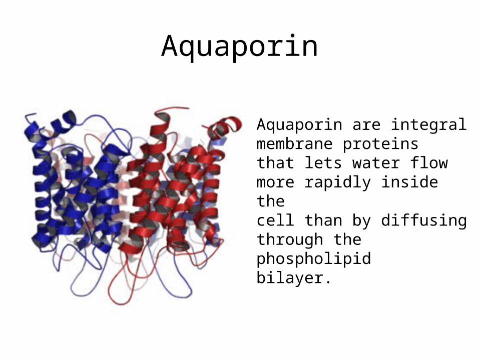
Aquaporin
Aquaporin are integral membrane proteinsthat lets water flow more rapidly inside the cell than by diffusing through the phospholipidbilayer.

Passive Transport

Passive Transport
It is the movement of substances down a concentration gradient and does not require energy use. - Diffusion- Osmosis- Dialysis- Facilitated diffusion- Simple diffusion

Diffusion

OSMOSIS


Facilitated diffusion
• Movement of large molecules• High to low regions of concentration• Channel proteins: tunnel shape
transporting small charged molecules• Carrier proteins: transports non
charged molecules with a specific shape

Facilitated Diffusion


Active Transport
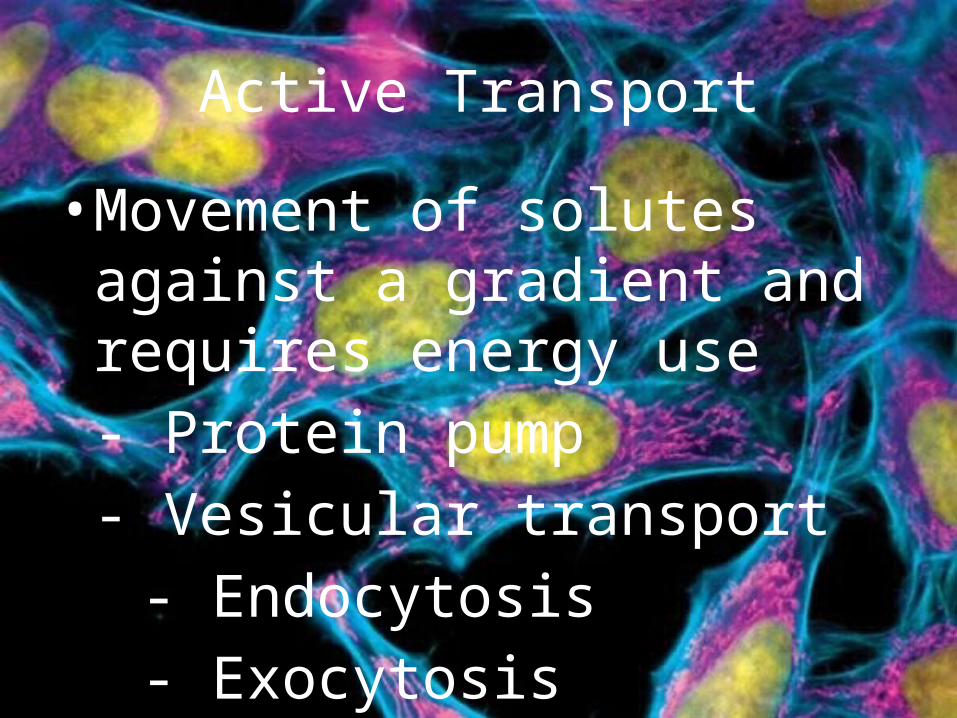
Active Transport
• Movement of solutes against a gradient and requires energy use- Protein pump- Vesicular transport
- Endocytosis- Exocytosis

Sodium and Potassium Pump


Exocytosis
A vesicle from inside the cell moves to the cell membrane. The vesicle fuses to the membrane and the contents are secreted.


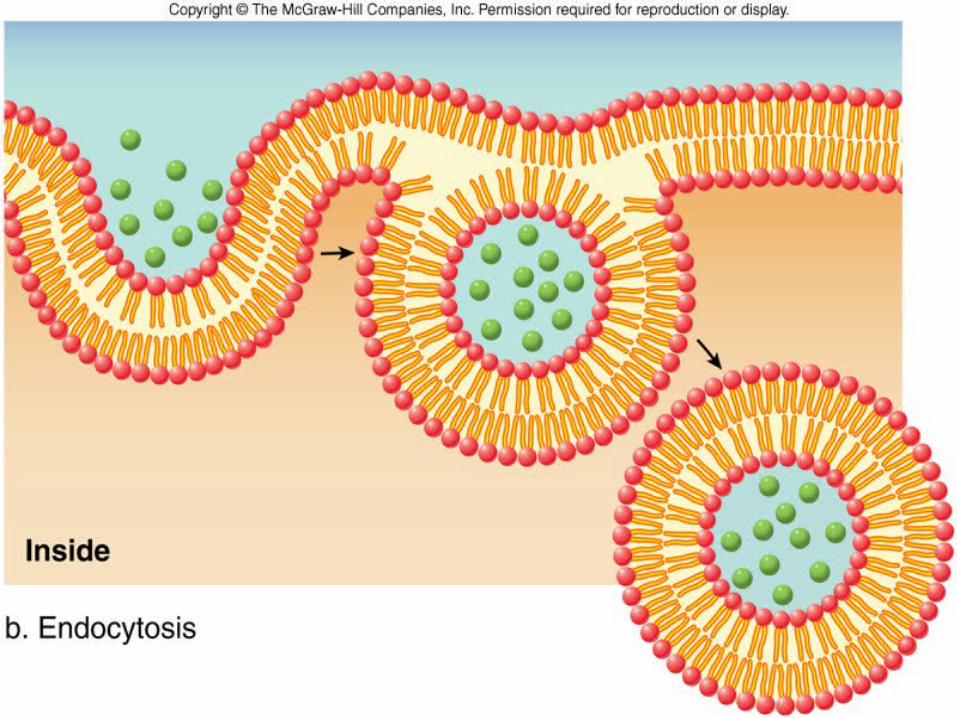
Endocytosis
• Pinocytosis: plasma membrane folds inward, forming a pinocytic vesicle
• Phagocytosis: plasma membrane engulfs the solid material
• Receptor mediated endocytosis: specific molecules bind to specialized receptors

PHAGOCYTOSIS


Cell Membrane Potential

• All eukaryotic cells maintain a non-zero transmembrane potential.
• Negative voltage inside, positive voltage outside (-40mV to -80mV)
• Membrane potential provides power• Used for transmitting signals

• The resting membrane potential is the electrical gradient across the cell membrane.
• Resting: the membrane potential has reached a steady state and is not changing.
• Potential: the electrical gradient created by the active transport of ions is a source of stored or potential energy.

Measuring Resting Membrane Potential
• Micropipets filled with solutions conduct charge. It is inserted into cell
• The voltmeter measures the potential difference (mV)
• The reference electrode is placed in the extracellular fluid. The extracellular fluid is designated as the ground and assigned a charge of 0 mV.

Nerst Equation
• The equilibrium potential is calculated using the Nerst equation:
in
oution [I]
[I]ln
FzRT
E (mV)

Resting Membrane Potential in Real Cells
• Most cells are 40x more permeable to K+ than Na+.
• The resting membrane potential is closer to -70 mV because a small amount of Na+ leaks into the cell.
• The Na+ is pumped out and the K+ pumped in by the Na+/K+-ATPase.

Goldman Equation
• Used to calculate the membrane potential resulting from all the participating ions when Vm is not changing:
outClinNainK
inCloutNaoutKm
][ClP][NaP][KP
][ClP][NaP][KPlnV
zFRT