cell-to-cell trafficking of macromolecules through plasmodesmata
TRANSCRIPT
The Plant Cell, Vol. 5, 1783-1794, December 1993 O 1993 American Society of Plant Physiologists
Cell-to-Cell Trafficking of Macromolecules through Plasmodesmata Potentiated by the Red Clover Necrotic Mosaic Virus Movement Protein
Toru Fujiwara,avl Donna Giesman-Cookmeyer,b Biao Ding,' Steven A. Lommel,b and William J. Lucasai2 a Section of Plant Biology, Division of Biological Sciences, University of California, Davis, California 95616
Department of Plant Pathology, Box 7616, North Carolina State University, Raleigh, North Carolina 27695
Direct evidence is presented for cell-to-cell trafficking of macromolecules via plasmodesmata in higher plants. The fluores- cently labeled 35-kD movement protein of red clover necrotic mosaic virus (RCNMV) trafficked rapidly from cell to cell when microinjected into cowpea leaf mesophyll cells. Furthermore, this protein potentiated rapid cell-to-cell trafficking of RCNMV RNA, but not DNA. Electron microscopic studies demonstrated that the 35-kD movement protein does not unfold the RCNMV RNA molecules. Thus, if unfolding of RNA is necessary for cellto-cell trafficking, it may well involve participation of endogenous cellular factors. These findings support the hypothesis that trafficking of macromolecules is a normal plasmodesmal function, which has been usurped by plant viruses for their cell-to-cell spread.
INTRODUCTION
Plasmodesmata are plasma membrane-lined cylindrical pores that traverse the cell wall. Each plasmodesma has appressed endoplasmic reticulum in the center that forms an endomem- brane continuum between neighboring cells. Proteinaceous particles are embedded in both the plasma membrane and the appressed endoplasmic reticulum membrane of this in- tercellular organelle, and spaces between these proteinaceous particles form a series of microchannels of ~ 2 . 5 nm in diameter within the plasmodesma (Ding et al., 1992b). Although it has long been held that plasmodesmata serve as potential routes for cell-to-cell communication and solute transport during plant growth and development (Gunning and Robards, 1976; Robards and Lucas, 1990; Lucas et ai., 1993), how plasmodes- mata function in these processes is not well understood. As established by dye-coupling studies, plasmodesmata normally have a size exclusion limit (SEL) of 800 to 1000 D (Tucker, 1982; Goodwin, 1983). Thus, it is generally accepted that transport through plasmodesmata is restricted to only small molecules, which would include metabolites, ions, and hormones.
Plant viral cell-tocell movement offers a powerful experimen- tal system for studying the function of plasmodesmata. It has been shown that some viruses appear to require capsid pro- tein for cell-to-cell movement, whereas others do not (Culver and Dawson, 1989; Petty and Jackson, 1990; Ziegler-Graff et al., 1991; Dalmay et al., 1992; Xiong et al., 1993). This sug- gests that in the latter, viral nucleic acids, not virions, move
Permanent address: Department of Agricultura1 Chemistry, The
To whom correspondence should be addressed. University of Tokyo, Tokyo 113, Japan.
through plasmodesmata during the infection process. In ad- dition, many known plant viruses encode a specific protein, the movement protein (MP), that mediates in the cell-to-cell spread of viruses during infection (Atabekov and Taliansky, 1990). Severa1 viral MPs have been localized to plasmodes- mata in infected plants as well as in transgenic plants that express MP genes (Tomenius et ai., 1987; Linstead et al., 1988; Shanks et al., 1989; Atkins et al., 1991; Ding et al., 1992a; Moore et ai., 1992), and the 30-kD MP of the tobacco mosaic virus (TMV) has been demonstrated to up-regulate the SEL of plas- modesmata in TMV MP transgenic tobacco plants (Wolf et al., 1989). Furthermore, a number of MPs have been found to bind RNA and single-stranded DNA in a cooperative but nonspecific manner in vitro (Citovsky et al., 1990,1991; Osman et al., 1992; Schoumacher et al., 1992; Giesman-Cookmeyer and Lommel, 1993). It has been hypothesized that such binding leads to unfolding, or relaxation of secondary structures, of viral nu- cleic acids to form an extended, linear, protein-nucleic acid complex that is then targeted to plasmodesmata for cell-to-cell transport (Citovsky et ai., 1990, 1991, 1992; Citovsky and Zambryski, 1991; Citovsky, 1993). These studies suggest that macromolecules can traffic (a presumably active and selec- tive process, in contrast with passive diffusion) cell to cell through plasmodesmata (see also Fisher et al., 1992; Sakuth et al., 1993).
In this study, direct evidence is presented showing that a viral MP and nucleic acids traffic from cell to cell via plasmodes- mata in higher plants. The fluorescently labeled 35-kD MP of red clover necrotic mosaic virus (RCNMV), when microinjected into mesophyll cells of a cowpea leaf, moved rapidly from cell
1784 The Plant Cell
to cell. The protein also increased the SEL of plasmodesmata to 10-fold higherthan the normal value. Furthermore, this pro- tein potentiated plasmodesmal trafficking of the RCNMV RNA, but not single- or double-stranded DNA. Interestingly, electron microscopic studies established that although the RCNMV MP bound to and potentiated trafficking of the RCNMV RNA, it did not unfold and extend these molecules in vitro. Thus, if unfolding of RCNMV RNA is a prerequisite for trafficking through plasmodesmata, it may well involve the participation of endogenous cellular factors.
Based on these findings, we propose that trafficking of mac- romolecules is a normal function of plasmodesmata, which was presumably usurped during coevolution by plant viruses to facilitate cell-to-cell spread of the viral genetic material. For plant growth and development, plasmodesmal trafficking of informational macromolecules potentially provides a means for supracellular (i.e., above the leve1 of individual cells) regu- lation of gene expression and coordination of physiological function(s) (Lucas et al., 1993).
RESULTS
RCNMV MP Produced in Escherichia coli lncreases Plasmodesmal SEL
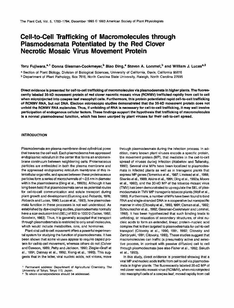
Previously, it was demonstrated that the 30-kD TMV MP in- creased the SEL of mesophyll plasmodesmata from the normal 800 D to greater than 10 kD in transgenic tobacco expressing this protein (Wolf et al., 1989). To determine whether the 35- kD RCNMV MP also modifies the plasmodesmal SEL, we took an alternative approach and directly coinjected the RCNMV MP produced in E. coli and a 9.4-kD fluorescein-conjugated dextran (F-dextran) into mesophyll cells of mature cowpea leaves. As illustrated in Figure lA, when the 9.4-kD F-dextran alone was microinjected into a mesophyll cell, it remained in the injected cell even after 20 min. However, when coinjected with the RCNMV MP, the 9.4-kD F-dextran-spread into the neighboring cells within 30 sec and then further into more dis- tant cells (up to three or more cells away) in less than 5 min (Figures 1B and íC). This experiment established that the RCNMV MP produced in E. coli was biologically active and that it functioned to interact with the cowpea mesophyll plasmodesmata to increase their permeability in a manner analogous to that of TMV MP expressed in transgenic tobacco plants.
This finding was further supported by studies of 12 RCNMV MP mutants. These mutants were generated by alanine scan- ning mutagenesis, in which clusters of two or three charged residues dispersed throughout the protein were all changed to alanines by site-directed mutagenesis (Giesman-Cookmeyer and Lommel, 1993). These MP mutants were named accord- ing to the position of the first amino acid in the mutagenized cluster (except for mutant 278, which was named based on the position of the second amino acid in this cluster to avoid
confusion with TMV MP transgenic tobacco line 277; Giesman- Cookmeyer and Lommel, 1993). As shown in Table 1, when MP mutants 27 to 31, 204, 242, 280, 301, and 305 were coin- jected with the 9.4-kD F-dextran, the F-dextran moved from cell to cell as rapidly as when the wild-type MP was coinjected. By contrast, when MP mutants 122, 128, 144, 161, 278, and 291 were coinjected, the 9.4-kD F-dextran remained in the in- jected cell, even after 20 min (see Figure 1D).
Interestingly, all of the RCNMV MP mutants that elicited an increase in plasmodesmal SEL also potentiated RCNMV cell- tocell movement in two host plants, cowpea and Nicotiana ben- thamiana, whereas all of the mutants that failed to increase the plasmodesmal SEL did not permit spread of the virus in either host (Table 1). These data demonstrate that increasing the SEL of plasmodesmata is aspecific function of the RCNMV MP, which is presumably required to establish effective viral cell-to-cell movement.
RCNMV MP Trafficks from Cell to Cell
Previous studies on TMV did not determine whether the MP itself trafficked from cell to cell, but only that plasmodesmal SEL was increased by the MP to allow diffusion of biologically inactive large dyes such as F-dextran (Wolf et al., 1989). The observation that the 9.4-kD F-dextran, when coinjected with RCNMV MP, moved into cells that were located three or more cells away from the injected site in cowpea mesophyll (cf. Fig- ure 1C) suggested that the MP itself also moved cell to cell to open plasmodesmal microchannels in those distant cells.
To directly test whether the RCNMV MP is indeed able to move from cell to cell, we labeled the 35-kD RCNMV MP puri- fied from E. coli with the fluorescent dye fluorescein isothiocyanate (FITC) and microinjected it into cowpea leaf mesophyll cells. As shown in Figure 2, the fluorescence spread from the injected cell into surrounding cells in less than 3 min after injection. By contrast, when MP mutant 278, which was defective in both increasing the plasmodesmal SEL and ef- fecting viral cell-to-cell spread, was similarly labeled and microinjected, the fluorescence remained in the injected cell even after 20 min (Figure 2D). In addition to the fact that free FITC molecules were removed from the protein fractions used for microinjection, these data indicate that the cell-tocell spread of the fluorescence represents cell-to-cell trafficking of the wild- type RCNMV MP.
Although we cannot provide direct cytological data to show that the cell-to-cell trafficking of the RCNMV MP is indeed via plasmodesmata, evidence from other RNA plant viruses indi- cates that plasmodesmata are the sole target sites across the cell wall for the viral MPs (Tomenius et al., 1987; Linstead et al., 1988; Shanks et al., 1989; Atkins et al., 1991; Ding et al., 1992a; Moore et al., 1992). Moreover, as shown above, the RCNMV MP increases by 10-fold the SEL of plasmodesmata when microinjected into cowpea leaf mesophyll cells. Based on these data, we conclude that the cell-to-cell trafficking of the FITC-labeled RCNMV MP occurred via plasmodesmata. .

Plasmodesmal Trafficking of Macromolecules 1785
Figure 1. Cell-to-Cell Movement of 9.4-kD F-Dextran Potentiated by RCNMV MP in Cowpea Mesophyll Cells.
The intensities of fluorescence are presented as false-color images. As shown in the color bar in (B), white represents the highest intensity whileblack is background.(A) The 9.4-kD F-dextran, injected alone, remains in the injected mesophyll cell (image taken 20 min after initial injection).(B) and (C) Thirty seconds and 5 min, respectively, after coinjection of the 9.4-kD F-dextran and wild-type RCNMV MP.(D) Fifteen minutes after coinjection of 9.4-kD F-dextran with RCNMV mutant MP 278.Bar in (B) = 50 urn.
RCNMV MP Potentiates Cell-to-Cell Trafficking ofRCNMV RNA
Because the coat protein is not required for cell-to-cell spreadof RCNMV (Xiong et al., 1993), it seems that the RCNMV RNAtrafficks cell to cell via plasmodesmata. To provide a direct testof this hypothesis, we labeled the in vitro-transcribed 3.9- and1.4-kb genomic RNAs of RCNMV (RNA 1 and RNA 2, respec-tively) with the nucleotide-specific fluorescent dye TOTO-1iodide (Glazer and Rye, 1992) and microinjected them into cow-pea mesophyll cells. As shown in Figure 3A, when the labeled
RNA 2 was microinjected alone, the fluorescence remainedin the injected cell after 5 min. Observations of such injectedcells indicated that the fluorescently labeled RCNMV RNA re-mained within the injected cell for periods greater than 20 to30 min. However, when unlabeled RCNMV MP was coinjectedalong with fluorescently labeled RCNMV RNA 2, the fluores-cence began to spread from the injected cell into surroundingcell(s) within 30 sec (Figure 3B). After 3 min, the fluorescenceassociated with RCNMV RNA 2 was detected in cells approxi-mately three cells away from the injection site (Figure 3C).Similar results were obtained when fluorescently labeled

1786 The Plant Cell
Table 1. Summatv of Functions ldentified of Wild-Type and Mutant Forms of RCNMV MP
Mutant
WTa 27 to 31 122 128 144 161 204 242 278 280 291 301 305
RNA bindingb 100 20 10 11 32 28 32 56 109 42 59 100 25 (% of wr)
- - Cooperativityb + - - + + + + + + + + Vira1 infectionC + + - - - - + + - ( + I - + + Pd SELd + + RNA traffickinge + + RNA unfolding' - - DNA traffickingg - a WT, wild-type.
These data are taken from Giesman-Cookmeyer and Lommel (1993) for the purpose of direct comparison with data obtained during the present study. (+) indicates cooperativity of MP binding to RNA (-) indicates lack of cooperativity for such binding. These data were obtained from studies on both N. benthamiana (Giesman-Cookmeyer and Lommel, 1993) and cowpea. (+) indicates wild-
type or mutant MP-potentiated viral infection; (-) indicates inability of a particular MP mutant to effect viral infection. For MP mutant 280, it has to revert to wild type to effect viral infection. See text for details.
(+) indicates RCNMV MP-induced increase in plasmodesmal (Pd) SEL, as demonstrated by 9.4-kD Fdextran cell-to-cell movement; ( - ) in- dicates no increase in Pd SEL. Four to five plants were used for each RCNMV MP (wild type or mutant) study. Original experimental data (number of injections showing RCNMV MP-potentiated 9.4-kD Fdextran movement per total number of injections) are as follows: Fdextran injected alone (0/9); Fdextran coinjected with the wild-type MP (7/10), 27 to 31 (5/7), 122 (015). 128 (0/5), 144 (4/18), 161 (0/6), 204 (4n). 242 (4/6), 278 (0/4), 280 (6/7), 291 (0/6), 301 (5/7), and 305 (5/7). e (+) indicates RCNMV MP-mediated cell-to-cell trafficking of RCNMV RNA 2 through plasmodesmata; (-) indicates no trafficking. Four to five plants were used for each RCNMV MP (wild type or mutant) study. Original experimental data (number of injections showing RCNMV MP-medi- ated RNA 2 trafficking per total number of injections) are as follows: RNA 2 injected alone (0/10); RNA 2 coinjected with wild-type MP (10/14), 27 to 31 (5/7), 122 (0/7), 128 (0/6), 144 (OB), 161 (0/6), 204 (5/6), 242 (7/9), 278 (0/12), 280 (0/6), 291 (0/6), 301 (4/6), and 305 (6n). See text for detailed discussion of RCNMV MP mutant 280. ( - ) indicates no unfolding of RCNMV RNA 1 and RNA 2 after binding to the RCNMV MP, as determined by electron microscopy. Two to
four experiments were conducted in each case. g ( - ) indicates no RCNMV MP-potentiated trafficking of single- or double-stranded DNA. Eight and 14 injection experiments were conducted for single- and double-stranded DNAs, respectively, and in none of these experiments did either of the DNAs show cell-to-cell trafficking in the presence of RCNMV MP.
- - - - + + - + - + + - - - - + + - - - + +
- - - - - - - - - - -
RCNMV RNA 1 was coinjected with unlabeled RCNMV MP (Figures 3D, 3E and 3F). Because TOTO-1 iodide only fluo- resces when bound to nucleotides (Glazer and Rye, 1992), the observed cell-to-cell spread of the fluorescence must be due to trafficking of RCNMV RNA 1 and RNA 2, which is poten- tiated by the coinjected RCNMV MP, rather than to diffusion of free TOTO-1 iodide. Because plasmodesmata are the sole targeting sites across the cell wall for viral MPs, the RCNMV MP-potentiated cell-to-cell trafficking of RCNMV RNA must be via plasmodesmata.
When the RCNMV MP mutants 27 to 31,204,242,301, and 305 were coinjected with fluorescently labeled RNA 2, the fluorescence also spread rapidly from cell to cell (Table 1). By contrast, when the RCNMV MP mutants 122, 128, 144, 161, 278, 280, and 291 were coinjected with the labeled RNA 2, the fluorescence remained in the injected cell, even after 20 min (Table 1 and Figure 3G). These data, as well as the fact that RNA did not traffic from cell to cell when injected alone, exclude the possibility that breakdown products of RCNMV RNA molecules diffused cell to cell.
Interestingly, the RCNMV MP mutant 280, which did not potentiate RNA trafficking, did appear to potentiate both cell-to-cell movement of RCNMV during infection (Giesman-
Cookmeyer and Lommel, 1993) and an increase in plasmodes- mal SEL (Table 1). Thus, an increase in the plasmodesmal SEL is necessary, but not sufficient, for RNA cell-to-cell traffick- ing. In a previous study (Giesman-Cookmeyer and Lommel, 1993), it was shown that in infected plants, this mutation al- ways reverted to the wild-type sequence, and only wild-type progeny virions were recovered. Hence, MP mutant 280 did not actually effect viral spread, but a reversion to the wild-type MP occurred during replication, and this was the form of the MP that was then able to effect cell-to-cell spread of the virus. These data, therefore, establish that increasing the plasmodes- mal SEL and potentiating plasmodesmal transport of RNA are two genetically distinct functions associated with the RCNMV MP that are both required for cell-tocell movement of RCNMV.
In recent studies, it was shown that RCNMV MP binds single- stranded DNA cooperatively in vitro (Osman et al., 1992; Giesman-Cookmeyer and Lommel, 1993), and thus experi- ments were conducted to test whether the RCNMV MP could also potentiate plasmodesmal trafficking of DNA molecules. We labeled single-stranded DNA (produced from the infectious transcript vector pBluescript KS+ containing the coding se- quence for RCNMV RNA 1) and double-stranded DNA (pBluescript KS+) with TOTO-1 iodide and coinjected either

Plasmodesmal Trafficking of Macromolecules 1787
one of them with unlabeled RCNMV MP into cowpea mesophyllcells. As shown in Figures 3H and 31, the single- and double-stranded DNA did not move from the injected cell into surround-ing cells, even after 20 min. These results demonstrate thatRCNMV MP-potentiated trafficking of nucleic acids via plas-modesmata is a selective process.
In view of the correlation between the abilities to modifyplasmodesmal SEL, to traffic RCNMV RNA (1 or 2), and topotentiate cell-to-cell spread of the virus for a given RCNMVMP mutant, we concluded that potentiating plasmodesmaltrafficking of RCNMV RNA, like modifying plasmodesmal SEL,is an essential function of the RCNMV MP.
Figure 2. Cell-to-Cell Trafficking of RCNMV MP in Cowpea Mesophyll Cells.
Details relating to fluorescence intensity are as given in Figure 1.(A) Bright-field image of a portion of the cowpea mesophyll after the epidermis was removed, revealing the cell (outlined by white dots) into whichthe fluorescently labeled RCNMV MP was injected.(B) and (C) Thirty seconds and 180 sec, respectively, after injection of fluorescently labeled RCNMV MP into the cell marked in (A).(D) Twenty minutes after the fluorescently labeled RCNMV MP mutant 278 was injected into a mesophyll cell.Bar in (C) = 50 urn.

1788 The Plant Cell
Figure 3. RCNMV MP-Potentiated Cell-to-Cell Trafficking of Fluorescently Labeled RCNMV RNA 1 and RNA 2 between Cowpea Mesophyll Cells.
Fluorescence intensities are as given in Figure 1.(A) Injection of RNA 2 alone into a mesophyll cell. RNA 2 did not move out of injected cells but was slowly degraded over time (image was taken5 min after injection). Injection of RNA 1 alone gave a similar result (data not shown).(B) and (C) Thirty and 180 sec, respectively, after coinjection of RNA 2 and wild-type RCNMV MP into a mesophyll cell.(D), (E), and (F) Forty, 80, and 180 seconds, respectively, after coinjection of RCNMV RNA 1 and wild-type RCNMV MR(G) Twenty minutes after coinjection of RNA 2 with RCNMV mutant MP 278.(H) Twenty minutes after coinjection of fluorescently labeled single-stranded DNA (produced from the infectious transcript vector of RCNMV RNA1) and wild-type RCNMV MP.(I) Twenty minutes after coinjection of fluorescently labeled double-stranded DNA (pBluescript KS+) and RCNMV MP.Bars in (E) and (F) = 50 urn.

Plasmodesmal Trafficking of Macromolecules 1789
RCNMV MP Does Not Unfold RCNMV RNA
As shown above, increasing the plasmodesmal SEL is required,but not sufficient, to permit cell-to-cell trafficking of RCNMVRNA. Presumably, one of the essential steps of the RNAtrafficking process involves interaction(s) between RNA andRCNMV MP and/or certain cellular factors. Based on studiesof TMV and cauliflower mosaic virus MPs, it has been pro-posed that cooperative binding of MP to a viral nucleic acidmolecule unfolds the latter to form an extended and linearprotein-nucleic acid complex that is then targeted to plasmo-desmata for transport (Citovsky et al., 1990, 1991, 1992;Citovsky and Zambryski, 1991; Citovsky, 1993). However, al-though RCNMV MP is able to bind RNA and single-strandedDNA in vitro in a nonspecific and cooperative manner (Osmanet al., 1992; Giesman-Cookmeyer and Lommel, 1993), suchin vitro cooperative binding is not correlated to the ability ofRCNMV MP either to effect viral cell-to-cell movement duringinfection (Giesman-Cookmeyer and Lommel, 1993) or to poten-tiate cell-to-cell RNA trafficking (Table 1). Thus, in vitroprotein-RNA binding assays alone appear to be insufficientto explain the mechanisms of RCNMV MP function in vivo.It should be noted that previous in vitro studies on the TMVand cauliflower mosaic virus MP binding properties were notperformed with RNA and DNA species that are known to betrafficked through plasmodesmata or that are of plant viral origin(Citovsky et al., 1990,1991,1992). Given these considerations,we undertook an electron microscopic characterization of thestructures of complexes formed between RCNMV MP andRCNMV RNA 2 and between RCNMV MP and RCNMV RNA 1to determine whether RCNMV MP indeed unfolds RCNMVRNA in vitro and, if it does, whether a direct correlation existsbetween the abilities of the RCNMV MP mutants to unfold RNAand to potentiate RNA cell-to-cell trafficking.
The in vitro-transcribed RNA 1 and RNA 2 were fluores-cently labeled with TOTO-1 iodide. Protein binding experimentswere performed as previously described (Giesman-Cookmeyerand Lommel, 1993). The buffer and incubation conditions forthe binding experiments were also essentially the same asthose used for the RCNMV MP/RNA microinjection experi-ments. Furthermore, for each binding experiment, an aliquotof the reaction mixture was used for electron microscopic study,and the remaining mixture was loaded onto an agarose gel,as shown in Figure 4. This strategy permitted a direct correla-tion of electron microscopic observations to the gel shiftprofiles. It should be pointed out here that incorporation ofTOTO-1 iodide into RNA molecules did not appear to affectRCNMV MP binding to RCNMV RNA as compared to bindingassay using radiolabeled RNA (Giesman-Cookmeyer andLommel, 1993).
As shown in Figure 5A, when RNA 2 molecules spread inthe presence of 10% formamide were visualized in the elec-tron microscope, they appeared as short, linear strandswith an average measured length of 165 ± 26 nm (50 RNAmolecules were measured). This would suggest that the "base-to-base" spacing for the measured RNA 2 was ~1.2 A. In the
presence of RCNMV MP at a saturating concentration, i.e.,the protein concentration at which all of the RNA moleculeswere bound by the protein as shown in gel shift studies (seeFigure 4A), the RNA 2-MP complexes also appeared as shortstrands similar to the RNA 2 alone (Figure 5B). These strandshad an average measured length of 157 ± 29 nm (50 RNAmolecules were measured), which was not different from thevalue obtained for the RNA 2 alone. To prove that the RCNMVMP was indeed bound to the RNA 2 molecules, we treatedthe reaction mixture with RNase A. We hypothesized that ifthe RCNMV MP was bound to the single-stranded regions ofRNA 2, it should protect these regions from RNase A diges-tion. Double-stranded regions, such as stem-loops at the 3'end (Lommel et al., 1988), would presumably be protected with-out MP binding. Both gel shift and electron microscopicexaminations demonstrated that after RNase A treatment, theRNA 2 molecules were degraded in the absence of the RCNMVMP, whereas they were protected in the presence of theRCNMV MP (Figures 4B, 5C, and 5D).
Because the measured length of RNA 2 molecules was notaltered after RCNMV MP binding, it was possible that the RNA2 molecules were already unfolded and extended in the ab-sence of RCNMV MP so that binding of this protein did nothave any further effect on these molecules. Alternatively, itwas possible that RNA 2 molecules were folded, but RCNMV
1 2 3 4 5 6 7 8 9 1 0
Figure 4. In Vitro RNA Binding Gel Shift Assay.(A) Binding of wild-type RCNMV MP to fluorescently labeled RCNMVRNA 2. In lanes 1 to 8, 0.0, 0.05, 0.1, 0.2, 0.4, 0.8, 1.5, and 3.0 ng ofprotein, respectively, were used to bind 50 ng of RNA 2. In lanes 9and 10,0.0 and 3.0 ng of protein were used, respectively, and the bind-ing reaction was conducted in 10% formamide. The solid dart indicatesthe position of RNA 2 bound by the RCNMV MP. The open dart pointsto the position of loading wells of the agarose gel, and the fluores-cence present in these wells is from aggregates of some RNA 2molecules that are formed in the presence of RCNMV MP, as deter-mined by electron microscopy.(B) Protection of fluorescently labeled RNA 2 by the wild-type RCNMVMP against RNase A digestion. The solid dart indicates the positionof RNA 2 bound by the RCNMV MP.

1790 The Plant Cell
&&^mm*m?&w$.i.< - - •• \•>..l .'.-• - - . - ^ r ; •,• -v •,'-.--/y,* **,'- •^•V-' • - ; ' - ' •" . > '
£$S&^ ^^ i •: • .''• '•'/ '*•',„ ^•i* \ *"* '—.* ' ^ ' "»•• i * /»"** I: /••* '* «•• •**! •* '» " ' " » " ' 1 * '• v » >j| ' j *»* *•*-" • cT'T ,^* N » • " * ' ' «^ "* *' *' • • * r f
Figure 5. Electron Microscopic Characterization of RCNMV RNA Molecules and Wild-Type RCNMV MP-RNA Complexes.
(A) RNA 2 alone spread in the presence of 10% formamide. The arrow points to an RNA 2 molecule.(B) RNA 2-RCNMV MP complexes spread in the presence of 10% formamide.(C) RNA 2 after RNase A digestion spread in 10% formamide.(D) RNA 2-RCNMV MP complexes after RNase A treatment spread in 10% formamide.(E) RNA 2 alone spread in 80% formamide.(F) RNA 1 alone spread in 10% formamide. An RNA 1 molecule is indicated by the arrow.(G) RNA 1-RCNMV MP complexes spread in 10% formamide.(H) RNA 1 alone, spread in 80% formamide.Black stipples in the background are cytochrome c. Bar in (H) = 0.2 urn.
MP binding did not cause unfolding of these molecules. Totest these possibilities, RNA 2 molecules were spread in 80%formamide for electron microscopic examination. Such a highconcentration of formamide would melt out random in-tramolecular base-to-base interactions and extend RNAmolecules (Ferguson and Davis, 1978). As shown in Figure5E, RNA 2 molecules became elongated in the presence of80% formamide, having an average measured length of 270± 52 nm (66 RNA molecules were measured) or a base-to-base distance of ~1.9 A. This represents a 36% increase overthe value obtained for RNA 2 molecules spread in 10% for-mamide. Obviously, RCNMV MP binding did not unfold andextend RNA 2 molecules, an observation which is in disagree-ment with that reported for the TMV MP-hepatitis delta virus(HDV) RNA complex (Citovsky et al., 1992).
These findings were further supported by the characteriza-tion of RNA 1 molecules and RNA 1-RCNMV MP complexes.
When RNA 1 molecules alone were spread in 10% formamideand examined in the electron microscope, they appeared highlystructured and folded (Figure 5F). After binding to the RCNMVMP (in the presence of 10% formamide), the RNA 1 moleculesstill retained this highly structured form and were clearly notunfolded and extended (Figure 5G). When spread alone in thepresence of 80% formamide, all of the RNA 1 molecules losttheir complex structural features and appeared as extended,linear entities (Figure 5H). Length measurement of theseRNA1 molecules yielded a value of 865 ± 116 nm, represent-ing a base-to-base distance of ~2.2 A (30 RNA moleculeswere measured).
These data demonstrate unequivocally that RCNMV MP didnot unfold RCNMV RNA molecules in vitro, although it wasindeed bound to them. Here it is important to point out thatthe experimental conditions we used in this study to spreadRCNMV RNA molecules as well as RCNMV MP-RNA

Plasmodesmal Trafficking of Macromolecules 1791
complexes for electron microscopic characterizations were similar to those used to spread the HDV molecules and TMV MP-HDV complexes (Citovsky et al., 1992). In particular, 10°/o formamide, which does not denature RNA molecules, was used in both studies. Our gel shift assays indicated that the presence of 10% formamide did not affect the RNA binding characteris- tics of the RCNMV MP (Figure 4A).
We tested all of the 12 RCNMV mutant MPs to determine what effect, if any, mutations at certain amino acids might have on the structure of the RCNMV MP-RNA complex. In all cases, the mutant MPs had no apparent effect on the appearance of either RNA 1 or RNA 2 molecules (data not shown). Hence, none of the mutant MPs was capable of RNA unfolding (Table 1). The differences among these RCNMV mutant MPs in their abil- ities to potentiate plasmodesmal trafficking of RCNMV RNA must, therefore, be due to differences in other functional aspects, e.g., in their ability to induce an increase in plasmodes- mal SEL, to recognize RCNMV RNA molecules, or to target viral RNA to plasmodesmata.
Because unfolding RCNMV RNA in vitro is not a function of RCNMV MP, the RCNMV MP may interact with RCNMV RNA in a manner that remains to be determined to potentiate cell- to-cell trafficking of these FINA molecules. Alternatively, un- folding of RNA, if necessary for trafficking, may well involve participation of cellular factors.
DISCUSSION
Our results establish that macromolecules, such as viral pro- teins and nucleic acids, are able to traffic from cell to cell via plasmodesmata in higher plants. Because such trafficking events take place within a few seconds of the test molecule being introduced into the target cell, we propose that endoge- nous mechanisms exist for macromolecular trafficking through plasmodesmata, and that such trafficking must be going on at all times. In a parallel study on the bean dwarf mosaic vi- rus, it was demonstrated that the fluorescently labeled BL1 movement protein and nucleic acids of this virus also trafficked cell to cell (A. Noueiry, W.J. Lucas, and R. Gilbertson, manu- script submitted). Collectively, these findings bring us one step closer to developing an understanding of the complex functions performed by plasmodesmata, as well as an understanding of viral cell-to-cell movement and systemic infection and dis- ease. We suggest that selective trafficking of macromolecules is a normal function of plasmodesmata and that some plant viruses have usurped this function to enable the spread of their own genetic material from cell to cell. It is possible that plant viruses acquired the genetic information encoding for their MPs from endogenous plant movement protein genes during coevo- lution (Lucas and Wolf, 1993).
Although the detailed mechanism(s) remains to be eluci- dated, plasmodesmal trafficking of macromolecules apparently shares certain parallel features with nucleocytoplasmic
transport (Lucas et al., 1993). The fact that proteins of more than 30 kD and RNA molecules of 4 kb traffic rapidly from cell to cell indicates that the normal plasmodesmal SEL of 8CO to 1000 D, as established by dyecoupling studies, represents the limit for passive diffusion of small molecules (see also Ding et al., 1993). If the protein molecule is in the globular form and moves through the cytoplasmic annulus, passage of the 35- kD RCNMV MP would require that the diameter of microchan- nels within the plasmodesma be dilated from the normal 2.5 nm to .v8 to 9 nm. Such an increase in the physical dimension of these plasmodesmal microchannels is conceivably tem- porary and highly controlled, much like the transport channels within the nuclear pore complex (Akey and Goldfarb, 1989; Ding et al., 1993; Lucas et al., 1993).
Like nucleocytoplasmic transport, plasmodesmal traffick- ing of macromolecules is a selective process: whereas RCNMV MP potentiates trafficking of RNA, but not any DNA (Figure 3), bean dwarf mosaic virus BL1 protein potentiates traffick- ing of double-stranded DNA, but not single-stranded DNA or RNA (A. Noueiry, W.J. Lucas, and R. Gilbertson, manuscript submitted). The mechanisms underlying such selectivity re- main to be elucidated.
The finding that RCNMV MP does not unfold the RCNMV RNA molecules in vitro, although it does bind to them in vitro and potentiate their trafficking through plasmodesmata in vivo, suggests that RCNMV MP interacts with these RNA molecules in an unidentified manner to permit their targeting to and trafficking through plasmodesmata. Alternatively, i f unfolding is required at any step during trafficking, it may well involve the participation of cellular factors (i.e., certain plasmodesmal proteins). At present, we do not know whether the RNA mole- cule alone, an RNA-MP complex, an RNA-cellular factor complex, or an RNA-MP-cellular factor complex trafficks through plasmodesmata.
Our preliminary results showed that RCNMV MP-potentiated cell-to-cell trafficking of RNA did not appear to be sequence specific. A 3.5-kb RNA was transcribed in vitro from the Scal- linearized plasmid pBSIL9, which contains sequences from the isocitrate lyase gene of Brassica napos and the E. coli plas- mid pES (Comai et ai., 1989). Like RCNMV RNA 1 and RNA 2, this nonviral RNA trafficked cell to cell in the presence of RCNMV MP (data not shown). This lack of sequence specific- ity is consistent with resultsobtained from in vitro RNA binding studies with RCNMV MP (Osman et al., 1992; Giesman- Cookmeyer and Lommel, 1993). It is possible that the three RNAs share a structural feature, rather than a sequence ele- ment, that is recognized by the RCNMV MP. This and other possibilities will be tested further.
It is of special interest that although the RCNMV MP does not potentiate plasmodesmal trafficking of single-stranded DNA, it is able to bind such DNA cooperatively in vitro (Osman et al., 1992; Giesman-Cookmeyer and Lommel, 1993). This finding, therefore, reveals a leve1 of specificity of RCNMV MP function in vivo which is not observed in vitro. Hence, the in vitro RNA binding assay alone provides limited information on the function of the MP, and the biological relevance of the
1792 The Plant Cell
nucleic acid binding activity in vitro by plant vira1 MPs remains To assess the effect of RCNMV MP on the plasmodesmal size ex- to be determined.
Although the RCNMV MP, with a molecular mass of 35 kD, readily trafficked from cell to cell, it rarely facilitated cell-to- cell movement of F-dextrans larger than 9.4 kD. Presumably, the F-dextran movement through plasmodesmata is via pas- sive diffusion that is limited solely by the effective pore size of the microchannels, whereas macromolecular trafficking via plasmodesmata, like nucleocytoplasmic transport of macro- molecules, is achieved by an actively controlled process (Silver, 1991; Lucas et al., 1993).
Our analysis of the RCNMV MP mutants suggests that RCNMV MP possesses a number of distinct functional do- mains. For example, increasing plasmodesmal SEL and potentiating RNA trafficking through plasmodesmata are two genetically distinct functions. Precise mapping of these functional domains must await the development of a three- dimensional structural model and more detailed mutational analyses 01 this MP.
The biological significance of plasmodesmal trafficking of macromolecules can be far-reaching. As previously discussed
clusion limit (SEL), a 5 mM solution of 9.4-kD fluorescein-conjugated dextran (F-dextran) (Molecular Probes, Eugene, OR) was prepared in 5 mM KHC03, filtered through a 0.5 pm-pore nylon syringe filter, and stored at 4°C. For experiments in which 9.4-kD F-dextran and RCNMV MP were to be coinjected, the injection medium was prepared by mix- ing equal volumes of F-dextran and MP.
To determine whether RCNMV MP trafficks cell tocell, the RCNMV MPs (wild-type and mutants) purified from E. coli were fluorescently labeled with fluorescein isothiocyanate (FITC) following the method of A. Noueiry, W.J. Lucas, and R. Gilbertson (manuscript submitted).
RNA 1, RNA 2, and single-stranded DNA (7.2 kb) were produced as previously described (Xiong and Lommel, 1991; Giesman- Cookmeyer and Lommel, 1993). The plasmid pBluescript KS+ (3.0 kb) was purified from E. coli (Sambrook et al., 1989). The nucleic acids were labeled with the nucleotide-specific fluorescent dye TOTO-1 io- dide (Molecular Probes Inc.) as follows. Briefly, a 5-pL nucleic acid (0.5 to 2 pgIpL) solution was mixed with 0.5 pL of lUTO-1 iodide and 5 pLof 61 buffer. The mixture was incubated for 1 hr at room tempera- ture and was then diluted in 40 pL of B1 and stored at -2OOC prior to use. The nucleic acid solution was injected alone or was mixed with an equal volume of RCNMV MP (5 mglmL) prior to being used in in- jection experiments.
(Lucas et al., 1993), whereas nucleocytoplasmic transport of macromolecules provides a direct means of regulating gene eXpreSSiOn in individual eukaryotic cells, plasmodesmal In Vitro Assay of RCNMV MP Binding to RCNMV RNA trafficking of informational macromolecules could potentially exert an elevated, supracellular coordination of these events in higher plants. The selectivity in terms oftrafficking ofspecific target molecules would establish a considerable advantage over the diffusion of small molecules. Future studies will con-
On identifying these putative endogenous plant proteins that are capable Of moving through plasmodesmata to control plant development and physiological Processes.
In vitro-transcribed RCNMV RNA 2 was fluorescently labeled by in- cubating 1.25 p!4 of RNA with 1 PL of 1 mM TOTO-1 iodide in 15 WL of Tris-EDTA (TE) for 1 hr at room temperature. The mixture was diluted to 25 nglpL by adding 35 pL of B1 buffer. RNA (50 ng) was then in- cubated with wild-type RCNMV MP in 8 pL of 61 buffer for 10 min on ice. After incubation, a 2-pL aliquot was removed from the reaction mixturewhich contained 3.0 pg of protein (lane8, Figure 4)for spread- ing for electron rnicroscopic characterization, and 2 FL of 50% glycero1/0.25 M EDTA was then added to each of the remaining reac- tion mixtures. The samples were loaded onto a 1% agarose gel and subjected to a current of 40 mamps for 30 min in TBE buffer vris- borate-EDTA) at room temperature. The gel was photographed on a FOTODYNE UV (FOTODYNE Inc.) light source using Fdaroid Type 665 film. For RNaseA treatment, the agarose gel described in Figure 4A was incubated for 1 hr in 10 pglmL RNaseA at room temperature be- fere being photographed as described above.
METHODS
Plant Material
Cowpea (Vigna unguiculata) plants were grown in a growth chamber under a temperature regime of 25"C/18"C (dayhight) with a 16-hr pho- toperiod at a PAR leve1 of 230 to 280 pmol m-2 sec-l. The first or second fully expanded trifoliate leaf was used for microinjection Electmn Mlcroscopy experiments.
Miclolnjectlon
Attached to the plant, the leaf was fixed on the microscope stage adaxial side up, and a small portion of the lower epidermis was peeled off. Distilled water was applied to the exposed region immediately. De- tails regarding the instrumentation and procedure for microinjection were as previously described (Ding et al., 1992a).
The wild-type and mutants of red clover necrotic mosaic virus (RCNMV) movement protein (MP) were expressed in Escherichia coli and stored in 61 buffer (10 mM Tris-HCI, pH 8.0, 1 mM EDTA, 10% glycerol, and 200 mM NaCI) as previously described (Giesman- Cookmeyer and Lommel, 1993). Protein concentrations (wild-type and mutants) were between 1.0 and 3.0 mglmL.
A 2-pL aliquot of the reaction mixture obtained from the binding ex- periment, which involved 50 ng of RNA and 3.0 pg of RCNMV MP (lane 8, Figure 4A), or from control reaction without RCNMV MP; asdescribed in Figure 4, was added to 8 pL of B1 buffer. For spreading in 10% for- mamide (as per Citovsky et al., 1992), this diluted mixture was added to a solution containing 28 pL of glass-distilled H20, 5 pL of TE (2 M Tris, pH 8.3,0.2 M EDTA), 5 pL formamide (highest purity grade; Elec- tron Microscopy Sciences, Philadelphia), and 3 pL of cytochrome c (Sigma) (4 mg/mL in H20). This preparation was then spread onto glass-distilled H20 to form a monolayer film of macromolecules. For spreading in 80% formamide, 5 pL of an RNA sample was added to a solution containing 40 pL formamide, 3 pL TE, and 2 pL cyto- chrome c. This preparation was spread onto 50% formamide buffered by TE (0.25 M Tris, pH 8.30.025 M EUA). Formvarcoated copper grids

Plasmodesmal Trafficking of Macromolecules 1793
were used to pick up the monolayerfilm. Grids were immediately soaked in a solution of uranyl acetate (0.1 @mL in 90% ethanol) for 10 sec and then transferred to 90% ethanol for an additional 10 sec before being air dried. Specimens were examined on a transmission elec- tron microscope (model41OLS; Philips, Eindhoven, The Netherlands). Micrographs were taken at a magnification of 10,400. Length mea- surement was conducted on straight RNA molecules or RNA-MP complexes.
ACKNOWLEDGMENTS
We thank Kelly Matsudaira and John Harada of the Section of Plant Biology, University of California, Davis, for assistance so generously rendered during the course of this study. We thank John Crowe and Richard Harrison of the Zoology Department of the University of Califor- nia, Davis, for the use of their electron microscope facility. This research was supported by Grant No. DCB-90-05722 from the National Science Foundation to W.J.L. and by grants from the North Carolina Biotech- nology Center (Research Triangle Park, NC) and the United States Department of Agriculture (No. 91-37303-6431) to S.A.L. T.F. thanks the Research lnstitute of lnnovative Technology for the Earth (RITE) of Japan for a fellowship.
Received September 22, 1993; accepted October 22, 1993.
REFERENCES
Akey, C.W., and Goldfarb, D.S. (1989). Protein import through the nu- clear porecomplex is a multistep process. J. Cell Biol. 109,971-982.
Atabekov, J.G., and Taliansky, M.E. (1990). Expression of a plant virus- coded transport function by different vira1 genomes. Adv. Virus Res.
Atkins, D., Hull, R., Wells, B., Roberts, K., Moore, P., and Beachy, R.N. (1991). The tobacco mosaic virus 30K movement protein in trans- genic tobacco plants is localized to plasmodesmata. J. Gen. Virol.
Citovsky, V. (1993). Probing plasmodesmal transport with plant viruses. Plant Physiol. 102, 1071-1076.
Citovsky, V., and Zambryski, P. (1991). How do plant virus nucleic acids move through intercellular connections? BioEssays 13, 373-379.
Cltovsky, V., Knorr, D., Schuster, G., and Zambryski, P. (1990). The P30 movement protein of tobacco mosaic virus is a single-strand nucleic acid binding protein. Cell 60, 637-647.
Cltovsky, V., Knorr, D., and Zambryski, P. (1991). Gene 1, a potential cell-to-cell movement locus of cauliflower mosaic virus, encodes an RNA binding protein. Proc. Natl. Acad. Sci. USA. 88, 2476-2480.
Cltovsky, V., Wong, M.L., Shaw, A.L., Prasad, B.V.V., andZambryski, P. (1992). Visualization and characterization of tobacco mosaic vi- rus movement protein binding to single-stranded nucleic acids. Plant Cell 4, 397-411.
Comai, L., Dietrich, R. A., Maslyar, D.J., Baden, C.S., and Harada, J.J. (1989). Coordinate expression of transcriptionally regulated isoci- trate lyase and malate synthase genes in Brassica napus L. Plant Cell 1, 293-300.
38, 201-248.
72, 209-211.
Culver, J.N., and Dawson, W.O. (1989). Tobacco mosaic virus coat protein: An elicitor of the hypersensitive reaction but not required for the development of mosaic symptoms in Nicotiena sybsfris. Virol-
Dalmay, T., Rubino, L., Burgyan, J., and Russo, M. (1992). Replica- tion and movement of a coat protein mutant of cymbidium ringspot tombusvirus. MOI. Plant-Microbe Interact. 5, 379-383.
Ding, B., Haudenshield, J.S., Hull, R.J., Wolf, S., Beachy, R.N., and Lucas, W.J. (1992a). Secondary plasmodesmata are specific sites of localization of the tobacco mosaic virus movement protein in transgenic tobacco plants. Plant Cell 4, 915-928.
Ding, B., Turgeon, R., and Parthasarathy, M.V. (1992b). Substruc- ture of freeze substituted plasmodesmata. Protoplasma 169,28-41.
Dlng, B., Haudenshield, J.S., Willmitzer, L., and Lucas, W.J. (1993). Correlation between arrested secondary plasmodesmal develop- ment and onset of accelerated leaf senescence in yeast acid invertase transgenic tobacco plants. Plant J. 4, 179-189.
Ferguson, J., and Davis, R.W. (1978). Quantitative electron micros- copy of nucleic acids. In Advanced Techniques in Biological Electron Microscopy I I . J. K. Koehler, ed (New York: Springer-Verlag), pp.
Fisher, D.B., Wu, Y., and Ku, M.S.B. (1992). Turnover of soluble pro- teins in the wheat sieve tube. Plant Physiol. 100, 1433-1441.
Giesman-Cookmeyer, D., and Lommel, S.A. (1993). Alanine scan- ning mutagenesis of a plant virus movement protein identifies three functional domains. Plant Cell 5, 973-982.
Glazer, A.N., and Rye, H.S. (1992). Stable dye-DNA intercalation com- plexes as reagents for high-sensitivity fluorescence detection. Nature
Goodwln, P.B. (1983). Molecular size limit for movement in the sym- plast of the Elodea leaf. Planta 157, 124-130.
Gunning, B.E.S., and Robards, A.W., eds (1976). lntercellular Com- munication in Plants: Studies on Plasmodesmata (New York: Springer-Verlag).
Linstead, P.J., Hills, G.J., Plaskitt, K.A., Wilson, I.G., Harker, C.L., and Maule, A.J. (1988). The subcellular location of the gene 1 product of cauliflower mosaic virus is consistent with a function associated with virus spread. J. Gen Virol. 69, 1809-1818.
Lommel, S.A., Weston-Fina, M., Xiong, i!., and Lomonossoff, G.P. (1988). The nucleotide sequence and gene organization of red clo- ver necrotic mosaic virus RNA-2. Nucl. Acids Res. 16, 8587-8602.
Lucas, W.J., and Wolf, S. (1993). Plasmodesmata: The intercellular organelles of green plants. Trends Cell Biol. 3, 308-315.
Lucas, W.J., Dlng, B., and van der Schoot, C. (1993). Plasmodes- mata and the supracellular nature of plants. New Phytol. 125,
Moore, P.J., Fenczik, C.A., Deom, C.M., and Beachy, R.N. (1992). Developmental changes in plasmodesmata in transgenic tobacco expressing the movement protein of tobacco mosaic virus. Pro- toplasma 170, 115-127.
Osman, T.A.M., Hayes, R.J., and Buck, K.W. (1992). Cooperative bind- ing of the red clover necrotic mosaic virus movement protein to single-stranded nucleic acids. J. Gen. Virol. 73, 223-227.
Petty, I.T.D., and Jackson, A.O. (1990). Mutational analysis of barley stripe mosaic virus RNA beta. Virology 179, 712-718.
Robards, A.W., and Lucas, W.J. (1990). Plasmodesmata. Annu. Rev. Plant Physiol. Plant MOI. Biol. 41, 369-419.
Ogy 173, 755-758.
123-171.
359, 859-861.
435-476.

1794 The Plant Cell
Sakuth, T., Schobert, C., Pecsvaradi, A., Eichholz, A., Komor, E., and Orlich, G. (1993). Specific proteins in the sieve-tube exudate of Ricinus communis L. seedlings: Separation, characterization and in vivo labelling. Planta 191, 207-213.
Sambrook, J., Fritsch, E.F., and Maniatis, T. (1989). Molecular Clon- ing: A Laboratory Manual, 2nd ed. (Cold Spring Harbor, NY Cold Spring Harbor Laboratory).
Schoumacher, F., Erny, C., Berna, A., Godefroy-Colburn, T., and Stussl-Garaud, C. (1992). Nucleic acid-binding properties of the alfalfa mosaic virus movement protein produced in yeast. Virology
Shanks, M., Tomenius, K., Clapham, O., Huskisson, N.S., Barker, P.J., Wllson, I.G., Maule, A.J., and Lomonossoff, G.P. (1989). Iden- tification and subcellular localization of a putative cell-twell transport
SilVer, P.A. (1991). How Proteins enterthe nucleus. Cell 64,489-497. Tomenius, K., Clapham, D., and Meshi, T. (1987). Localization by
immunogold cytochemistry of the virus-coded 30K protein in
plasmodesmata of leaves infected with tobacco mosaic virus. Virol- ogy 160, 363-371.
E .~ . (1982). Translocation in the staminal hairs of Setcreasea purpurea. 1. A study of cell ultrastructure and cell-to-cell passage of molecular probes. Protoplasma 113, 193-201.
Wolf, S., Deom, C.M., Beachy, R.N., and Lucas, W.J. (1989). Move- ment protein of tobacco mosaic virus modifies plasmodesmatal size exclusion limit. Science 246, 377-379.
Xiong, z., and bmmel, S.A. (1991). Red clover necrotic mosaic vi- rus infectious transcripts synthesized in vitro. Virology 182,388-392.
Xiong, Z., Klm, K.H., Giesman-Cookmeyer, D., and Lommel, S.A. (1993). The roles of the red clover necrotic mosaic virus capsid and cell-to-cell movement proteins in systemic infection. Virology 192,
Ziegler-Graff, V., Guilfotd, P.J., and Baulcombe, D.C. (1991). Tobacco rattle virus RNA-1 29K gene product potentiates vira1 movement and also affects symptom induction in tobacco. Virology 182, 145-155.
188, 896-899.
protein from red clover mottle virus. Virology 173, 400-407. 27-32.