cells · 7 cell structure 8 cells and their environment 9 photosynthesis and cellular respiration...
TRANSCRIPT

Sex chromosomes of a human male: Y (left) and X (right)
UNIT 3Cells 7 Cell Structure
8 Cells and Their Environment
9 Photosynthesis and Cellular Respiration
10 Cell Growth and Division
Macrophage (purple) attack on a cancer cell (yellow)
hb08se_3ucl_opn.indd 146 10/23/06 12:57:27 PM hb08se_3ucl_opn.indd 1 10/23/06 12:57:36 PM
146 UNIT 3 Cells
hb08te_3uni_opn.indd 2 11/17/06 2:55:40 PM

UN
IT 3
Human motor neuron
hb08se_3ucl_opn.indd 1 10/23/06 12:57:36 PM
UN
IT 3
UNIT 3 Cells 146A
hb08te_3uni_opn.indd 3 11/17/06 2:55:56 PM
hb08se_3ucl_opn.indd Sec1:147 10/23/06 12:57:59 PM
Microtubules (green) and chromosomes (blue) in a dividing cell
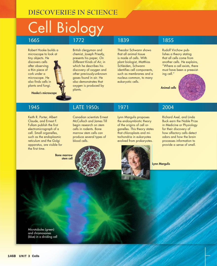
1665
DISCOVERIES IN SCIENCE
Cell BiologyRobert Hooke builds a microscope to look at tiny objects. He discovers cells after observing a thin piece of cork under a microscope. He also finds cells in plants and fungi.
1772
British clergyman and chemist, Joseph Priestly, presents his paper, On Different Kinds of Air, in which he describes his discovery of oxygen and other previously-unknown gases found in air. He also demonstrates that oxygen is produced by plants.
1839
Theodor Schwann shows that all animal tissue is made of cells. With plant biologist, Matthias Schleiden, Schwann identifies cell components, such as membranes and a nucleus common, to many eukaryotic cells.
1855
Rudolf Virchow pub-lishes a theory stating that all cells come from another cells. He explains, “Where a cell exists, there must have been a preexist-ing cell.”
1945
Keith R. Porter, Albert Claude, and Ernest F. Fullam publish the first electromicrograph of a cell. Small organelles, such as the endoplasmic reticulum and the Golgi apparatus, are visible for the first time.
LATE 1950S
Canadian scientists Ernest McCulloch and James Till begin research on stem cells in rodents. Bone marrow stem cells can produce several types of blood cells.
1971
Lynn Margulis proposes the endosymbiotic theory of the origins of cell or-ganelles. This theory states that chloroplasts and mi-tochondria in eukaryotes evolved from prokaryotes.
2004
Richard Axel, and Linda Buck earn the Noble Prize in Medicine or Physiology for their discovery of how olfactory cells detect odors and how the brain processes information to provide a sense of smell.
Hooke’s microscope
Animal cells
Bone marrowstem cell
Lynn Margulis
hb08se_3ucl_opn.indd 2 10/23/06 12:57:43 PM
146B UNIT 3 Cells
hb08te_3uni_opn.indd 4 11/17/06 2:56:05 PM

UN
IT 3
BIOLOGY CAREER
Cell BiologistShubha Govind
Shubha Govind is a professor of biology at City College, City University of New York. Govind consid-ers her most important scientific contribution to be developing a model system for using genetic tools to study the molecular basis of host-parasite interaction in fruit flies. She is studying how blood cells of fruit flies are formed and how they guard against infec-tions when flies are attacked by parasites. She is also studying how parasites have evolved to overcome the immune reactions of the fly.
Govind grew up in India, and her family traveled a lot. As she traveled, she was impressed with the di-versity of flora and fauna in different parts of the country. By the time she reached middle school, she knew that she wanted to be a biologist.
Apart from science, Govind enjoys reading, listening to music and spending time with family and friends.
Freeze fracture of cell
hb08se_3ucl_opn.indd Sec1:147 10/23/06 12:57:59 PM
UN
IT 3
UNIT 3 Cells 147
hb08te_3uni_opn.indd 5 11/17/06 2:56:19 PM
00 min.
00 min.
Chapter Planner
Chapter Review and Assessment Resources
FastTrackC H A P T E R
Teach Key IdeasStandardsCHAPTER OPENER, pp. 148–149
SECTION 1 Introduction to Cells, pp. 151–155
V The Discovery of Cells
V Looking at Cells
V Cell Features
Bellringer Transparency
Transparencies A10 Metric Units of Length and Equivalents • A4 Objects Size and Magnifying Power of Microscopes • A5 Compound Light Microscope • B2 Relationship Between Surface Area and Volume • B8 Structure of Lipid Bilayer • B9 Organelles
Visual Concepts Types of Microscopes• Magnification and Resolution • Parts of a Light Microscope • Cell Theory • Cell Membrane• Cytoplasm • Ribosomes • Internal Organization of a Cell • Parts of a Prokaryotic Cell • Parts of a Cell Wall
SECTION 2 Inside the Eukaryotic Cell, pp. 156–161
V The Framework of the Cell
V Directing Cellular Activity
V Protein Processing
V Storage and Maintenance
V Energy Production
Bellringer Transparency
Transparencies B13 Processing of Proteins • B12 Mitochondrion
Visual Concepts Cytoskeleton • Nucleus of a Cell • Endoplasmic Reticulum (ER) and Ribosomes • Golgi Apparatus • Mitochondrion • Chloroplasts • Vacuoles
SECTION 3 From Cell to Organism, pp. 162–166
V Diversity in Cells
V Levels of Organization
V Body Types
Bellringer Transparency
Transparencies B4 Animal Cells
Visual Concepts Structure of Cilia and Flagella• Comparing Prokaryotes and Eukaryotes • Parts of an Animal Cell • Comparing Plant and Animal Cells • Parts of a Plant Cell
7 Cell Structure
148A CHAPTER 7 Cell Structure
Thorough instruction will require the times shown.
SE Super Summary, p. 168SE Chapter Review, p. 169SE Standardized Test Prep, p. 171
Review Resources
Chapter Tests A and B
Holt Online Assessment
Basic LearnersTE Magnification, p. 152TE What Am I?, p. 158TE 3-D Representations, p. 159TE Body Systems, p. 164
Directed Reading Worksheets*
Active Reading Worksheets*
Lab Manuals, Level A*
\ Study Guide*
Note-taking Workbook*
Special Needs Activities and Modified Tests*
Advanced LearnersTE Specimen Size, p. 153TE Mitochondria, p. 160 TE Behavior of Cell Colonies, p. 165
Critical Thinking Worksheets*
Concept Mapping Worksheets*
Science Skills Worksheets*
Lab Datasheets, Level C*
45 min.
15 min.
60 min.
90 min.
See also PowerPoint ® Resources
National Science Education Standards
LSCell 1, LSCell 2,
LSCell 3, LSCell 4,
LSCell 6, LSGene 1,
LSEvol 4, UCP1,
UCP2, UCP5, HNS1,
HNS2, HNS3
LSCell 1, LSCell 2,
LSCell 3, LSCell 4,
LSCell 5, LSGene 1,
LSMat 2, LSMat 4,
UCP1, UCP5, SI1
LSCell 4, LSCell 5,
LSCell 6, LSMat 4,
LSMat 6, UCP1,
UCP5, SI1, SI2
hb08te_csf_plg.indd 2 11/30/06 4:31:03 PM

Key
SE Student Edition
TE Teacher’s Edition
Chapter Resource File
Workbook
Transparency
CD or CD-ROM
* Datasheet or blackline
master available
Also available
in Spanish All resources listed below are also available on the Teacher’s One-Stop Planner.
Resources for Differentiated Instruction
Hands-On Skills Development AssessmentSE Inquiry Lab Is It Alive?,
p. 149* TE Reading Toolbox Assessing
Prior Knowledge, p. 148SE Reading Toolbox, p. 150
TE Bacteria in Yogurt, p. 151SE Cell Shape, p. 152TE Prokaryote Diversity, p. 154
Quick Lab Modeling Cells: Surface Area to Volume*
Skills Practice Lab Using a Microscope*
TE Reading Toolbox Visual Literacy, p. 152
TE Math Skills Surface Area–to-Volume Ratio, p. 153
SE Reading Toolbox Word Parts, p. 155
TE Reading Toolbox Word Parts, p. 155
SE Section Review
TE Formative Assessment
Spanish Assessment*
Section Quiz
TE Amoeboid Movement, p. 156TE Demonstration Observing Cell Structure,
p. 157TE Lysosmal Malfunction, p. 158TE Filtering Out Toxins, p. 158TE History Connection, p. 159
SE Quick Lab Model Cell Parts, p. 160*
SE Reading Toolbox Process Chart, p. 158
TE Reading Toolbox Process Chart, p. 158
SE Section Review
TE Formative Assessment
Spanish Assessment*
Section Quiz
TE Diversity, p. 162TE Endosymbiosis, p. 163TE Career Development, p. 164TE Cryogenics, p. 165
SE Quick Lab Colonies On the Move, p. 165*
SE Skills Practice Lab Plant Cell Observation, p. 167*
TE Reading Toolbox Fold Notes, p. 162
SE Reading Toolbox Similes, p. 164
TE Reading Toolbox Similes, p. 164
SE Section Review
TE Formative Assessment
Spanish Assessment*
Section Quiz
Build student motivation with resources about high-interest applications.
Why It Matters
English LearnersTE Paired Reading, p. 157TE Reading Organizer, p. 163
Directed Reading Worksheets*
Active Reading Worksheets*
Lab Manuals, Level A*
Study Guide*
Note-taking Workbook*
Multilingual Glossary
Struggling ReadersTE TE Paired Reading, p. 157
Directed Reading Worksheets*
Active Reading Worksheets*
Lab Manuals, Level A*
Study Guide*
Note-taking Workbook*
Special Needs Activities and Modified Tests*
Special Education
StudentsTE Cell Model, p. 154
Directed Reading Worksheets*
Active Reading Worksheets*
Lab Manuals, Level A*
Study Guide*
Note-taking Workbook*
Special Needs Activities and Modified Tests*
Alternative AssessmentTE Fold Notes, p. 157
Science Skills Worksheets*
Section Quizzes*
Chapter Tests A, B, and C*
Chapter Planning Guide 148B
See also Lab Generator
See also Holt Online Assessment Resources
hb08te_csf_plg.indd 3hb08te_csf_plg.indd 3 8/13/07 3:43:41 PM8/13/07 3:43:41 PM
1 Introduction to CellsThe Discovery of Cells
Looking at Cells
Cell Features
2 Inside the Eukaryotic Cell The Framework of the Cell
Directing Cellular Activity
Protein Processing
Storage and Maintenance
Energy Production
3 From Cell to Organism Diversity in Cells
Levels of Organization
Body Types
Why It Matters
All living things are made of cells. Scientists study how cells work to understand life.
Preview
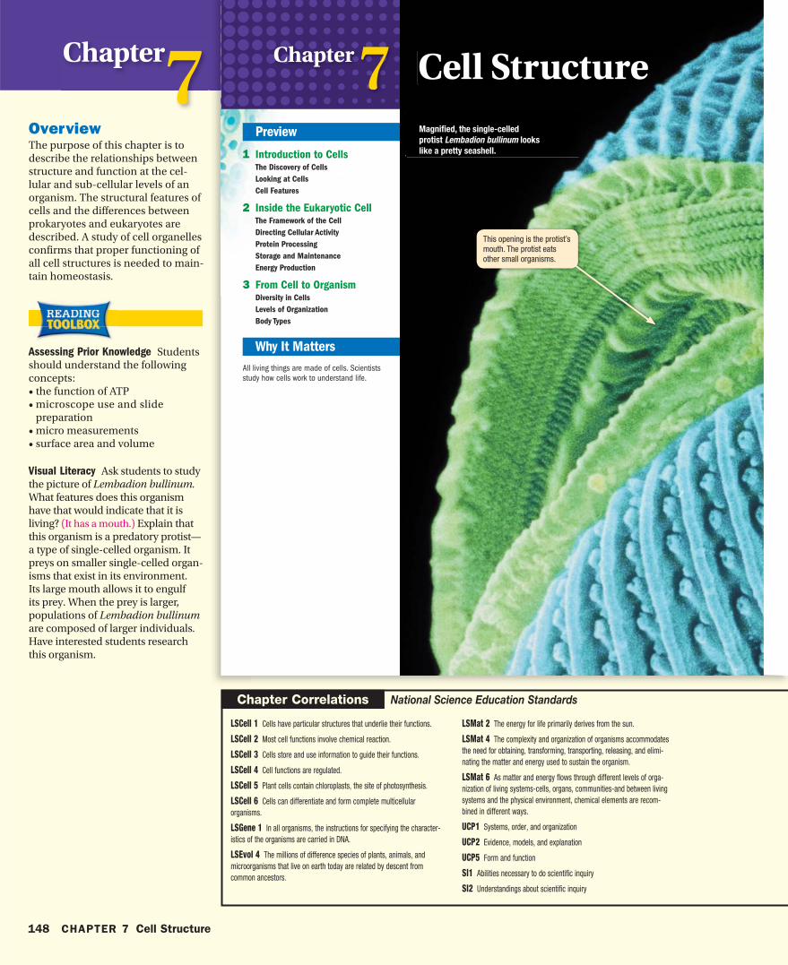
Chapter 7
This opening is the protist’s mouth. The protist eats other small organisms.
Magnified, the single-celled
protist Lembadion bullinum looks
like a pretty seashell .
Cell Structure
hb08se_csf_cho.indd 148 3/20/07 11:00:04 AM
148 CHAPTER 7 Cell Structure
Chapter7OverviewThe purpose of this chapter is to describe the relationships between structure and function at the cel-lular and sub-cellular levels of an organism. The structural features of cells and the differences between prokaryotes and eukaryotes are described. A study of cell organelles confirms that proper functioning of all cell structures is needed to main-tain homeostasis.
Assessing Prior Knowledge Students should understand the following concepts:• the function of ATP• microscope use and slide
preparation• micro measurements• surface area and volume
Visual Literacy Ask students to study the picture of Lembadion bullinum. What features does this organism have that would indicate that it is living? (It has a mouth.) Explain that this organism is a predatory protist— a type of single-celled organism. It preys on smaller single-celled organ-isms that exist in its environment. Its large mouth allows it to engulf its prey. When the prey is larger, populations of Lembadion bullinum are composed of larger individuals. Have interested students research this organism.
Chapter Correlations National Science Education Standards
LSCell 1 Cells have particular structures that underlie their functions.
LSCell 2 Most cell functions involve chemical reaction.
LSCell 3 Cells store and use information to guide their functions.
LSCell 4 Cell functions are regulated.
LSCell 5 Plant cells contain chloroplasts, the site of photosynthesis.
LSCell 6 Cells can differentiate and form complete multicellular organisms.
LSGene 1 In all organisms, the instructions for specifying the character-istics of the organisms are carried in DNA.
LSEvol 4 The millions of difference species of plants, animals, and microorganisms that live on earth today are related by descent from common ancestors.
LSMat 2 The energy for life primarily derives from the sun.
LSMat 4 The complexity and organization of organisms accommodates the need for obtaining, transforming, transporting, releasing, and elimi-nating the matter and energy used to sustain the organism.
LSMat 6 As matter and energy flows through different levels of orga-nization of living systems-cells, organs, communities-and between living systems and the physical environment, chemical elements are recom-bined in different ways.
UCP1 Systems, order, and organization
UCP2 Evidence, models, and explanation
UCP5 Form and function
SI1 Abilities necessary to do scientific inquiry
SI2 Understandings about scientific inquiry
hb08te_csf_cho.indd 148 4/16/07 6:09:01 PM
15 min
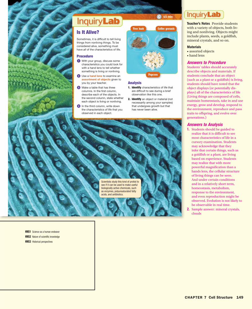
Scientists study this kind of protist to see if it can be used to make useful biologically active chemicals, such as enzymes, polyunsaturated fatty acids, and antibiotics.
Sometimes, it is difficult to tell living things from nonliving things. To be considered alive, something must have all of the characteristics of life.
Procedure1 With your group, discuss some
characteristics you could look for with a hand lens to tell whether something is living or nonliving.
2 Use a hand lens to examine an assortment of objects given to you by your teacher.
3 Make a table that has three columns. In the first column, describe each of the objects. In the second column, state whether each object is living or nonliving.
4 In the third column, write down the characteristics of life that you observed in each object.
Is It Alive?
Analysis1. Identify characteristics of life that
are difficult to see during a brief observation like this one.
2. Identify an object or material (not necessarily among your samples) that undergoes growth but that has never been alive.
Popcorn
Tree bark Coffee grounds
hb08se_csf_cho.indd 149 10/12/06 9:14:34 AM
CHAPTER 7 Cell Structure 149
Teacher’s Notes Provide students with a variety of objects, both liv-ing and nonliving. Objects might include plants, seeds, a goldfish, mineral crystals, and so on.
Materials
• assorted objects• hand lens
Answers to ProcedureStudents’ tables should accurately describe objects and materials. If students conclude that an object (such as a plant or a goldfish) is living, students should have noted that the object displays (or potentially dis-plays) all of the characteristics of life (Living things are composed of cells, maintain homeostasis, take in and use energy, grow and develop, respond to the environment, reproduce and pass traits to offspring, and evolve over generations.)
Answers to Analysis1. Students should be guided to
realize that it is difficult to see most characteristics of life in a cursory examination. Students may acknowledge that they infer that certain things, such as a goldfish or a plant, are living based on experience. Students may realize that with more powerful magnification than a hands lens, the cellular structure of living things can be seen. And under certain conditions and in a relatively short term, homeostasis, metabolism, response to the environment, and even reproduction might be observed. Evolution is not likely to be observable in real time.
2. Sample answer: mineral crystals, clouds
HNS1 Science as a human endeavor
HNS2 Nature of scientific knowledge
HNS3 Historical perspectives
hb08te_csf_cho.indd 149 4/3/07 8:11:10 AM

Using WordsWord Parts Knowing the meanings of word parts can help you figure out the meanings of words that you do not know.
Your Turn Use the information in the table to answer the questions that follow.
1. Use the table to write your own definition for organize.
2. What do you think an organelle does in a cell?
Using Science GraphicsProcess Chart Science is full of processes. A process chart shows the steps that a process takes to get from one point to another point. This tool can help you visualize a process and remember the steps.
Your Turn Make a process chart to help you remember the steps of protein packaging.
1. Draw a box. In the box, write the first step of the process.
2. Under the box, draw another box and an arrow connecting the two boxes. In the second box, write the next step of the process.
3. Keep adding boxes and arrows until each step of the process has been included.
Using LanguageSimiles Similes help relate new ideas to ideas that you already know. Often, similes use the terms like or as. For example, if you were describing a motorcycle to someone who had never seen one, you might say that it is like a bicycle that has a motor.
Your Turn Use information in the chapter to answer the questions that follow.
1. Find a simile to describe a cytoskeleton.
2. Write a simile to describe the function of a mitochondrion.
Word Parts
Word part Type Meaning
organ root a group of parts that work together
-ize suffix to make or become
-elle suffix small part
These reading tools can help you learn the material in this chapter. For more information
on how to use these and other tools, see Appendix: Reading and Study Skills.
hb08se_csf_cho.indd 150 9/12/06 9:29:51 AM
150 CHAPTER 7 Cell Structure
Using Words 1. to make a group of parts work
together2. An organelle functions like a small
organ, or is one of the working parts of an organ.
Using Language1. The cytoskeleton supports the cell,
much like your bones make up the skeleton that supports your body.
2. A mitochondria is like an electric plant that turns the energy of coal into electricity, a more easily used form of energy.
Using Science Graphics
Proteins are produced at the rough endoplasmic reticulum (ER).
ER membrane pinches off forming a vesicle around the protein.
Vesicles move to the Golgi apparatus.
Vesicle membrane fuses with the Golgi membrane.
Enzymes modify the proteins as they move through the Golgi apparatus.
Finished proteins are enclosed in new vesicles that bud from the surface of
the Golgi apparatus.
Vesicles move to the cell membrane.
Vesicle membrane fuses with the cell membrane and releases the protein
outside the cell.
hb08te_csf_cho.indd 150 11/16/06 9:30:26 AM
Introduction to Cells
Cells are the basic
unit of life. By studying
cells, biologists can
better understand life’s
processes.
cell membranecytoplasmribosome
Why It MattersKey Ideas Key Terms
Section
1
All life-forms on our planet are made up of cells. The bacteria that live in our gut and the cells that make up our body are built from the same chemical machinery. This machinery allows living things to obtain and use energy, to respond to their environment, and to reproduce. In all organisms, cells have the same basic structure.
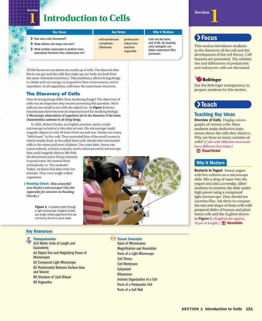
The Discovery of CellsHow do living things differ from nonliving things? The discovery of cells was an important step toward answering this question. Most cells are too small to see with the naked eye. As Figure 1 shows, microscopes have become an important tool for studying biology. V Microscope observations of organisms led to the discovery of the basic
characteristics common to all living things.
In 1665, Robert Hooke, an English scientist, used a crude microscope to look at a thin slice of cork. His microscope could magnify objects to only 30 times their normal size. Hooke saw many “little boxes” in the cork. They reminded him of the small rooms in which monks lived, so he called them cells. Hooke later discovered cells in the stems and roots of plants. Ten years later, Anton van Leeuwenhoek, a Dutch scientist, used a more powerful microscope that could magnify objects 300-fold. He discovered many living creatures in pond water. He named them animalcules, or “tiny animals.” Today, we know that they were not animals. They were single-celled organisms.
V Reading Check How powerful was Hooke’s microscope? (See the Appendix for answers to Reading Checks.)
prokaryoteeukaryotenucleusorganelle
V V How were cells discovered?
V V What defines cell shape and size?
V V What enables eukaryotes to perform more
specialized functions than prokaryotes do?
Figure 1 A student looks through a light microscope. Euglena (inset) are single-celled organisms that are commonly found in pond water.
hb08se_csf_s01.indd 151 3/20/07 11:00:39 AM
SECTION 1 Introduction to Cells 151
Section1This section introduces students to the discovery of the cell and the development of the cell theory. Cell features are presented. The similari-ties and differences of prokaryotic and eukaryotic cells are discussed.
Use the Bellringer transparency to prepare students for this section.
Teaching Key IdeasOverview of Cells Display micro-graphs of various cells. Have students make deductive state-ments about the cells they observe. Why are there so many varieties of cells? (Cells with different structures have different functions.) Visual/Verbal
Bacteria in Yogurt Smear yogurt with live cultures on a microscope slide. Mix a drop of water into the yogurt and add a coverslip. Allow students to examine the slide under high power using a compound light microscope. They should see Lactobacillus. Ask them to compare the size and shape of these cells with prepared slides of human and plant tissue cells and the Euglena shown in Figure 1. (Euglena are approx. 25μm in length.) Kinesthetic
Key Resources
Visual Concepts
Types of Microscopes
Magnifi cation and Resolution
Parts of a Light Microscope
Cell Theory
Cell Membrane
Cytoplasm
Ribosomes
Internal Organization of a Cell
Parts of a Prokaryotic Cell
Parts of a Cell Wall
Transparencies
A10 Metric Units of Length and
Equivalents
A4 Object Size and Magnifying Power of
Microscopes
A5 Compound Light Microscope
B2 Relationship Between Surface Area
and Volume
B8 Structure of Lipid Bilayer
B9 Organelles
hb08te_csf_s01.indd 151 4/16/07 6:13:59 PM
Cell Theory It took more than 150 years for scientists to fully appreciate the discoveries of Hooke and Leeuwenhoek. By the 1830s, microscopes were powerful enough to resolve structures only 1 μm apart. In 1838, Matthias Schleiden, a German botanist, concluded that cells make up every part of a plant. A year later, Theodor Schwann, a German zoologist, discovered that animals are also made up of cells. In 1858, Rudolph Virchow, a German physician, proposed that cells come only from the division of existing cells. The observa-tions of Schleiden, Schwann, and Virchow form the cell theory:
• All living things are made up of one or more cells.
• Cells are the basic units of structure and function in organisms.
• All cells arise from existing cells.
The cell theory has withstood the rigorous examination of cells by scientists equipped with today’s high-powered microscopes. As new tools and techniques are invented, scientists will learn more about the characteristics of cells.
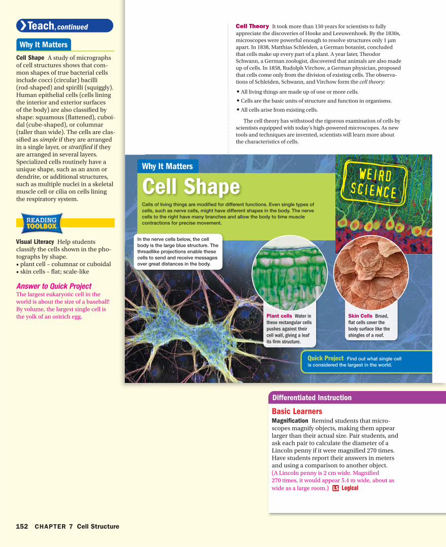
Cell ShapeWhy It Matters
Cells of living things are modified for different functions. Even single types of cells, such as nerve cells, might have different shapes in the body. The nerve cells to the right have many branches and allow the body to time muscle contractions for precise movement.
Quick Project Find out what single cell is considered the largest in the world.
In the nerve cells below, the cell body is the large blue structure. The threadlike projections enable these cells to send and receive messages over great distances in the body.
Plant cells Water in
these rectangular cells
pushes against their
cell wall, giving a leaf
its firm structure.
Skin Cells Broad,
flat cells cover the
body surface like the
shingles of a roof.
hb08se_csf_s01.indd 152 9/12/06 9:30:47 AM
152 CHAPTER 7 Cell Structure
Cell Shape A study of micrographs of cell structures shows that com-mon shapes of true bacterial cells include cocci (circular) bacilli (rod-shaped) and spirilli (squiggly). Human epithelial cells (cells lining the interior and exterior surfaces of the body) are also classified by shape: squamous (flattened), cuboi-dal (cube-shaped), or columnar (taller than wide). The cells are clas-sified as simple if they are arranged in a single layer, or stratified if they are arranged in several layers. Specialized cells routinely have a unique shape, such as an axon or dendrite, or additional structures, such as multiple nuclei in a skeletal muscle cell or cilia on cells lining the respiratory system.
Visual Literacy Help students classify the cells shown in the pho-tographs by shape. • plant cell – columnar or cuboidal• skin cells – flat; scale-like
Answer to Quick ProjectThe largest eukaryotic cell in the world is about the size of a baseball! By volume, the largest single cell is the yolk of an ostrich egg.
Basic LearnersMagnification Remind students that micro-scopes magnify objects, making them appear larger than their actual size. Pair students, and ask each pair to calculate the diameter of a Lincoln penny if it were magnified 270 times. Have students report their answers in meters and using a comparison to another object. (A Lincoln penny is 2 cm wide. Magnified 270 times, it would appear 5.4 m wide, about as wide as a large room.) Logical
hb08te_csf_s01.indd 152 11/16/06 9:30:35 AM
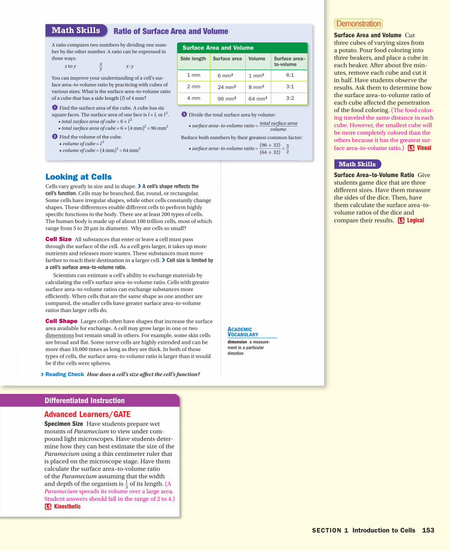
Surface Area and Volume
Side length Surface area Volume Surface area–to-volume
1 mm 6 mm2 1 mm3 6:1
2 mm 24 mm2 8 mm3 3:1
4 mm 96 mm2 64 mm3 3:2
Looking at CellsCells vary greatly in size and in shape. V A cell’s shape reflects the
cell’s function. Cells may be branched, flat, round, or rectangular. Some cells have irregular shapes, while other cells constantly change shapes. These differences enable different cells to perform highly specific functions in the body. There are at least 200 types of cells. The human body is made up of about 100 trillion cells, most of which range from 5 to 20 μm in diameter. Why are cells so small?
Cell Size All substances that enter or leave a cell must pass through the surface of the cell. As a cell gets larger, it takes up more nutrients and releases more wastes. These substances must move farther to reach their destination in a larger cell. V Cell size is limited by
a cell’s surface area–to-volume ratio.
Scientists can estimate a cell’s ability to exchange materials by calculating the cell’s surface area–to-volume ratio. Cells with greater surface area–to-volume ratios can exchange substances more efficiently. When cells that are the same shape as one another are compared, the smaller cells have greater surface area–to-volume ratios than larger cells do.
Cell Shape Larger cells often have shapes that increase the surface area available for exchange. A cell may grow large in one or two dimensions but remain small in others. For example, some skin cells are broad and flat. Some nerve cells are highly extended and can be more than 10,000 times as long as they are thick. In both of these types of cells, the surface area–to-volume ratio is larger than it would be if the cells were spheres.
V Reading Check How does a cell’s size affect the cell’s function?
ACADEMIC VOCABULARY
dimension a measure-
ment in a particular
direction
Ratio of Surface Area and Volume
A ratio compares two numbers by dividing one num-ber by the other number . A ratio can be expressed in three ways:
x to y x _ y x : y
You can improve your understanding of a cell’s sur-face area–to-volume ratio by practicing with cubes of various sizes. What is the surface area–to-volume ratio of a cube that has a side length (l) of 4 mm?
1 Find the surface area of the cube. A cube has six square faces. The surface area of one face is l × l, or l 2. • total surface area of cube = 6 × l 2
• total surface area of cube = 6 × (4 mm)2 = 96 mm2
2 Find the volume of the cube. • volume of cube = l 3
• volume of cube = (4 mm)3 = 64 mm3
3 Divide the total surface area by volume:
• surface area–to-volume ratio = total surface area __ volume
Reduce both numbers by their greatest common factor:
• surface area–to-volume ratio = (96 ÷ 32)
_ (64 ÷ 32)
= 3 _ 2
Math Skills
hb08se_csf_s01.indd 153 9/12/06 9:31:11 AM
SECTION 1 Introduction to Cells 153
Surface Area and Volume Cut three cubes of varying sizes from a potato. Pour food coloring into three beakers, and place a cube in each beaker. After about five min-utes, remove each cube and cut it in half. Have students observe the results. Ask them to determine how the surface area–to-volume ratio of each cube affected the penetration of the food coloring. (The food color-ing traveled the same distance in each cube. However, the smallest cube will be more completely colored than the others because it has the greatest sur-face area–to-volume ratio.) Visual
Surface Area–to-Volume Ratio Give students game dice that are three different sizes. Have them measure the sides of the dice. Then, have them calculate the surface area–to-volume ratios of the dice and compare their results. Logical
Advanced Learners/GATESpecimen Size Have students prepare wet mounts of Paramecium to view under com-pound light microscopes. Have students deter-mine how they can best estimate the size of the Paramecium using a thin centimeter ruler that is placed on the microscope stage. Have them calculate the surface area–to-volume ratio of the Paramecium assuming that the width and depth of the organism is 1 __ 3 of its length. (A Paramecium spreads its volume over a large area. Student answers should fall in the range of 2 to 4.)
Kinesthetic
hb08te_csf_s01.indd 153 11/16/06 9:30:45 AM

CSFS01011A
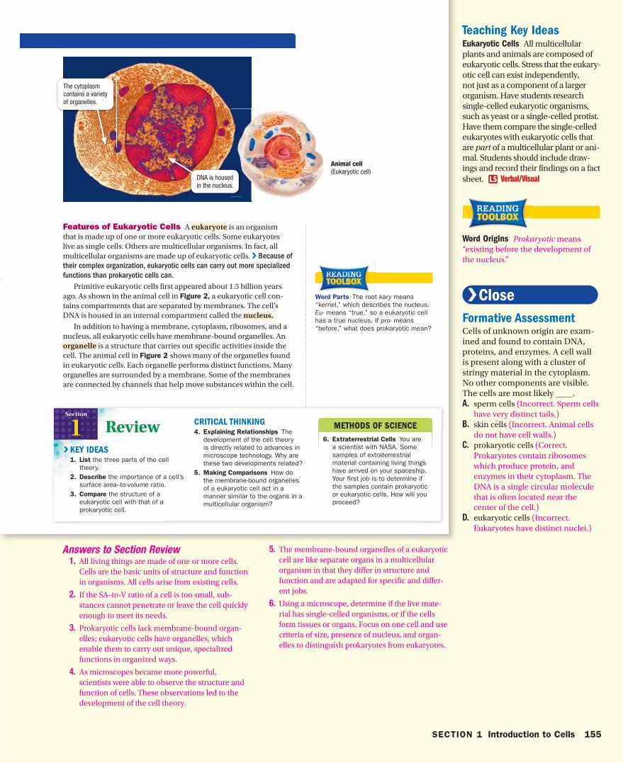
Cell FeaturesAll cells—from bacteria to those in a berry, bug, or bunny—share common structural features. All cells have a cell membrane, cyto-plasm, ribosomes, and DNA. The cell membranecell membrane is the cell’s outer boundary. It acts as a barrier between the outside environment and the inside of the cell. The cytosol, the fluid inside the cell, is full of dissolved particles. The cytoplasmcytoplasm includes this fluid and almost all of the structures that are suspended in the fluid. Many ribosomes are found in the cytoplasm. A ribosomeribosome is a cellular structure on which proteins are made. All cells also have DNA, the genetic material. DNA provides instructions for making proteins, regulates cellular activ-ities, and enables cells to reproduce.
Features of Prokaryotic Cells The bacterium shown in Figure 2 is an example of a prokaryote,prokaryote, an organism that is a single prokaryotic cell. A prokaryotic cell is quite simple in its organiza-tion. The genetic material is a single loop of DNA, which looks like a tangled string and usually lies near the center of the cell. Ribosomes and enzymes share the cytoplasm with the DNA.
Prokaryotic cells have a cell wall that surrounds the cell mem-brane and that provides structure and support. Some prokaryotic cell walls are surrounded by a capsule, a structure that enables prokary-otes to cling to surfaces, including teeth, skin, and food.
Scientists think that the first prokaryotes may have lived 3.5 bil-lion years ago or more. For millions of years, prokaryotes were the only organisms on Earth. They were very simple and small (1 to 2 μm in diameter). Like their ancestors, modern prokaryotes are also very small (1 to 15 μm), and they live in a wide range of habitats. Prokaryotes make up a very large and diverse group of cells.
V Reading Check What is a ribosome?
cell membrane a phospholipid layer that covers a cell’s surface and acts as a barrier between the inside of a cell and the cell’s environment
cytoplasm (SIET oh PLAZ uhm) the region of the cell within the membrane
ribosome (RIE buh SOHM) a cell organelle where protein synthesis occurs
prokaryote a single-celled organism that does not have a nucleus or membrane-bound organelles
eukaryote an organism made up of cells that have a nucleus and membrane-bound organelles
nucleus in a eukaryotic cell, a membrane-bound organelle that contains the cell’s DNA
organelle one of the small bodies that are found in the cytoplasm of a cell and that are specialized to perform a specific function
Figure 2 The cytoplasm of a prokaryotic cell (left) is made up of everything that is inside the cell membrane, including ribosomes and a loop of DNA. The cytoplasm of a eukaryotic cell (right) is made up of many different structures that are surrounded by membranes.
www.scilinks.orgTopic: Cell Features Code: HX80238
Features of Prokaryotic and Eukaryotic Cells
The loop of DNA is clustered but not surrounded by a membrane.
A cell wall covers the cell membrane.
Bacterium(Prokaryoticcell )
hb08se_csf_s01.indd 154 3/1/07 7:49:28 AM
154 CHAPTER 7 Cell Structure
Teaching Key IdeasInterpreting Graphics Draw stu-dents’ attention to Figure 2. Tell students that any color shown in electron micrographs is usually digi-tally applied; that is, the colors are “painted” by computers. These col-ors are artificial; the cell is not col-ored as is shown in the micrograph. Ask students why colorized electron micrographs might be useful. (to highlight or point out structures) Ask students which structures are shown in Figure 2. (cell wall and DNA)What other structures are in the cytoplasm of this cell that is not vis-ible in the micrograph? (ribosomes)
Visual
Prokaryote Diversity Modern prokaryotes include archaea (archaebacteria) and bacteria (eubac-teria). They can be found in nearly any envi-ronment on Earth, including volcanic vents at the bottom of the ocean and ice in Arctic and Antarctic regions. Some archaeae seem to thrive in harsh conditions. These types of organisms are referred to as extremophiles. Many bacteria live in or on the bodies of other organisms, including humans. They can cause bacterial infection, but in most cases the organisms are harmless or even beneficial to the host.
Special Education StudentsCell Model Have students use food to make a model of a eukaryotic cell. Students should use their understanding of the organelle structures to choose appropriate foods to represent them. Gelatin (cytoplasm) could be poured into a large, clear cellophane bag (cell membrane). Organelles can be represented by kidney beans (mitochondria), poppy seeds (ribosomes), sections of lasagna noodles (ER and Golgi apparatus), peppercorns (lysosomes), spa-ghetti noodles (cytoskeleton), and an orange (nucleus). Kinesthetic
hb08te_csf_s01.indd 154 4/3/07 8:12:13 AM
METHODS OF SCIENCE
6. Extraterrestrial Cells You are a scientist with NASA. Some samples of extraterrestrial material containing living things have arrived on your spaceship. Your first job is to determine if the samples contain prokaryotic or eukaryotic cells. How will you proceed?
ReviewSection
1KEY IDEAS1. List the three parts of the cell
theory. 2. Describe the importance of a cell’s
surface area–to-volume ratio. 3. Compare the structure of a
eukaryotic cell with that of a prokaryotic cell.
CRITICAL THINKING4. Explaining Relationships The
development of the cell theory is directly related to advances in microscope technology. Why are these two developments related?
5. Making Comparisons How do the membrane-bound organelles of a eukaryotic cell act in a manner similar to the organs in a multicellular organism?
V
Features of Eukaryotic Cells A eukaryoteeukaryote is an organism that is made up of one or more eukaryotic cells. Some eukaryotes live as single cells. Others are multicellular organisms. In fact, all multicellular organisms are made up of eukaryotic cells. V Because of
their complex organization, eukaryotic cells can carry out more specialized
functions than prokaryotic cells can.
Primitive eukaryotic cells first appeared about 1.5 billion years ago. As shown in the animal cell in Figure 2, a eukaryotic cell con-tains compartments that are separated by membranes. The cell’s DNA is housed in an internal compartment called the nucleus.nucleus. In addition to having a membrane, cytoplasm, ribosomes, and a nucleus, all eukaryotic cells have membrane-bound organelles. An organelleorganelle is a structure that carries out specific activities inside the cell. The animal cell in Figure 2 shows many of the organelles found in eukaryotic cells. Each organelle performs distinct functions. Many organelles are surrounded by a membrane. Some of the membranes are connected by channels that help move substances within the cell.
Word Parts The root kary means “kernel,” which describes the nucleus. Eu- means “true,” so a eukaryotic cell has a true nucleus. If pro- means “before,” what does prokaryotic mean?
DNA is housed in the nucleus.
The cytoplasm contains a variety of organelles.
Animal cell(Eukaryotic cell )
hb08se_csf_s01.indd 155 3/1/07 7:49:48 AM
SECTION 1 Introduction to Cells 155
Teaching Key IdeasEukaryotic Cells All multicellular plants and animals are composed of eukaryotic cells. Stress that the eukary-otic cell can exist independently, not just as a component of a larger organism. Have students research single-celled eukaryotic organisms, such as yeast or a single-celled protist. Have them compare the single-celled eukaryotes with eukaryotic cells that are part of a multicellular plant or ani-mal. Students should include draw-ings and record their findings on a fact sheet. Verbal/Visual
Word Origins Prokaryotic means “existing before the development of the nucleus.”
Formative AssessmentCells of unknown origin are exam-ined and found to contain DNA, proteins, and enzymes. A cell wall is present along with a cluster of stringy material in the cytoplasm. No other components are visible. The cells are most likely ____.A. sperm cells (Incorrect. Sperm cells
have very distinct tails.)B. skin cells (Incorrect. Animal cells
do not have cell walls.)C. prokaryotic cells (Correct.
Prokaryotes contain ribosomes which produce protein, and enzymes in their cytoplasm. The DNA is a single circular molecule that is often located near the center of the cell.)
D. eukaryotic cells (Incorrect. Eukaryotes have distinct nuclei.)
Answers to Section Review 1. All living things are made of one or more cells.
Cells are the basic units of structure and function in organisms. All cells arise from existing cells.
2. If the SA–to-V ratio of a cell is too small, sub-stances cannot penetrate or leave the cell quickly enough to meet its needs.
3. Prokaryotic cells lack membrane-bound organ-elles; eukaryotic cells have organelles, which enable them to carry out unique, specialized functions in organized ways.
4. As microscopes became more powerful, scientists were able to observe the structure and function of cells. These observations led to the development of the cell theory.
5. The membrane-bound organelles of a eukaryotic cell are like separate organs in a multicellular organism in that they differ in structure and function and are adapted for specific and differ-ent jobs.
6. Using a microscope, determine if the live mate-rial has single-celled organisms, or if the cells form tissues or organs. Focus on one cell and use criteria of size, presence of nucleus, and organ-elles to distinguish prokaryotes from eukaryotes.
hb08te_csf_s01.indd 155 4/3/07 8:12:36 AM
Cytoskeletonfibers
Nucleus
Organelles
Ribosomes
Inside the Eukaryotic CellSection
2Knowing how cells work
helps you understand
how your body functions
and what goes wrong
when you get sick.
Why It MattersKey Ideas Key Terms
The cytoplasm of a eukaryotic cell is packed with all sorts of struc-tures and molecules. Molecules can be concentrated in certain parts of the cell because of the membranes that divide the cytoplasm into compartments. This organization enables each organelle to perform highly sophisticated and specialized functions.
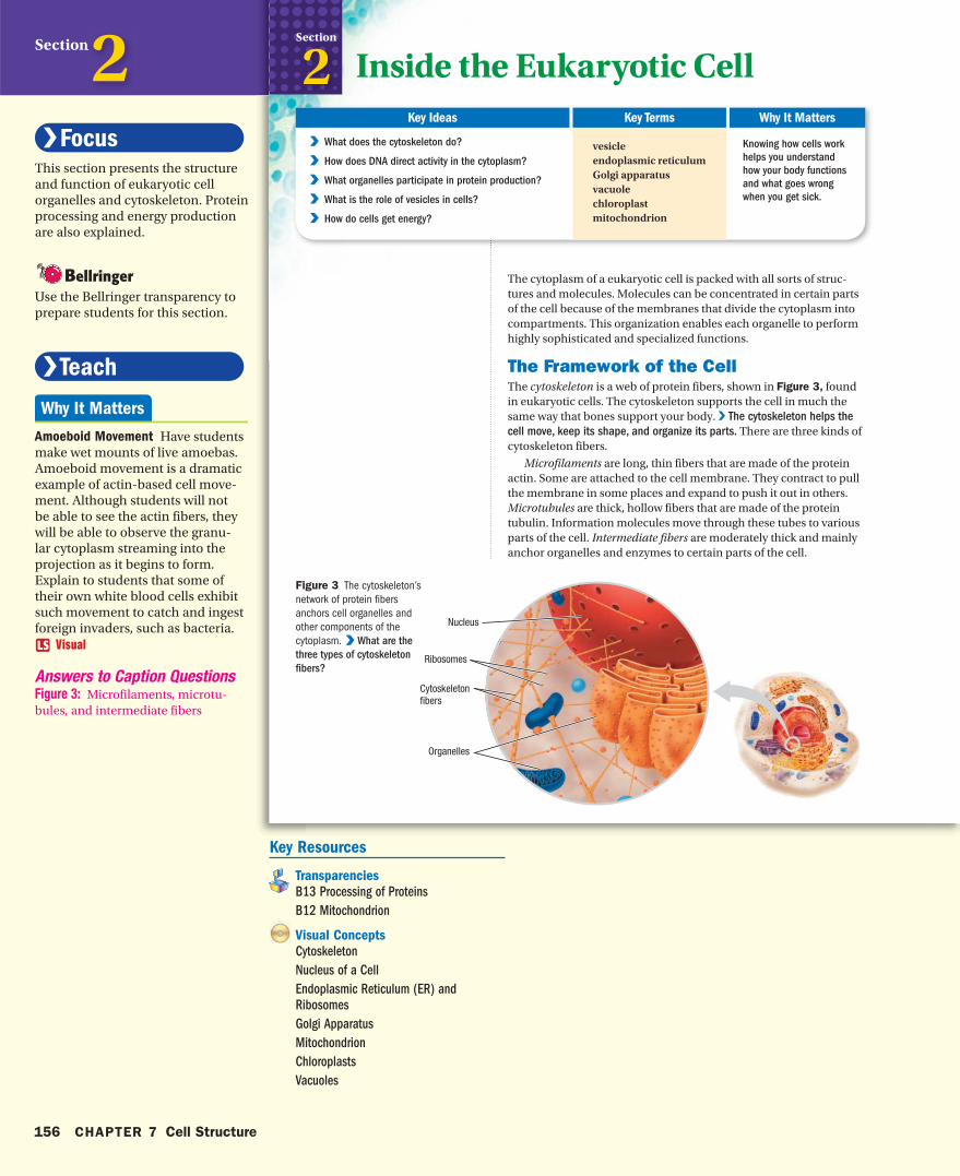
The Framework of the CellThe cytoskeleton is a web of protein fibers, shown in Figure 3, found in eukaryotic cells. The cytoskeleton supports the cell in much the same way that bones support your body. V The cytoskeleton helps the
cell move, keep its shape, and organize its parts. There are three kinds of cytoskeleton fibers.
Microfilaments are long, thin fibers that are made of the protein actin. Some are attached to the cell membrane. They contract to pull the membrane in some places and expand to push it out in others. Microtubules are thick, hollow fibers that are made of the protein tubulin. Information molecules move through these tubes to various parts of the cell. Intermediate fibers are moderately thick and mainly anchor organelles and enzymes to certain parts of the cell.
vesicleendoplasmic reticulumGolgi apparatusvacuolechloroplastmitochondrion
V V What does the cytoskeleton do?
V V How does DNA direct activity in the cytoplasm?
V V What organelles participate in protein production?
V V What is the role of vesicles in cells?
V V How do cells get energy?
Figure 3 The cytoskeleton’s network of protein fibers anchors cell organelles and other components of the cytoplasm. V V What are the
three types of cytoskeleton
fibers?
hb08se_csf_s02.indd 156 3/20/07 11:01:26 AM
156 CHAPTER 7 Cell Structure
Section2This section presents the structure and function of eukaryotic cell organelles and cytoskeleton. Protein processing and energy production are also explained.
Use the Bellringer transparency to prepare students for this section.
Amoeboid Movement Have students make wet mounts of live amoebas. Amoeboid movement is a dramatic example of actin-based cell move-ment. Although students will not be able to see the actin fibers, they will be able to observe the granu-lar cytoplasm streaming into the projection as it begins to form. Explain to students that some of their own white blood cells exhibit such movement to catch and ingest foreign invaders, such as bacteria.
Visual
Answers to Caption QuestionsFigure 3: Microfilaments, microtu-bules, and intermediate fibers
Key Resources
Transparencies
B13 Processing of Proteins
B12 Mitochondrion
Visual Concepts
Cytoskeleton
Nucleus of a Cell
Endoplasmic Reticulum (ER) and
Ribosomes
Golgi Apparatus
Mitochondrion
Chloroplasts
Vacuoles
hb08te_csf_s02.indd 156 4/16/07 6:14:47 PM
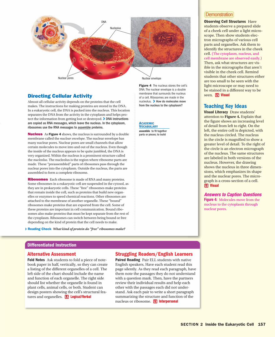
Nucleolus
Nuclear envelope Nuclear pores
DNA
Directing Cellular ActivityAlmost all cellular activity depends on the proteins that the cell makes. The instructions for making proteins are stored in the DNA. In a eukaryotic cell, the DNA is packed into the nucleus. This location separates the DNA from the activity in the cytoplasm and helps pro-tect the information from getting lost or destroyed. V DNA instructions
are copied as RNA messages, which leave the nucleus. In the cytoplasm,
ribosomes use the RNA messages to assemble proteins.
Nucleus As Figure 4 shows, the nucleus is surrounded by a double membrane called the nuclear envelope. The nuclear envelope has many nuclear pores. Nuclear pores are small channels that allow certain molecules to move into and out of the nucleus. Even though the inside of the nucleus appears to be quite jumbled, the DNA is very organized. Within the nucleus is a prominent structure called the nucleolus. The nucleolus is the region where ribosome parts are made. These “preassembled” parts of ribosomes pass through the nuclear pores into the cytoplasm. Outside the nucleus, the parts are assembled to form a complete ribosome.
Ribosomes Each ribosome is made of RNA and many proteins. Some ribosomes in a eukaryotic cell are suspended in the cytosol, as they are in prokaryotic cells. These “free” ribosomes make proteins that remain inside the cell, such as proteins that build new organ-elles or enzymes to speed chemical reactions. Other ribosomes are attached to the membrane of another organelle. These “bound” ribosomes make proteins that are exported from the cell. Some of these proteins are important in cell communication. Bound ribo-somes also make proteins that must be kept separate from the rest of the cytoplasm. Ribosomes can switch between being bound or free depending on the kind of protein that the cell needs to make.
V Reading Check What kind of protein do “free” ribosomes make?
Figure 4 The nucleus stores the cell’s DNA. The nuclear envelope is a double membrane that surrounds the nucleus of a cell. Ribosomes are made in the nucleolus. V How do molecules move
from the nucleus to the cytoplasm?
ACADEMIC VOCABULARY
assemble to fit together
parts or pieces; to build
hb08se_csf_s02.indd 157 10/12/06 9:15:25 AM
SECTION 2 Inside the Eukaryotic Cell 157
Observing Cell Structures Have students observe a prepared slide of a cheek cell under a light micro-scope. Then show students elec-tron micrographs of various cell parts and organelles. Ask them to identify the structures in the cheek cell. (The cytoplasm, nucleus, and cell membrane are observed easily.)Then, ask what structures are vis-ible in the micrographs that aren’t visible in the cheek cell. Remind students that other structures either are too small to be seen with the light microscope or may need to be stained in a different way to be seen. Visual
Teaching Key IdeasVisual Literacy Draw students’ attention to Figure 4. Explain that the figure shows an increasing level of detail from left to right. On the left, the entire cell is depicted, with the nucleus circled. The nucleus in the circle is magnified to show a greater level of detail. To the right of the circle is an electron micrograph of the nucleus. The same structures are labeled in both versions of the nucleus. However, the drawing shows the nucleus in three dimen-sions, which emphasizes its shape and the nuclear pores. The micro-graph is a cross-section of a cell.
Visual
Answers to Caption QuestionsFigure 4: Molecules move from the nucleus to the cytoplasm through nuclear pores.
Struggling Readers/English LearnersPaired Reading Pair ELL students with native English speakers. Have each student read this page silently. As they read each paragraph, have them note the passages they do not understand with a question mark. Then, have the partners review their individual results and help each other with the passages each did not under-stand. Ask each pair to write a short paragraph summarizing the structure and function of the nucleus or ribosome. Interpersonal
Alternative AssessmentFold Notes Ask students to fold a piece of note-book paper in half, vertically, so they can create a listing of the different organelles of a cell. The left side of the chart should include the name and function of each organelle. The right side should list whether the organelle is found in plant cells, animal cells, or both. Student can design posters showing the cell’s structural fea-tures and organelles. Logical/Verbal
hb08te_csf_s02.indd 157 11/16/06 9:29:35 AM
Protein ProcessingThe proteins produced by cells have many uses. The proteins that are sent outside the cell must be kept separate from the rest of the cytoplasm. To achieve this separation, the cell packages the proteins in vesicles. A vesiclevesicle is a small, often spherical-shaped sac that is formed by a membrane.
In a eukaryotic cell, two structures are mainly responsible for modifying, packaging, and transporting proteins for use outside the cell. V The endoplasmic reticulum and the Golgi apparatus are organelles
that prepare proteins for extracellular export.
Endoplasmic Reticulum The endoplasmic reticulumendoplasmic reticulum (ER) is a system of internal membranes that moves proteins and other sub-stances through the cell. The membrane of the ER is connected to the outer membrane of the nuclear envelope.
Rough ER Ribosomes are attached to some parts of the surface of the ER. This rough ER has a bumpy appearance when viewed with an electron microscope, as shown in Figure 5. 1 As proteins are made, they cross the ER membrane, entering the ER . Then, the ER mem-brane pinches off to form a vesicle around the proteins.
Smooth ER The rest of the ER, called smooth ER, has no attached ribosomes. Thus it appears smooth when viewed with an electron microscope. Enzymes of the smooth ER performs various functions, such as making lipids and breaking down toxic substances.
Golgi Apparatus The GolgiGolgi apparatusapparatus is a set of flattened, membrane-bound sacs. Cell products enter one side of the Golgi apparatus, which modifies, sorts, and packages them for distribution.
Repackaging Vesicles that contain newly made proteins move through the cytoplasm from the ER to the Golgi apparatus. 2 The vesicle membrane fuses with the Golgi membrane. Inside the Golgi apparatus, enzymes modify the proteins as they move through the organelle. On the other side, the finished proteins are enclosed in new vesicles that bud from the surface of the Golgi apparatus.
Exporting Many of these vesicles then migrate to the cell mem-brane. 3 As the vesicle membrane fuses with the cell membrane, the completed proteins are released to the outside the cell.
Storage and MaintenanceVesicles have many functions in the cell. Some transport materials within the cell. Others have important storage roles. V Vesicles help
maintain homeostasis by storing and releasing various substances as the
cell needs them.
Lysosome A lysosome is a vesicle that contains specific enzymes that break down large molecules. These enzymes can digest food particles to provide nutrients for the cell. They also help recycle materials in the cell by digesting old, damaged, or unused organelles. Lysosomes work by fusing with other vesicles. Lysosomes, made by the Golgi apparatus, prevent the enzymes from destroying the cell.
vesicle a small cavity or sac that contains materials in a eukaryotic cell
endoplasmic reticulum (EN doh PLAZ mik ri TIK yuh luhm) a system of membranes that is found in a cell’s cytoplasm and that assists in the production, processing, and transport of proteins and in the production of lipids
Golgi apparatus (GOHL jee) a cell organelle that helps make and package materials to be transported out of the cell
www.scilinks.orgTopic: Proteins Code: HX 81241
Process Chart Make a process chart that shows how the cell digests food particles.
hb08se_csf_s02.indd 158 9/12/06 9:29:58 AM
158 CHAPTER 7 Cell Structure
Filtering Out Toxins Enzymes found in the abundant smooth ER of the liver help detoxify drugs and environmental pollutants. Detoxification usually involves a series of chemical reactions within the smooth ER of the liver that make the drug more water soluble, so it can be excreted in the urine. The ingestion of many drugs, including barbiturates and alcohol, trigger an increase in the amount of smooth ER and accompanying enzymes in liver cells. Have students research this detoxifying function of the smooth ER. Verbal
Process Chart Have students refer to the Reading Tool Box activity at the beginning of the chapter to create a process chart explaining the protein sequence. Logical/Visual
Lysosomal Malfunctions Tay-Sachs disease is caused by the deficiency of a lysosomal enzyme that digests lipids. As a result, cells in the brain fill with lipids, eventually causing death. Pompe’s disease results when a lysosomal enzyme that breaks down glycogen is absent. Glycogen is the energy storage compound for the body, so it’s abundant in muscle. Without the enzyme to break down glycogen, the lyso-somes in the heart and skeletal muscle quickly accumulate large amounts of glycogen. These muscles are progressively weakened, especially the heart.
Basic LearnersWhat Am I? Have each student choose one cell part or organelle and write a “What Am I?” essay. The essay should be written in the first person and should describe the structure and function of that cell part. Encourage stu-dents to be creative in their writing styles. Have students read their essays aloud in class. The class should guess the structure or organelle described in the essay. Verbal/Interpersonal
hb08te_csf_s02.indd 158 11/21/06 12:35:52 PM
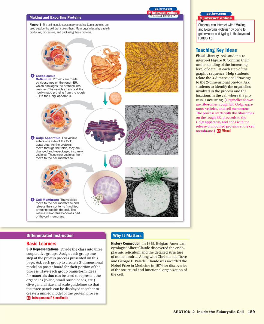
Making and Exporting ProteinsKeyword: HX8CSFF5
1 Endoplasmic Reticulum Proteins are made by ribosomes on the rough ER, which packages the proteins into vesicles. The vesicles transport the newly made proteins from the rough ER to the Golgi apparatus.
2 Golgi Apparatus The vesicle enters one side of the Golgi apparatus. As the proteins move through the folds, they are changed and repackaged into new vesicles. These new vesicles then move to the cell membrane.
3 Cell Membrane The vesicles move to the cell membrane and release their contents (modified proteins) outside the cell. The vesicle membrane becomes part of the cell membrane.
Figure 5 The cell manufactures many proteins. Some proteins are used outside the cell that makes them. Many organelles play a role in producing, processing, and packaging these proteins.
hb08se_csf_s02.indd 159 10/12/06 9:15:26 AM
SECTION 2 Inside the Eukaryotic Cell 159
Basic Learners3-D Representations Divide the class into three cooperative groups. Assign each group one step of the protein process presented on this page. Ask each group to create a 3-dimensional model on poster board for their portion of the process. Have each group brainstorm ideas for materials that can be used to represent the organelles (twine, small round beads, etc.). Give general size and scale guidelines so that the three panels can be displayed together to create a unified model of the protein process.
Intrapersonal/ Kinesthetic
Teaching Key IdeasVisual Literacy Ask students to interpret Figure 6. Confirm their understanding of the increasing level of detail at each step of the graphic sequence. Help students relate the 3-dimensional drawings to the 2-dimensional photos. Ask students to identify the organelles involved in the process and the locations in the cell where the pro-cess is occurring. (Organelles shown are ribosomes, rough ER, Golgi appa-ratus, vesicles, and cell membrane. The process starts with the ribosomes on the rough ER, proceeds to the Golgi apparatus, and ends with the release of modified proteins at the cell membrane.) Visual
Students can interact with “Making and Exporting Proteins” by going to go.hrw.com and typing in the keyword HX8CSFF5.
History Connection In 1945, Belgian-American cytologist Albert Claude discovered the endo-plasmic reticulum and the detailed structure of mitochondria. Along with Christian de Duve and George E. Palade, Claude was awarded the Nobel Prize in Medicine in 1974 for discoveries of the structural and functional organization of the cell.
hb08te_csf_s02.indd 159 11/16/06 9:29:48 AM
Chloroplast
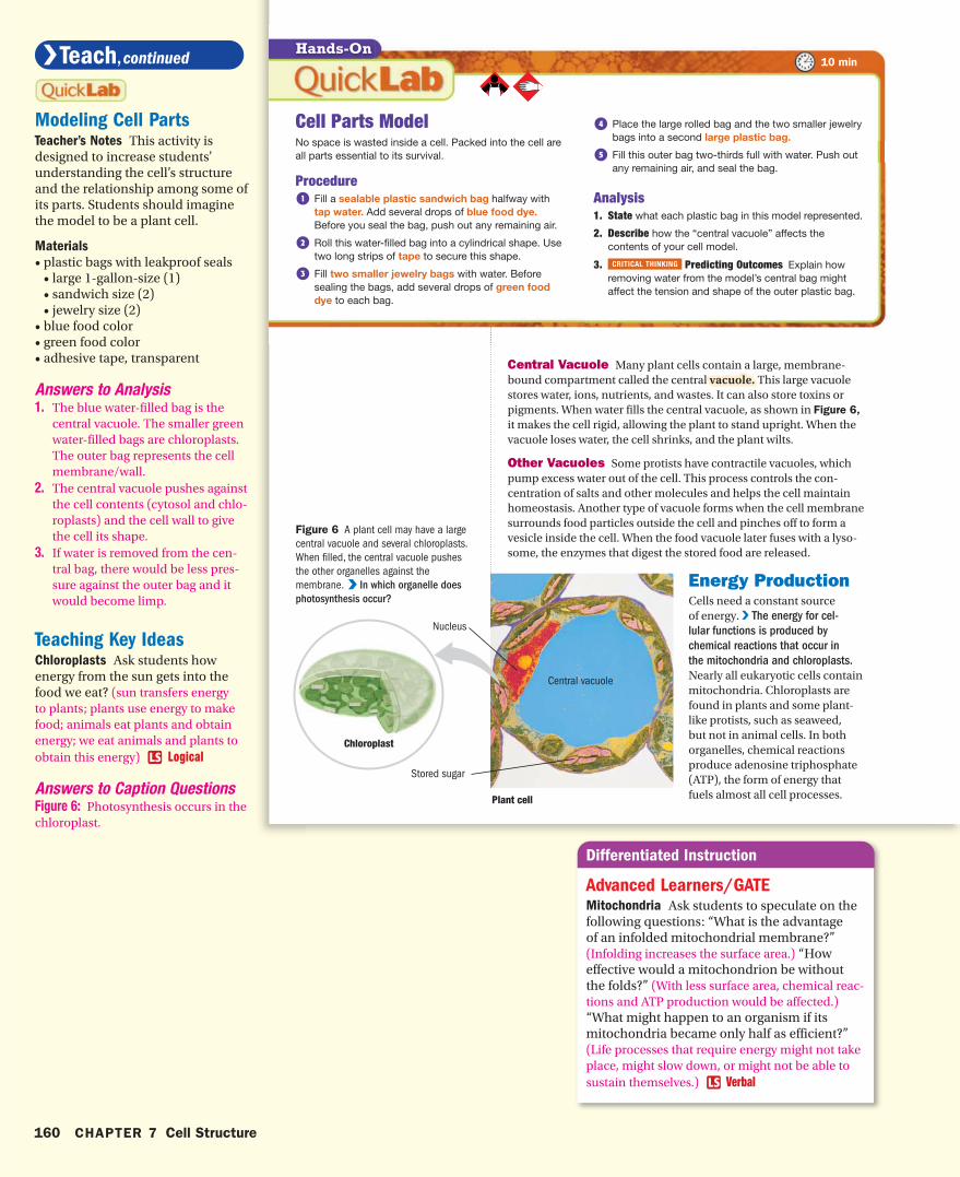
Central Vacuole Many plant cells contain a large, membrane-bound compartment called the central vacuole.vacuole. This large vacuole stores water, ions, nutrients, and wastes. It can also store toxins or pigments. When water fills the central vacuole, as shown in Figure 6, it makes the cell rigid, allowing the plant to stand upright. When the vacuole loses water, the cell shrinks, and the plant wilts.
Other Vacuoles Some protists have contractile vacuoles, which pump excess water out of the cell. This process controls the con-centration of salts and other molecules and helps the cell maintain homeostasis. Another type of vacuole forms when the cell membrane surrounds food particles outside the cell and pinches off to form a vesicle inside the cell. When the food vacuole later fuses with a lyso-some, the enzymes that digest the stored food are released.
Energy ProductionCells need a constant source of energy. V The energy for cel-
lular functions is produced by
chemical reactions that occur in
the mitochondria and chloroplasts. Nearly all eukaryotic cells contain mitochondria. Chloroplasts are found in plants and some plant-like protists, such as seaweed, but not in animal cells. In both organelles, chemical reactions produce adenosine triphosphate (ATP), the form of energy that fuels almost all cell processes.
Cell Parts ModelNo space is wasted inside a cell. Packed into the cell are all parts essential to its survival.
Procedure1 Fill a sealable plastic sandwich bag halfway with
tap water. Add several drops of blue food dye. Before you seal the bag, push out any remaining air.
2 Roll this water-filled bag into a cylindrical shape. Use two long strips of tape to secure this shape.
3 Fill two smaller jewelry bags with water. Before sealing the bags, add several drops of green food dye to each bag.
4 Place the large rolled bag and the two smaller jewelry bags into a second large plastic bag.
5 Fill this outer bag two-thirds full with water. Push out any remaining air, and seal the bag.
Analysis1. State what each plastic bag in this model represented.
2. Describe how the “central vacuole” affects the contents of your cell model.
3. CRITICAL THINKING Predicting Outcomes Explain how removing water from the model’s central bag might affect the tension and shape of the outer plastic bag.
Figure 6 A plant cell may have a large central vacuole and several chloroplasts. When filled, the central vacuole pushes the other organelles against the membrane. V V In which organelle does
photosynthesis occur?
Hands-On10 min
Plant cell
Nucleus
Central vacuole
Stored sugar
hb08se_csf_s02.indd 160 9/12/06 9:30:09 AM
160 CHAPTER 7 Cell Structure
Modeling Cell PartsTeacher’s Notes This activity is designed to increase students’ understanding the cell’s structure and the relationship among some of its parts. Students should imagine the model to be a plant cell.
Materials
• plastic bags with leakproof seals • large 1-gallon-size (1) • sandwich size (2) • jewelry size (2)• blue food color• green food color• adhesive tape, transparent
Answers to Analysis1. The blue water-filled bag is the
central vacuole. The smaller green water-filled bags are chloroplasts. The outer bag represents the cell membrane/wall.
2. The central vacuole pushes against the cell contents (cytosol and chlo-roplasts) and the cell wall to give the cell its shape.
3. If water is removed from the cen-tral bag, there would be less pres-sure against the outer bag and it would become limp.
Teaching Key IdeasChloroplasts Ask students how energy from the sun gets into the food we eat? (sun transfers energy to plants; plants use energy to make food; animals eat plants and obtain energy; we eat animals and plants to obtain this energy) Logical
Answers to Caption QuestionsFigure 6: Photosynthesis occurs in the chloroplast.
Advanced Learners/GATEMitochondria Ask students to speculate on the following questions: “What is the advantage of an infolded mitochondrial membrane?” (Infolding increases the surface area.) “How effective would a mitochondrion be without the folds?” (With less surface area, chemical reac-tions and ATP production would be affected.) “What might happen to an organism if its mitochondria became only half as efficient?” (Life processes that require energy might not take place, might slow down, or might not be able to sustain themselves.) Verbal
hb08te_csf_s02.indd 160 11/16/06 9:56:50 AM

Outermembrane
Innermembrane
Chloroplasts A chloroplastchloroplast is an organelle that uses light energy to make sugar from carbon dioxide and water. As Figure 6 shows, plant cells may have several chloroplasts. Each chloroplast is sur-rounded by a pair of membranes. Inside the inner membrane are many stacks of flattened sacs. The ATP-producing chemical reactions take place on the membranes of these sacs.
Mitochondria A mitochondrionmitochondrion is an organelle that uses energy from organic compounds to make ATP. Although some ATP is made in the cytosol, most of a cell’s ATP is made inside mitochondria. Cells that have a high energy requirement, such as muscle cells, may contain hundreds or thousands of mitochondria. As Figure 7 shows, a mitochondrion has a smooth outer membrane. It also has a greatly folded inner membrane, which divides the organelle into two com-partments. Many ATP-producing enzymes are located on the inner membrane.
V Reading Check In what kinds of cells are mitochondria found?
ALTERNATIVE ASSESSMENT
8. Analogy Compare the organelles of a eukaryotic cell to the parts of a city. For example, the lysosome could be a recycling center.
ReviewSection
2KEY IDEAS1. Compare the functions of the three
types of cytoskeletal fibers. 2. Describe the nucleus. 3. Trace a protein’s path through the
cell, from assembly to export. 4. Contrast vesicles and vacuoles.
5. Compare the role of mitochondria and chloroplasts.
CRITICAL THINKING6. Constructing Explanations Is it
accurate to say that organelles are floating freely in the cytosol? Why or why not?
7. Real World Research Tay-Sachs disease, and explain what goes wrong in diseased cells.
V
vacuole (VAK yoo OHL) a fluid-filled vesicle found in the cytoplasm of plant cells or protists
chloroplast an organelle found in plant and algae cells where photosynthesis occurs
mitochondrion (MIET oh KAHN dree uhn) in eukaryotic cells, the cell organelle that is surrounded by two membranes and that is the site of cellular respiration
Figure 7 A mitochondrion uses the energy in organic molecules to make ATP for the cell. The mitochondrion has two membranes. V V Where in the
mitochondrion is ATP produced?
hb08se_csf_s02.indd 161 3/1/07 7:50:29 AM
SECTION 2 Inside the Eukaryotic Cell 161
Mitochondria in Plant Cells Plant cells, as well as animal cells and almost all other eukaryotic cells, contain mitochondria. Some students think that because plants perform photosynthesis, they do not contain mitochon-dria. However, it is important for students to understand that the products of photosyn-thesis among other things are used to fuel the manufacture of ATP in the mitochondria. Mitochondrial ATP synthesis takes place in parts of plants that do not undergo photosyn-thesis, such as the roots, and at night in parts of plants that do undergo photosynthesis, such as the leaves.
Formative AssessmentWhat is the function of the mitochondria?A. make carbohydrates (Incorrect.
Chloroplasts use the sun’s energy, carbon dioxide and water to pro-duce carbohydrates.)
B. make ATP (Correct. The mitochon-drion makes ATP, which is impor-tant for energy transfer in living things.)
C. make cytoplasm (Incorrect. Mitochondria have no role in cyto-plasm formation.)
D. move proteins through the cell (Incorrect. The rough ER, the Golgi apparatus, and vesicles move pro-teins through the cell.)
Answers to Section Review 1. The cell moves and changes its shape by the
action of actin microfilaments. Microtubules move molecules to different parts of the cell. Intermediate fibers help anchor organelles and enzymes in certain parts of the cell.
2. A double membrane with many pores surrounds the nucleus. The nucleus contains most of the cell’s DNA and a nucleolus, where ribosome parts are made.
3. ribosomes, rough ER, vesicles, Golgi apparatus, vesicles, and cell membrane
4. Vesicles are generally smaller and hold one par-ticular type of molecule. Vacuoles are larger and may hold a variety of different substances.
5. Both mitochondria and chloroplasts are the sites of chemical reactions that produce ATP, the energy currency of cells.
6. Organelles exist within the network of protein fibers that make up the cytoskeleton. This restricts their movement.
7. Nerve cells in affected children accumulate high levels of a glycolipid called ganglioside. The con-dition results from a missing lysosomal enzyme. The lysosomes of these cells fill with fragments of membranes that contain the undigested gan-glioside causing nerve cells to not function. As a result, affected children have mental deteriora-tion, paralysis, and will die within three years.
8. Answers will vary. Sample answer: the mitochon-dria could be electrical power plants.
Answers to Caption QuestionsFigure 7: ATP is produced on the inner membrane.
hb08te_csf_s02.indd 161 4/3/07 8:15:03 AM
Ribosome
Cell membrane
Pili
Flagellum
Cell wall
DNA
Diverse organisms have
unique cells and cellular
organization.
organ systemcolonial
organism
Why It MattersKey Ideas Key Terms
More than 50 million types of organisms live on Earth. Each organ-ism is made up of different types of cells. Differences in cells enable organisms to adapt to their natural environments.
Diversity in CellsProkaryotes are always unicellular and limited in size. Eukaryotes are often larger and can be either unicellular or multicellular. Prokaryotic cells lack a nucleus and membrane-bound organelles, which are found in eukaryotic cells. Within both types, cells can have a variety of shapes and structures. Recall that a cell’s shape reflects its func-tion. VV The different organelles and features of cells enable organisms to
function in unique ways in different environments.
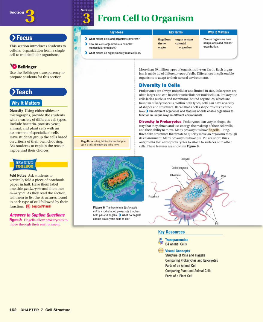
Diversity in Prokaryotes Prokaryotes can vary in shape, the way that they obtain and use energy, the makeup of their cell walls, and their ability to move. Many prokaryotes have flagellaflagella—long, threadlike structures that rotate to quickly move an organism through its environment. Many prokaryotes have pili. Pili are short, thick outgrowths that allow prokaryotes to attach to surfaces or to other cells. These features are shown in Figure 8.
flagellumtissueorgan
V V What makes cells and organisms different?
V V How are cells organized in a complex
multicellular organism?
V V What makes an organism truly multicellular?
From Cell to OrganismSection
3
Figure 8 The bacterium Escherichia coli is a rod-shaped prokaryote that has both pili and flagella. V V What do flagella
enable prokaryotic cells to do?
flagellum a long, hairlike structure that grows out of a cell and enables the cell to move
hb08se_csf_s03.indd 162 3/20/07 11:02:16 AM
162 CHAPTER 7 Cell Structure
Section3This section introduces students to cellular organization from a single cell to multicellular organisms.
Use the Bellringer transparency to prepare students for this section.
Diversity Using either slides or micrographs, provide the students with a variety of different cell types. Include bacteria, protists, yeast, animal, and plant cells with an assortment of specialized cells. Have students group the cells based on criteria of their own choosing. Ask students to explain the reason-ing behind their choices.
Fold Notes Ask students to vertically fold a piece of notebook paper in half. Have them label one side prokaryote and the other eukaryote. As they read the section, tell them to list the structures found in each type of cell followed by their function. Logical/Visual
Answers to Caption QuestionsFigure 8: Flagella allow prokaryotes to move through their environment.
Key Resources
Transparencies
B4 Animal Cells
Visual Concepts
Structure of Cilia and Flagella
Comparing Prokaryotes and Eukaryotes
Parts of an Animal Cell
Comparing Plant and Animal Cells
Parts of a Plant Cell
hb08te_csf_s03.indd 162 4/16/07 6:15:52 PM
Mitochondrion
Vesicle
Ribosomes
Golgiapparatus
Smooth ER Rough ER
Cell membrane
NucleusNucleolus
Cytoskeleton fibers
Chloroplast
Cell membrane
Vesicle Golgi apparatus
Smooth ER
Rough ER
Nucleus
Nucleolus
Cytoskeleton fibers
Cell wall
Mitochondrion
Ribosome
Central vacuole
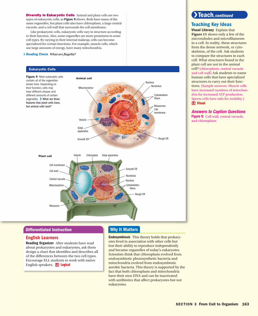
Figure 9 Most eukaryotic cells contain all of the organelles shown here. Depending on their function, cells may have different shapes and different amounts of certain organelles. V What are three
features that plant cells have,
but animal cells lack?
Animal cell
Plant cell
Eukaryotic Cells
Diversity in Eukaryotic Cells Animal and plant cells are two types of eukaryotic cells, as Figure 9 shows. Both have many of the same organelles, but plant cells also have chloroplasts, a large central vacuole, and a cell wall that surrounds the cell membrane.
Like prokaryotic cells, eukaryotic cells vary in structure according to their function. Also, some organelles are more prominent in some cell types. By varying in their internal makeup, cells can become specialized for certain functions. For example, muscle cells, which use large amounts of energy, have many mitochondria.
V Reading Check What are flagella?
hb08se_csf_s03.indd 163 9/12/06 9:29:03 AM
SECTION 3 From Cell to Organism 163
Teaching Key IdeasVisual Literacy Explain that Figure 11 shows only a few of the microtubules and microfilaments in a cell. In reality, these structures form the dense network, or cyto-skeleton, of the cell. Ask students to compare the structures in each cell. What structures found in the plant cell are not in the animal cell? (chloroplasts, central vacuole and cell wall) Ask students to name human cells that have specialized structures to carry out their func-tions. (Sample answers: Muscle cells have increased numbers of mitochon-dria for increased ATP production. Sperm cells have tails for mobility.)
Visual
Answers to Caption QuestionsFigure 9: Cell wall, central vacuole, and chloroplasts
English LearnersReading Organizer After students have read about prokaryotes and eukaryotes, ask them design a chart that identifies and describes all of the differences between the two cell types. Encourage ELL students to work with native English speakers. Logical
Endosymbiosis This theory holds that prokary-otes lived in association with other cells but lost their ability to reproduce independently and became organelles of today’s eukaryotes. Scientists think that chloroplasts evolved from endosymbiotic photosynthetic bacteria and mitochondria evolved from endosymbiotic aerobic bacteria. This theory is supported by the fact that both chloroplasts and mitochondria have their own DNA and can be inactivated with antibiotics that affect prokaryotes but not eukaryotes.
hb08te_csf_s03.indd 163 11/16/06 9:31:14 AM
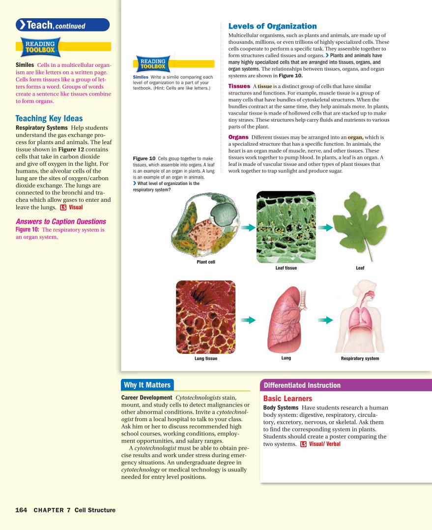
Levels of OrganizationMulticellular organisms, such as plants and animals, are made up of thousands, millions, or even trillions of highly specialized cells. These cells cooperate to perform a specific task. They assemble together to form structures called tissues and organs. V Plants and animals have
many highly specialized cells that are arranged into tissues, organs, and
organ systems. The relationships between tissues, organs, and organ systems are shown in Figure 10.
Tissues A tissuetissue is a distinct group of cells that have similar structures and functions. For example, muscle tissue is a group of many cells that have bundles of cytoskeletal structures. When the bundles contract at the same time, they help animals move. In plants, vascular tissue is made of hollowed cells that are stacked up to make tiny straws. These structures help carry fluids and nutrients to various parts of the plant.
Organs Different tissues may be arranged into an organ,organ, which is a specialized structure that has a specific function. In animals, the heart is an organ made of muscle, nerve, and other tissues. These tissues work together to pump blood. In plants, a leaf is an organ. A leaf is made of vascular tissue and other types of plant tissues that work together to trap sunlight and produce sugar.
Figure 10 Cells group together to make tissues, which assemble into organs. A leaf is an example of an organ in plants. A lung is an example of an organ in animals. V What level of organization is the
respiratory system?
Plant cellLeaf tissue Leaf
Lung tissue Lung Respiratory system
Similes Write a simile comparing each level of organization to a part of your textbook. (Hint: Cells are like letters.)
hb08se_csf_s03.indd 164 9/12/06 9:29:05 AM
164 CHAPTER 7 Cell Structure
Similes Cells in a multicellular organ-ism are like letters on a written page. Cells form tissues like a group of let-ters forms a word. Groups of words create a sentence like tissues combine to form organs.
Teaching Key IdeasRespiratory Systems Help students understand the gas exchange pro-cess for plants and animals. The leaf tissue shown in Figure 12 contains cells that take in carbon dioxide and give off oxygen in the light. For humans, the alveolar cells of the lung are the sites of oxygen/carbon dioxide exchange. The lungs are connected to the bronchi and tra-chea which allow gases to enter and leave the lungs. Visual
Answers to Caption QuestionsFigure 10: The respiratory system is an organ system.
Career Development Cytotechnologists stain, mount, and study cells to detect malignancies or other abnormal conditions. Invite a cytotechnol-ogist from a local hospital to talk to your class. Ask him or her to discuss recommended high school courses, working conditions, employ-ment opportunities, and salary ranges. A cytotechnologist must be able to obtain pre-cise results and work under stress during emer-gency situations. An undergraduate degree in cytotechnology or medical technology is usually needed for entry level positions.
Basic LearnersBody Systems Have students research a human body system: digestive, respiratory, circula-tory, excretory, nervous, or skeletal. Ask them to find the corresponding system in plants. Students should create a poster comparing the two systems. Visual/ Verbal
hb08te_csf_s03.indd 164 11/16/06 9:31:21 AM
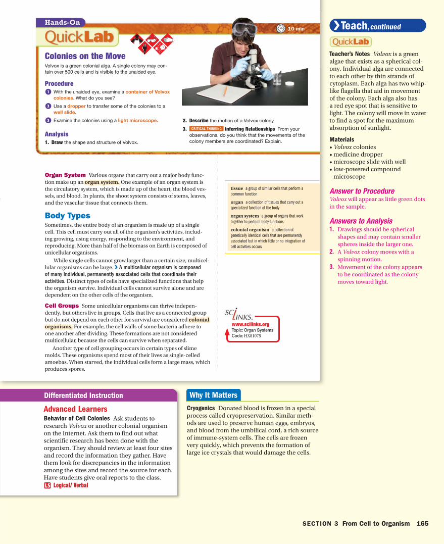
Colonies on the MoveVolvox is a green colonial alga. A single colony may con-tain over 500 cells and is visible to the unaided eye.
Procedure1 With the unaided eye, examine a container of Volvox
colonies. What do you see?
2 Use a dropper to transfer some of the colonies to a well slide.
3 Examine the colonies using a light microscope.
Analysis1. Draw the shape and structure of Volvox.
2. Describe the motion of a Volvox colony.
3. CRITICAL THINKING Inferring Relationships From your observations, do you think that the movements of the colony members are coordinated? Explain.
Organ System Various organs that carry out a major body func-tion make up an organ system.organ system. One example of an organ system is the circulatory system, which is made up of the heart, the blood ves-sels, and blood. In plants, the shoot system consists of stems, leaves, and the vascular tissue that connects them.
Body TypesSometimes, the entire body of an organism is made up of a single cell. This cell must carry out all of the organism’s activities, includ-ing growing, using energy, responding to the environment, and reproducing. More than half of the biomass on Earth is composed of unicellular organisms.
While single cells cannot grow larger than a certain size, multicel-lular organisms can be large. V A multicellular organism is composed
of many individual, permanently associated cells that coordinate their
activ ities. Distinct types of cells have specialized functions that help the organism survive. Individual cells cannot survive alone and are dependent on the other cells of the organism.
Cell Groups Some unicellular organisms can thrive indepen-dently, but others live in groups. Cells that live as a connected group but do not depend on each other for survival are considered colonial colonial organisms.organisms. For example, the cell walls of some bacteria adhere to one another after dividing. These formations are not considered multicellular, because the cells can survive when separated.
Another type of cell grouping occurs in certain types of slime molds. These organisms spend most of their lives as single-celled amoebas. When starved, the individual cells form a large mass, which produces spores.
tissue a group of similar cells that perform a common function
organ a collection of tissues that carry out a specialized function of the body
organ system a group of organs that work together to perform body functions
colonial organism a collection of genetically identical cells that are permanently associated but in which little or no integration of cell activities occurs
10 minHands-On
www.scilinks.orgTopic: Organ SystemsCode: HX81075
hb08se_csf_s03.indd 165 3/20/07 11:02:24 AM
SECTION 3 From Cell to Organism 165
Advanced LearnersBehavior of Cell Colonies Ask students to research Volvox or another colonial organism on the Internet. Ask them to find out what scientific research has been done with the organism. They should review at least four sites and record the information they gather. Have them look for discrepancies in the information among the sites and record the source for each. Have students give oral reports to the class.
Logical/ Verbal
Teacher’s Notes Volvox is a green algae that exists as a spherical col-ony. Individual alga are connected to each other by thin strands of cytoplasm. Each alga has two whip-like flagella that aid in movement of the colony. Each alga also has a red eye spot that is sensitive to light. The colony will move in water to find a spot for the maximum absorption of sunlight.
Materials
• Volvox colonies• medicine dropper• microscope slide with well• low-powered compound
microscope
Answer to Procedure Volvox will appear as little green dots in the sample.
Answers to Analysis1. Drawings should be spherical
shapes and may contain smaller spheres inside the larger one.
2. A Volvox colony moves with a spinning motion.
3. Movement of the colony appears to be coordinated as the colony moves toward light.
Cryogenics Donated blood is frozen in a special process called cryopreservation. Similar meth-ods are used to preserve human eggs, embryos, and blood from the umbilical cord, a rich source of immune-system cells. The cells are frozen very quickly, which prevents the formation of large ice crystals that would damage the cells.
hb08te_csf_s03.indd 165 4/3/07 8:16:15 AM
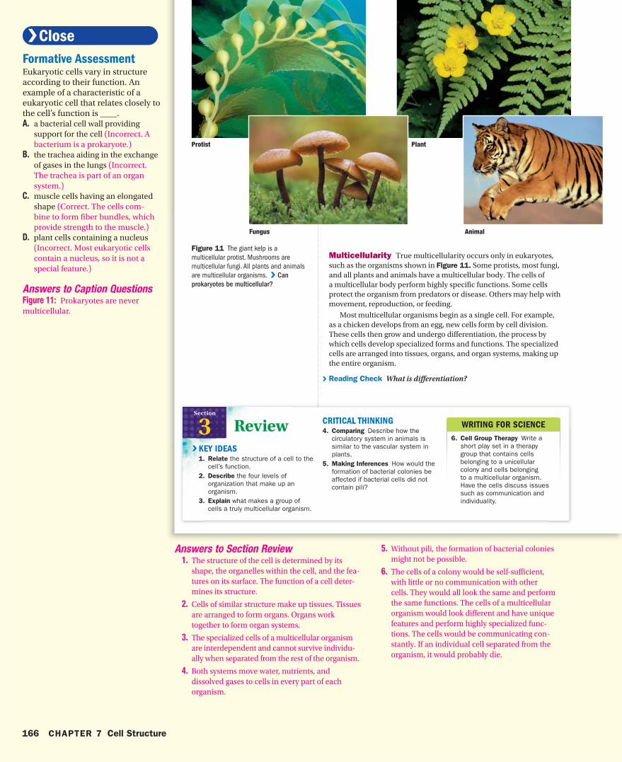
WRITING FOR SCIENCE
6. Cell Group Therapy Write a short play set in a therapy group that contains cells belonging to a unicellular colony and cells belonging to a multicellular organism. Have the cells discuss issues such as communication and individuality.
ReviewSection
3KEY IDEAS1. Relate the structure of a cell to the
cell’s function. 2. Describe the four levels of
organization that make up an organism.
3. Explain what makes a group of cells a truly multicellular organism.
CRITICAL THINKING4. Comparing Describe how the
circulatory system in animals is similar to the vascular system in plants.
5. Making Inferences How would the formation of bacterial colonies be affected if bacterial cells did not contain pili?
V
Figure 11 The giant kelp is a multicellular protist. Mushrooms are multicellular fungi. All plants and animals are multicellular organisms. V Can
prokaryotes be multicellular?
Multicellularity True multicellularity occurs only in eukaryotes, such as the organisms shown in Figure 11. Some protists, most fungi, and all plants and animals have a multicellular body. The cells of a multicellular body perform highly specific functions. Some cells protect the organism from predators or disease. Others may help with movement, reproduction, or feeding.
Most multicellular organisms begin as a single cell. For example, as a chicken develops from an egg, new cells form by cell division. These cells then grow and undergo differentiation, the process by which cells develop specialized forms and functions. The specialized cells are arranged into tissues, organs, and organ systems, making up the entire organism.
V Reading Check What is differentiation?
Protist Plant
Fungus Animal
hb08se_csf_s03.indd 166 9/12/06 9:29:23 AM
166 CHAPTER 7 Cell Structure
Formative AssessmentEukaryotic cells vary in structure according to their function. An example of a characteristic of a eukaryotic cell that relates closely to the cell’s function is ____.A. a bacterial cell wall providing
support for the cell (Incorrect. A bacterium is a prokaryote.)
B. the trachea aiding in the exchange of gases in the lungs (Incorrect. The trachea is part of an organ system.)
C. muscle cells having an elongated shape (Correct. The cells com-bine to form fiber bundles, which provide strength to the muscle.)
D. plant cells containing a nucleus (Incorrect. Most eukaryotic cells contain a nucleus, so it is not a special feature.)
Answers to Caption QuestionsFigure 11: Prokaryotes are never multicellular.
Answers to Section Review 1. The structure of the cell is determined by its
shape, the organelles within the cell, and the fea-tures on its surface. The function of a cell deter-mines its structure.
2. Cells of similar structure make up tissues. Tissues are arranged to form organs. Organs work together to form organ systems.
3. The specialized cells of a multicellular organism are interdependent and cannot survive individu-ally when separated from the rest of the organism.
4. Both systems move water, nutrients, and dissolved gases to cells in every part of each organism.
5. Without pili, the formation of bacterial colonies might not be possible.
6. The cells of a colony would be self-sufficient, with little or no communication with other cells. They would all look the same and perform the same functions. The cells of a multicellular organism would look different and have unique features and perform highly specialized func-tions. The cells would be communicating con-stantly. If an individual cell separated from the organism, it would probably die.
hb08te_csf_s03.indd 166 11/16/06 9:31:35 AM
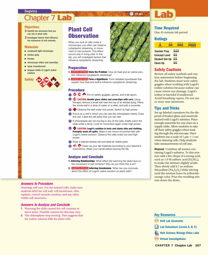
Inquiry
Objectives V V Identify the structures that you
can see in plant cells.
V V Investigate factors that influence
the movement of cell contents.
MaterialsW compound light microscope
W elodea sprig
W forceps
W microscope slides and coverslips
W lamp, incandescent
W dropper bottle of Lugol’s iodine
solution
Safety
Plant Cell ObservationWhen you look at cells under a microscope, you often can observe cytoplasmic streaming, or move-ment of cell contents. This effect occurs only in living cells. In this lab, you will investigate factors that influence cytoplasmic streaming.
Preparation1. SCIENTIFIC METHODS State the Problem How do heat and an iodine solu-
tion influence cytoplasmic streaming?
2. SCIENTIFIC METHODS Form a Hypothesis Form testable hypotheses that explain how heat and iodine influence cytoplasmic streaming.
Procedure1 Put on safety goggles, gloves, and a lab apron.
2 CAUTION: Handle glass slides and coverslips with care. Using forceps, remove a small leaf near the top of an elodea sprig. Place the whole leaf in a drop of water on a slide, and add a coverslip.
3 Observe the leaf under low power. Switch to high power.
4 Focus on a cell in which you can see the chloroplasts clearly. Draw this cell. Label the cell parts that you can see.
5 If chloroplasts are not moving in any of the cells, briefly warm the slide under a lamp. Look for movement again under high power.
6 CAUTION: Lugol’s solution is toxic and stains skin and clothing. Promptly wash off spills. Make a wet mount of another leaf with Lugol’s iodine solution. Observe the cells under low and high power.
7 Draw a stained elodea cell and label all visible parts.
8 Clean up your lab materials according to your teacher’s instructions. Wash your hands before leaving the lab.
Analyze and Conclude1. Inferring Relationships What effect did warming the slide have on
the movement of cell contents? Why do you think this is so?
2. SCIENTIFIC METHODS Inferring Conclusions What can you conclude about the effect of Lugol’s iodine solution on plant cells?
45 min
hb08se_csf_lab.indd 167 3/20/07 11:02:48 AM
CHAPTER 7 Chapter Lab 167
Time RequiredOne 45-minute lab period
Ratings
Teacher Prep
Concept Level
Student Set-Up
Clean Up
Safety CautionsReview all safety symbols and cau-tion statements before beginning the lab. Students must wear safety goggles when working with Lugol’s iodine solution because iodine can cause severe eye damage. Lugol’s iodine is harmful if swallowed. Avoid breathing vapors. Do not use or store near ammonia.
Tips and TricksSet up labeled containers for the dis-posal of broken glass and materials stained with Lugol’s solution. Place enough materials for one class on a supply table. Allow students to take off their safety goggles when look-ing through the microscope. Have students use a scale of 1 μm = 1 cm when drawing cells. Help students take measurements of cell size.
Disposal Combine all wastes con-taining Lugol’s solution. To this mix-ture add a few drops of a strong acid, such as 1.0 M sulfuric acid (H2SO4), to make the mixture slightly acidic. Then slowly add 0.1 m sodium thiosulfate (Na2S2O3) while stirring until the mixture loses its yellowish-orange color. Pour the resulting mix-ture down the drain.
Answers to ProcedureDrawings will vary. For the stained cells, make sure students label the cell wall, cell membrane, chlo-roplasts, central vacuole, nucleus, and any other visible cell structures.
Answers to Analyze and Conclude1. Warming the slide caused the cell contents to
move more. Possible reasons for this may vary.2. The chloroplasts stop moving. This suggests that
the iodine solution kills the plant cells.Key Resources
Holt Lab Generator
Lab Datasheet (Levels A, B, C)
Holt Science Biology Video Labs
Virtual Investigations
hb08te_csf_lab.indd 167 4/3/07 8:16:58 AM
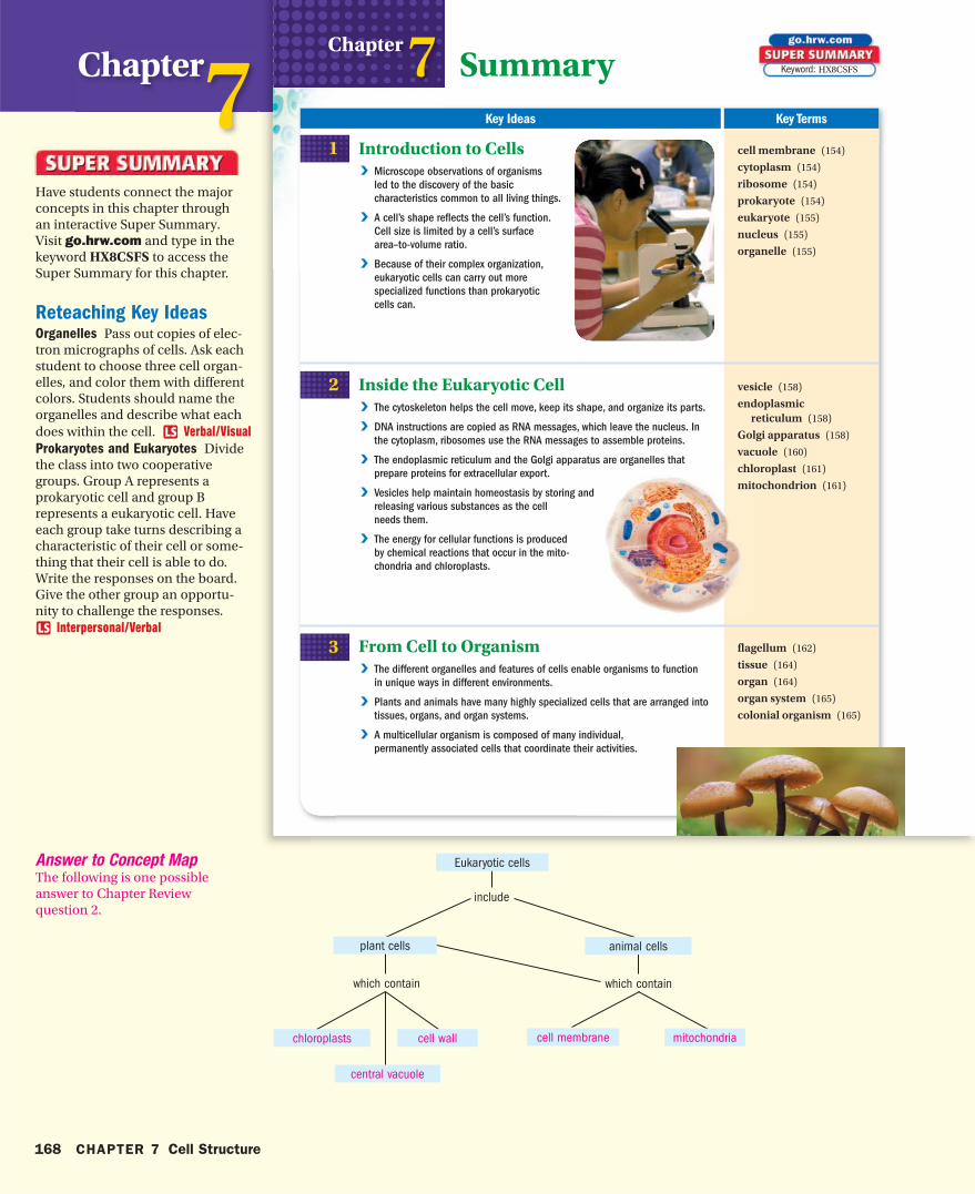
Summary Key Ideas Key Terms
1
2
3
Chapter 7Introduction to Cells V Microscope observations of organisms
led to the discovery of the basic
characteristics common to all living things.
V A cell’s shape reflects the cell’s function.
Cell size is limited by a cell’s surface
area–to-volume ratio.
V Because of their complex organization,
eukaryotic cells can carry out more
specialized functions than prokaryotic
cells can.
Inside the Eukaryotic Cell V The cytoskeleton helps the cell move, keep its shape, and organize its parts.
V DNA instructions are copied as RNA messages, which leave the nucleus. In
the cytoplasm, ribosomes use the RNA messages to assemble proteins.
V The endoplasmic reticulum and the Golgi apparatus are organelles that
prepare proteins for extracellular export.
V Vesicles help maintain homeostasis by storing and
releasing various substances as the cell
needs them.
V The energy for cellular functions is produced
by chemical reactions that occur in the mito-
chondria and chloroplasts.
From Cell to Organism V The different organelles and features of cells enable organisms to function
in unique ways in different environments.
V Plants and animals have many highly specialized cells that are arranged into
tissues, organs, and organ systems.
V A multicellular organism is composed of many individual,
permanently associated cells that coordinate their activities.
cell membrane (154)
cytoplasm (154)
ribosome (154)
prokaryote (154)
eukaryote (155)
nucleus (155)
organelle (155)
vesicle (158)
endoplasmic reticulum (158)
Golgi apparatus (158)
vacuole (160)
chloroplast (161)
mitochondrion (161)
flagellum (162)
tissue (164)
organ (164)
organ system (165)
colonial organism (165)
Keyword: HX8CSFS
hb08se_csf_chr.indd 168 9/12/06 9:30:20 AM
168 CHAPTER 7 Cell Structure
Chapter7Have students connect the major concepts in this chapter through an interactive Super Summary. Visit go.hrw.com and type in the keyword HX8CSFS to access the Super Summary for this chapter.
Reteaching Key IdeasOrganelles Pass out copies of elec-tron micrographs of cells. Ask each student to choose three cell organ-elles, and color them with different colors. Students should name the organelles and describe what each does within the cell. Verbal/VisualProkaryotes and Eukaryotes Divide the class into two cooperative groups. Group A represents a prokaryotic cell and group B represents a eukaryotic cell. Have each group take turns describing a characteristic of their cell or some-thing that their cell is able to do. Write the responses on the board. Give the other group an opportu-nity to challenge the responses.
Interpersonal/Verbal
Answer to Concept MapThe following is one possible answer to Chapter Review question 2.
include
which containwhich contain
Eukaryotic cells
cell wall
animal cells
cell membrane
plant cells
mitochondriachloroplasts
central vacuole
hb08te_csf_chr.indd 168 11/16/06 9:28:58 AM
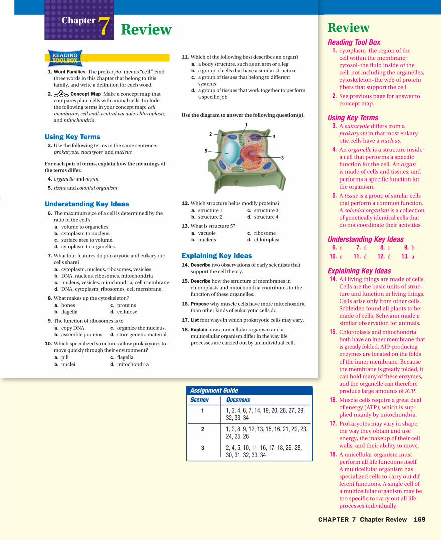
1
2
53
4
Review
1. Word Families The prefix cyto- means “cell.” Find three words in this chapter that belong to this family, and write a definition for each word.
2. Concept Map Make a concept map that compares plant cells with animal cells. Include the following terms in your concept map: cell membrane, cell wall, central vacuole, chloroplasts, and mitochondria.
Using Key Terms 3. Use the following terms in the same sentence:
prokaryote, eukaryote, and nucleus.
For each pair of terms, explain how the meanings of the terms differ.
4. organelle and organ
5. tissue and colonial organism
Understanding Key Ideas 6. The maximum size of a cell is determined by the
ratio of the cell’s a. volume to organelles. b. cytoplasm to nucleus. c. surface area to volume. d. cytoplasm to organelles.
7. What four features do prokaryotic and eukaryotic cells share?a. cytoplasm, nucleus, ribosomes, vesiclesb. DNA, nucleus, ribosomes, mitochondriac. nucleus, vesicles, mitochondria, cell membraned. DNA, cytoplasm, ribosomes, cell membrane.
8. What makes up the cytoskeleton? a. bones c. proteins b. flagella d. cellulose
9. The function of ribosomes is toa. copy DNA. c. organize the nucleus. b. assemble proteins. d. store genetic material.
10. Which specialized structures allow prokaryotes to move quickly through their environment? a. pili c. flagella b. nuclei d. mitochondria
Chapter 7 11. Which of the following best describes an organ?
a. a body structure, such as an arm or a legb. a group of cells that have a similar structurec. a group of tissues that belong to different
systemsd. a group of tissues that work together to perform
a specific job
Use the diagram to answer the following question(s).
12. Which structure helps modify proteins?a. structure 1 c. structure 3b. structure 2 d. structure 4
13. What is structure 5?a. vacuole c. ribosomeb. nucleus d. chloroplast
Explaining Key Ideas 14. Describe two observations of early scientists that
support the cell theory.
15. Describe how the structure of membranes in chloroplasts and mitochondria contributes to the function of these organelles.
16. Propose why muscle cells have more mitochondria than other kinds of eukaryotic cells do.
17. List four ways in which prokaryotic cells may vary.
18. Explain how a unicellular organism and a multicellular organism differ in the way life processes are carried out by an individual cell.
hb08se_csf_chr.indd 169 9/12/06 9:30:27 AM
CHAPTER 7 Chapter Review 169
ReviewReading Tool Box 1. cytoplasm–the region of the
cell within the membrane; cytosol–the fluid inside of the cell, not including the organelles; cytoskeleton–the web of protein fibers that support the cell
2. See previous page for answer to concept map.
Using Key Terms 3. A eukaryote differs from a
prokaryote in that most eukary-otic cells have a nucleus.
4. An organelle is a structure inside a cell that performs a specific function for the cell. An organ is made of cells and tissues, and performs a specific function for the organism.
5. A tissue is a group of similar cells that perform a common function. A colonial organism is a collection of genetically identical cells that do not coordinate their activities.
Understanding Key Ideas 6. c 7. d 8. c 9. b
10. c 11. d 12. d 13. a
Explaining Key Ideas 14. All living things are made of cells.
Cells are the basic units of struc-ture and function in living things. Cells arise only from other cells. Schleiden found all plants to be made of cells; Schwann made a similar observation for animals.
15. Chloroplasts and mitochondria both have an inner membrane that is greatly folded. ATP-producing enzymes are located on the folds of the inner membrane. Because the membrane is greatly folded, it can hold many of these enzymes, and the organelle can therefore produce large amounts of ATP.
16. Muscle cells require a great deal of energy (ATP), which is sup-plied mainly by mitochondria.
17. Prokaryotes may vary in shape, the way they obtain and use energy, the makeup of their cell walls, and their ability to move.
18. A unicellular organism must perform all life functions itself. A multicellular organism has specialized cells to carry out dif-ferent functions. A single cell of a multicellular organism may be too specific to carry out all life processes individually.
Assignment GuideSECTION QUESTIONS
1 1, 3, 4, 6, 7, 14, 19, 20, 26, 27, 29, 32, 33, 34
2 1, 2, 8, 9, 12, 13, 15, 16, 21, 22, 23, 24, 25, 26
3 2, 4, 5, 10, 11, 16, 17, 18, 26, 28, 30, 31, 32, 33, 34
hb08te_csf_chr.indd 169 11/16/06 9:29:04 AM
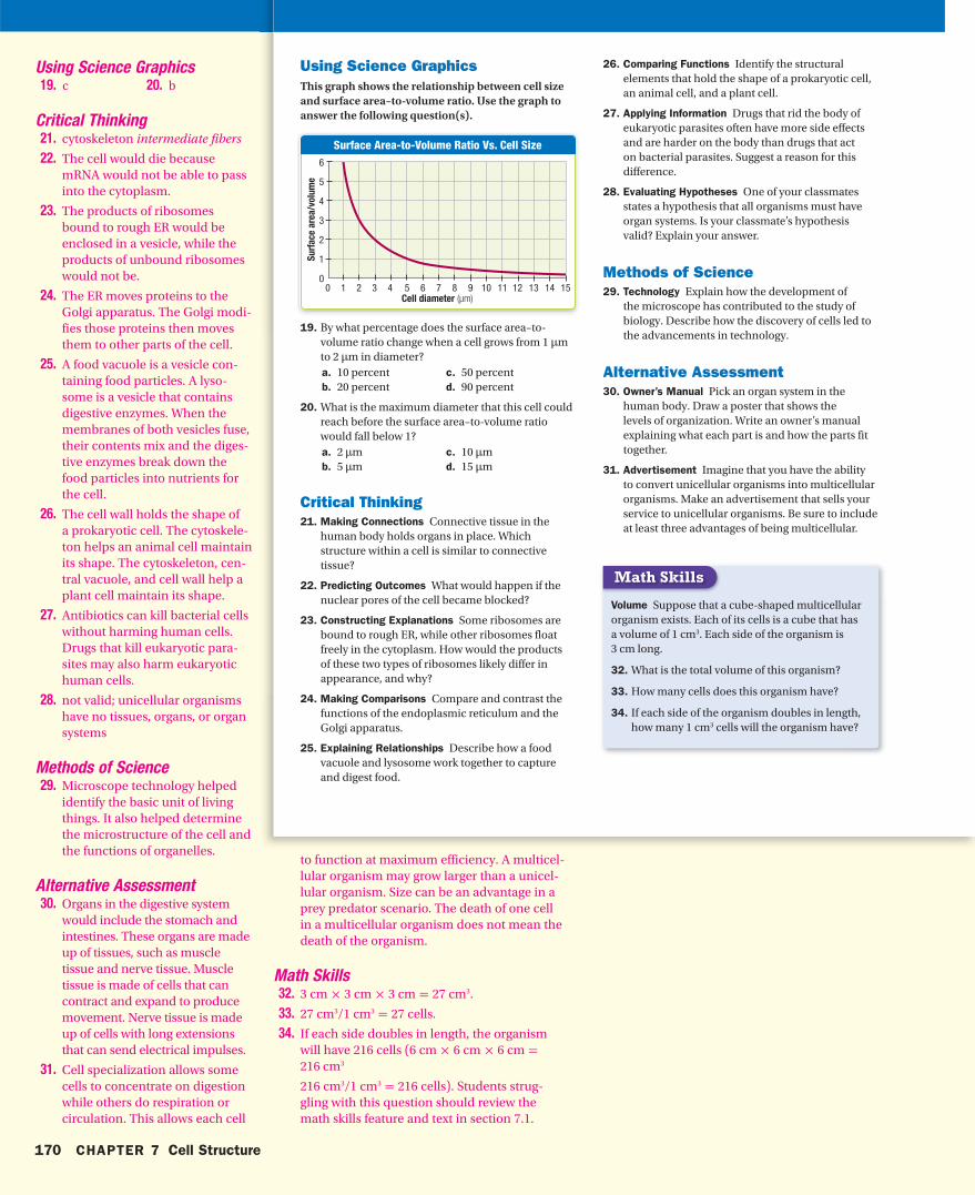
Surface Area-to-Volume Ratio Vs. Cell Size
0 1 2 3 4
6
5
4
3
2
1
015105 6 7 8 9 11 12 13 14
Su
rfa
ce a
rea
/vo
lum
e
Cell diameter (μm)
Using Science GraphicsThis graph shows the relationship between cell size and surface area–to-volume ratio. Use the graph to answer the following question(s).
19. By what percentage does the surface area–to-volume ratio change when a cell grows from 1 μm to 2 μm in diameter?a. 10 percent c. 50 percent b. 20 percent d. 90 percent
20. What is the maximum diameter that this cell could reach before the surface area–to-volume ratio would fall below 1?a. 2 μm c. 10 μmb. 5 μm d. 15 μm
Critical Thinking 21. Making Connections Connective tissue in the
human body holds organs in place. Which structure within a cell is similar to connective tissue?
22. Predicting Outcomes What would happen if the nuclear pores of the cell became blocked?
23. Constructing Explanations Some ribosomes are bound to rough ER, while other ribosomes float freely in the cytoplasm. How would the products of these two types of ribosomes likely differ in appearance, and why?
24. Making Comparisons Compare and contrast the functions of the endoplasmic reticulum and the Golgi apparatus.
25. Explaining Relationships Describe how a food vacuole and lysosome work together to capture and digest food.
26. Comparing Functions Identify the structural elements that hold the shape of a prokaryotic cell, an animal cell, and a plant cell.
27. Applying Information Drugs that rid the body of eukaryotic parasites often have more side effects and are harder on the body than drugs that act on bacterial parasites. Suggest a reason for this difference.
28. Evaluating Hypotheses One of your classmates states a hypothesis that all organisms must have organ systems. Is your classmate’s hypothesis valid? Explain your answer.
Methods of Science 29. Technology Explain how the development of
the microscope has contributed to the study of biology. Describe how the discovery of cells led to the advancements in technology.
Alternative Assessment 30. Owner’s Manual Pick an organ system in the
human body. Draw a poster that shows the levels of organization. Write an owner’s manual explaining what each part is and how the parts fit together.
31. Advertisement Imagine that you have the ability to convert unicellular organisms into multicellular organisms. Make an advertisement that sells your service to unicellular organisms. Be sure to include at least three advantages of being multicellular.
Volume Suppose that a cube-shaped multicellular organism exists. Each of its cells is a cube that has a volume of 1 cm3. Each side of the organism is 3 cm long.
32. What is the total volume of this organism?
33. How many cells does this organism have?
34. If each side of the organism doubles in length, how many 1 cm3 cells will the organism have?
Math Skills
hb08se_csf_chr.indd 170 10/12/06 9:16:01 AM
170 CHAPTER 7 Cell Structure
Using Science Graphics 19. c 20. b
Critical Thinking21. cytoskeleton intermediate fibers
22. The cell would die because mRNA would not be able to pass into the cytoplasm.
23. The products of ribosomes bound to rough ER would be enclosed in a vesicle, while the products of unbound ribosomes would not be.
24. The ER moves proteins to the Golgi apparatus. The Golgi modi-fies those proteins then moves them to other parts of the cell.
25. A food vacuole is a vesicle con-taining food particles. A lyso-some is a vesicle that contains digestive enzymes. When the membranes of both vesicles fuse, their contents mix and the diges-tive enzymes break down the food particles into nutrients for the cell.
26. The cell wall holds the shape of a prokaryotic cell. The cytoskele-ton helps an animal cell maintain its shape. The cytoskeleton, cen-tral vacuole, and cell wall help a plant cell maintain its shape.
27. Antibiotics can kill bacterial cells without harming human cells. Drugs that kill eukaryotic para-sites may also harm eukaryotic human cells.
28. not valid; unicellular organisms have no tissues, organs, or organ systems
Methods of Science 29. Microscope technology helped
identify the basic unit of living things. It also helped determine the microstructure of the cell and the functions of organelles.
Alternative Assessment 30. Organs in the digestive system
would include the stomach and intestines. These organs are made up of tissues, such as muscle tissue and nerve tissue. Muscle tissue is made of cells that can contract and expand to produce movement. Nerve tissue is made up of cells with long extensions that can send electrical impulses.
31. Cell specialization allows some cells to concentrate on digestion while others do respiration or circulation. This allows each cell
to function at maximum efficiency. A multicel-lular organism may grow larger than a unicel-lular organism. Size can be an advantage in a prey predator scenario. The death of one cell in a multicellular organism does not mean the death of the organism.
Math Skills 32. 3 cm × 3 cm × 3 cm = 27 cm3.
33. 27 cm3/1 cm3 = 27 cells.
34. If each side doubles in length, the organism will have 216 cells (6 cm × 6 cm × 6 cm = 216 cm3
216 cm3/1 cm3 = 216 cells). Students strug-gling with this question should review the math skills feature and text in section 7.1.
hb08te_csf_chr.indd 170 11/16/06 9:29:09 AM
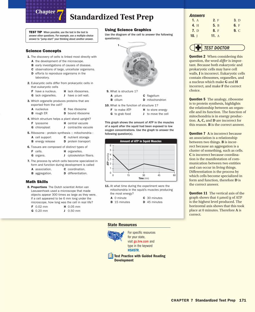
1
Amount of ATP in Squid Muscles
0
6
5
4
3
2
1
0453015 60
AT
P (μ
mol
/g)
Time (min)
Standardized Test PrepChapter 7
TEST TIP When possible, use the text in the test to
answer other questions. For example, use a multiple-choice
answer to “jump-start” your thinking about another answer.
Science Concepts 1. The discovery of cells is linked most directly with
A the development of the microscope. B early investigations of causes of disease.C observations of large, unicellular organisms.D efforts to reproduce organisms in the
laboratory.
2. Eukaryotic cells differ from prokaryotic cells in that eukaryotic cells F have a nucleus. H lack ribosomes.G lack organelles. J have a cell wall.
3. Which organelle produces proteins that are exported from the cell?A nucleolus C free ribosomeB rough ER D bound ribosome
4. Which structure helps a plant stand upright?F lysosome H central vacuole G chloroplast J contractile vacuole
5. Ribosome : protein synthesis :: mitochondria :A cell support C nutrient storageB energy release D protein transport
6. Tissues are composed of distinct types ofF cells. H organelles.G organs. J cytoskeleton fibers.
7. The process by which cells become specialized in form and function during development is called A association. C coordination.B aggregation. D differentiation.
Math Skills 8. Proportions The Dutch scientist Anton van
Leeuwenhoek used a microscope that made objects appear 300 times as large as they were. If a cell appeared to be 6 mm long under the microscope, how long was the cell in real life? F 0.02 mm H 0.05 mmG 0.20 mm J 0.50 mm
Using Science GraphicsUse the diagram of the cell to answer the following question(s).
9. What is structure 1? A pilum C flagellum B cilium D mitochondrion
10. What is the function of structure 1? F to make ATP H to store energyG to grab food J to move the cell
This graph shows the amount of ATP in the muscles of a squid after the squid had been exposed to low oxygen concentrations. Use the graph to answer the following question(s).
11. At what time during the experiment were the mitochondria in the squid’s muscles producing the most energy?A 0 minute C 30 minutesB 15 minutes D 45 minutes
hb08se_csf_stp.indd 171 9/12/06 9:30:39 AM
CHAPTER 7 Standardized Test Prep 171
State Resources
For specifi c resources for your state, visit go.hrw.com and type in the keyword HSHSTR.
Test Practice with Guided Reading
Development
Answers 1. A 2. F 3. D
4. H 5. B 6. F
7. D 8. F 9. C
10. J 11. A
Question 2 When considering this question, the word differ is impor-tant. Because both eukaryotic and prokaryotic cells may have cell walls, J is incorrect. Eukaryotic cells contain ribosomes, organelles, and a nucleus which make G and H incorrect, and make F the correct choice.
Question 5 The analogy, ribosome is to protein synthesis, highlights the relationship between an organ-elle and its function. The function of mitochondria is in energy produc-tion. A, C, and D are incorrect for this reason. B is the correct answer.
Question 7 A is incorrect because an association is a relationship between two things. B is incor-rect because an aggregation is a cluster of something, such as cells. C is incorrect because coordina-tion is the manifestation of com-munication between two entities and can occur in living things. Differentiation is the process by which cells become specialized in form and function, therefore D is the correct answer.
Question 11 The vertical axis of the graph shows that 4 μmol/g of ATP is the highest level produced. The horizontal axis shows that this took place at 0 minutes. Therefore A is correct.
hb08te_csf_stp.indd 171 11/21/06 12:39:04 PM