cells: a review - homepage | wiley · ˜ review and expand on key knowledge from units 1 and 2 ˜...
TRANSCRIPT
KEY KNOWLEDGE
This chapter is designed to enable students to: ■ review and expand on key knowledge from Units 1 and 2 ■ understand that cells are the basic units of structure and function of living organisms
■ understand and apply the concept of surface-area-to-volume ratio ■ list the de� ning characteristics of prokaryotic and eukaryotic cells ■ recognise the plasma membrane as the boundary separating the cell from its external environment
■ describe the various modes of transport across the plasma membrane ■ identify cell organelles in eukaryotic cells and recognise their various functions.
1 Cells: a review
CHAPTER
FIGURE 1.1 This prize-winning image shows eukaryotic cells stained with � uorescent probes. The actin � laments (purple) and the microtubules (yellow) are part of the cytoskeleton of these cells. The nucleus is stained green. Note that the cell on the left appears to be in the process of dividing. In this chapter, we will explore the infrastructure of eukaryotic cells (both plant and animal), relate the structure of cell organelles to their various life-sustaining functions, and examine aspects of the emergence of more complex multicellular organisms. (Image courtesy of Torsten Wittmann.)
c01CellsAReview 1 26 October 2016 9:40 AM
UNCORRECTED PAGE P
ROOFS

Nature of biology 22
c01CellsAReview 2 24 October 2016 10:42 AM
Cells: the basic units of life Cells are the basic structural and functional units of life, and all living organisms are built of one or more cells. Cells, with only a very few excep-tions, are too small to be seen with an unaided eye. Th eir existence was not recognised until after the development of the fi rst simple microscopes. Th is enabled the fi rst observations of cells to be made, in the 1660s. However, the recognition of cells as the basic unit of life did not occur until almost 200 years later.
Cells: how big?Cells are typically microscopic (not visible with an unaided eye). Only a few single cells are large enough to be seen with an unaided human eye, for example, human egg cells with diameters about 0.1 mm and the common amoeba (Amoeba proteus), a unicellular organism with an average size ranging from 0.25 to 0.75 mm. (You would see an amoeba as about the size of a full stop on this page.) Contrast this with one of the smallest bacteria, Pelagibacter ubique, consisting of a cell just 0.2 µm diameter. How many of these bacteria could fi t across an amoeba that is 0.5 mm wide?• Most animal cells fall within the size range of 10 to
40 μm. Among the smallest human cells are red blood cells with diameters for normal cells in the range of 6 to 8 μm.
• Plantcellstypicallyfallintherangeof10to100μm.• Microbialcells,bothbacterialandarchaeal,aremuch
smaller than plant and animal cells. Most bacterial cells have diameters in the range of 0.4 to 2.0 µm and 0.5 to 5 µm in length. On average, microbial cells are about 10 times smaller than plant and animal cells, with sizes typically in the few micrometres range. A non-living microworld exists beyond that of
microbes. Th is is occupied by viruses, which are non-cellular particles that are generally regarded as belonging to the grey area between living and non-living. Why? Because viruses do not have a cellular structure, they cannot carry out metabolic activities in isolation and they cannot self-replicate. (Viruses can replicate only inside and with the assistance of living cells.) Viruses range in diameter from 20 to 300 nanometres (nm). (Refer to chapter 6, page 245, for more detail about viruses, in particular their role in human diseases.) Figure 1.2 shows a sample of the range of sizes seen in selected cells. (Other non-cellular struc-tures are included for size comparison.)
Microbial cells are relatively much smaller than the cells of animals and plants, so some animal bacterial infections can involve the invasion of bacterial cells into the cells of the host, where they multiply. Examine fi gure 1.3 of a human lung fi broblast and note the pres-ence of numerous bacterial cells in a single cell. Th is image highlights the size diff erence between microbial cells and the cells of animals (and plants).
fiGuRe 1.2 Diagrams, at increasing levels of magnifi cation, showing cells, cell organelles and viruses. Note the extreme differences in size. (a) Some human cells showing variation in cell size (b) A bacterial cell with a mitochondrion, a cell organelle and other small cells shown for comparison (c) A mitochondrion compared with three viruses and a ribosome
Human egg130 μm
Sperm cell60 × 5 μm
(a)
(b)
(c)
Yeast cell3 × 4 μm
Mitochondrion4 × 0.8 μm
Mitochondrion
Ribosome30 nm
In�uenza virus130 nm
Measles virus220 nm
HIV130 nm
Skin cell30 μm
Red blood cell8 μm
Yeast cellE coli bacterium3 × 0.6 μm
Red blood cell
1 millimetre (mm) = 1000 micrometres (µm)
1 micrometre (µm) = 1000 nanometres (nm)
UNCORRECTED PAGE P
ROOFS
3CHaPter 1 Cells: a review
c01CellsAReview 3 24 October 2016 10:42 AM
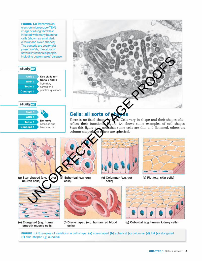
fiGuRe 1.3 Transmission electron microscope (TEM) image of a lung fi broblast infected with many bacterial cells (shown as small dark circular and ovoid shapes). The bacteria are Legionella pneumophila, the cause of several infections in people, including Legionnaires’ disease.
Cells: all sorts of shapesTh ere is no fi xed shape for cells. Cells vary in shape and their shapes often refl ect their functions. Figure 1.4 shows some examples of cell shapes. Scan this fi gure and note that some cells are thin and fl attened, others are column-shaped, yet others are spherical.
fiGuRe 1.4 Examples of variations in cell shape: (a) star-shaped (b) spherical (c) columnar (d) fl at (e) elongated (f) disc-shaped (g) cuboidal
(a) Star-shaped (e.g. motor neuron cells)
(e) Elongated (e.g. human smooth muscle cells)
(f) Disc-shaped (e.g. human red blood cells)
(g) Cuboidal (e.g. human kidney cells)
(b) Spherical (e.g. egg cells)
(c) Columnar (e.g. gut cells)
(d) Flat (e.g. skin cells)
unit 3 Key skills for units 3 and 4 Summary screen and practice questions
aOs 1
topic 1
concept 1
unit 3
do moreCatalase and temperature
aOs 1
topic 1
concept 1
UNCORRECTED PAGE P
ROOFS

Nature of biology 24
c01CellsAReview 4 24 October 2016 10:42 AM
Look at fi gure 1.4a. Note the long axon, which is a distinctive feature of motor neuron cells. Th ese cells transmit nerve impulses from a person’s spinal cord to voluntary muscles throughout the body. In this case, the shape of the nerve cell is fi tted to its conductive function. Can you estimate the approxi-mate length of a motor neuron that has its cell body in the lower spinal cord with its axon reaching to your big toe?
Look at fi gure 1.4e. Note the spindle-shaped smooth muscle cells. Smooth muscle cells contain special proteins that criss-cross the cell, and when these proteins contract the smooth muscle fi bres shorten. Th e spindle shape of these cells is suited to their contractile function. Bundles of smooth muscle cells are found in the gut wall, in the walls of blood vessels, in ducts of secretory glands and in the wall of the uterus. Th ese bundles of smooth muscle cells can gen-erate sustained involuntary contractions in these organs.
Microbial cells also vary in shape (see fi gure 1.5). Note that some bacteria are rod-shaped, such as the gut-dwelling bacterium Escherichia coli; some are corkscrew-shaped, such as Borrelia burgdorferi, the caus ative agent of Lyme disease; while others are more or less spherical, such as Streptococcus pneumonia, the cause of many infections, including pneumonia.
2.5 μm
(b)
2.2 μm
(a)
2.9 μm
(c)
fiGuRe 1.5 Bacterial cells come in many shapes. Some are (a) rod-shaped bacilli (singular: bacillus) (b) spiral-shaped and (c) spherical cocci (singular: coccus).
Odd fact
Motor neurons in animals, such as the giant squid (Architeuthis sp.), may be as long as 12 metres.
unit 3 Cells of living organisms Summary screen and practice questions
aOs 1
topic 2
concept 1
unit 3
see moreLiving organisms are made of cells
aOs 1
topic 2
concept 1
UNCORRECTED PAGE P
ROOFS

5CHaPter 1 Cells: a review
c01CellsAReview 5 24 October 2016 10:42 AM
Not all cells have a fi xed shape. For example, some cells are able to move actively, and these self-propelled cells do not have fi xed shapes because their outer boundary is their fl exible plasma membrane. So, as these cells move, their shapes change. Examples of cells capable of active self-propelled movement include:• cancer cells that migrate into capillaries and move around the body when
a malignant tumour undergoes metastasis (see fi gure 1.6a). Th e thread-like protrusions (known as fi lopodia) that fold out from the plasma membrane of cancer cells make a cancer cell self-mobile and able to migrate from a primary tumour and invade other tissues.
• white blood cells that can squeeze from capillaries into the surrounding tissues where they travel to attack infectious microbes (refer to fi gure 1.12, p. 13)
• amoebas as they move across surfaces (see fi gure 1.6b).Some other cells that have a fi xed shape because of the presence of a rigid
cell wall outside their plasma membranes can self-propel. However, this ability depends on the presence of cilia or fl agella to power their movement. For example, the green alga, Chlamydomonas sp., moves due to the beating of its two fl agella (see fi gure 1.6c).
(a)1
4 5
2 3(b)
(c)
fiGuRe 1.6 (a) Cancer cells. Note the many thread-like projections (fi lopodia) that enable these cancer cells to be mobile or self-propelling. The ability to move is an important factor in the spread of a malignant cancer. (b) Outlines showing the changing shape of an amoeba as it moves (c) Scanning EM image of Chlamydomonas reinhardtii, a single-celled green alga. Note the presence of two fl agella that make this organism able to self-propel. What is the reason for the fi xed shape of this organism? Like all algae, this organism has a rigid cell wall that defi nes its shape.
UNCORRECTED PAGE P
ROOFS
Nature of biology 26
c01CellsAReview 6 24 October 2016 10:42 AM
Cells: why so small? Why are cells microscopically small? Would it be more effi cient to have a larger macroscopic unit to carry out cellular processes rather than many smaller units occupying the same space? To answer these questions we need to look at the concept of surface-area-to-volume ratio.
Surface-area-to-volume ratioEvery living cell must maintain its internal environment within a narrow range of conditions, such as pH and the concentrations of ions and chemical com-pounds. At the same time, a cell must carry out a variety of functions that are essential for life. Th ese functions include trapping a source of energy, obtaining the chemical building blocks needed for cellular repair, growth and reproduc-tion, taking up water and nutrients, and removing wastes. • Th ese essential functions require a constant exchange of material between
the cell and its external environment. • Th e site of exchange where materials are moved into or out of a cell is the
plasma membrane, also termed the cell membrane. Th e plasma membrane must enable enough exchange between the external and internal environ-ments to support these life functions.
• Th e exchange of materials must occur at rates suffi cient to ensure that sub-stances are delivered fast enough into cells to meet their nutrient needs and that wastes are removed fast enough from the cells to avoid their accumulation. A critical issue in keeping a cell alive is the surface area of plasma mem-
brane available to supply material to or remove wastes from the metabolically active cytoplasm of the cell. Th is can be quantifi ed by a measure termed the surface-area-to-volume ratio, abbreviated SA:V ratio. Th is ratio provides a key clue to the answer to the question: why are cells so small?
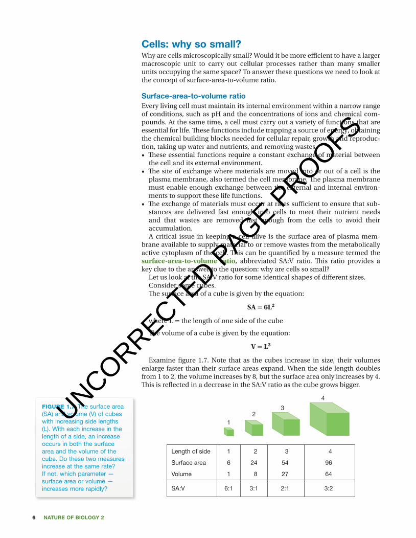
Let us look at the SA:V ratio for some identical shapes of diff erent sizes.Consider some cubes. Th e surface area of a cube is given by the equation:
SA = 6L2
where L = the length of one side of the cube
Th e volume of a cube is given by the equation:
V = L3
Examine fi gure 1.7. Note that as the cubes increase in size, their volumes enlarge faster than their surface areas expand. When the side length doubles from 1 to 2, the volume increases by 8, but the surface area only increases by 4. Th is is refl ected in a decrease in the SA:V ratio as the cube grows bigger.
Length of side 1
1
Surface area 6
Volume 1
SA:V 6:1
2
2
24
8
3:1
3
3
54
27
2:1
4
4
96
64
3:2
fiGuRe 1.7 The surface area (SA) and volume (V) of cubes with increasing side lengths (L). With each increase in the length of a side, an increase occurs in both the surface area and the volume of the cube. Do these two measures increase at the same rate? If not, which parameter — surface area or volume — increases more rapidly?
UNCORRECTED PAGE P
ROOFS
7CHaPter 1 Cells: a review
c01CellsAReview 7 24 October 2016 10:42 AM
Th is generalisation applies to other shapes; that is, the SA:V ratio of a smaller object is higher than that of a larger object with the same shape. Th e higher the SA:V ratio, the greater effi ciency of two-way exchange of materials across the plasma membrane; that is, effi cient uptake and output of dissolved material is favoured by a high SA:V ratio.
Th e same principle applies to cells. As cells increase in size through an increase in cytoplasm, both their surface areas and volumes increase, but not at the same rate. Th e internal volumes of cells expand at a greater rate than the areas of their plasma membrane. Th is means that the growth of an individual cell is accompanied by a relative decrease in the area of its plasma membrane.
Th e metabolic needs of a cell increase in proportion to the volume of meta-bolically active cytoplasm. But, the inputs/outputs of materials to meet these needs increase only in proportion to the cell surface area. So, as a cell increases in cytoplasmic volume, its metabolic needs increase faster than the cell’s ability to transport the materials into and out of the cell to meet those needs. Th e continued decrease in SA:V ratio as metabolically active cells increase in size places an upper limit on cell size. Th is is the one clue to why metabolically active cells are so small.
In general, the rate at which nutrients enter and wastes leave a cell is inversely proportional to the cell size, as measured in meta bolically active cytoplasm; in other words, the larger the cell, the slower the rate of move-ment of nutrients into, and wastes out of, a cell. Beyond a given cell size, the two-way exchange of materials across the plasma membrane cannot occur fast enough to sustain the volume of the cell contents. If that cell is to carry out the functions necessary for living, it must divide into smaller cells or die.
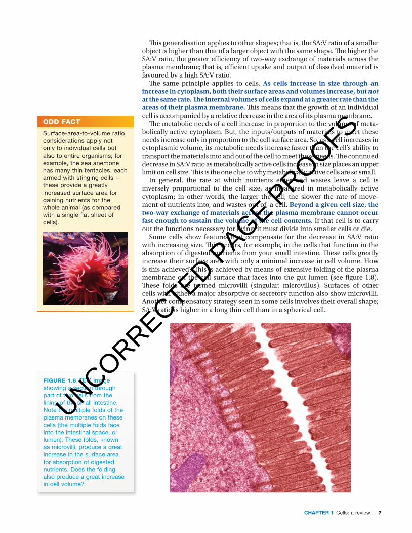
Some cells show features that compensate for the decrease in SA:V ratio with increasing size. Th is occurs, for example, in the cells that function in the absorption of digested nutrients from your small intestine. Th ese cells greatly increase their surface area with only a minimal increase in cell volume. How is this achieved? Th is is achieved by means of extensive folding of the plasma membrane on the cell surface that faces into the gut lumen (see fi gure 1.8). Th ese folds are termed microvilli (singular: microvillus). Surfaces of other cells with either a major absorptive or secretory function also show microvilli. Another compensatory strategy seen in some cells involves their overall shape; SA:V ratio is higher in a long thin cell than in a spherical cell.
fiGuRe 1.8 TEM image showing a section through part of two cells from the lining of the small intestine. Note the multiple folds of the plasma membranes on these cells (the multiple folds face into the intestinal space, or lumen). These folds, known as microvilli, produce a great increase in the surface area for absorption of digested nutrients. Does the folding also produce a great increase in cell volume?
Odd fact
Surface-area-to-volume ratio considerations apply not only to individual cells but also to entire organisms; for example, the sea anemone has many thin tentacles, each armed with stinging cells — these provide a greatly increased surface area for gaining nutrients for the whole animal (as compared with a single fl at sheet of cells).
UNCORRECTED PAGE P
ROOFS

Nature of biology 28
c01CellsAReview 8 24 October 2016 10:42 AM
key ideas
■ Cells are the basic structural and functional units of life. ■ Cells are typically too small to be seen by an unaided eye. ■ The unit of measurement used for cell size is the micrometre (µm), one millionth of a metre.
■ Microbial cells are much smaller than plant and animal cells. ■ The metabolic needs of a cell are determined by its metabolically active cytoplasmic volume.
■ The ability of a cell to meet its metabolic needs is determined by the surface area of the cell.
■ As a cell increases in size, its internal volume expands at a greater rate than the area of its plasma membrane.
■ The surface-area-to-volume ratio (SA:V ratio) of a smaller object is higher than that of a larger object with the same shape.
■ The continued decrease in SA:V ratio as metabolically active cells increase in size places an upper limit on cell size.
Quick check
1 Identify whether each of the following statements is true or false:a Cells are typically too small to be seen with an unaided eye.b Bacterial cells are typically larger than animal cells.c Viral particles are smaller than microbial cells.d As a given shape increases in size, its surface-area-to-volume ratio
increases.e Beyond a given cell size, the two-way exchange of materials across the
cell surface cannot occur at a rate sufficient to meet the needs of a cell. 2 Two spheres (A and B) have different diameters, with A being larger than B.
Which has the higher SA:V ratio?
Prokaryotes: no nuclear envelope!The microscopically tiny creatures that we call ‘microbes’ are a diverse group of organisms. In fact, the microbes comprise two different classification groups, namely bacteria and archaea. The cells of all these microbes can be readily dis-tinguished from the cells of the other major groups of living organisms: fungi, plants and animals. The key distinguishing feature of archaea and bacteria is that their cells lack a membrane-bound nucleus (see figure 1.9a). Cells with this characteristic are described as prokaryotic cells and organisms dis-playing this feature are called prokaryotes. Prokaryotes are generally assumed to be the oldest existing form of life on planet Earth. The absence of a distinct nucleus does not mean that prokaryotes, such as archaea and bacteria, lack genetic material. Like all other kinds of organism, archaea and bacteria have DNA in their cells, but the DNA in prokaryotic cells is dispersed, not enclosed within a separate membrane-bound compartment.
In contrast, the cells of all other organisms — protists, fungi, plants and animals — have a definite nucleus (see figure 1.9b). The nucleus is enclosed by a double membrane, called the nuclear envelope. Organisms with this feature are termed eukaryotes and their cells are described as being eukaryotic. The nucleus of a eukaryotic cell contains DNA, the genetic material of cells. In addition, eukaryotic cells contain many membrane-bound cell organelles that are not present in prokaryotic cells (see table 1.1).
Odd fact
A single cell lining the small intestine may have up to 10 000 microvilli on its apical surface facing into the gut lumen. How would this affect the surface area available for absorption of digested nutrients, compared with a cell with no microvilli?
UNCORRECTED PAGE P
ROOFS

9CHaPter 1 Cells: a review
c01CellsAReview 9 24 October 2016 10:42 AM
(a) (b)
fiGuRe 1.9 Images of cells (not to same magnifi cation) (a) Image of a prokaryotic cell, which is in the process of dividing. Note that the cell does not have a discrete nucleus enclosed within a membrane. Instead, the genetic material (stained orange) is dispersed. (b) A confocal fl uorescence microscope view of human breast cells, examples of eukaryotic cells. The position of the nucleus that encloses the genetic material is shown by the discrete blue area within each cell. The cells have been treated with special stains to highlight two different cytoskeleton proteins: vimentin (green), found in cells within cancerous tissue, and keratin (red). (Image (b) courtesy of Judith Kinnear)
Comparing prokaryotes with eukaryotesFigure 1.10 shows the structure of a typical prokaryotic cell in comparison with a eukaryotic cell. Note that a prokaryotic cell has a simple architecture in con-trast to a eukaryotic cell, which has a more elaborate structure owing to the presence of many membrane-enclosed compartments within the cell.
fiGuRe 1.10 Diagram showing a basic comparison of a prokaryotic cell (left) and a eukaryotic cell (right). The key distinction between these cells is the presence, only in eukaryotic cells, of membrane-bound organelles, in particular the nucleus that contains the genetic material, DNA. (Cells are not drawn to scale.)
Table 1.1 outlines a comparison between the structures of prokaryotic and eukaryotic cells. Th e critical diff erence is the absence of membrane-enclosed organelles in prokaryotes, in contrast to eukaryotic cells in which the nucleus and many other cell organelles are membrane-enclosed.
In general, prokaryotic cells are about 10 times smaller than eukaryotic cells. However, size is not an absolute distinction; there are some rare exceptions:• Large prokaryotic cells exist, such as the giant bacterium Th iomargarita
namibiensis (0.1 × 0.3 mm in diameter), which lives in the muddy sea fl oor off the coast of Namibia.
• Relatively small eukaryotic cells exist, such as the single-celled green alga Ostreococcus tauri, which is just 0.8 µm in diameter.
pro = before + karyon = kernel, nucleus
eu = well, good + karyon = kernel, nucleus
UNCORRECTED PAGE P
ROOFS
Nature of biology 210
c01CellsAReview 10 24 October 2016 10:42 AM
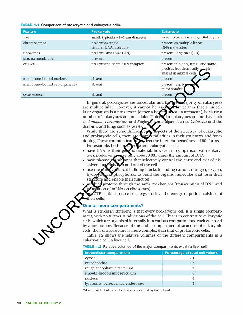
table 1.1 Comparison of prokaryotic and eukaryotic cells.
feature Prokaryote eukaryote
size small: typically ~1−2 µm diameter larger: typically in range 10–100 µm
chromosomes present as singlecircular DNA molecule
present as multiple linearDNA molecules
ribosomes present: small size (70s) present: large size (80s)
plasma membrane present present
cell wall present and chemically complex present in plants, fungi, and some protists, but chemically simple; absent in animal cells
membrane-bound nucleus absent present
membrane-bound cell organelles absent present; e.g. lysosomes, mitochondria
cytoskeleton absent present
In general, prokaryotes are unicellular and the great majority of eukaryotes are multicellular. However, it cannot be assumed for certain that a unicel-lular organism is a prokaryote (either a bacterium or an archaean), because a number of eukaryotes are unicellular. Unicellular eukaryotes are protists, such as Amoeba, Paramecium and Euglena, some algae such as Chlorella and the diatoms, and fungi such as yeasts.
While there are some differences in aspects of the structure of eukaryotic and prokaryotic cells, there are many similarities in their structures and func-tioning. These common features reflect the inter-connectedness of life forms.
For example, both prokaryotic and eukaryotic cells:• have DNA as their genetic material; however, in comparison with eukary-
otes, prokaryotes have only about 0.001 times the amount of DNA• have plasma membranes that selectively control the entry and exit of dis-
solved materials into and out of the cell• use the same chemical building blocks including carbon, nitrogen, oxygen,
hydrogen and phosphorus, to build the organic molecules that form their structure and enable their function
• produce proteins through the same mechanism (transcription of DNA and translation of mRNA on ribosomes)
• use ATP as their source of energy to drive the energy-requiring activities of their cells.
one or more compartments?What is strikingly different is that every prokaryotic cell is a single compart-ment, with no further subdivisions of the cell. This is in contrast to eukaryotic cells, which are organised internally into various compartments, each enclosed by a membrane. Because of the multi-compartmental structure of eukaryotic cells, their ultrastructure is more complex than that of prokaryotic cells.
Table 1.2 shows the relative volumes of the different compartments in a eukaryotic cell, a liver cell.
table 1.2 Relative volumes of the major compartments within a liver cell
intracellular compartment Percentage of total cell volume*cytosol 54mitochondria 22rough endoplasmic reticulum 9smooth endoplasmic reticulum 6nucleus 6lysosomes, peroxisomes, endosomes 3
*More than half of the cell volume is occupied by the cytosol.
UNCORRECTED PAGE P
ROOFS
11CHaPter 1 Cells: a review
c01CellsAReview 11 24 October 2016 10:42 AM
The multicompartment structure of a eukaryotic cell enables it to maintain different conditions within each membrane-enclosed compartment that are suitable for the particular function of that compartment. Think about a house that is subdivided into rooms with different functions: you shower in the bath-room, not in the kitchen; the stove is in the kitchen, not in the bedroom. A eukaryotic cell can be likened to a house — its many compartments are like different rooms where different tasks are carried out. (Later in this chapter, we will explore some of the various compartments in eukaryotic cells.)
key ideas
■ Prokaryotic cells lack a membrane-bound nucleus, and organisms lacking a nuclear envelope are termed prokaryotes — bacteria and archaea.
■ Prokaryotes differ from eukaryotes in the absence of membrane-bound cell organelles of any kind.
■ Eukaryotic cells are typically about ten times larger than prokaryotic cells. ■ Eukaryotic cells have a membrane-bound nucleus in addition to other membrane-bound organelles.
■ Organisms built of cells that have a nucleus enclosed within a nuclear envelope are termed eukaryotes — protists, fungi, plants and animals.
Quick check
3 Identify whether each of the following statements is true or false:a Prokaryotes are unicellular organisms, comprising bacteria and archaea.b The presence of a membrane-bound nucleus in its cells provides
evidence that an organism is a eukaryote. c All eukaryotes are multicellular organisms.d The various compartments within eukaryotic cells would be expected to
have identical conditions.4 List two similarities between prokaryotic and eukaryotic cells.5 A unicellular organism was found in a sample of pond water. Is it reasonable
to conclude that this organism must be either a bacterium or an archaeon? Briefly explain.
Plasma membrane: the gatekeeperThe cells of all living organisms have a boundary that separates their internal environment from their surroundings. From single-celled organisms, such as amoebae or bacteria, to multicellular organisms, such as mushrooms, palm trees and human beings, each of their cells has an active boundary called the plasma membrane, also known as the cell membrane.
The plasma membrane forms the outer boundary of the living compartment of every cell. Within this compartment, conditions can be established that differ from those in the external environment and that support the living state. The plasma membrane can exclude some substances from entering the cell, while permitting entry of other substances and elimination of yet other sub-stances. Without such a boundary, life could not exist, and indeed could not have evolved.
The plasma membrane boundary can be thought of as a busy gatekeeper selectively controlling the entry and exit of materials into and out of cells. As such, the plasma membrane is said to be semipermeable or selectively permeable, meaning that it allows only some substances to cross it — in or out.
unit 3 structure of the plasma membraneSummary screen and practice questions
aOs 1
topic 2
concept 2
UNCORRECTED PAGE P
ROOFS

Nature of biology 212
c01CellsAReview 12 24 October 2016 10:42 AM
This gatekeeper function ensures that materials required by the cell are supplied and that excesses and wastes are removed, both entry and exit occur-ring at rates sufficient to maintain the internal environment of the cell within narrow limits. This is quite a cellular balancing act! In addition to its role in transportation of materials into and out of the cell, the plasma membrane plays other important cellular roles (see p. 16).
The cells of fungi, plants and many bacteria and archaea have rigid cell walls outside their plasma membranes. The cells of animals do not have a cell wall. Cell walls do not control which materials enter or leave cells; instead, cell walls provide strength and give a fixed shape to those cells that possess them (see chapter 2). A cell wall is fully permeable so that gases and dissolved solutes can pass freely across it. It is only when these substances reach the plasma membrane that their passage may be blocked.
Structure of the plasma membraneSmall, but vitally important, the plasma membrane is just 8 nanometres (nm) wide and so is only visible using a transmission electron microscope (TEM). A TEM image of the plasma membrane has a ‘train track’ appearance with two dark lines separated by a more lightly stained region. These images were important clues in elucidating the structure of the plasma membrane (see Biochallenge, p. 34).
The plasma membrane has two major components:1. phospholipids. These are the main structural component of the plasma
membrane (see below).2. proteins. Most proteins are embedded in the plasma membrane, while
others are attached at the membrane surface (see next page). Let’s consider each of these components in turn.
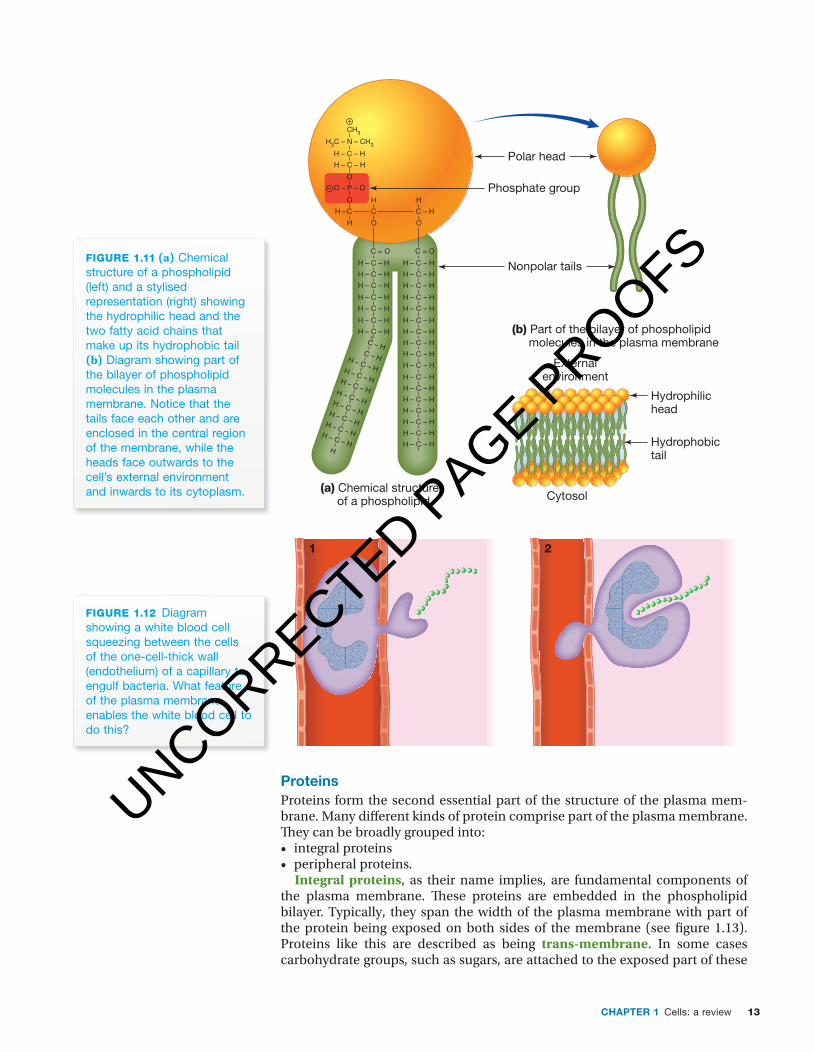
PhospholipidsThe plasma membrane consists of a double layer (bilayer) of phospholipids. Each phospholipid molecule consists of two fatty acid chains joined to a phosphate-containing group. The phosphate-containing group of a phospho-lipid molecule constitutes its water-loving (hydrophilic) head. The fatty acid chains constitute the water-fearing (hydrophobic) tail of each phospholipid molecule.
Examine figure 1.11. Notice that the two layers of phospholipids are arranged so that the hydrophilic heads are exposed at both the external environment of the cell and at the cytosol (the internal environment of the cell). In contrast, the two layers of hydrophobic tails face each other in the central region of the plasma membrane. Water and lipids do not mix.
At human body temperature, the fatty acid chains in the inner portion of the plasma membrane are not solid. Instead, they are viscous fluids — think about thick oil or very soft butter — this makes the plasma membrane flexible, soft and able to move freely. This property of the plasma membrane is very important as it enables cells to change shape (provided they do not have a cell wall outside the plasma membrane). For example, red blood cells are about 8 micrometres (μm) in diameter. When circulating red blood cells reach a capillary bed, they must deform themselves by bending and stretching in order to squeeze through capillaries, some of which have diameters as narrow as 5 micrometres. Likewise, when white blood cells reach sites of infection, they must squeeze out of small gaps between the single layer of cells that forms the capillary walls (see figure 1.12). Shape changes by animal cells are only possible because of the flexible nature of the lipids in the plasma membrane. Flexibility and shape changes are not possible for cells with cell walls.
unit 3 a selectively permeable membraneSummary screen and practice questions
aOs 1
topic 2
concept 3
Odd fact
In addition to phospholipids, the plasma membrane of animal cells also contains cholesterol as part of its structure.
Hydrophilic (water-loving) molecules dissolve readily in water.
Lipophilic substances dissolve readily in organic solvents such as benzene.
Hydrophobic (water-fearing) molecules are usually lipophilic (lipid-loving).
UNCORRECTED PAGE P
ROOFS
13CHaPter 1 Cells: a review
c01CellsAReview 13 24 October 2016 10:42 AM
(a) Chemical structure of a phospholipid
C = O
H – C – H
H – C – H
H
C – H
H
C
H – C – H
H – C – H
H – C – H
H – C – H
H – C – H
H – C – H
H – C – H
H – C – H
H – C – H
H – C – H
H – C – H
H – C – H
H – C – H
H – C – H
H – C – H
C – HC – HH – C – HH – C – HH – C – HH – C – HH – C – HH – C – HH – C – HH – C – H
H
C = O
O
H – C – H
H –
H – C – H
H – C – H
H – C – H
O
O – P – O
O
H – C – H
H – C – H
H – C – H
H3C – N – CH3
CH3
H – C – H
H – C – H
O
C
H
+
–
Nonpolar tails
Phosphate group
(b) Part of the bilayer of phospholipid molecules in the plasma membrane
Polar head
Hydrophilichead
Hydrophobictail
Externalenvironment
Cytosol
fiGuRe 1.11 (a) Chemical structure of a phospholipid (left) and a stylised representation (right) showing the hydrophilic head and the two fatty acid chains that make up its hydrophobic tail (b) Diagram showing part of the bilayer of phospholipid molecules in the plasma membrane. Notice that the tails face each other and are enclosed in the central region of the membrane, while the heads face outwards to the cell’s external environment and inwards to its cytoplasm.
1 2
fiGuRe 1.12 Diagram showing a white blood cell squeezing between the cells of the one-cell-thick wall (endothelium) of a capillary to engulf bacteria. What feature of the plasma membrane enables the white blood cell to do this?
ProteinsProteins form the second essential part of the structure of the plasma mem-brane. Many diff erent kinds of protein comprise part of the plasma membrane. Th ey can be broadly grouped into:• integral proteins • peripheral proteins.
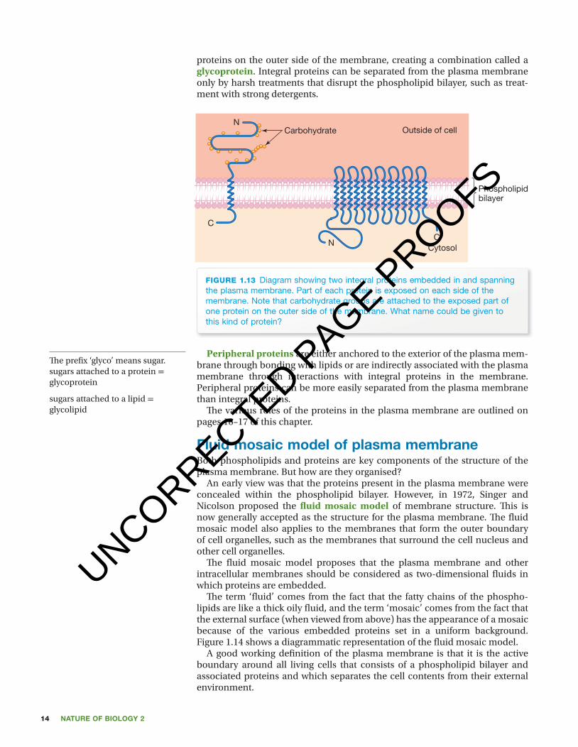
Integral proteins, as their name implies, are fundamental components of the plasma membrane. Th ese proteins are embedded in the phospholipid bilayer. Typically, they span the width of the plasma membrane with part of the protein being exposed on both sides of the membrane (see fi gure 1.13). Proteins like this are described as being trans-membrane. In some cases carbohydrate groups, such as sugars, are attached to the exposed part of these
UNCORRECTED PAGE P
ROOFS
Nature of biology 214
c01CellsAReview 14 24 October 2016 10:42 AM
proteins on the outer side of the membrane, creating a combination called a glycoprotein. Integral proteins can be separated from the plasma membrane only by harsh treatments that disrupt the phospholipid bilayer, such as treat-ment with strong detergents.
Carbohydrate Outside of cell
CytosolC
C
N
N
Phospholipidbilayer
fiGuRe 1.13 Diagram showing two integral proteins embedded in and spanning the plasma membrane. Part of each protein is exposed on each side of the membrane. Note that carbohydrate groups are attached to the exposed part of one protein on the outer side of the membrane. What name could be given to this kind of protein?
Peripheral proteins are either anchored to the exterior of the plasma mem-brane through bonding with lipids or are indirectly associated with the plasma membrane through interactions with integral proteins in the membrane. Peripheral proteins can be more easily separated from the plasma membrane than integral proteins.
Th e various roles of the proteins in the plasma membrane are outlined on pages 16–17 of this chapter.
fluid mosaic model of plasma membraneBoth phospholipids and proteins are key components of the structure of the plasma membrane. But how are they organised?
An early view was that the proteins present in the plasma membrane were concealed within the phospholipid bilayer. However, in 1972, Singer and Nicolson proposed the fl uid mosaic model of membrane structure. Th is is now generally accepted as the structure for the plasma membrane. Th e fl uid mosaic model also applies to the membranes that form the outer boundary of cell organelles, such as the membranes that surround the cell nucleus and other cell organelles.
Th e fl uid mosaic model proposes that the plasma membrane and other intracellular membranes should be considered as two-dimensional fl uids in which proteins are embedded.
Th e term ‘fl uid’ comes from the fact that the fatty chains of the phospho-lipids are like a thick oily fl uid, and the term ‘mosaic’ comes from the fact that the external surface (when viewed from above) has the appearance of a mosaic because of the various embedded proteins set in a uniform background. Figure 1.14 shows a diagrammatic representation of the fl uid mosaic model.
A good working defi nition of the plasma membrane is that it is the active boundary around all living cells that consists of a phospholipid bilayer and associated proteins and which separates the cell contents from their external environment.
Th e prefi x ‘glyco’ means sugar.sugars attached to a protein = glycoprotein
sugars attached to a lipid = glycolipid
UNCORRECTED PAGE P
ROOFS
15CHaPter 1 Cells: a review
c01CellsAReview 15 24 October 2016 10:42 AM
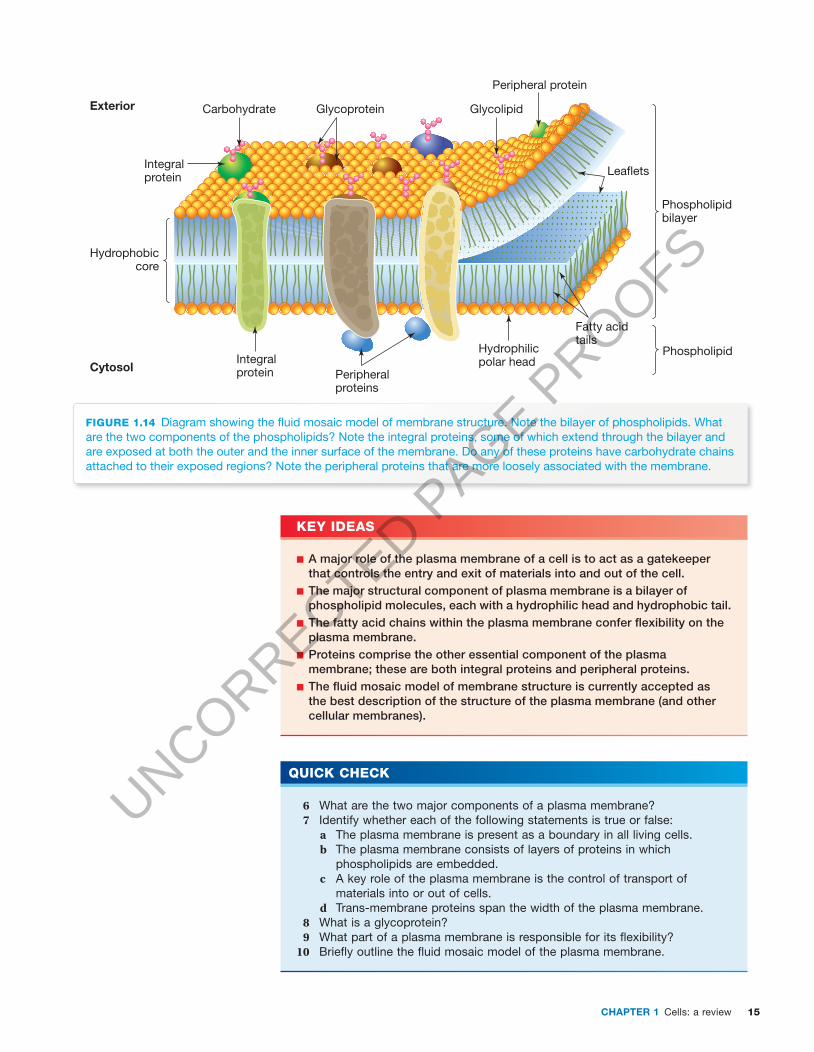
fiGuRe 1.14 Diagram showing the fl uid mosaic model of membrane structure. Note the bilayer of phospholipids. What are the two components of the phospholipids? Note the integral proteins, some of which extend through the bilayer and are exposed at both the outer and the inner surface of the membrane. Do any of these proteins have carbohydrate chains attached to their exposed regions? Note the peripheral proteins that are more loosely associated with the membrane.
Carbohydrate
Integralprotein
Exterior
CytosolIntegralprotein
Hydrophobiccore
Glycoprotein Glycolipid
Lea�ets
Phospholipidbilayer
Phospholipid
Fatty acidtails
Peripheral protein
Peripheralproteins
Hydrophilicpolar head
key ideas
■ A major role of the plasma membrane of a cell is to act as a gatekeeper that controls the entry and exit of materials into and out of the cell.
■ The major structural component of plasma membrane is a bilayer of phospholipid molecules, each with a hydrophilic head and hydrophobic tail.
■ The fatty acid chains within the plasma membrane confer fl exibility on the plasma membrane.
■ Proteins comprise the other essential component of the plasma membrane; these are both integral proteins and peripheral proteins.
■ The fl uid mosaic model of membrane structure is currently accepted as the best description of the structure of the plasma membrane (and other cellular membranes).
Quick check
6 What are the two major components of a plasma membrane? 7 Identify whether each of the following statements is true or false:
a The plasma membrane is present as a boundary in all living cells.b The plasma membrane consists of layers of proteins in which
phospholipids are embedded.c A key role of the plasma membrane is the control of transport of
materials into or out of cells.d Trans-membrane proteins span the width of the plasma membrane.
8 What is a glycoprotein? 9 What part of a plasma membrane is responsible for its fl exibility?10 Briefl y outline the fl uid mosaic model of the plasma membrane.
UNCORRECTED PAGE P
ROOFS
Nature of biology 216
c01CellsAReview 16 24 October 2016 10:42 AM
functions of the plasma membrane The plasma membrane carries out several important functions for a cell. The plasma membrane:1. is an active and selective boundary2. denotes cell identity3. receives external signals 4. transports materials.
the active boundaryThe plasma membrane forms the active boundary of a cell, separating the cell from its external environment and from other cells; it allows the passage of some substances only. The plasma membrane forms the boundary of a com-partment in which the internal environment of a living cell can be held within a narrow range of conditions that are different from those of the external environment.
Within the cell, similar membranes form the active boundaries of cell organ-elles, including the nucleus, the endoplasmic reticulum, the Golgi apparatus and lysosomes. In other cell organelles, such as mitochondria and chloro-plasts, membranes form both the external boundary and part of the internal structure. Because of the presence of their membrane boundaries, mem-brane-bound cell organelles can maintain internal environments that differ from those in the surrounding cytosol and can perform different functions. This is a critical role of cell membranes.
Cell identityGlycoproteins on the outer plasma membrane function as cell surface markers, also known as antigens or cell identity tags. Each cell type has a dif-ferent combination of surface markers. In mammals, these markers enable the immune system to identify these cells as ‘self’ and distinguish them from foreign cells. Glycolipids on the plasma membrane play a role in tissue recognition.
receiving external signalsCells receive signals from their external environment. In the case of a multicel-lular organism, the signal may originate from another cell within that organism. In the case of unicellular organisms, the signal may come from other organisms in its neighbourhood or from its external environment. These external signals are often chemical compounds, for example, hormones.
Trans-membrane proteins on the outer surface of the plasma membrane are the receptors for these signals, and each cell has many different kinds of receptor protein. The signal binds to the receptor protein and this binding alters the shape of the receptor protein and starts a specific response in the cell. For example, Saccharomyces cereviseae is a single-celled yeast used in winemaking, brewing and bread making. During one stage of the yeast life cycle, haploid yeast cells exist in one of two different mating types, desig-nated ‘a’ and ‘α’ (alpha). When one type is ready to mate, it releases a small chemical, called a mating factor, into its environment. The mating factor is a signal to nearby yeast cells of the other mating type that it is ready to mate (see figure 1.15). The signal from an a-type yeast cell can be received by recep-tors on the surface of the plasma membrane of yeast cells of the other mating type. The signal binds to a specific receptor and causes a change in the behav-iour in the receiver yeast cell — it changes its shape and moves towards the
Odd fact
The human blood group A and B antigens are present on red blood cells as glycolipids and as glycoproteins — they differ by one sugar group.
Cell signalling is treated in detail in chapter 5.
UNCORRECTED PAGE P
ROOFS

17CHaPter 1 Cells: a review
c01CellsAReview 17 24 October 2016 10:42 AM
source of the signal. Th e originator of the signal also changes shape in res-ponse. Th e end result is the fusion of the two haploid yeast cells to form one diploid yeast cell.
I’m signalling thatI’m ready to mate.
Signalreceived.
α
aa
α
a
a/α
α
fiGuRe 1.15 Two mating types (a and α) of haploid cells occur during the life cycle of yeast. Chemical signals released by a yeast cell of one mating type can travel to surface protein receptors on neighbouring cells of yeasts of the other mating type. This signal is an invitation to mate. Reception of the signal causes a change in the behaviour of the receiver cell.
transportAll cells must take in or expel a range of substances and the plasma mem-brane forms a selectively permeable barrier between a cell and its external environment. An impermeable barrier allows no substances to cross it; a fully permeable barrier allows all substances to cross it, while a selectively per-meable barrier allows some substances to cross it but precludes the passage of others.
Some substances can cross the hydrophobic phospholipid bilayer of the plasma membrane. Other substances can cross the plasma membrane, but only with the assistance of special trans-membrane proteins, collectively called transporters, that are embedded in the plasma membrane. In the next section, the various modes by which molecules can be transported into and out of cells will be explored, including those that involve transporter proteins.
key ideas
■ The plasma membrane performs a range of cellular functions. ■ Functions involving the plasma membrane include creating a compartment that separates the cell from its external environment, receiving external signals as part of communication between cells, providing cell surface markers that identify the cell and transporting materials across the plasma membrane.
■ Transport proteins in the plasma membrane enable movement of substances that cannot cross the lipid bilayer of the membrane.
elesson Mechanisms of membrane transporteles-2463
UNCORRECTED PAGE P
ROOFS
Nature of biology 218
c01CellsAReview 18 24 October 2016 10:42 AM
Quick check
11 Is the plasma membrane impermeable, selectively permeable or fully permeable?
12 Identify two functions of the plasma membrane.13 What kind of proteins act as cell identity tags?14 What advantage might result from creating several membrane-enclosed
compartments within a cell? 15 What is the role of receptor proteins in the plasma membrane?16 Give an example of a problem that arises from the malfunction of a protein
transporter in the plasma membrane.
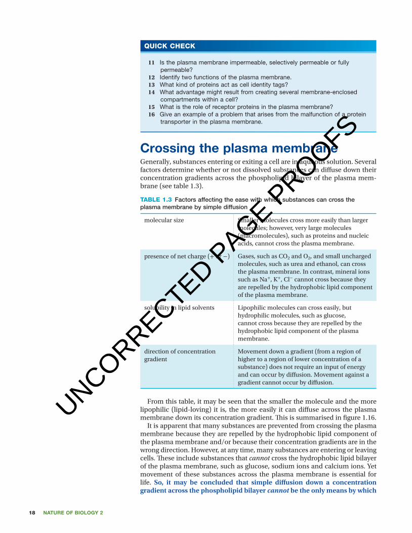
Crossing the plasma membrane Generally, substances entering or exiting a cell are in aqueous solution. Several factors determine whether or not dissolved substances can diffuse down their concentration gradients across the phospholipid bilayer of the plasma mem-brane (see table 1.3).
table 1.3 Factors affecting the ease with which substances can cross the plasma membrane by simple diffusion
molecular size Smaller molecules cross more easily than larger molecules; however, very large molecules (macromolecules), such as proteins and nucleic acids, cannot cross the plasma membrane.
presence of net charge (+ or −) Gases, such as CO2 and O2, and small uncharged molecules, such as urea and ethanol, can cross the plasma membrane. In contrast, mineral ions such as Na+, K+, Cl− cannot cross because they are repelled by the hydrophobic lipid component of the plasma membrane.
solubility in lipid solvents Lipophilic molecules can cross easily, but hydrophilic molecules, such as glucose, cannot cross because they are repelled by the hydrophobic lipid component of the plasma membrane.
direction of concentration gradient
Movement down a gradient (from a region of higher to a region of lower concentration of a substance) does not require an input of energy and can occur by diffusion. Movement against a gradient cannot occur by diffusion.
From this table, it may be seen that the smaller the molecule and the more lipophilic (lipid-loving) it is, the more easily it can diffuse across the plasma membrane down its concentration gradient. This is summarised in figure 1.16.
It is apparent that many substances are prevented from crossing the plasma membrane because they are repelled by the hydrophobic lipid component of the plasma membrane and/or because their concentration gradients are in the wrong direction. However, at any time, many substances are entering or leaving cells. These include substances that cannot cross the hydrophobic lipid bilayer of the plasma membrane, such as glucose, sodium ions and calcium ions. Yet movement of these substances across the plasma membrane is essential for life. So, it may be concluded that simple diffusion down a concentration gradient across the phospholipid bilayer cannot be the only means by which
UNCORRECTED PAGE P
ROOFS

19CHaPter 1 Cells: a review
c01CellsAReview 19 24 October 2016 10:42 AM
substances cross the plasma membrane. Instead, additional means of entry to and exit from cells must exist. Th ese additional means of transport for dis-solved substances, such as facilitated diff usion and active transport, involve the proteins of the plasma membrane (see the following section).
Gases
Smallunchargedpolarmolecules
Water
Largeunchargedpolarmolecules
Chargedpolarmolecules
Amino acidsATPGlucose 6-phosphate
Glucose
Ethanol
CO2
H2ONH2
K+, Mg2+, Ca2+,CI−, HCO3
−,HPO4
2−
NH2C
O
N2
O2
Ions
UreafiGuRe 1.16 Diagram showing the semipermeable nature of a phospholipid bilayer membrane. The membrane is fully permeable to some substances, partially permeable to others and impermeable to yet other substances. Note that the term ‘polar’ refers to molecules with an unequal distribution of electrons such that one side of a molecule has more electrons (and so is more negative) than the other side that has fewer electrons (and so is more positive). This is different from ions that have lost or gained an electron and so have a net electric charge.
Various ways of crossing the boundaryMovement of substances across the plasma membrane into or out of cells can occur by several mechanisms:1. Simple diff usion is the means of transport of small lipophilic substances.
Water can also move across the plasma membrane by diff usion; this is a special case of diff usion known as osmosis.
2. Facilitated diff usion involves protein transporters and is the means of trans-port of dissolved hydrophilic substances down their concentration gradients.
3. Active transport involves protein transporters known as pumps and is the means of transport of dissolved hydrophilic substances against their con-centration gradients.
4. Endocytosis/exocytosis are the means of bulk transport of macromol-ecules and liquids.
Simple diffusionSimple diff usion is the movement of substances across the phospholipid bilayer from a region of higher concentration to one of lower concentration of that substance; that is, down its concentration gradient (see fi gure 1.17a).
unit 3 Movement across the membrane: simple diffusionSummary screen and practice questions
aOs 1
topic 2
concept 4
UNCORRECTED PAGE P
ROOFS
Nature of biology 220
c01CellsAReview 20 24 October 2016 10:42 AM
Movement down a concentration gradient by simple diff usion does not require any input of energy. It is the gradient that drives the diff usion (like letting a ball roll down a slope). Th e end point of simple diff usion is reached when equal concentrations of the substance are reached on both sides of the plasma membrane.
Substances that move easily across the plasma membrane by simple dif-fusion are small lipophilic molecules that can dissolve in the lipid bilayer. Among these substances are steroid hormones, alcohol and lipophilic drugs.
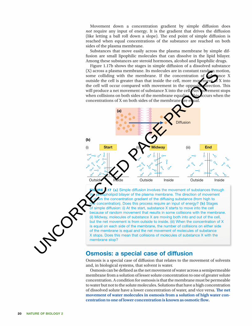
Figure 1.17b shows the stages in simple diff usion of a dissolved substance (X) across a plasma membrane. Its molecules are in constant random motion, some colliding with the membrane. If the concentration of substance X outside the cell is greater than that inside the cell, more movement of X into the cell will occur compared with movement in the opposite direction. Th is will produce a net movement of substance X into the cell. Net movement stops when collisions on both sides of the membrane equalise. Th is occurs when the concentrations of X on both sides of the membrane are equal.
fiGuRe 1.17 (a) Simple diffusion involves the movement of substances through the phospholipid bilayer of the plasma membrane. The direction of movement is down the concentration gradient of the diffusing substance (from high to low concentration). Does this process require an input of energy? (b) Stages of simple diffusion: (i) At the start, substance X starts to move into the cell because of random movement that results in some collisions with the membrane. (ii) Midway, molecules of substance X are moving both into and out of the cell, but the net movement is from outside to inside. (iii) When the concentration of X is equal on each side of the membrane, the number of collisions on either side of the membrane is equal and the net movement of molecules of substance X stops. Does this mean that collisions of molecules of substance X with the membrane stop?
Diffusion
(a)
Start Midway End(i)
(b)
(ii) (iii)
Outside Inside Outside Inside Outside Inside
osmosis: a special case of diffusion Osmosis is a special case of diff usion that relates to the movement of solvents and, in biological systems, that solvent is water.
Osmosis can be defi ned as the net movement of water across a semipermeable membrane from a solution of lesser solute concentration to one of greater solute concentration. A condition for osmosis is that the membrane must be permeable to water but not to the solute molecules. Solutions that have a high concentration of dissolved solute have a lower concentration of water, and vice versa. Th e net movement of water molecules in osmosis from a solution of high water con-centration to one of lower concentration is known as osmotic fl ow.
UNCORRECTED PAGE P
ROOFS
21CHaPter 1 Cells: a review
c01CellsAReview 21 24 October 2016 10:42 AM
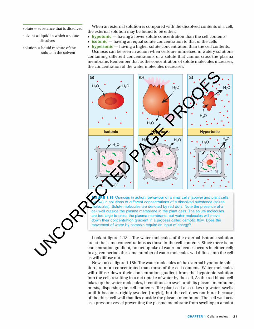
When an external solution is compared with the dissolved contents of a cell, the external solution may be found to be either:• hypotonic — having a lower solute concentration than the cell contents• isotonic — having an equal solute concentration to that of the cells• hypertonic — having a higher solute concentration than the cell contents.
Osmosis can be seen in action when cells are immersed in watery solutions containing diff erent concentrations of a solute that cannot cross the plasma membrane. Remember that as the concentration of solute molecules increases, the concentration of the water molecules decreases.
fiGuRe 1.18 Osmosis in action: behaviour of animal cells (above) and plant cells (below) in solutions of different concentrations of a dissolved substance (solute molecules). Solute molecules are denoted by red dots. Note the presence of a cell wall outside the plasma membrane in the plant cells. The solute molecules are too large to cross the plasma membrane, but water molecules will move down their concentration gradient in a process called osmotic fl ow. Does the movement of water by osmosis require an input of energy?
Isotonic Hypotonic Hypertonic
H2O H2O H2O H2O
H2O
H2O
H2O
H2O
H2OH2O H2OH2O
(a) (b) (c)
Look at fi gure 1.18a. Th e water molecules of the external isotonic solution are at the same concentrations as those in the cell contents. Since there is no concentration gradient, no net uptake of water molecules occurs in either cell; in a given period, the same number of water molecules will diff use into the cell as will diff use out.
Now look at fi gure 1.18b. Th e water molecules of the external hypotonic solu-tion are more concentrated than those of the cell contents. Water molecules will diff use down their concentration gradient from the hypotonic solution into the cell, resulting in a net uptake of water by the cell. As the red blood cell takes up the water molecules, it continues to swell until its plasma membrane bursts, dispersing the cell contents. Th e plant cell also takes up water, swells until it becomes rigidly swollen (turgid), but the cell does not burst because of the thick cell wall that lies outside the plasma membrane. Th e cell wall acts as a pressure vessel preventing the plasma membrane from swelling to a point
solute = substance that is dissolved
solvent = liquid in which a solute dissolves
solution = liquid mixture of the solute in the solvent
UNCORRECTED PAGE P
ROOFS

Nature of biology 222
c01CellsAReview 22 24 October 2016 10:42 AM
of bursting. Net entry of water molecules into the plant cell finally stops as a result of the increasing outward pressure of the cell contents that opposes the net inward flow of water.
Finally look at figure 1.18c. The water molecules of the external hypertonic solution are at a lower concentration than those in the cell contents. Water molecules will diffuse down their concentration gradient from the cells into the external solution, resulting in a net loss of water from the cells. The red blood cell shrinks, becoming crenated. The plant cell within its plasma mem-brane shrinks away from its cell wall.The dehydrated personA person suffering from a prolonged bout of diarrhoea is severely dehydrated and may need to be admitted to hospital. Normally, the cells lining the small and large intestine absorb the large volume of fluid and dissolved salts that enter the gut daily (see Odd fact). Several bacterial infections, including Staphylococcus sp., can inhibit this absorption. As a result, most of the fluid and the dissolved salts pass into the large intestine and are expelled from the body in large volumes of watery diarrhoea, resulting in dehydration and salt loss. Worsening dehydration is serious because it produces a decrease in blood volume that, if untreated, may lead to cardiovascular failure.
Initial treatment of severe dehydration is replacement of the lost fluid and salts. The rehydration therapy involves an isotonic solution of saline (salt) solu-tion plus glucose. This solution can be administered either orally (by mouth) or by direct infusion into the bloodstream (by intravenous infusion, or IV).
Why does this treatment act as a rehydration therapy? Glucose stimulates the absorption of sodium by the gut cells. The uptake of sodium and glucose from the gut into the extracellular fluid creates a hypertonic internal environment that causes water to move by osmosis from the gut across the cells lining the gut, and so returns water to the extracellular fluid and from there to the body cells.
facilitated diffusionFacilitated diffusion is an example of protein-mediated transport. Facilitated diffusion is so named because the diffusion across the membrane is enabled or facilitated by special protein transporters in the plasma membrane.
Like simple diffusion, facilitated diffusion does not require an input of energy. Like simple diffusion, facilitated diffusion moves substances down their concentration gradients. However, facilitated diffusion of dissolved sub-stances requires the action of protein transporters that are embedded in the cell membrane.
Facilitated diffusion enables molecules that cannot diffuse across the phospholipid bilayer to move across the plasma membrane through the agency of transporter proteins. These transporters are either channel proteins or carrier proteins. Are transporters required in simple diffusion? (Refer to figure 1.19.)
Channel proteinsOne group of transport proteins involved in facilitated diffusion are the channel proteins. Each channel protein is trans-membrane and has a central water-filled pore through which dissolved substances can pass down their con-centration gradient. Different channel proteins are specific for the diffusion of charged particles or polar molecules.
Each channel protein consists of a narrow water-filled pore in the plasma membrane (see figure 1.19b). By providing water-filled pores, channel proteins create a hydrophilic passage across the plasma membrane that bypasses the phospholipid bilayer and facilitates the diffusion of charged particles, such as sodium and potassium ions, and small polar molecules.
Odd fact
Each day about 2 litres of fluid are taken into the gut in food and drinks. This is increased by a further volume of about 7 litres from secretions, including those of the salivary glands, stomach, liver and pancreas.
unit 3 Movement across the membrane: facilitated diffusionSummary screen and practice questions
aOs 1
topic 2
concept 5
In the case of ions, which carry either + or − charge(s), it is more correct to say that, rather than moving down their concentration gradients, they move down their electrochemical gradients. UNCORRECTED P
AGE PROOFS
23CHaPter 1 Cells: a review
c01CellsAReview 23 24 October 2016 10:42 AM
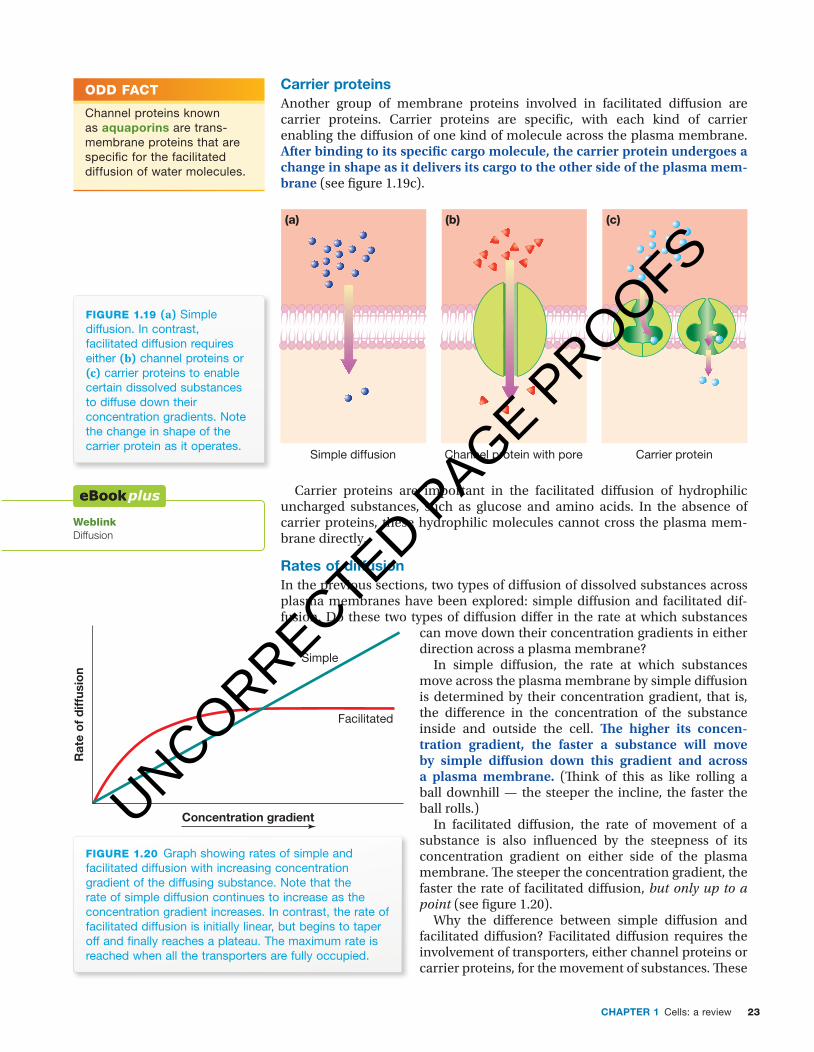
Carrier proteinsAnother group of membrane proteins involved in facilitated diff usion are carrier proteins. Carrier proteins are specifi c, with each kind of carrier enabling the diff usion of one kind of molecule across the plasma membrane. After binding to its specifi c cargo molecule, the carrier protein undergoes a change in shape as it delivers its cargo to the other side of the plasma mem-brane (see fi gure 1.19c).
Simple diffusion Carrier proteinChannel protein with pore
(a) (b) (c)
fiGuRe 1.19 (a) Simple diffusion. In contrast, facilitated diffusion requires either (b) channel proteins or (c) carrier proteins to enable certain dissolved substances to diffuse down their concentration gradients. Note the change in shape of the carrier protein as it operates.
Carrier proteins are important in the facilitated diff usion of hydrophilic uncharged substances, such as glucose and amino acids. In the absence of carrier proteins, these hydrophilic molecules cannot cross the plasma mem-brane directly.
rates of diffusionIn the previous sections, two types of diff usion of dissolved substances across plasma membranes have been explored: simple diff usion and facilitated dif-fusion. Do these two types of diff usion diff er in the rate at which substances
can move down their concentration gradients in either direction across a plasma membrane?
In simple diff usion, the rate at which substances move across the plasma membrane by simple diff usion is determined by their concentration gradient, that is, the diff erence in the concentration of the substance inside and outside the cell. Th e higher its concen-tration gradient, the faster a substance will move by simple diff usion down this gradient and across a plasma membrane. (Th ink of this as like rolling a ball downhill — the steeper the incline, the faster the ball rolls.)
In facilitated diff usion, the rate of movement of a substance is also infl uenced by the steepness of its concentration gradient on either side of the plasma membrane. Th e steeper the concentration gradient, the faster the rate of facilitated diff usion, but only up to a point (see fi gure 1.20).
Why the diff erence between simple diff usion and facilitated diff usion? Facilitated diff usion requires the involvement of transporters, either channel proteins or carrier proteins, for the movement of substances. Th ese
Odd fact
Channel proteins known as aquaporins are trans-membrane proteins that are specifi c for the facilitated diffusion of water molecules.
WeblinkDiffusion
Concentration gradient
Rat
e o
f d
iffus
ion
Simple
Facilitated
fiGuRe 1.20 Graph showing rates of simple and facilitated diffusion with increasing concentration gradient of the diffusing substance. Note that the rate of simple diffusion continues to increase as the concentration gradient increases. In contrast, the rate of facilitated diffusion is initially linear, but begins to taper off and fi nally reaches a plateau. The maximum rate is reached when all the transporters are fully occupied.
UNCORRECTED PAGE P
ROOFS

Nature of biology 224
c01CellsAReview 24 24 October 2016 10:42 AM
channel and carrier proteins are present in limited numbers on the plasma membrane. Because their numbers are limited, eventually a concentration of the diff using substance will be reached when all the transporters are saturated (fully occupied). So, as the concentration gradient increases, the rate of facil-itated diff usion of a substance will at fi rst increase, then become slower, and fi nally will reach a plateau. Th e plateau is the maximum rate of facilitated dif-fusion. When this is reached, all the transporter molecules are fully occupied.
active transportActive transport is the process of moving substances across the plasma mem-brane against the direction that they would travel by diff usion; that is, active transport moves dissolved substances from a region of low concentration to a region of high concentration of those substances. Active transport can occur only with an input of energy, and the energy source is typically adeno-sine triphosphate (ATP).
Special transport proteins embedded across the plasma membrane carry out the process of active transport. Th e proteins involved are called pumps and each diff erent pump transports one (or sometimes two) specifi c substance(s). Important pumps are proteins with both a trans-port function and an enzyme function. Th e enzyme part of the pump catalyses an energy- releasing reaction:
ATP → ADP + Pi + energy
Th e transport part of the pump uses this energy to move small polar molecules and ions across the plasma membrane against their con-centration gradients. During this process, the protein of the pump undergoes a shape change (see fi gure 1.21).
Cells use pumps to move materials that they need by active transport. Active transport is essential for the key function of cells including
pH balance, regulation of cell volume and uptake of needed nutrients. Examples of active transport include:• uptake of dissolved mineral ions from water in the soil by plant root hair
cells against their concentration gradient • production of acidic secretions (pH of nearly 1) by stomach cells that have
a low internal concentration of hydrogen ions (H+) but produce secretions (gastric juice) with an extremely high concentration of hydrogen ions
• uptake of glucose from the small intestine into the cells lining the intestine against its concentration gradient, using the glucose–sodium pump
• maintenance of the diff erence in the concentrations of sodium and pot-assium ions that exist inside and outside cells (see table 1.4) by action of the sodium–potassium pump, which actively transports these ions against their concentration gradients (see below). Some pumps actively transport a single dissolved substance against its con-
centration gradient. Other pumps transport two substances simultaneously, for example, the sodium–potassium pump. Th e importance of the sodium–potassium pump is highlighted by the fact that, in the human body overall, about 25% of the body’s ATP is expended in keeping the sodium–potassium pump operating. For brain cells, the fi gure is even higher, about 70%.
Why is this pump needed? Table 1.4 provides a clue to the answer.
fiGuRe 1.21 Diagram showing a simplifi ed representation of active transport. A pump is a trans-membrane protein that is both a carrier and an ATPase enzyme. The enzyme component of the pump catalyses the energy-releasing reaction that powers active transport.
Active transportATP
Odd fact
Compared with the concentration of H+ ions in the contents of stomach cells, the gastric juice in the stomach has about a concentration three million times higher. This is achieved by active transport of these ions out of the stomach cells against their concentration gradient.
unit 3 Movement across themembrane:active transportSummary screen and practice questions
aOs 1
topic 2
concept 6
UNCORRECTED PAGE P
ROOFS

25CHaPter 1 Cells: a review
c01CellsAReview 25 24 October 2016 10:42 AM
table 1.4 Approximate concentrations of sodium and potassium ions in the cytosol of cells and in the surrounding extracellular fluid
ion inside cell outside cell
sodium (Na+) 10 mM 142 mM
potassium (K+) 150 mM 5 mM
Table 1.4 shows the concentrations of sodium and potassium ions inside and outside cells. Note that these concentrations differ greatly. In what direction would sodium ions tend to flow passively? What about potassium ions? How are these concentration differences maintained? The different concentrations are maintained by the sodium–potassium pump, which is present in all animal cells and constantly expends energy in active transport, pushing sodium ions out of the cell and pulling potassium ions in. For each ATP molecule expended, the pump pushes three sodium ions out and drags two potassium ions in. This process of active transport compensates for the constant passive diffusion of sodium ions into cells and of potassium ions out of cells, both down their con-centration gradients.
The sodium–potassium pump plays a key role in excitable cells, such as nerve cells and muscle cells. During the transmission of a nerve impulse, sodium ion channels open and sodium ions rapidly flood into the nerve cell by facilitated diffusion. After the nerve impulse has passed, the sodium channels close and the sodium–potassium pump then restores the concentrations of sodium and potassium ions to their resting levels by actively pushing sodium ions across the membrane out of the cell and dragging potassium ions into the cell (refer back to table 1.4). Restoring these concentrations involves active transport against the concentration gradients of these ions.
When transport goes wrong . . .The importance of transport proteins in moving substances becomes apparent if they do not operate as expected, as may be seen in cystic fibrosis and in cholera infections.
Cystic fibrosisSymptoms seen in people affected by the inherited disorder cystic fibrosis result from a defect in one transporter protein on the plasma membrane of cells. This protein is a channel that normally allows chloride ions (Cl−) to move out of cells. Cystic fibrosis affects various organs, including the lungs, the pan-creas and the skin. A faulty chloride ion channel protein, such as occurs in cystic fibrosis, blocks the movement of chloride ions. This affects the various organs as follows: • Lungs. Normally, the inner surfaces of a person’s lungs are covered with
a thin layer of mucus. This mucus is important in a healthy lung because it traps microbes and other particles, and it is constantly removed from the lungs by the beating action of hair-like projections (cilia) that line the airways. Cough or clear your throat and that mucus and those trapped particles are gone. In the lungs, the chloride ion channel normally moves chloride ions out of lung cells. In cystic fibrosis, however, the defect in the transporter stops i the movement of Cl− ions out of the cells into the lung cavity ii the consequential flow of sodium (Na+) ions that move in response to
the electrochemical gradient created by the movement of the negative chloride ions into the lung cavity
iii osmotic flow of water into the lungs that normally thins the mucus.
Odd fact
The Danish scientist Jens Skou was awarded the Nobel Prize in chemistry for his discovery of the ion-transporting enzyme Na+, K+ ATPase, otherwise known as the sodium–potassium pump.
unit 3
do moreMovement across membranes
aOs 1
topic 2
concept 6
Odd fact
Before accurate genetic testing for cystic fibrosis became available, an early test was the so-called ‘sweat test’ that measured salt levels in a baby’s sweat.
UNCORRECTED PAGE P
ROOFS

NATURE OF BIOLOGY 226
c01CellsAReview 26 26 October 2016 9:40 AM
� ese e� ects of the faulty chloride ion channel mean that the mucus in the lungs is abnormally thick, sticky and di� cult to move and, rather than being easily cleared, the mucus remains in the lungs a� ecting breathing and acting as a potential source of infection. • Sweat glands in skin. In the skin, the chloride ion channel is normally
involved in reabsorbing salt (NaCl) from � uid within cells of the sweat glands before it is released as sweat. When the chloride ion (Cl–) transporter is blocked, reabsorption does not occur and very salty sweat is produced.
• Pancreas. Pancreatic enzymes are normally involved in the digestion of some foods. In cystic � brosis, these enzymes are unable to enter the gut because abnormally thick mucus blocks the narrow duct that connects the pancreas to the small intestine. Without these enzymes, food cannot be fully digested. However, replacement enzymes in the form of tablets, powders or capsules can replace the enzymes normally released by the pancreas (see � gure 1.22).
Cholera� e pores of some channel proteins are permanently open, while the pores in other channel proteins open only in response to a speci� c signal. � e chloride ion channel on the plasma membranes of cells lining the intestine is not always open. Normally chloride ions are retained within the cells lining the intestine. Only when this channel is opened can chloride ions move from the cells into the cavity of the intestine.
Cholera results from a bacterial infection of Vibrio cholera. A toxin produced by these bacteria causes the chloride ion channels in the cells lining the intestine to be locked in the ‘open’ position. � is results in a � ood of chloride ions into the intestinal space that is followed by a � ow of sodium ions (down the resulting electrochemical gradient that is created). In turn, the increased concentration
of salt in the gut creates a hyperosmotic environ-ment that draws water into the gut by osmosis. � e continuous secretion of water into the intestine causes the production of large volumes of watery diarrhoea. If left untreated the water loss caused by this diarrhoea can be fatal within hours. Cholera epidemics have caused many deaths (see � gure 1.23). Cholera can be spread by water that is contaminated by contact with untreated sewage or by the faeces of an infected person. Cholera can be spread by food that is washed in or mixed with water contaminated by cholera bacteria, or by food that is inappropriately handled by a person infected with cholera.
FIGURE 1.23 Cholera epidemics and pandemics have taken many lives. During the period 1899 to 1923, a cholera pandemic that began in India spread across the globe and reached as far as Russia and Eastern Europe. This French publication from 1912 illustrates the public fear and the widespread deaths that this cholera pandemic caused.
FIGURE 1.22 Capsules and tablets containing the missing pancreatic enzymes (lipase, protease, amylase) are taken by people affected by cystic � brosis. Note the enzyme granules in the capsules. (Image courtesy of Marjory Martin.)
UNCORRECTED PAGE P
ROOFS

27CHaPter 1 Cells: a review
c01CellsAReview 27 24 October 2016 10:42 AM
bulk transport of solids and liquidsTo this point, we have been concerned with move-ment of dissolved substances across the plasma membrane. In addition, small solid particles and liquids in bulk can be moved across the plasma mem-brane into or out of cells. Figure 1.24 gives a summary of how bulk material can enter cells.
endocytosis: getting inSolid particles can be taken into a cell. For example, several cells of the immune system can engulf disease-causing bacteria cells, enclosing them within lysosomal sacs where they are destroyed. Unicellular protists, such as Amoeba and Paramecium, obtain their energy for living in the form of relatively large ‘food’ particles, which they engulf and enclose within a sac where the food is digested (see fi gure 1.25a).
Pseudopods
Food particle
(a)
Lysosomes containingdigestive enzymes
Entrapment
Amoeba
Engulfment
Digestion
Absorption
Foodvacuole
Digestedfood
Absorbedfood
Food
Cytosol
Phagocyticvesicle
Outside cell(b)
Lipidbilayer
fiGuRe 1.25 (a) Transport of a solid food particle across the membrane of an amoeba (b) Endocytosis occurs when part of the plasma membrane forms around food particles to form a phagocytic vesicle (or phagosome). This vesicle then moves into the cytosol where it fuses with a lysosome, a bag of digestive enzymes. The same process is also part of the body’s immune defence against infectious microbes.
If material is solid If material is �uid
Endocytosisbulk transport of
material into a cell
phagocytosisfrom the Greek
phagos = ‘eating’and cyto = ‘cell’
pinocytosisfrom the Greek
pinus = ‘drinking’and cyto = ‘cell’
the process is called the process is called
fiGuRe 1.24 Endocytosis — a summary
UNCORRECTED PAGE P
ROOFS
Nature of biology 228
c01CellsAReview 28 24 October 2016 10:42 AM
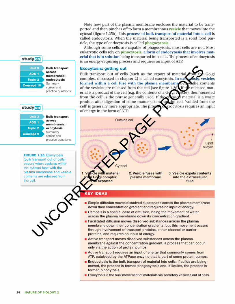
Note how part of the plasma membrane encloses the material to be trans-ported and then pinches off to form a membranous vesicle that moves into the cytosol (fi gure 1.25b). Th is process of bulk transport of material into a cell is called endocytosis. When the material being transported is a solid food par-ticle, the type of endocytosis is called phagocytosis.
Although some cells are capable of phagocytosis, most cells are not. Most eukaryotic cells rely on pinocytosis, a form of endocytosis that involves mat-erial that is in solution being transported into cells. Th e process of endocytosis is an energy-requiring process and requires an input of ATP.
exocytosis: getting out Bulk transport out of cells (such as the export of material from the Golgi complex, discussed in chapter 2) is called exocytosis. In exocytosis, vesicles formed within a cell fuse with the plasma membrane before the contents of the vesicles are released from the cell (see fi gure 1.26). If the released mat-erial is a product of the cell (e.g. the contents of a Golgi vesicle), then ‘secreted from the cell’ is the phrase generally used. If the released material is a waste product after digestion of some matter taken into the cell, ‘voided from the cell’ is generally more appropriate. Th e process of exocytosis requires an input of energy in the form of ATP.
fiGuRe 1.26 Exocytosis (bulk transport out of cells) occurs when vesicles within the cytosol fuse with the plasma membrane and vesicle contents are released from the cell.
Cytosol
1. Vesicle with materialfrom Golgi complex
to be exported
2. Vesicle fuses withplasma membrane
3. Vesicle expels contentsinto the extracellular
fluid
Outside cell
Lipidbilayer
key ideas
■ Simple diffusion moves dissolved substances across the plasma membrane down their concentration gradient and requires no input of energy.
■ Osmosis is a special case of diffusion, being the movement of water across the plasma membrane down its concentration gradient.
■ Facilitated diffusion moves dissolved substances across the plasma membrane down their concentration gradients, but this movement occurs through involvement of transport proteins, either channel or carrier proteins, and requires no input of energy.
■ Active transport moves dissolved substances across the plasma membrane against the concentration gradient, a process that can occur only via the action of protein pumps.
■ Active transport requires an input of energy that commonly comes from ATP, catalysed by the ATPase enzyme that is part of some protein pumps.
■ Endocytosis is the bulk transport of material into cells; if solids are being moved, the process is termed phagocytosis and, if liquids, the process is termed pinocytosis.
■ Exocytosis is the bulk movement of materials via secretory vesicles out of cells.
unit 3 bulk transport across membranes: endocytosisSummary screen and practice questions
aOs 1
topic 2
concept 10
unit 3 bulk transport across membranes: exocytosisSummary screen and practice questions
aOs 1
topic 2
concept 9
UNCORRECTED PAGE P
ROOFS

29CHaPter 1 Cells: a review
c01CellsAReview 29 24 October 2016 10:42 AM
Quick check
17 What is the process by which bulk materials are exported out of cells? 18 Consider passive diffusion and facilitated diffusion:
a Identify one difference between these processes.b Identify one similarity that they share.
19 Identify one difference between diffusion and active transport.20 Which transport process relies on the involvement of either a carrier or a
channel protein?21 By which process do cells of the stomach lining manage to move hydrogen
ions out of the cells to produce a highly acidic gastric secretion? 22 What process is involved in the movement of water down its concentration
gradient and across a layer of cells from outside the body to inside?
the role of different organelles in the export of protein from the cellribosomes: protein factoriesCells make a range of proteins for many purposes: for example, manufacture of haemoglobin, an oxygen-transporting protein by developing human red blood cells in the bone marrow; manufacture of the contractile proteins actin and myosin by the muscle cells; and manufacture of the hormone insulin and diges-tive enzymes including lipases by diff erent cells of the pancreas. Insulin is an example of a protein that is exported from the cell in which it was manufactured.
Ribosomes are the site in the cell where proteins are made. It is on the ribo-somes that amino acids are assembled and joined into polypeptide chains or proteins. Th e diameter of a ribosome is only about 0.03 µm. Because of their very small size, ribosomes can be seen only through an electron microscope (see fi gure 1.27a). However, ribosomes are present in very large numbers in a cell. Ribosomes in animal and plant cells are composed of about two-thirds RNA and one-third protein.
ri
(a)
Ribosomes
Transport channel
(b)
fiGuRe 1.27 (a) TEM image of a section of cell showing the rough endoplasmic reticulum (er) with ribosomes (ri). Note also the nucleus (n) inside its nuclear membrane or envelope (ne). Ribosomes are only about 0.03 µm in diameter so they appear as tiny dots in this image. Are ribosomes enclosed in a membrane? (b) 3D representation of the endoplasmic reticulum with ribosomes
Ribosomes
UNCORRECTED PAGE P
ROOFS

Nature of biology 230
c01CellsAReview 30 24 October 2016 10:42 AM
Within a cell, many ribosomes are attached to membranous channels known as the endoplasmic reticulum (see next section). Other ribosomes are found free in the cytosol. Proteins made by ‘free’ ribosomes are for local use within the cell.
rough endoplasmic reticulumTh e endoplasmic reticulum (ER) is an interconnected system of membrane- enclosed fl attened channels. Figure 1.28 shows part of the channels of the endoplasmic reticulum in a eukaryotic cell. Refer also to fi gure 1.27a. Where the ER has ribosomes attached to the outer surface of its channels, it is known as rough endoplasmic reticulum. Proteins produced by ribosomes on the endoplasmic reticulum are generally exported from the cell.
fiGuRe 1.28 False coloured scanning electron micrograph of part of a eukaryotic cell showing the channels of rough ER (pink). Note the many tiny bead-like structures attached to the outside of the ER channels. What are these structures?
Odd fact
Ribosomes can join amino acids into a protein chain at the rate of about 200 per minute.
Endoplasmicreticulum
Odd fact
An estimated 13 million ribosomes are attached to the rough ER in a typical human liver cell.
UNCORRECTED PAGE P
ROOFS
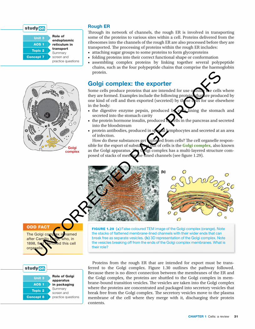
31CHaPter 1 Cells: a review
c01CellsAReview 31 24 October 2016 10:42 AM
rough erTh rough its network of channels, the rough ER is involved in transporting some of the proteins to various sites within a cell. Proteins delivered from the ribosomes into the channels of the rough ER are also processed before they are transported. Th e processing of proteins within the rough ER includes: • attaching sugar groups to some proteins to form glycoproteins• folding proteins into their correct functional shape or conformation• assembling complex proteins by linking together several polypeptide
chains, such as the four polypeptide chains that comprise the haemoglobin protein.
golgi complex: the exporterSome cells produce proteins that are intended for use outside the cells where they are formed. Examples include the following proteins that are produced by one kind of cell and then exported (secreted) by those cells for use elsewhere in the body:• the digestive enzyme pepsin, produced by cells lining the stomach and
secreted into the stomach cavity• the protein hormone insulin, produced by cells in the pancreas and secreted
into the bloodstream• protein antibodies, produced in special lymphocytes and secreted at an area
of infection.How do these substances get exported from cells? Th e cell organelle respon-
sible for the export of substances out of cells is the Golgi complex, also known as the Golgi apparatus. Th e Golgi complex has a multi-layered structure com-posed of stacks of membrane-lined channels (see fi gure 1.29).
(b)(a)
fiGuRe 1.29 (a) False coloured TEM image of the Golgi complex (orange). Note the stacks of fl attened membrane-lined channels with their wider ends that can break free as separate vesicles. (b) 3D representation of the Golgi complex. Note the vesicles breaking off from the ends of the Golgi complex membranes. What is their role?
Proteins from the rough ER that are intended for export must be trans-ferred to the Golgi complex. Figure 1.30 outlines the pathway followed. Because there is no direct connection between the membranes of the ER and the Golgi complex, the proteins are shuttled to the Golgi complex in mem-brane-bound transition vesicles. Th e vesicles are taken into the Golgi complex where the proteins are concentrated and packaged into secretory vesicles that break free from the Golgi complex. Th e secretory vesicles move to the plasma membrane of the cell where they merge with it, discharging their protein contents.
unit 3 Role of endoplasmic reticulum in transport Summary screen and practice questions
aOs 1
topic 2
concept 7
Golgicomplex
Odd fact
The Golgi complex is named after Camillo Golgi, who, in 1898, fi rst identifi ed this cell organelle.
unit 3 Role of Golgi apparatus in packagingSummary screen and practice questions
aOs 1
topic 2
concept 8
UNCORRECTED PAGE P
ROOFS

Nature of biology 232
c01CellsAReview 32 24 October 2016 10:42 AM
Roughendoplasmicreticulum
Ribosomes
Secretoryvesicle
Golgicomplex
Membranefusion occurring
Transitionvesicle
Cytoplasmof cell
Discharge byexocytosis; for example,a hormone
fiGuRe 1.30 The secretory export pathway for proteins. Packages of protein are transferred from the rough ER in transition vesicles to the Golgi complex where they are taken in. From the Golgi complex, the secretory vesicles with their protein cargo move to the plasma membrane of the cell, merge with it and discharge their contents.
key ideas
■ Ribosomes are cell organelles where proteins are manufactured. ■ The endoplasmic reticulum (ER) is made of a series of membrane-bound channels.
■ Rough ER is so named because of the presence of ribosomes on the external surface of its membranes.
■ Rough ER is involved in processing of proteins and in their transport. ■ The Golgi complex packages substances into vesicles for export from a cell.
Quick check
23 Identify whether each of the following is true or false:a The RNA of the ribosomes is made in the nucleolus.b The folding of a protein into its functional 3D shape takes place on the
ribosomes.c Ribosomes are membrane-bound organelles that form part of the cell
cytoplasm.d The channels of the Golgi complex are connected to those of the ER.
24 A scientist wishes to examine ribosomes in a liver cell.a Where should the scientist look: in the nucleus or the cytoplasm?b What kind of microscope is likely to be used by the scientist: a light
microscope or a transmission electron microscope? Explain.
UNCORRECTED PAGE P
ROOFS
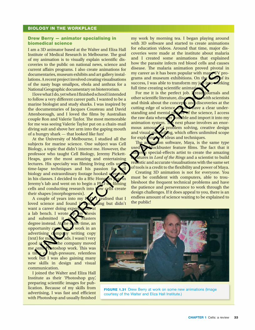
33CHAPTER 1 Cells: a review
c01CellsAReview 33 26 October 2016 9:40 AM
BIOLOGY IN THE WORKPLACE
Drew Berry — animator specialising in biomedical scienceI am a 3D animator based at the Walter and Eliza Hall Institute of Medical Research in Melbourne. � e goal of my animation is to visually explain scienti� c dis-coveries to the public on national news, science and current a� airs programs. I also create animations for documentaries, museum exhibits and art gallery instal-lations. A recent project involved creating visualisations of the nasty bugs smallpox, ebola and anthrax for a National Geographic documentary on bioterrorism.
I love what I do, yet when I � nished school I intended to follow a very di� erent career path. I wanted to be a marine biologist and study sharks. I was inspired by the documentaries of Jacques Cousteau and David Attenborough, and I loved the � lms by Australian couple Ron and Valerie Taylor. � e most memorable for me was seeing Valerie Taylor put on a chain-mail diving suit and shove her arm into the gaping mouth of a hungry shark — that looked like fun!
At the University of Melbourne, I studied all the subjects for marine science. One subject was Cell Biology, a topic that didn’t interest me. However, the professor who taught Cell Biology, Jeremy Pickett-Heaps, gave the most amazing and entertaining lectures. His specialty was � lming living cells using time-lapse techniques and his passion for cell biology and extra ordinary footage hooked everyone in his classes. I decided to do a BSc Honours year in Jeremy’s lab and went on to begin a PhD on � lming cells and conducting research into how cells create their shapes (morphogenesis).
A couple of years into my PhD, I realised that I loved science and found it fascinating but didn’t want a career doing experiments at a lab bench. I wrote up my thesis and submitted it as a Masters degree instead. At the same time, an opportunity came up to work in an advertising company writing copy (text) for magazine ads. I wasn’t very good at it and the company moved me onto Photoshop work. � is was a time of high-pressure, relentless work but I was also gaining many new skills in design and visual communication.
I joined the Walter and Eliza Hall Institute as their ‘Photoshop guy’, preparing scienti� c images for pub-lication. Because of my skills from advertising, I was fast and e� cient with Photoshop and usually � nished
my work by morning tea. I began playing around with 3D software and started to create animations for education videos. Around that time, major dis-coveries were made at the institute about malaria and I created some animations that explained how the parasite infects red blood cells and causes disease. � e malaria animation proved pivotal in my career as it has been popular with many TV pro-grams and museum exhibitions. On the basis of its success, I was able to transform my job into working full time creating scienti� c animations.
For me it is the perfect job. I read journals and other scienti� c literature, discuss ideas with scientists and think about the concepts and discoveries at the cutting edge of science. Once I have a clear under-standing and mental picture of the science, I access the raw data wherever possible and import it into my animation system. � e next phase involves an enor-mous amount of problem solving, creative design and visual story telling, which o� ers unlimited scope for exploring new ideas and techniques.
� e animation software, Maya, is the same type used for blockbuster feature � lms. � e fact that it allows a special-e� ects artist to create the amazing creatures in Lord of the Rings and a scientist to build realistic and accurate visualisations with the same set of tools is a credit to the � exibility and power of Maya.
Creating 3D animation is not for everyone. You must be con� dent with computers, able to trou-bleshoot the frequent technical problems and have the patience and perseverance to work through the design challenges. If it does appeal to you, there is an endless amount of science waiting to be explained to the public!
FIGURE 1.31 Drew Berry at work on some new animations (Image courtesy of the Walter and Eliza Hall Institute.)
UNCORRECTED PAGE P
ROOFS
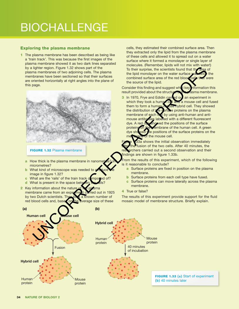
34 Nature of biology 2
BIOCHALLENGE
c01CellsAReview 34 24 October 2016 10:42 AM
exploring the plasma membrane
1 The plasma membrane has been described as being like a ‘train track’. This was because the fi rst images of the plasma membrane showed it as two dark lines separated by a lighter region. Figure 1.32 shows part of the plasma membranes of two adjoining cells. The plasma membranes have been sectioned so that their surfaces are oriented horizontally at right angles into the plane of this page.
fiGuRe 1.32 Plasma membrane
a How thick is the plasma membrane in nanometres? In micrometres?
b What kind of microscope was needed to produce the image in fi gure 1.32?
c What are the ‘rails’ of the train track composed of?d What is present in the space between the rails?
2 Key information about the nature of the plasma membrane came from an experiment carried out in 1925 by two Dutch scientists. They took a known number of red blood cells and, based on the average size of these
cells, they estimated their combined surface area. Then they extracted only the lipid from the plasma membrane of these cells and allowed it to spread out on a water surface where it formed a monolayer or single layer of molecules. (Remember, lipids will not mix with water!) To their surprise, the scientists found that the area of the lipid monolayer on the water surface was twice the combined surface area of the red blood cells that were the source of the lipid.
Consider this fi nding and suggest what key information this result provided about the structure of the plasma membrane.
3 In 1970, Frye and Edidin carried out an experiment in which they took a human cell and a mouse cell and fused them to form a human–mouse hybrid cell. They showed the distribution of the surface proteins on the plasma membrane of each cell by using anti-human and anti-mouse antibodies labelled with a different fl uorescent dye. A red dye showed the positions of the surface proteins on the membrane of the human cell. A green dye showed the positions of the surface proteins on the membrane of the mouse cell.
Figure 1.33a shows the initial observation immediately after the fusion of the two cells. After 40 minutes, the researchers carried out a second observation and their fi ndings are shown in fi gure 1.33b.
From the results of this experiment, which of the following is it reasonable to conclude?
a Surface proteins are fi xed in position on the plasma membrane.
b Surface proteins from each cell type have fused.c Surface proteins can move laterally across the plasma
membrane.
4 True or false?
The results of this experiment provide support for the fl uid mosaic model of membrane structure. Briefl y explain.
Human cell Mouse cell
Hybrid cell
Hybrid cell
Humanprotein
Humanprotein
Fusion
Mouseprotein
40 minutesof incubation
Mouseprotein
(a) (b)
fiGuRe 1.33 (a) Start of experiment (b) 40 minutes later
UNCORRECTED PAGE P
ROOFS
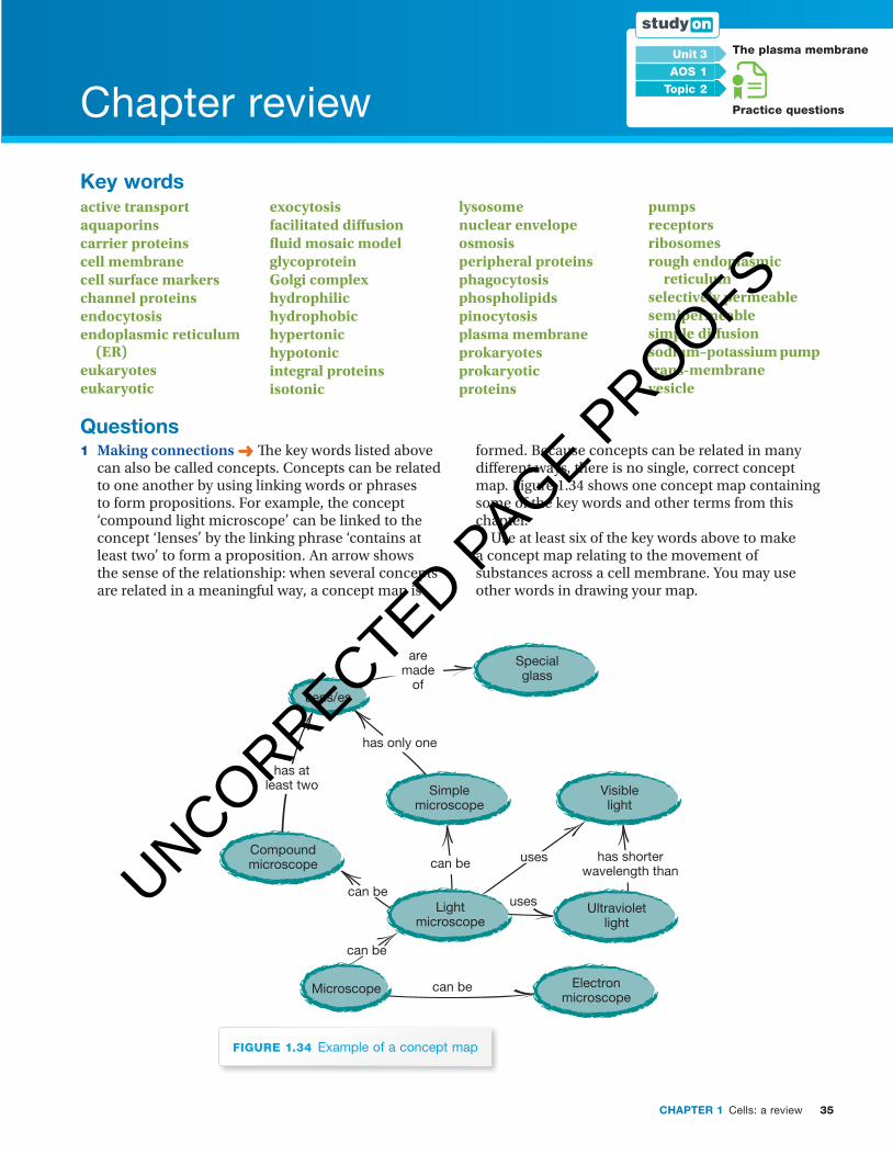
35CHaPter 1 Cells: a review
Chapter review
c01CellsAReview 35 24 October 2016 10:42 AM
unit 3 the plasma membrane
Practice questions
aOs 1
topic 2
Key wordsactive transportaquaporinscarrier proteinscell membranecell surface markerschannel proteinsendocytosisendoplasmic reticulum
(ER)eukaryoteseukaryotic
exocytosisfacilitated diff usion fl uid mosaic modelglycoproteinGolgi complex hydrophilic hydrophobichypertonichypotonic integral proteinsisotonic
lysosomenuclear envelopeosmosisperipheral proteinsphagocytosisphospholipidspinocytosisplasma membraneprokaryotesprokaryotic proteins
pumpsreceptorsribosomes rough endoplasmic
reticulum selectively permeablesemipermeablesimple diff usionsodium–potassium pumptrans-membranevesicle
Questions 1 Making connections ➜ Th e key words listed above
can also be called concepts. Concepts can be related to one another by using linking words or phrases to form propositions. For example, the concept ‘compound light microscope’ can be linked to the concept ‘lenses’ by the linking phrase ‘contains at least two’ to form a proposition. An arrow shows the sense of the relationship: when several concepts are related in a meaningful way, a concept map is
formed. Because concepts can be related in many diff erent ways, there is no single, correct concept map. Figure 1.34 shows one concept map containing some of the key words and other terms from this chapter.
Use at least six of the key words above to make a concept map relating to the movement of substances across a cell membrane. You may use other words in drawing your map.
Lens/es
Simplemicroscope
Visiblelight
Compoundmicroscope
Electronmicroscope
Lightmicroscope
Ultravioletlight
Microscope
Specialglass
has only one
aremade
of
can be
can be
uses
uses has shorterwavelength than
can be
can be
has atleast two
fiGuRe 1.34 Example of a concept map
UNCORRECTED PAGE P
ROOFS

36 Nature of biology 2
c01CellsAReview 36 24 October 2016 10:42 AM
2 Applying your understanding ➜ Consider the information in table 1.5.
table 1.5 Data for three different shapes, each having the same volume. (Where necessary, figures have been rounded.)
Cell Shape DimensionsSurface
area VolumeSa:V ratio
A flat sheet 10 × 10 × 0.1 204 10 20.4
B cube 2.15 × 2.15 × 2.15 28 10 2.8
C sphere diameter: 1.67 22 10 2.2
a If these shapes represented cells, which cell (A, B or C) would be most efficient in moving required materials into and removing wastes from the cell? Explain.
b Which cell would be least efficient? Explain.c Can you suggest a biological consequence of your
conclusion?d Identify one way in which a cell might retain its
overall shape, but greatly increase its surface area with a minimal increase in volume. (Clue: This strategy is used by cells involved in absorption of material, such as those lining the small intestine.)
3 Analysing information and drawing conclusions ➜ Consider the data in table 1.6.
table 1.6 Data for two sets of cells of identical shape but of decreasing sizes.
Cell Shape DimensionsSurface
area VolumeSa:V ratio
P flat sheet 10 × 10 × 0.1 204 10 20.4
Q flat sheet 5 × 5 × 0.05 51 1.25 40.8
R flat sheet 1 × 1 × 0.01 2.04 0.01 204
H sphere diameter: 10 314.2 523.6 0.6
K sphere diameter: 5 78.5 65.5 1.2
L sphere diameter: 1.0 3.14 0.52 6
Note: relative to the first shape in each set, the dimensions of other members of the set are scaled down by a factor of 2 and by a factor of 10.
a A student stated the same shape scaled down should retain the same surface-area-to-volume ratio, the student’s reason being ‘the shapes stay the same’. Do you agree with this student? Explain your decision.
b With regard to the information in table 1.6, identify how scaling a shape (up or down) affects
the SA:V ratio of a given shape by completing the following sentences:
i If the size of a given shape is doubled, its SA:V ratio is . . .
ii If the size of a given shape is halved, its SA:V ratio is . . .
c A particular shape has an SA:V ratio of 10. i What would happen to this ratio if this shape
were scaled up by a factor of 5? ii What would happen to this ratio if this shape
were scaled down by a factor of 2? d Sphere (M) has a diameter of 0.5 units. Refer to
table 1.6 and predict its SA:V ratio. e Consider a different shape, such as a cube or a
pyramid, that is changed in scale. Would its SA:V ratio be expected to follow a similar or a different pattern to that shown by the flat sheets and the spheres?
4 Communicating understanding ➜ Two cells (P and Q) have the same volume, but the surface area of cell P is 10 times greater than that of cell Q.a Placed in the same environment, which cell
would be expected to take up dissolved material at a greater rate? Why?
b What might reasonably be inferred about the shapes of these two cells?
c Which measure — surface area or volume — determines the rate at which essential materials can be supplied to a cell?
d Which measure — surface area or volume — determines the needs of a cell for essential materials?
e Briefly explain why the surface-area-to-volume ratio provides a clue as to why cells are microscopically small?
5 Analysing information and drawing conclusions ➜ Identify the following statements as true or false. For (d) only, briefly justify your choice.a Osmotic flow of water occurs from a region of
high to low solute concentration. b Simple diffusion does not require the
involvement of transporter proteins.c Facilitated diffusion requires the involvement of a
protein pump.d The movement by diffusion of charged ions, such
as Na+ and Cl−, across the plasma membrane is blocked by the lipid bilayer in the middle of the plasma membrane.
e Water is moved into and out of cells by active transport.
f Solid particles cannot cross the plasma membrane.
g Plant cells immersed in a hypertonic solution would be expected to burst.
6 Applying your understanding ➜ Sucrose cannot cross the plasma membranes of red blood cells,
UNCORRECTED PAGE P
ROOFS

37CHaPter 1 Cells: a review
c01CellsAReview 37 24 October 2016 10:42 AM
but glucose can. Red blood cells are immersed in the following solutions:
■ a hypertonic sucrose solution ■ a hypertonic glucose solution ■ a hypotonic sucrose solution ■ a hypotonic glucose solution.
a Which solution would be expected to cause the greatest water loss and shrinkage of the red blood cells? Explain.
b Which solution, if any, might cause the red blood cells to burst? Explain.
7 Making valid comparisons ➜a Name the two types of diffusion that are
involved in the movement of dissolved substances across the plasma membrane.
b Identify two similarities in these two types of diffusion.
c Identify how these two types of diffusion differ.
d A student stated ‘Surely two types of diffusion are unnecessary’. Indicate whether you agree or disagree with this statement and give a reason for your decision.
e Identify one key difference between diffusion and active transport of a substance.
8 Analysing information and drawing conclusions ➜ Suggest a possible explanation for the following observations:a Proteins can move laterally across the plasma
membrane.b A person with cystic fibrosis is at high risk of
lung infections.c Lipophilic substances cross the plasma
membrane by simple diffusion, but not charged particles.
d A baby with cystic fibrosis produces abnormally salty sweat.
e People infected with cholera suffer severe diarrhoea.
9 Making valid predictions based on your knowledge ➜ An artificial membrane, composed of a phospholipid bilayer only, was manufactured. Its behaviour was compared with that of a natural plasma membrane.
Predict if these two membranes might behave in a similar or a different manner when tested for their ability to allow the following dissolved substances to cross them:
■ small lipophilic substances ■ charged particles, such as sodium ions ■ glucose ■ proteins.
Briefly justify each of your decisions. 10 Performing calculations ➜ The width of an
average head hair from a Caucasian is about 0.6 mm. Refer back to figure 1.2a and estimate
about how many red blood cells could fit across the width of such a hair.
11 Making an estimation ➜ Use your knowledge of the structure of the plasma membrane to suggest which of the following dimensions is most likely to designate the width and length of one phospholipid molecule:
■ 0.9 × 3.4 nm ■ 0.9 × 3.4 µm OR 0.9 × 3.4 mm.
Briefly explain your choice. 12 Applying your knowledge and understanding ➜
Nerve impulses involve several movements of sodium ions in different directions across the plasma membrane of a nerve cell as follows: a Before a nerve impulse occurs, sodium ions
are more concentrated in the extracellular fluid outside the cell than inside the cell. By what means does the cell maintain this difference?
b During transmission of the nerve impulse, sodium ions flood into the nerve cell from the extracellular fluid. By what means do these ions enter the cell?
c After the impulse has passed, the original concentration of sodium ions is restored to its high concentration outside the cell by a process that moves sodium ions out of the cell. By what means does this restoration occur?
13 Discussion question ➜ In Haiti, a disastrous earthquake in January 2010 killed and injured hundreds of thousands of people, left even more homeless, destroyed buildings and damaged infrastructure including roads, telecommunications, water supplies and water treatment plants. Homeless people sought shelter in camps that soon became overcrowded. Cholera infections broke out and developed into an epidemic. By March 2016, more than 770 000 cases of cholera have been reported, resulting in more than 9000 deaths, and the epidemic continues.a What is the causative agent of cholera?b What causes the particular effects of a cholera
infection? c By what means is cholera spread?d What possible health consequences would
be expected from the destruction of the water supplies and the re-housing of large numbers of people in temporary camps?
e In an attempt to halt the spread of cholera, measures have been introduced, including:
■ repair of water treatment plants ■ distribution of water purification tablets and
hygiene kits to families
UNCORRECTED PAGE P
ROOFS
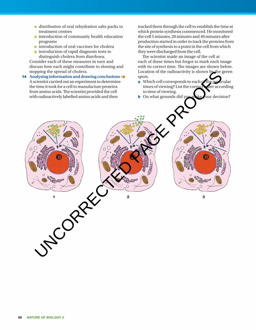
38 Nature of biology 2
c01CellsAReview 38 24 October 2016 10:42 AM
■ distribution of oral rehydration salts packs to treatment centres
■ introduction of community health education programs
■ introduction of oral vaccines for cholera ■ introduction of rapid diagnosis tests to
distinguish cholera from diarrhoea. Consider each of these measures in turn and
discuss how each might contribute to slowing and stopping the spread of cholera.
14 Analysing information and drawing conclusions ➜ A scientist carried out an experiment to determine the time it took for a cell to manufacture proteins from amino acids. The scientist provided the cell with radioactively labelled amino acids and then
tracked them through the cell to establish the time at which protein synthesis commenced. He monitored the cell 5 minutes, 20 minutes and 40 minutes after production started in order to track the proteins from the site of synthesis to a point in the cell from which they were discharged from the cell.
The scientist made an image of the cell at each of these times but forgot to mark each image with its correct time. The images are shown below. Location of the radioactivity is shown by the green spots.a Which cell corresponds to each of the particular
times of viewing? List the correct order according to time of viewing.
b On what grounds did you make your decision?
1 2 3
UNCORRECTED PAGE P
ROOFS