cells. scientists hooke-saw cork cells under a microscope van leeuweenhoek – saw living bacteria...
TRANSCRIPT
Cells

Scientists
• Hooke-saw cork cells under a microscope• Van Leeuweenhoek – saw living bacteria• Pasteur – studied bacteria and developed the
germ theory that said that ‘germs’ cause disease. He also developed the first vaccines.
• Koch – rules to test if a germ is the cause of a specific disease
• Margulis – tested DNA in mitochondria and found it was the same as bacteria DNA

Cell Theory
• All living organisms are composed of one or more living cells
• Cells are the basic units of life
• All cells come from preexisting cells

Microscopes
• Compound Light Microscope – series of glass lens that can be no better than 1000X, but can be used to view living cells
• Electron microscopes – aims a beam of electrons at thin slices of cells (dead)– Transmission electron microscope (TEM)– Scanning electron microscope (SEM)

Microscopes
Cell size
• As cell size increases, the surface area to volume ratio decreases
• Rates of chemical exchange may then be inadequate for cell size
• Cell size, therefore, remains small

Cell Size
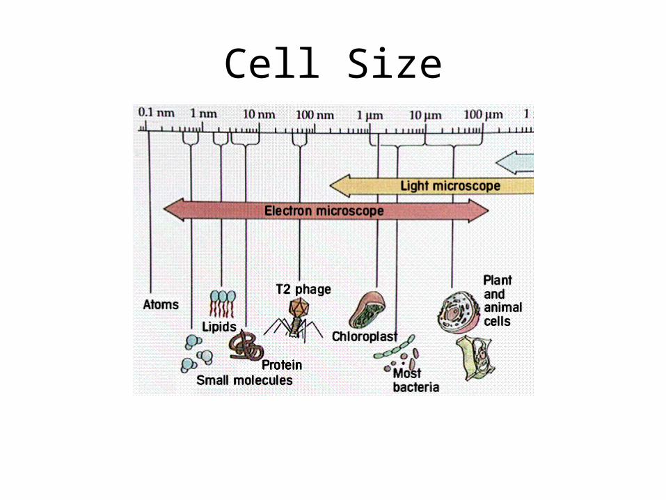
Cell Size

Basic Cell Types
• Prokaryotes – cells without a nucleus or other membrane bound organelles– Example: most unicellular organisms, e.g.,
bacteria
• Eukaryotes – cells with a nucleus and other membrane bound organelles (ER, mitochondria, Golgi apparatus, chloroplast, lysosome)

Plasma Membrane

The Lipid Bilayer
• Lipids of the bilayer– fluid or liquid-crystalline state
• Proteins move within the membrane

Fig. 5-2b, p. 108
Integral(transmembrane)
protein
(b) Fluid mosaic model. According to this model, a cellmembrane is a fluid lipid bilayer with a constantly changing“mosaic pattern“ of associated proteins.
Phospholipidbilayer
Peripheralprotein
Hydrophilicregion of protein
Hydrophobicregion of protein

Cytoplasm
• Environment inside cell membrane
• Cytoskeleton – supporting network of long, protein fibers that form a network and anchor for the cell organelles

Membrane structure
• Phospholipids~ membrane fluidity• Cholesterol~ membrane
stabilization• “Mosaic” Structure~ proteins• Membrane carbohydrates ~ cell
to cell recognition;

Nucleus
• Genetic material...•chromatin•chromosomes
• Nucleolus:; ribosome synthesis• Double membrane envelope
with pores• Protein synthesis

Ribosomes
• Protein manufacture

Endomembrane system, I
• Endoplasmic reticulum (ER)• Smooth ER
•no ribosomes; •synthesis of lipids
• Rough ER•with ribosomes;
•synthesis of proteins
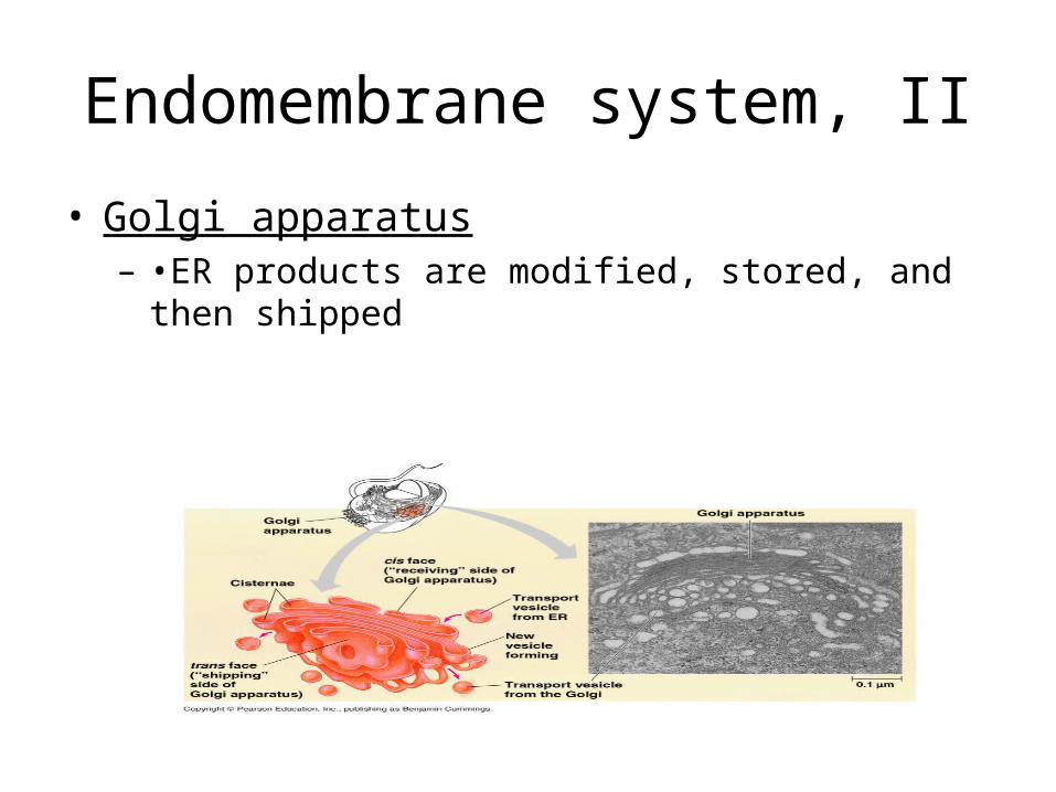
Endomembrane system, II
• Golgi apparatus– •ER products are modified, stored, and then shipped

Endomembrane system, III
• Lysosomes •sac of hydrolytic enzymes; digestion of macromolecules
• Tay-Sachs disease~ lipid-digestion disorder

Other membranous organelles, I
• Mitochondria • quantity in cell correlated with metabolic activity;
•cellular respiration •contain own
DNA

Other membranous organelles, II
• Chloroplast (doubled membranous plastid) •photosynthesis •own DNA

Peroxisomes
• Metabolism of fatty acids; detoxification of alcohol (liver)

Cellular Transport
• Passive Transport – does not require energy– Diffusion– Across a membrane
• Osmosis• Facilitated diffusion
• Active Transport – requires energy– Sodium/Potassium pump– Transport of Large Particles
• Endocytosis• Exocytosis

Diffusion

Osmosis

Water balance
• Osmoregulation~ control of water balance
• Hypertonic~ higher concentration of solutes
• Hypotonic~ lower concentration of solutes
• Isotonic~ equal concentrations of solutes
• Cells with Walls:• Turgid (very firm)
• Flaccid (limp)
• Plasmolysis~ plasma membrane pulls away from cell wall
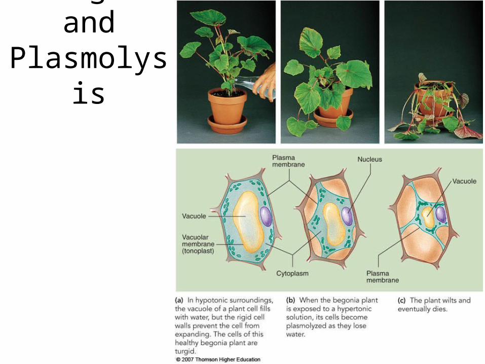
Turgor and Plasmolysis

Facilitated Diffusion

Types of Active Transport
• Sodium-potassium pump• Exocytosis~ secretion of
macromolecules by the fusion of vesicles with the plasma membrane
• Endocytosis~ import of macromolecules by forming new vesicles with the plasma
membrane
•phagocytosis•pinocytosis
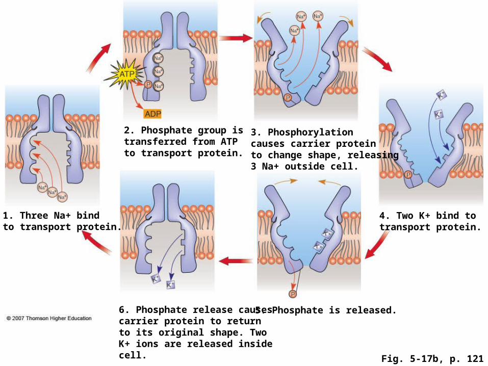
Fig. 5-17b, p. 121
1. Three Na+ bindto transport protein.
2. Phosphate group istransferred from ATPto transport protein.
3. Phosphorylationcauses carrier proteinto change shape, releasing3 Na+ outside cell.
4. Two K+ bind totransport protein.
5. Phosphate is released.6. Phosphate release causescarrier protein to returnto its original shape. TwoK+ ions are released insidecell.

Phagocytosis
Large particles enter cell