cellular and transcriptional response of - pedro alvarez
TRANSCRIPT
Published: April 28, 2011
r 2011 American Chemical Society 4988 dx.doi.org/10.1021/es1042673 | Environ. Sci. Technol. 2011, 45, 4988–4994
ARTICLE
pubs.acs.org/est
Cellular and Transcriptional Response of Pseudomonas stutzerito Quantum Dots under Aerobic and Denitrifying ConditionsYu Yang,† Huiguang Zhu,‡ Vicki L. Colvin,‡ and Pedro J. Alvarez*,†
†Department of Civil and Environmental Engineering, Rice University, Houston, Texas 77005, United States‡Department of Chemistry, Rice University, Houston, Texas 77005, United States
bS Supporting Information
’ INTRODUCTION
More than one-half century has passed since Richard Feynmanpresented the perspective of nanomaterial manipulation andnanotechnology application.1 Nanotechnology is now leading acreative and epochal revolution covering various subjects, such aspharmaceutics, electronics, physics, chemistry, and biology.2�4
With the increasing worldwide usage of manufactured nano-materials (MNMs), their accidental or incidental releases tothe environmental seem inevitable. Accordingly, the potentialadverse effect of MNMs to different types of biological molecules,cells, and organisms has received significant recent attention.5�11
However, considerable uncertainty remains about the potentialimpacts to microbial ecosystem services (e.g., nutrient cycling)and the associated risks to ecosystem health.
The nitrogen cycle is one of the fundamental biogeochemicalprocesses on the earth and denitrification is a key component thatis critically implicated in water quality, agricultural productivity,and climate change. This form of anaerobic respiration (which isperformed by a wide range of Bacteria and Archaea) involves thereduction of nitrate (NO3
�) or nitrite (NO2�) (both common
water pollutants) coupled with the production of dinitrogen gas(N2) through intermediate nitrogen oxides (NO and N2O).
12�14
Consequently, denitrification results in a loss of nitrogen(a common limiting nutrient) from aquatic and soil systems withconcomitant production of a greenhouse gas (N2O).
15 Pseudo-monas stutzeri is one of the most studied denitrifying bacteria,16,17
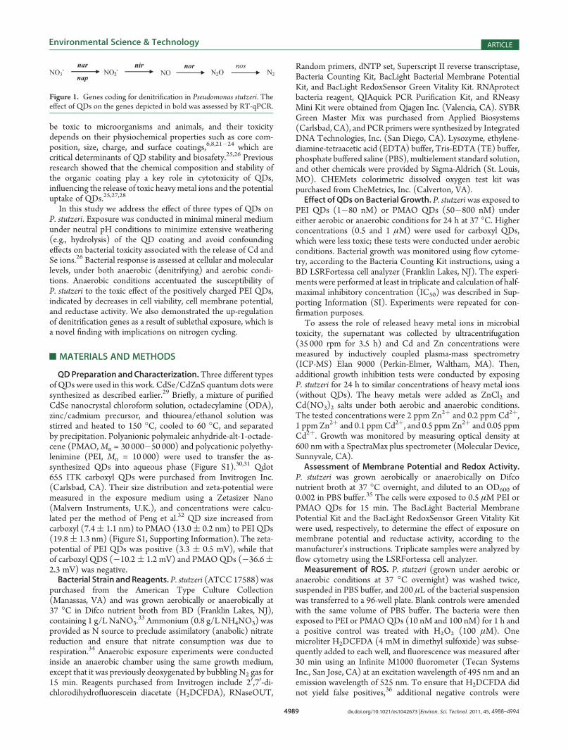
and its denitrification genes have been discerned and sequenced(Figure 1).17 Thus, P. stutzeri is a convenient model bacterium toinvestigate the potential impact of MNMs to denitrificationprocesses and associated bacterial response, which has not yetbeen addressed in the literature.
Quantum dots (QDs) offer valuable functionality for bioimag-ing, solar cells, electronics, and drug (or gene) delivery, sincethese semiconductor nanocrystals possess uniform sizes anddistinct optical and electrical properties.2,18�20 Some QDs can
Received: December 20, 2010Accepted: April 15, 2011Revised: April 11, 2011
ABSTRACT: Pseudomonas stutzeri was exposed to quantum dots (QDs) withthree different surface coatings (anionic polymaleic anhydride-alt-1-octadecene(PMAO), cationic polyethylenimine (PEI), and carboxyl QDs) under both aerobicand anaerobic (denitrifying) conditions. Under aerobic conditions, toxicity(assessed per growth inhibition) increased from PMAO to carboxyl to PEI QDs.The positive charge of PEI facilitated direct contact with negatively chargedbacteria, which was verified by TEM analysis. Both PMAO and PEI QDs hinderedenergy transduction (indicated by a decrease in cell membrane potential), and thiseffect was most pronounced with PEI QDs under denitrifying conditions. Up-regulation of denitrification genes (i.e., nitrate reductase narG, periplasmic nitratereductase napB, nitrite reductase nirH, and NO reductase norB) occurred uponexposure to subinhibitory PEI QD concentrations (1 nM). Accordingly, denitri-fication activity (assessed per respiratory nitrate consumption in the presence ofammonia) increased during sublethal PEI QD exposure. However, cell viability(including denitrification) was hindered at 10 nM or higher PEI QD concentra-tions. Efflux pump genes czcB and czcC were induced by PEI QDs underdenitrifying conditions, even though Cd and Se dissolution from QDs did notreach toxic levels (exposure was at pH 7 tominimize hydrolysis of QD coatings and the associated release of metal constituents). Up-regulation of the superoxide dismutase (stress) gene sodB occurred only under aerobic conditions, likely due to intracellularproduction of reactive oxygen species (ROS). The absence of ROS under denitrifying conditions suggests that the antibacterialactivity of QDs was not due to ROS production alone. Overall, this work forewarns about unintended potential impacts todenitrification as a result of disposal and incidental releases of QDs, especially those with positively charged coatings (e.g., PEI QDs).
4989 dx.doi.org/10.1021/es1042673 |Environ. Sci. Technol. 2011, 45, 4988–4994
Environmental Science & Technology ARTICLE
be toxic to microorganisms and animals, and their toxicitydepends on their physiochemical properties such as core com-position, size, charge, and surface coatings,6,8,21�24 which arecritical determinants of QD stability and biosafety.25,26 Previousresearch showed that the chemical composition and stability ofthe organic coating play a key role in cytotoxicity of QDs,influencing the release of toxic heavy metal ions and the potentialuptake of QDs.25,27,28
In this study we address the effect of three types of QDs onP. stutzeri. Exposure was conducted in minimal mineral mediumunder neutral pH conditions to minimize extensive weathering(e.g., hydrolysis) of the QD coating and avoid confoundingeffects on bacterial toxicity associated with the release of Cd andSe ions.26 Bacterial response is assessed at cellular and molecularlevels, under both anaerobic (denitrifying) and aerobic condi-tions. Anaerobic conditions accentuated the susceptibility ofP. stutzeri to the toxic effect of the positively charged PEI QDs,indicated by decreases in cell viability, cell membrane potential,and reductase activity. We also demonstrated the up-regulationof denitrification genes as a result of sublethal exposure, which isa novel finding with implications on nitrogen cycling.
’MATERIALS AND METHODS
QDPreparation and Characterization.Three different typesof QDs were used in this work. CdSe/CdZnS quantum dots weresynthesized as described earlier.29 Briefly, a mixture of purifiedCdSe nanocrystal chloroform solution, octadecylamine (ODA),zinc/cadmium precursor, and thiourea/ethanol solution wasstirred and heated to 150 �C, cooled to 60 �C, and separatedby precipitation. Polyanionic polymaleic anhydride-alt-1-octade-cene (PMAO,Mn = 30 000�50 000) and polycationic polyethy-lenimine (PEI, Mn = 10 000) were used to transfer the as-synthesized QDs into aqueous phase (Figure S1).30,31 Qdot655 ITK carboxyl QDs were purchased from Invitrogen Inc.(Carlsbad, CA). Their size distribution and zeta-potential weremeasured in the exposure medium using a Zetasizer Nano(Malvern Instruments, U.K.), and concentrations were calcu-lated per the method of Peng et al.32 QD size increased fromcarboxyl (7.4( 1.1 nm) to PMAO (13.0( 0.2 nm) to PEI QDs(19.8( 1.3 nm) (Figure S1, Supporting Information). The zeta-potential of PEI QDs was positive (3.3 ( 0.5 mV), while thatof carboxyl QDS (�10.2( 1.2 mV) and PMAOQDs (�36.6(2.3 mV) was negative.Bacterial Strain andReagents. P. stutzeri (ATCC 17588) was
purchased from the American Type Culture Collection(Manassas, VA) and was grown aerobically or anaerobically at37 �C in Difco nutrient broth from BD (Franklin Lakes, NJ),containing 1 g/LNaNO3.
33 Ammonium (0.8 g/LNH4NO3) wasprovided as N source to preclude assimilatory (anabolic) nitratereduction and ensure that nitrate consumption was due torespiration.34 Anaerobic exposure experiments were conductedinside an anaerobic chamber using the same growth medium,except that it was previously deoxygenated by bubbling N2 gas for15 min. Reagents purchased from Invitrogen include 20,70-di-chlorodihydrofluorescein diacetate (H2DCFDA), RNaseOUT,
Random primers, dNTP set, Superscript II reverse transcriptase,Bacteria Counting Kit, BacLight Bacterial Membrane PotentialKit, and BacLight RedoxSensor Green Vitality Kit. RNAprotectbacteria reagent, QIAquick PCR Purification Kit, and RNeasyMini Kit were obtained from Qiagen Inc. (Valencia, CA). SYBRGreen Master Mix was purchased from Applied Biosystems(Carlsbad, CA), and PCR primers were synthesized by IntegratedDNA Technologies, Inc. (San Diego, CA). Lysozyme, ethylene-diamine-tetraacetic acid (EDTA) buffer, Tris-EDTA (TE) buffer,phosphate buffered saline (PBS), multielement standard solution,and other chemicals were provided by Sigma-Aldrich (St. Louis,MO). CHEMets colorimetric dissolved oxygen test kit waspurchased from CheMetrics, Inc. (Calverton, VA).Effect of QDs on Bacterial Growth. P. stutzeri was exposed to
PEI QDs (1�80 nM) or PMAO QDs (50�800 nM) undereither aerobic or anaerobic conditions for 24 h at 37 �C. Higherconcentrations (0.5 and 1 μM) were used for carboxyl QDs,which were less toxic; these tests were conducted under aerobicconditions. Bacterial growth was monitored using flow cytome-try, according to the Bacteria Counting Kit instructions, using aBD LSRFortessa cell analyzer (Franklin Lakes, NJ). The experi-ments were performed at least in triplicate and calculation of half-maximal inhibitory concentration (IC50) was described in Sup-porting Information (SI). Experiments were repeated for con-firmation purposes.To assess the role of released heavy metal ions in microbial
toxicity, the supernatant was collected by ultracentrifugation(35 000 rpm for 3.5 h) and Cd and Zn concentrations weremeasured by inductively coupled plasma-mass spectrometry(ICP-MS) Elan 9000 (Perkin-Elmer, Waltham, MA). Then,additional growth inhibition tests were conducted by exposingP. stutzeri for 24 h to similar concentrations of heavy metal ions(without QDs). The heavy metals were added as ZnCl2 andCd(NO3)2 salts under both aerobic and anaerobic conditions.The tested concentrations were 2 ppm Zn2þ and 0.2 ppm Cd2þ,1 ppm Zn2þ and 0.1 ppmCd2þ, and 0.5 ppm Zn2þ and 0.05 ppmCd2þ. Growth was monitored by measuring optical density at600 nm with a SpectraMax plus spectrometer (Molecular Device,Sunnyvale, CA).Assessment of Membrane Potential and Redox Activity.
P. stutzeri was grown aerobically or anaerobically on Difconutrient broth at 37 �C overnight, and diluted to an OD600 of0.002 in PBS buffer.35 The cells were exposed to 0.5 μM PEI orPMAO QDs for 15 min. The BacLight Bacterial MembranePotential Kit and the BacLight RedoxSensor Green Vitality Kitwere used, respectively, to determine the effect of exposure onmembrane potential and reductase activity, according to themanufacturer’s instructions. Triplicate samples were analyzed byflow cytometry using the LSRFortessa cell analyzer.Measurement of ROS. P. stutzeri (grown under aerobic or
anaerobic conditions at 37 �C overnight) was washed twice,suspended in PBS buffer, and 200 μL of the bacterial suspensionwas transferred to a 96-well plate. Blank controls were amendedwith the same volume of PBS buffer. The bacteria were thenexposed to PEI or PMAOQDs (10 nM and 100 nM) for 1 h anda positive control was treated with H2O2 (100 μM). Onemicroliter H2DCFDA (4 mM in dimethyl sulfoxide) was subse-quently added to each well, and fluorescence was measured after30 min using an Infinite M1000 fluorometer (Tecan SystemsInc., San Jose, CA) at an excitation wavelength of 495 nm and anemission wavelength of 525 nm. To ensure that H2DCFDA didnot yield false positives,36 additional negative controls were
Figure 1. Genes coding for denitrification in Pseudomonas stutzeri. Theeffect of QDs on the genes depicted in bold was assessed by RT-qPCR.
4990 dx.doi.org/10.1021/es1042673 |Environ. Sci. Technol. 2011, 45, 4988–4994
Environmental Science & Technology ARTICLE
prepared withQDs andH2DCFDA alone (no cells). BackgroundQD fluorescence was subtracted from the signal, and ROS-induced fluorescence increases was compared with the fluores-cence of control samples. All the samples were replicated at leastsix times.Effect of QDs on Gene Expression. P. stutzeri was exposed
during 24 h to 10 nM PEI or 250 nM PMAOQDs under aerobicconditions, and to 1 nM PEI and 250 nM PMAO underanaerobic (denitrifying) conditions. Lower concentrations wereused for conditions of higher susceptibility to QD toxicity. Theexpression of czcB, czcC, sodB, narG, napB, nirH, and norB geneswas then quantified by reverse transcriptase quantitative PCE(RT-qPCR), using the housekeeping gene gapA (which codes forD-glyceraldehyde-3-phosphate dehydrogenase) as an internalstandard. Genes czcB and czcC, located in the cobalt�zinc�cadmium efflux system czcABC, encode a resistance pro-tein and an efflux transporter membrane fusion protein, respec-tively. Gene nirH encodes nitrite reductase H protein and isassociated with denitrification, as well as norB, coding for NOreductase (Figure 1). Both napB and narG encode differentnitrate reductase subunits, located in periplasm and membranerespectively. Superoxide dismutase gene sodB is protectingbacteria against oxidative stress caused by superoxide andhydroxyl radicals.37 All treatments and RT-qPCR analyses foreach sample were run in triplicate. Additional experimentaldetails and gene sequences are described in the SI.
’RESULTS AND DISCUSSION
Release of QD Constituents. Cd and Zn ions were releasedfrom QDs, and higher QD concentrations resulted in higherdissolved metal concentrations after 24 h exposure as illustratedfor PEI (Table 1) and PMAO QDs (Table S1). For a given QDconcentration, lower concentrations of total dissolved Cd and Znwere present in the growth medium under aerobic conditions,possibly due to more biosorption associated with higher biomassyield and faster growth under aerobic conditions.38 The leachingof these heavy metals fromQDs occurred to a much lesser extentthan previously reported26 due to differences in exposure con-ditions (e.g., pH 7 tominimize hydrolysis of the organic coating),and did not significantly contribute to bacterial toxicity. The factthat toxicity was primarily exerted by QDs rather than releasedmetals is evident since (1) total dissolved Cd and Zn concentra-tions were less than 0.2 and 1.2 mg/L, respectively, and (2) theseconcentrations (when added as metal salts) were not toxic underaerobic or denitrifying conditions (Figure S2). Other studieshave corroborated that P. stutzeri is quite resistant to even higherconcentrations of Zn, Se, and Cd ions.39�41
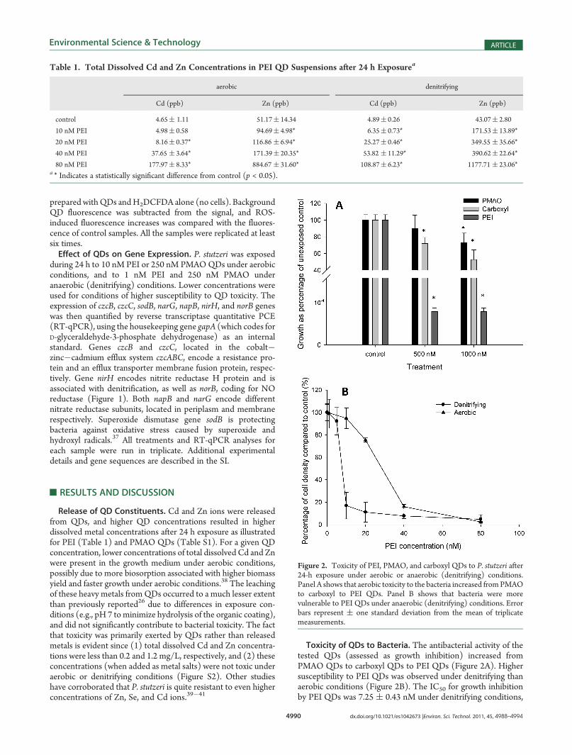
Toxicity of QDs to Bacteria. The antibacterial activity of thetested QDs (assessed as growth inhibition) increased fromPMAO QDs to carboxyl QDs to PEI QDs (Figure 2A). Highersusceptibility to PEI QDs was observed under denitrifying thanaerobic conditions (Figure 2B). The IC50 for growth inhibitionby PEI QDs was 7.25 ( 0.43 nM under denitrifying conditions,
Figure 2. Toxicity of PEI, PMAO, and carboxyl QDs to P. stutzeri after24-h exposure under aerobic or anaerobic (denitrifying) conditions.Panel A shows that aerobic toxicity to the bacteria increased fromPMAOto carboxyl to PEI QDs. Panel B shows that bacteria were morevulnerable to PEI QDs under anaerobic (denitrifying) conditions. Errorbars represent ( one standard deviation from the mean of triplicatemeasurements.
Table 1. Total Dissolved Cd and Zn Concentrations in PEI QD Suspensions after 24 h Exposurea
aerobic denitrifying
Cd (ppb) Zn (ppb) Cd (ppb) Zn (ppb)
control 4.65( 1.11 51.17( 14.34 4.89( 0.26 43.07( 2.80
10 nM PEI 4.98( 0.58 94.69( 4.98* 6.35( 0.73* 171.53( 13.89*
20 nM PEI 8.16( 0.37* 116.86 ( 6.94* 25.27( 0.46* 349.55 ( 35.66*
40 nM PEI 37.65 ( 3.64* 171.39( 20.35* 53.82 ( 11.29* 390.62( 22.64*
80 nM PEI 177.97( 8.33* 884.67 ( 31.60* 108.87( 6.23* 1177.71 ( 23.06*a * Indicates a statistically significant difference from control (p < 0.05).

4991 dx.doi.org/10.1021/es1042673 |Environ. Sci. Technol. 2011, 45, 4988–4994
Environmental Science & Technology ARTICLE
compared to 26.45 ( 1.08 nM under aerobic conditions. Nosignificant aerobic or anaerobic growth inhibition was observedwhen P. stutzeri was exposed to the least toxic PMAO QD up to800 nM (Figure S3). Whether lower susceptibility under aerobicconditions was due to higher energy harvesting and faster growthcapabilities remains to be determined.The higher toxicity of PEI QDs is probably due to their
positive charge (zeta-potential = 3.3( 0.5 mV), which enhancesdirect contact by electrostatic attraction to the negatively chargedbacteria (zeta-potential = �0.03 ( 0.01 mV). TEM analysis(method details are included in the Supporting Information(SI)) confirmed that more PEI QDs associated with P. stutzerithan PMAO QDs (Figure 3), which predominantly remainedsuspended without attaching to the cells. The relatively smallamount of PMAOQDs observed on the bacteria (Figure 3B) waslikely deposited when the solution was dried during the samplepreparation.ROS Production. ROS—whether produced exogenously (by
redox or photocatalytic reactions) or endogenously (by theimmune response)—is commonly considered as a potentialmechanism of MNM toxicity (i.e., oxidative stress).42 ROS weredetected in aerobic incubations with PEI QDs at 100 nM, but notat 10 nM (Table 1). No ROS were detected under denitrifyingconditions due to the absence of O2, which is a required ROSprecursor.43 Thus, the observed anaerobic toxicity of PEI QDscannot be attributed to ROS production. No ROS was detected
either upon aerobic or denitrifying exposure to (relatively nontoxic) PMAO QDs.Reductase Activity and Membrane Potential. Exposure to
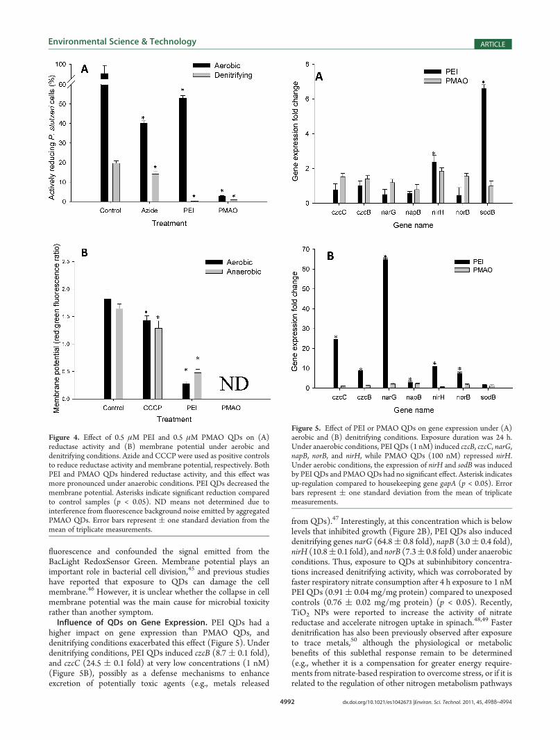
both PEI and PMAO QDs decreased the percentage of activelyrespiring cells under both aerobic and anaerobic conditions(Figure 4A), indicating the potential for QDs to hinder electrontransport phosphorylation.44 However, aerobic reductase activitywas inhibited to a greater extent in the presence of PMAO QDs(100 nM) than with PEI QDs (10 nM), implying that the highertoxicity of positively charged PEI QDs was not solely due tointerference with respiration. Lower reductase activities weregenerally observed under denitrifying conditions (Figure 4A),especially in the presence of the more toxic PEI QDs. Thisprobably reflects that P. stutzeri uses a shorter nitrate-basedelectron transport chain with fewer reductases compared to theaerobic respiration mode.15
Cell membrane potential was affected by exposure to someQDs, but not by the respiration (aerobic versus denitrifying)mode. Exposure to PEI QDs (but not PMAOQDs) resulted in asignificant decrease in cell membrane potential (Figure 4B),which is indicative of inhibition of the generation of the protonmotive force (PMF) and associated energy transduction.36,44
This effect is consistent with the high degree of microbial growthinhibition exerted by PEI QDs (Figure 2A). Changes in mem-brane potential could not be reliably assessed in treatments withPMAO QDs because their aggregation increased background
Figure 3. TEM images of (A) P. stutzeri unexposed control and those exposed to (B) PMAO and (C) PEI QDs. More PEI QDs were attached tobacteria than PMAO QDs. White arrows point to the QDs. Duplicate samples were used for TEM analysis.
4992 dx.doi.org/10.1021/es1042673 |Environ. Sci. Technol. 2011, 45, 4988–4994
Environmental Science & Technology ARTICLE
fluorescence and confounded the signal emitted from theBacLight RedoxSensor Green. Membrane potential plays animportant role in bacterial cell division,45 and previous studieshave reported that exposure to QDs can damage the cellmembrane.46 However, it is unclear whether the collapse in cellmembrane potential was the main cause for microbial toxicityrather than another symptom.Influence of QDs on Gene Expression. PEI QDs had a
higher impact on gene expression than PMAO QDs, anddenitrifying conditions exacerbated this effect (Figure 5). Underdenitrifying conditions, PEI QDs induced czcB (8.7 ( 0.1 fold),and czcC (24.5 ( 0.1 fold) at very low concentrations (1 nM)(Figure 5B), possibly as a defense mechanisms to enhanceexcretion of potentially toxic agents (e.g., metals released
from QDs).47 Interestingly, at this concentration which is belowlevels that inhibited growth (Figure 2B), PEI QDs also induceddenitrifying genes narG (64.8( 0.8 fold), napB (3.0( 0.4 fold),nirH (10.8( 0.1 fold), and norB (7.3( 0.8 fold) under anaerobicconditions. Thus, exposure to QDs at subinhibitory concentra-tions increased denitrifying activity, which was corroborated byfaster respiratory nitrate consumption after 4 h exposure to 1 nMPEI QDs (0.91( 0.04 mg/mg protein) compared to unexposedcontrols (0.76 ( 0.02 mg/mg protein) (p < 0.05). Recently,TiO2 NPs were reported to increase the activity of nitratereductase and accelerate nitrogen uptake in spinach.48,49 Fasterdenitrification has also been previously observed after exposureto trace metals,50 although the physiological or metabolicbenefits of this sublethal response remain to be determined(e.g., whether it is a compensation for greater energy require-ments from nitrate-based respiration to overcome stress, or if it isrelated to the regulation of other nitrogen metabolism pathways
Figure 4. Effect of 0.5 μM PEI and 0.5 μM PMAO QDs on (A)reductase activity and (B) membrane potential under aerobic anddenitrifying conditions. Azide and CCCP were used as positive controlsto reduce reductase activity and membrane potential, respectively. BothPEI and PMAO QDs hindered reductase activity, and this effect wasmore pronounced under anaerobic conditions. PEI QDs decreased themembrane potential. Asterisks indicate significant reduction comparedto control samples (p < 0.05). ND means not determined due tointerference from fluorescence background noise emitted by aggregatedPMAO QDs. Error bars represent ( one standard deviation from themean of triplicate measurements.
Figure 5. Effect of PEI or PMAO QDs on gene expression under (A)aerobic and (B) denitrifying conditions. Exposure duration was 24 h.Under anaerobic conditions, PEI QDs (1 nM) induced czcB, czcC, narG,napB, norB, and nirH, while PMAO QDs (100 nM) repressed nirH.Under aerobic conditions, the expression of nirH and sodB was inducedby PEI QDs and PMAOQDs had no significant effect. Asterisk indicatesup-regulation compared to housekeeping gene gapA (p < 0.05). Errorbars represent ( one standard deviation from the mean of triplicatemeasurements.
4993 dx.doi.org/10.1021/es1042673 |Environ. Sci. Technol. 2011, 45, 4988–4994
Environmental Science & Technology ARTICLE
such as the need for aminoacid synthesis to repair damagedproteins). Gene nar and nor code formembrane-associated proteinswhereas gene nap and nir code for periplasmic proteins.15,17
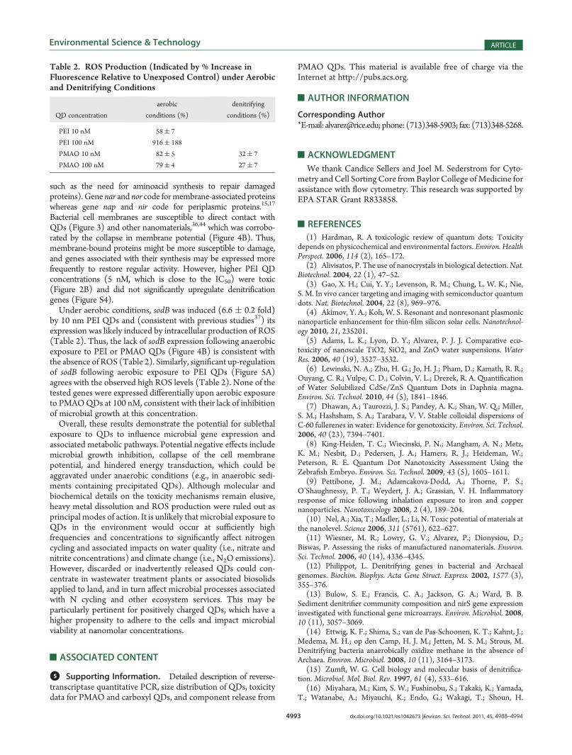
Bacterial cell membranes are susceptible to direct contact withQDs (Figure 3) and other nanomaterials,36,44 which was corrobo-rated by the collapse in membrane potential (Figure 4B). Thus,membrane-bound proteins might be more susceptible to damage,and genes associated with their synthesis may be expressed morefrequently to restore regular activity. However, higher PEI QDconcentrations (5 nM, which is close to the IC50) were toxic(Figure 2B) and did not significantly upregulate denitrificationgenes (Figure S4).Under aerobic conditions, sodB was induced (6.6 ( 0.2 fold)
by 10 nm PEI QDs and (consistent with previous studies37) itsexpression was likely induced by intracellular production of ROS(Table 2). Thus, the lack of sodB expression following anaerobicexposure to PEI or PMAO QDs (Figure 4B) is consistent withthe absence of ROS (Table 2). Similarly, significant up-regulationof sodB following aerobic exposure to PEI QDs (Figure 5A)agrees with the observed high ROS levels (Table 2). None of thetested genes were expressed differentially upon aerobic exposureto PMAOQDs at 100 nM, consistent with their lack of inhibitionof microbial growth at this concentration.Overall, these results demonstrate the potential for sublethal
exposure to QDs to influence microbial gene expression andassociated metabolic pathways. Potential negative effects includemicrobial growth inhibition, collapse of the cell membranepotential, and hindered energy transduction, which could beaggravated under anaerobic conditions (e.g., in anaerobic sedi-ments containing precipitated QDs). Although molecular andbiochemical details on the toxicity mechanisms remain elusive,heavy metal dissolution and ROS production were ruled out asprincipal modes of action. It is unlikely that microbial exposure toQDs in the environment would occur at sufficiently highfrequencies and concentrations to significantly affect nitrogencycling and associated impacts on water quality (i.e., nitrate andnitrite concentrations) and climate change (i.e., N2O emissions).However, discarded or inadvertently released QDs could con-centrate in wastewater treatment plants or associated biosolidsapplied to land, and in turn affect microbial processes associatedwith N cycling and other ecosystem services. This may beparticularly pertinent for positively charged QDs, which have ahigher propensity to adhere to the cells and impact microbialviability at nanomolar concentrations.
’ASSOCIATED CONTENT
bS Supporting Information. Detailed description of reverse-transcriptase quantitative PCR, size distribution of QDs, toxicitydata for PMAO and carboxyl QDs, and component release from
PMAO QDs. This material is available free of charge via theInternet at http://pubs.acs.org.
’AUTHOR INFORMATION
Corresponding Author*E-mail: [email protected];phone: (713)348-5903; fax: (713)348-5268.
’ACKNOWLEDGMENT
We thank Candice Sellers and Joel M. Sederstrom for Cyto-metry andCell Sorting Core fromBaylor College ofMedicine forassistance with flow cytometry. This research was supported byEPA STAR Grant R833858.
’REFERENCES
(1) Hardman, R. A toxicologic review of quantum dots: Toxicitydepends on physicochemical and environmental factors. Environ. HealthPerspect. 2006, 114 (2), 165–172.
(2) Alivisatos, P. The use of nanocrystals in biological detection.Nat.Biotechnol. 2004, 22 (1), 47–52.
(3) Gao, X. H.; Cui, Y. Y.; Levenson, R. M.; Chung, L. W. K.; Nie,S. M. In vivo cancer targeting and imaging with semiconductor quantumdots. Nat. Biotechnol. 2004, 22 (8), 969–976.
(4) Akimov, Y. A.; Koh, W. S. Resonant and nonresonant plasmonicnanoparticle enhancement for thin-film silicon solar cells. Nanotechnol-ogy 2010, 21, 235201.
(5) Adams, L. K.; Lyon, D. Y.; Alvarez, P. J. J. Comparative eco-toxicity of nanoscale TiO2, SiO2, and ZnO water suspensions. WaterRes. 2006, 40 (19), 3527–3532.
(6) Lewinski, N. A.; Zhu, H. G.; Jo, H. J.; Pham, D.; Kamath, R. R.;Ouyang, C. R.; Vulpe, C. D.; Colvin, V. L.; Drezek, R. A. Quantificationof Water Solubilized CdSe/ZnS Quantum Dots in Daphnia magna.Environ. Sci. Technol. 2010, 44 (5), 1841–1846.
(7) Dhawan, A.; Taurozzi, J. S.; Pandey, A. K.; Shan, W. Q.; Miller,S. M.; Hashsham, S. A.; Tarabara, V. V. Stable colloidal dispersions ofC-60 fullerenes in water: Evidence for genotoxicity. Environ. Sci. Technol.2006, 40 (23), 7394–7401.
(8) King-Heiden, T. C.; Wiecinski, P. N.; Mangham, A. N.; Metz,K. M.; Nesbit, D.; Pedersen, J. A.; Hamers, R. J.; Heideman, W.;Peterson, R. E. Quantum Dot Nanotoxicity Assessment Using theZebrafish Embryo. Environ. Sci. Technol. 2009, 43 (5), 1605–1611.
(9) Pettibone, J. M.; Adamcakova-Dodd, A.; Thorne, P. S.;O’Shaughnessy, P. T.; Weydert, J. A.; Grassian, V. H. Inflammatoryresponse of mice following inhalation exposure to iron and coppernanoparticles. Nanotoxicology 2008, 2 (4), 189–204.
(10) Nel, A.; Xia, T.; Madler, L.; Li, N. Toxic potential of materials atthe nanolevel. Science 2006, 311 (5761), 622–627.
(11) Wiesner, M. R.; Lowry, G. V.; Alvarez, P.; Dionysiou, D.;Biswas, P. Assessing the risks of manufactured nanomaterials. Environ.Sci. Technol. 2006, 40 (14), 4336–4345.
(12) Philippot, L. Denitrifying genes in bacterial and Archaealgenomes. Biochim. Biophys. Acta Gene Struct. Express. 2002, 1577 (3),355–376.
(13) Bulow, S. E.; Francis, C. A.; Jackson, G. A.; Ward, B. B.Sediment denitrifier community composition and nirS gene expressioninvestigated with functional gene microarrays. Environ. Microbiol. 2008,10 (11), 3057–3069.
(14) Ettwig, K. F.; Shima, S.; van de Pas-Schoonen, K. T.; Kahnt, J.;Medema, M. H.; op den Camp, H. J. M.; Jetten, M. S. M.; Strous, M.Denitrifying bacteria anaerobically oxidize methane in the absence ofArchaea. Environ. Microbiol. 2008, 10 (11), 3164–3173.
(15) Zumft, W. G. Cell biology and molecular basis of denitrifica-tion. Microbiol. Mol. Biol. Rev. 1997, 61 (4), 533–616.
(16) Miyahara, M.; Kim, S. W.; Fushinobu, S.; Takaki, K.; Yamada,T.; Watanabe, A.; Miyauchi, K.; Endo, G.; Wakagi, T.; Shoun, H.
Table 2. ROS Production (Indicated by % Increase inFluorescence Relative to Unexposed Control) under Aerobicand Denitrifying Conditions
QD concentration
aerobic
conditions (%)
denitrifying
conditions (%)
PEI 10 nM 58( 7
PEI 100 nM 916( 188
PMAO 10 nM 82( 5 32( 7
PMAO 100 nM 79( 4 27( 7
4994 dx.doi.org/10.1021/es1042673 |Environ. Sci. Technol. 2011, 45, 4988–4994
Environmental Science & Technology ARTICLE
Potential of Aerobic Denitrification by Pseudomonas stutzeri TR2 ToReduce Nitrous Oxide Emissions from Wastewater Treatment Plants.Appl. Environ. Microbiol. 2010, 76 (14), 4619–4625.(17) Lalucat, J.; Bennasar, A.; Bosch, R.; Garcia-Valdes, E.; Palleroni,
N. J. Biology of Pseudomonas stutzeri.Microbiol. Mol. Biol. Rev. 2006, 70(2), 510–547.(18) Bagalkot, V.; Zhang, L.; Levy-Nissenbaum, E.; Jon, S.; Kantoff,
P. W.; Langer, R.; Farokhzad, O. C. Quantum dot - Aptamer conjugatesfor synchronous cancer imaging, therapy, and sensing of drug deliverybased on Bi-fluorescence resonance energy transfer. Nano Lett. 2007,7 (10), 3065–3070.(19) Mora-Sero, I.; Gimenez, S.; Fabregat-Santiago, F.; Gomez, R.;
Shen, Q.; Toyoda, T.; Bisquert, J. Recombination in Quantum DotSensitized Solar Cells. Acc. Chem. Res. 2009, 42 (11), 1848–1857.(20) Chan, W. C. W.; Maxwell, D. J.; Gao, X. H.; Bailey, R. E.;
Han, M. Y.; Nie, S. M. Luminescent quantum dots for multiplexedbiological detection and imaging. Curr. Opin. Biotechnol. 2002, 13 (1),40–46.(21) Dumas, E.; Gao, C.; Suffern, D.; Bradforth, S. E.; Dimitrijevic,
N. M.; Nadeau, J. L. Interfacial Charge Transfer between CdTeQuantum Dots and Gram Negative Vs Gram Positive Bacteria. Environ.Sci. Technol. 2010, 44 (4), 1464–1470.(22) Gagne, F.; Auclair, J.; Turcotte, P.; Fournier, M.; Gagnon, C.;
Sauve, S.; Blaise, C. Ecotoxicity of CdTe quantum dots to freshwatermussels: Impacts on immune system, oxidative stress and genotoxicity.Aquat. Toxicol. 2008, 86 (3), 333–340.(23) Lovric, J.; Bazzi, H. S.; Cuie, Y.; Fortin, G. R. A.; Winnik, F. M.;
Maysinger, D. Differences in subcellular distribution and toxicity ofgreen and red emitting CdTe quantum dots. J. Mol. Med. 2005, 83 (5),377–385.(24) Schneider, R.; Wolpert, C.; Guilloteau, H.; Balan, L.; Lambert,
J.; Merlin, C. The exposure of bacteria to CdTe-core quantum dots: Theimportance of surface chemistry on cytotoxicity. Nanotechnology 2009,20, (22), 2255101.(25) Kirchner, C.; Liedl, T.; Kudera, S.; Pellegrino, T.; Javier, A. M.;
Gaub, H. E.; Stolzle, S.; Fertig, N.; Parak, W. J. Cytotoxicity ofcolloidal CdSe and CdSe/ZnS nanoparticles. Nano Lett. 2005, 5 (2),331–338.(26) Mahendra, S.; Zhu, H. G.; Colvin, V. L.; Alvarez, P. J. Quantum
Dot Weathering Results in Microbial Toxicity. Environ. Sci. Technol.2008, 42 (24), 9424–9430.(27) Hoshino, A.; Fujioka, K.; Oku, T.; Suga, M.; Sasaki, Y. F.; Ohta,
T.; Yasuhara, M.; Suzuki, K.; Yamamoto, K. Physicochemical propertiesand cellular toxicity of nanocrystal quantum dots depend on their surfacemodification. Nano Lett. 2004, 4 (11), 2163–2169.(28) Chang, E.; Thekkek, N.; Yu, W. W.; Colvin, V. L.; Drezek, R.
Evaluation of quantum dot cytotoxicity based on intracellular uptake.Small 2006, 2 (12), 1412–1417.(29) Zhu, H. G.; Prakash, A.; Benoit, D. N.; Jones, C. J.; Colvin, V. L.
Low temperature synthesis of ZnS and CdZnS shells on CdSe quantumdots. Nanotechnology 2010, 21, 255604.(30) Yu, W. W.; Chang, E.; Falkner, J. C.; Zhang, J. Y.; Al-Somali,
A. M.; Sayes, C. M.; Johns, J.; Drezek, R.; Colvin, V. L. Formingbiocompatible and nonaggregated nanocrystals in water using amphi-philic polymers. J. Am. Chem. Soc. 2007, 129 (10), 2871–2879.(31) Duan, H. W.; Nie, S. M. Cell-penetrating quantum dots based
on multivalent and endosome-disrupting surface coatings. J. Am. Chem.Soc. 2007, 129 (11), 3333–3338.(32) Yu, W. W.; Qu, L. H.; Guo, W. Z.; Peng, X. G. Experimental
determination of the extinction coefficient of CdTe, CdSe, and CdSnanocrystals. Chem. Mater. 2003, 15 (14), 2854–2860.(33) Heiss, B.; Frunzke, K.; Zumft, W. G. Formation of the N-N-
Bond from Nitric-Oxide by a Membrane-Bound Cytochrome Bc Com-plex of Nitrate-Respiring (Denitrifying) Pseudomonas-Stutzeri. J. Bac-teriol. 1989, 171 (6), 3288–3297.(34) Betlach, M. R.; Tiedje, J. M.; Firestone, R. B. Assimilatory
Nitrate Uptake in Pseudomonas-Fluorescens Studied Using N-13. Arch.Microbiol. 1981, 129 (2), 135–140.
(35) Lyon, D. Y.; Alvarez, P. J. J. Fullerene Water Suspension(nC(60)) Exerts Antibacterial Effects via ROS-Independent ProteinOxidation. Environ. Sci. Technol. 2008, 42 (21), 8127–8132.
(36) Lyon, D. Y.; Brunet, L.; Hinkal, G. W.; Wiesner, M. R.; Alvarez,P. J. Antibacterial activity of fullerene water suspensions (nC60) is notdue to ROS-mediated damage. Nano Lett. 2008, 8 (5), 1539–43.
(37) Fridovich, I. Superoxide Radical and Superoxide Dismutases.Annu. Rev. Biochem. 1995, 64, 97–112.
(38) Hassan, S. H. A.; Kim, S. J.; Jung, A. Y.; Joo, J. H.; Oh, S. E.;Yang, J. E. Biosorptive capacity of Cd(II) and Cu(II) by lyophilized cellsof Pseudomonas stutzeri. J. Gen. Appl. Microbiol. 2009, 55 (1), 27–34.
(39) Bhagat, R.; Srivastava, S. Growth response of Psedudomonasstutzeri RS34 to ethylenediaminetetraacetic acid (EDTA) and its inter-action with Zn. Indian J. Exp. Biol. 1993, 31, 509–594.
(40) Zawadzka, A. M.; Crawford, R. L.; Paszczynski, A. J. Pyridine-2,6-bis(thiocarboxylic acid) produced by Pseudomonas stutzeri KCreduces chromium(VI) and precipitates mercury, cadmium, lead andarsenic. Biometals 2007, 20 (2), 145–158.
(41) Zawadzka, A. M.; Crawford, R. L.; Paszczynski, A. J. Pyridine-2,6-bis(thiocarboxylic acid) produced by Pseudomonas stutzeri KCreduces and precipitates selenium and tellurium oxyanions. Appl.Environ. Microbiol. 2006, 72 (5), 3119–3129.
(42) Klaine, S. J.; Alvarez, P. J.; Batley, G. E.; Fernandes, T. F.;Handy, R. D.; Lyon, D. Y.; Mahendra, S.; McLaughlin, M. J.; Lead, J. R.Nanomaterials in the Environment: Behavior, Fate, Bioavailability, andEffects. Environ. Toxicol. Chem. 2008, 27 (9), 1825–1851.
(43) Cabiscol, E.; Tamarit, J.; Ros, J. Oxidative stress in bacteria andprotein damage by reactive oxygen species. Int. Microbiol. 2000, 3 (1),3–8.
(44) Lyon, D. Y.; Alvarez, P. J. Fullerene water suspension (nC60)exerts antibacterial effects via ROS-independent protein oxidation.Environ. Sci. Technol. 2008, 42 (21), 8127–32.
(45) Strahl, H.; Hamoen, L.W.Membrane potential is important forbacterial cell division. Proc. Natl. Acad. Sci. U.S.A. 2010, 107 (27),12281–12286.
(46) Priester, J. H.; Stoimenov, P. K.; Mielke, R. E.; Webb, S. M.;Ehrhardt, C.; Zhang, J. P.; Stucky, G. D.; Holden, P. A. Effects of SolubleCadmium Salts Versus CdSe Quantum Dots on the Growth ofPlanktonic Pseudomonas aeruginosa. Environ. Sci. Technol. 2009,43 (7), 2589–2594.
(47) Nies, D. H.; Silver, S. Ion Efflux Systems Involved in BacterialMetal Resistances. J. Ind. Microbiol. 1995, 14 (2), 186–199.
(48) An, Y.; Li, T.; Jin, Z.; Dong, M.; Xia, H.; Wang, X. Effect ofbimetallic and polymer-coated Fe nanoparticles on biological denitrifi-cation. Bioresour. Technol. 2010, 101 (24), 9825–8.
(49) Yang, F.; Hong, F.; You, W.; Liu, C.; Gao, F.; Wu, C.; Yang, P.Influences of nano-anatase TiO2 on the nitrogen metabolism of growingspinach. Biol. Trace Elem. Res. 2006, 110 (2), 179–90.
(50) Labbe, N.; Parent, S.; Villemur, R. Addition of trace metalsincreases denitrification rate in closed marine systems.Water Res. 2003,37 (4), 914–920.
’NOTE ADDED AFTER ASAP PUBLICATION
The first sentence of the Introduction section had RichardFeynman's name spelled incorrectly in the version of this paperpublished April 28, 2011. The correct version published May 2,2011.