cellular biochemical changes in selaginella tamariscina ... · pdf filecellular biochemical...
TRANSCRIPT
Tropical Life Sciences Research, 27(2), 37–52, 2016
© Penerbit Universiti Sains Malaysia, 2016
Cellular Biochemical Changes in Selaginella tamariscina (Beauv.) Spring and Sellaginella plana (Desv. ex Poir.) Heiron. as Induced by Desiccation 1,2Angelo Rellama Agduma* and 2Maribel Dionisio Sese 1Department of Biological Sciences, College of Arts and Sciences, University of Southern Mindanao, Kabacan, Cotabato 9407, Philippines 2Plant Biology Division, Institute of Biological Sciences, College of Arts and Sciences, University of the Philippines Los Baños, Laguna 4031, Philippines Published date: 17 August 2016 To cite this article: Angelo Rellama Agduma and Maribel Dionisio Sese. (2016). Cellular
biochemical changes in Selaginella tamariscina (Beauv.) Spring and Sellaginella plana (Desv. ex Poir.) Heiron. as induced by desiccation. Tropical Life Sciences Research 27(2): 37–52. doi: 10.21315/tlsr2016.27.2.4 To link to this article: http://dx.doi.org/10.21315/tlsr2016.27.2.4
Abstrak: Perubahan biokimia dalam dua spesies Selaginella iaitu S. tamariscina (Beauv.)
Spring dan S. plana (Desv. ex Poir.) Heiron., yang dicetuskan oleh pengeringan dan diikuti oleh rehidrasi telah dikaji. Tumbuhan telah dibenarkan untuk dehidrat secara semula jadi dengan menyekat pengairan sehingga kandungan air relatif (RWC) pucuk bernilai <10%. Kemudiannya, tumbuhan yang dehidrat disiram air sehingga mencapai keadaan rehidrasi sepenuhnya iaitu RWC 90% ataupun lebih. Sifat-sifat menahan pengeringan telah diperhatikan dalam S. tamariscina manakala sifat sensitif terhadap pengeringan dilihat pada S. plana. Integriti membran dikekalkan dalam S. tamariscina tetapi bukan dalam S. plana seperti yang dilihat dalam ukuran kebocoran elektrolit relatif semasa fasa pengeringan dan seterusnya semasa fasa rehidrasi. Analisa pigmen telah menunjukkan konservasi beberapa klorofil dan karotenoid semasa pengeringan dan mencapai tahap kawalan yang mengikuti proses rehidrasi dalam S. tamariscina. Kandungan pigmen yang sangat rendah telah dijumpai dalam S. plana semasa fasa pengeringan dan pigmen tersebut tidak dipulih semula semasa rehidrasi. Penentuan zat terlarut yang serasi menunjukkan kenaikan dalam kandungan gula dan proline dalam S. tamariscina kering sahaja, yang menunjukkan kewujudan jentera-jentera perlindungan biokimia dalam spesies ini dan ketidakhadirannya dalam S. plana semasa keadaan dehidrat. Data-data ini menunjukkan satu elemen penting untuk toleransi terhadap pengeringan dalam tumbuhan vaskular rendah ialah kebolehan melindungi tisu-tisu daripada kemusnahan serius akibat pengeringan yang teruk. Kata kunci: Elektrolit, Pigmen, Proline, Rehidrasi, Gula
Abstract: The biochemical changes in two Selaginella species namely, S. tamariscina (Beauv.) Spring and S. plana (Desv. ex Poir.) Heiron., as induced by desiccation and subsequent rehydration were explored. Plants were allowed to dehydrate naturally by withholding irrigation until shoot’s relative water content (RWC) reached <10%. After which, dehydrated plants were watered until fully rehydrated states were obtained which was about 90% RWC or more. Desiccation-tolerance characteristics were observed in S. tamariscina while desiccation-sensitivity features were seen in S. plana. Membrane integrity was maintained in S. tamariscina but not in S. plana as evidenced in the relative
*Corresponding author: [email protected]
Angelo Rellama Agduma and Maribel Dionisio Sese
38
electrolyte leakage measurements during desiccation phase and the subsequent rehydration stage. Pigment analyses revealed conservation of some chlorophylls and carotenoids during desiccation and reaching control levels following rehydration in S. tamariscina. Very low pigment contents were found in S. plana during desiccation phase and the pigments were not recovered during rehydration attempt. Meanwhile, compatible solute determination showed rise in total sugar and proline contents of desiccated S. tamariscina only, indicating presence of biochemical protection machineries in this species and absence of such in S. plana during dehydrating conditions. These data indicate that one key element for desiccation-tolerance in lower vascular plants is the ability to protect tissues from severe damages caused by intense desiccation. Keywords: Electrolyte, Pigment, Proline, Rehydration, Sugar
INTRODUCTION One of the most significant evolutionary forces is to be able to live in a very dry environment. Atmosphere with a relative humidity of 50% at 20°C equals to a water potential of –100 MPa (Alpert 2006). This also corresponds to drying to <0.1 g H2O g–1 dry biomass which is roughly equivalent to about 10% water content or less (Alpert 2005). Majority of modern day plants may not survive in such environment as it might result to the loss of their intracellular water content down to 90% (Alpert 2006). However, there are certain species of plants, called “resurrection” plants, having strong selection for desiccation tolerance.
Resurrection plants survive the loss of most of their water content down to <5% relative water content (RWC) until a quiescent stage is achieved (Peters et al. 2007). Since most of the protoplasmic water is lost, it is considered the severest form of water stress (Bartels 2005). Upon watering, the plants rapidly revive and return to their normal physiological functioning (Alpert 2005). They are also known as poikilohydric plants because they lack capability to prevent desiccation. They directly rely on the environment for their water status. As a consequence, their cells’ water content tend to reach equilibrium with that of the environment (Alpert 2000; Scott 2000; Pandey et al. 2010).
Numerous studies were conducted to investigate desiccation tolerance mechanisms in different resurrection plants. Most of the available information are derived from the rich data obtained from studies conducted in angiosperm species (Sherwin & Farrant 1996, 1998; Farrant et al. 1999, 2003; Farrant 2000; Cooper & Farrant 2002; Moore et al. 2007). Little attention has been given to desiccation tolerance in the lower group of vascular plants (Oliver et al. 2000). Although desiccation-tolerant pteridophytes have been documented, for example fern Polypodium polypodioides (Layton et al. 2010) and fern allies Selaginella lepidophylla (Brighigna et al. 2002), and S. bryopteris (Pandey et al. 2010), much still needs to be done (Oliver et al. 2005).
One of the most primitive taxa of vascular plants is Selaginella (Kenrick & Crane 1997). In the recent review by Setyawan (2011), this sole surviving genus of the spikemoss family, Selaginellaceae, has 700–750 species throughout the world. The genus has not been intensively studied in the Philippines; taxonomically, morphologically, and physiologically. Regarding the species of
Biochemical Changes in Selaginella Induced by Desiccation
39
Selaginella found in the country, no formal proclamation has been made on which are desiccation-tolerant or desiccation-intolerant. This implies that common and distinct physiological features of desiccation-tolerant and intolerant Selaginella in the Philippines are not yet fully worked out.
The aim of this work was threefold: (1) to investigate the biochemical responses of two species of Selaginella, namely S. tamariscina and S. plana, to desiccation and rehydration treatments; (2) to assess which species will perform better with respect to its capability to resume normal physiological functioning during rehydration stage; and (3) to elucidate different physiological strategies of the species to maintain cellular integrity and limit the damage caused by desiccation.
MATERIALS AND METHODS Plant Materials and Growing Conditions Commercially obtained S. tamariscina and S. plana were allowed to acclimatise and grow in a 50 kg substrate with an equal mixture of river sand, coir dust, and garden soil placed in a wooden tray. The substrate was kept damp supplemented weekly with half strength of Hoagland’s solution. They were maintained in screen house conditions with a daytime temperature range of 23°C to 42°C. Experimental Set-up
The experimental treatments were as follows: 1. Desiccated – plants dried to a constant air-dried state (10% RWC) or
below; 2. Rehydrated – plants which have been fully recovered at 90% RWC
or more; and 3. Control – well watered plants which had not been desiccated.
All measurements were performed on desiccated and rehydrated plants.
Same measurements were carried out in control plants twice. The first set of control measurements was done simultaneously with the desiccated plants while the second control set was done simultaneously with the rehydrated plants. This was purposely carried out to provide a point of comparison since plants experience different environmental conditions at different times. All measurements were done in triplicate wherein composite sampling was employed.
Dehydration and Rehydration Treatments Whole plants previously acclimatised were slowly dried by withholding water and allowing the plants to dry out naturally under ambient screen house condition until below 10% RWC was reached. The plants were left in the dry state for no longer than three days. Same plants were rehydrated up to approximately 90% RWC or more to be used for the next cycle of experimentation. Rehydration was carried out through overhead watering using a mist spray to simulate rainfall. The
Angelo Rellama Agduma and Maribel Dionisio Sese
40
plants were well watered on the first day and then the soil was kept damp by daily watering for the remainder of the experiment. Plant Relative Water Content Determination Hydration states were determined by measuring the relative water contents of the shoot. Fully expanded similarly-sized fronds were selected from at least three plants per replicate. Using a sharp razor blade, individual fronds were detached at the leaf base. Fronds were immediately weighed to get the initial mass (Mi). In order to obtain the mass at full turgor (Mt), fronds were floated in distilled water inside a closed Petri dish for 24 h. Surface water was eliminated by blotting the fronds dry with a paper towel. Mt was recorded and the leaf samples were subsequently dried at 80°C for 24 h, and the dry mass (Md) was obtained. All mass measurements were done using an analytical scale, with precision of 0.01 g. Values of Mi, Mt, and Md were used to calculate RWC using the equation (Barrs & Weatherley 1962):
RWC % = Mi–Md/Mt–Md × 100. Electrolyte Leakage Measurement Electrolyte leakage, which gives an indication of the degree of membrane integrity, was measured following the procedure of Wang et al. (2010) with modifications. Fronds (0.5 g) were rinsed three times in distilled water to remove the contents of the cut cells. The fronds were soaked in 25 mL distilled water and shaken at room temperature for 24 h after which aliquot for leachate measurement was taken. The electrical conductivity of the solution (C1) was determined using a conductivity instrument (D-2, Horiba Ltd., Kyoto). The samples in the tube was then placed in boiling water (100°C) for 10 min and allowed to cool to ambient temperature. The electrical conductivity of this solution (C2) was then measured to obtain the maximum conductivity. The electrical conductivity of the distilled water (C3) was also measured. The relative electrolyte leakage (REL) was calculated using the equation:
REL % = C1–C3/C2–C3 × 100. Chlorophyll and Carotenoid Measurements Approximately 0.5 g of leaf tissues was frozen with liquid nitrogen and homogenised using mortar and pestle. The homogenised samples were contained in a test tube with cover and the chlorophyll was extracted with 10 mL 80% acetone. The test tubes were wrapped with aluminum foil and left in room temperature overnight. The crude extract was centrifuged at 3000 g for 5 min. The supernatant was kept while the pellet was discarded. The absorbance of the supernatants was read at 663.6 nm, 646.6 nm, and 440.5 nm, which are the major absorption peaks of chlorophylls a, b, and carotenoids, respectively (Porra et al. 1989). The total chlorophyll (Chl a+b) and total carotenoid (Car) contents were calculated using extinction coefficients provided by Porra et al. (1989). The chlorophyll and carotenoid concentrations were then expressed on the basis of µg chl/g dry sample (µg g–1).
Biochemical Changes in Selaginella Induced by Desiccation
41
Total Soluble Sugar Content Determination The total soluble sugar content (TSS) of leaves from dry, rehydrated, and control plants was estimated following the phenol-sulfuric acid colorimetric assay by Dubois et al. (1956), modified and improved by the Institute of Plant Breeding Analytical Services Laboratory, University of the Philippines Los Baños. Fifty mg dried and ground sample was extracted by adding 5 mL 80% ethanol with occasional shaking for 10 min. The extract was centrifuged at 3000 g for another 10 min and the supernatant was decanted into a 100 mL volumetric flask. The extraction process was repeated twice and supernatant of these extractions were pooled. The supernatant was diluted with distilled water, made up to volume, and mixed well. One mL 5% phenol reagent was added and followed by 5 mL 96% sulfuric acid in an aliquot of 0.10 mL. The solution was mixed well and let to react for 10 min at room temperature. The absorbance of the solution was immediately read at 490 nm using a spectrophotometer (UV-2700, Shimadzu Corporation, Kyoto). A standard curve for sucrose was constructed to determine the total soluble sugar concentration in each sample and expressed in percentage of the dry sample.
Proline Content Determination Proline content was determined according to the method of Bates et al. (1973) and further modified by Cagampang and Rodriguez (1980). Dry powdered frond sample (50 mg) was extracted by the addition of 4 mL of chilled 3% sulphosalicylic acid solution and shaken for 30 min. The homogenate was filtered and the supernatant was collected in a glass tube. In a tube with 0.50 mL aliquot of the supernatant, 50 mL of 6M phosphoric acid, and 1 mL ninhydrin acid were added and shaken well. One mL glacial acetic acid was then added. The tube was incubated in boiling water bath for 20 min and then in an ice bath, then in room temperature. Absorbance of the solution was recorded at 520 nm against blank. A standard curve for proline was constructed to determine the proline concentration in each sample and expressed in mg/g dry sample (mg g–1). Statistical Analysis Data obtained from the biochemical parameters were presented as means of replicates. Data were subjected to Analysis of Variance (ANOVA) using SPSS® version 16.0 software (SPSS Inc., Chicago, Illinois, USA) at 5% level of significance to determine significant differences among the treatments in each species. Multiple comparisons of treatments were carried out using Tukey HSD at 5% level of significance.
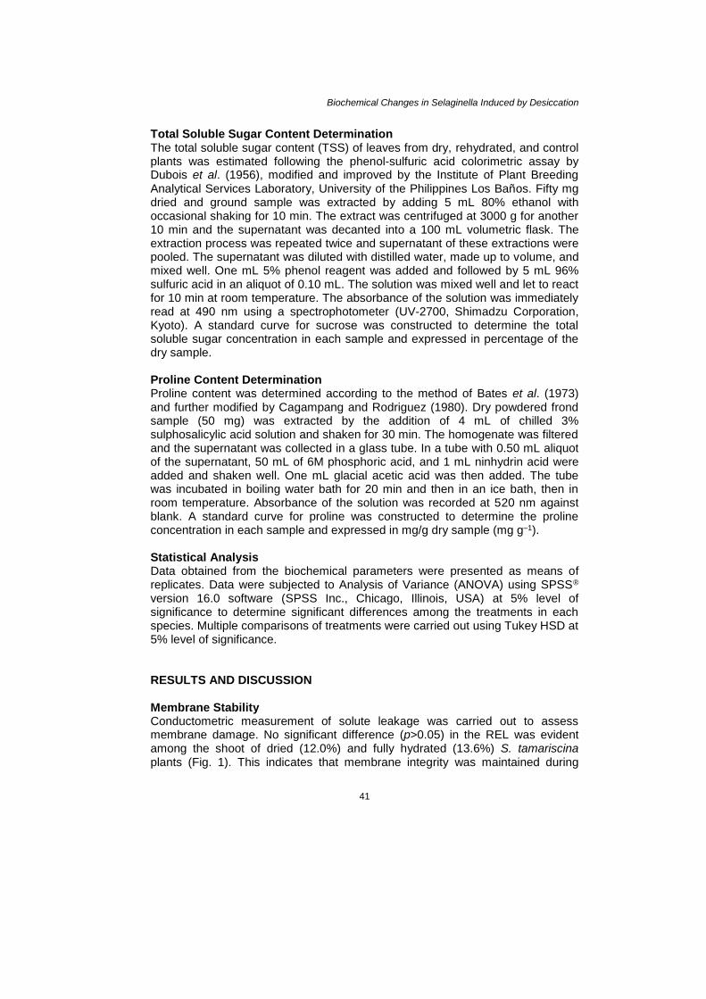
RESULTS AND DISCUSSION Membrane Stability Conductometric measurement of solute leakage was carried out to assess membrane damage. No significant difference (p>0.05) in the REL was evident among the shoot of dried (12.0%) and fully hydrated (13.6%) S. tamariscina plants (Fig. 1). This indicates that membrane integrity was maintained during
Angelo Rellama Agduma and Maribel Dionisio Sese
42
drying in this species. Meanwhile, there was reduction in electrolyte leakage in rehydrated S. tamariscina as evidenced in its lower REL (9.1%) than in desiccated plants. This means that membrane integrity was even more intact during the rehydration phase.
Figure 1: Effect of desiccation and rehydration on stability of cell membrane in
S. tamariscina and S. plana reflected as REL. Error bars represent standard deviation within the test group or treatment (p<0.05), obtained from three replicates.
Although placed in a different plant group, the same trend was observed
by Sherwin and Farrant (1996) when they monitored the rate of electrolyte leakage in the resurrection plant Craterostigma wilmsii. Leakages in control, dry, and rehydrated leaves were not variable. Similar to this species, protection mechanism was probably reinforced in S. tamariscina during rehydration as reflected in the lower REL noted in the rehydrated plants when compared with the dehydrated ones. This supports claim that as a desiccation tolerant plant rehydrates following dehydration, damage in the plasma membrane is immediately repaired. Crowe et al. (1992) asserted that drying causes the membrane to change from a liquid crystalline phase to gel phase during dehydration and then return to liquid crystalline phase during rehydration. These membrane phase transitions are the putative cause when leakage occurs in desiccation tolerant species (Oliver et al. 2005). In addition, rupture in the plasma membrane may occur as it contracts from the cell wall and by its attachment to it via the plasmodesmata during dehydration (Levitt 1980).
Highly significant rise in REL, however, was noted in both desiccated and rehydrated S. plana against the controls. It was also observed that there was a further substantial increase in REL upon rehydration attempt. This indicates that damage in the membrane incurred during the dehydration process was not
Biochemical Changes in Selaginella Induced by Desiccation
43
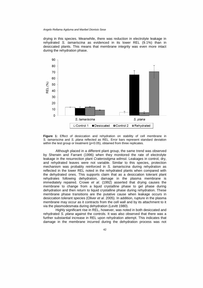
reversed or repaired during rehydration. In fact it would seem that leakage was even aggravated during rehydration in this species. Pigment Content Illustrated in Figure 2 is the total chlorophyll content (Chl a+b) in desiccated, rehydrated, and control plants. Statistical analysis showed that there was significant difference in the mean Chl a+b among test groups or treatments in S. tamariscina (p<0.05). The Chl a+b in desiccated plants dropped to 1133.8 µg g–1
of dry shoot tissues from 1770.9 µg g–1 (p<0.05). Hence, approximately 64.0% total chlorophyll of the fully hydrated (control) plants was retained during the desiccated state. When hydration resumed, however, total chlorophyll content (1617.5 µg g–1) roughly reached control levels. This indicates that chloroplasts of this species likely recovered and became functional during this state.
Figure 2: Total chlorophyll (Chl a+b) contents of S. tamariscina and S. plana following
desiccation and rehydration. Error bars represent standard deviation within the test group or treatment (p<0.05), obtained from three replicates.
On the other hand, it is apparent that there was significant loss in
Chl a+b in S. plana during the dry state (Fig. 2). Total chlorophyll content dropped from 3633.3 µg g–1 to 167.9 µg g–1 after desiccation (p<0.05), which was maintained even during the rehydration attempt. In this species, the total chlorophyll in rehydrated plants was not significantly different from the desiccated ones indicating that there was no element of regeneration (p>0.05).
Decrease in chlorophyll content of leaves is thought to be linked to the protection of plants against UV light and from damage as a result of oxygen free radical generation during desiccation (Sherwin & Farrant 1998). Chlorophyll content of the leaves did not drop too much in desiccated S. tamariscina indicating that no complete dismantling of photosynthetic apparatus was observed. It is assumed that S. tamariscina did not lose all its chlorophyll because the plant itself is protected from irradiation damage through different means. Since it was observed that it formed a ball upon desiccation, the
Angelo Rellama Agduma and Maribel Dionisio Sese
44
functional leaves were protected inside. The adaxial side of the leaves remained green while the abaxial side did not. It must be noted, however, that the total chlorophyll in desiccated S. tamariscina was significantly lower than the control suggesting degradation probably happened but was not severe.
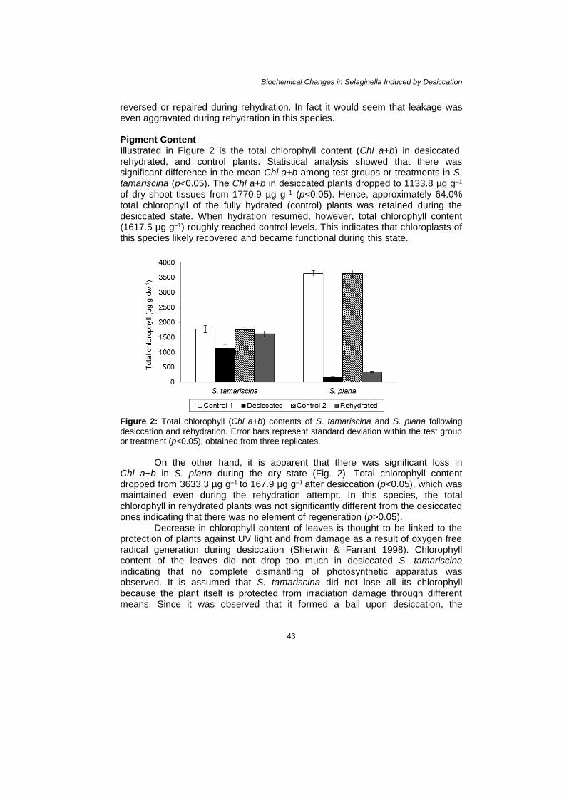
The changes in carotenoid content during dry state are shown in Figure 3. Compared to S. tamariscina, carotenoids in S. plana were almost degraded. From 122.9 µg g–1, carotenoids in S. plana declined to 9.9 µg g–1 or approximately 8.1% of the total (p<0.05). Whereas, approximately 41.9% of the total carotenoid content of control S. tamariscina (124.2 µg g–1) was retained in the dried state. Upon rehydration, control levels were even exceeded unlike in the case of S. plana.
Figure 3: Total carotenoid contents in shoot of S. tamariscina and S. plana following
desiccation and rehydration. Error bars represent standard deviation within the test group or treatment (p<0.05), obtained from three replicates.
Thus, chlorophyll and carotenoid results show that some of the
photosynthetic apparatus during desiccation in S. tamariscina was retained, if not completely upheld. Desiccation-tolerant plants can be classified through pigment analysis, whether they retain their photosynthetic pigments or not. Oliver et al. (2000) and Tuba et al. (1998) stated that desiccation-tolerant plants that retain their chlorophyll content during dry state are homoichlorophyllous while those that dismantle their chlorophyll are termed poikilochlorophyllous. On this basis, Farrant (2000) classified the resurrection angiosperms C. wilmsii and Myrothamnus flabellifolius as homoichlorophyllous species because they retained some of their chlorophyll content, 82% and 60% chlorophyll, respectively. Since S. tamariscina retained 64% of its chlorophyll in the dehydrated state, it can be classified then as homoichlorophyllous species. Upon rehydration, this partial loss of chlorophyll was regained. Being a desiccation-tolerant plant, S. tamariscina has innate characteristics protecting its photosynthetic apparatus. This can be supported by the observation that when non-irrigation of some plants
Biochemical Changes in Selaginella Induced by Desiccation
45
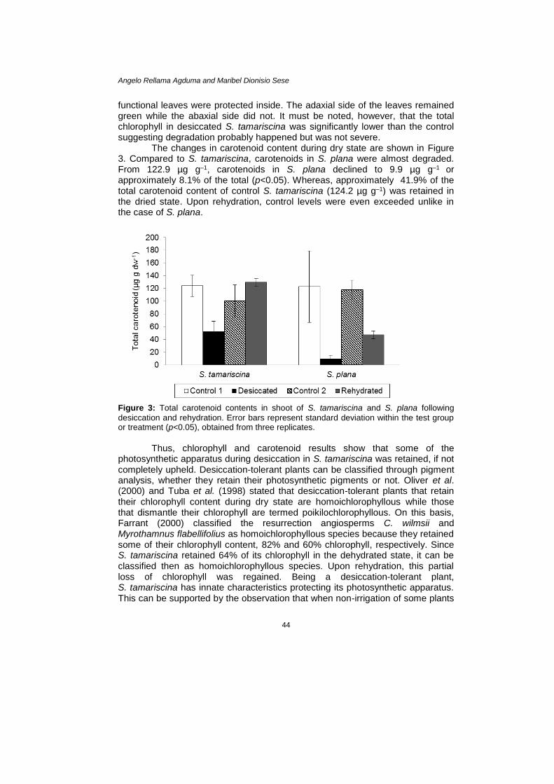
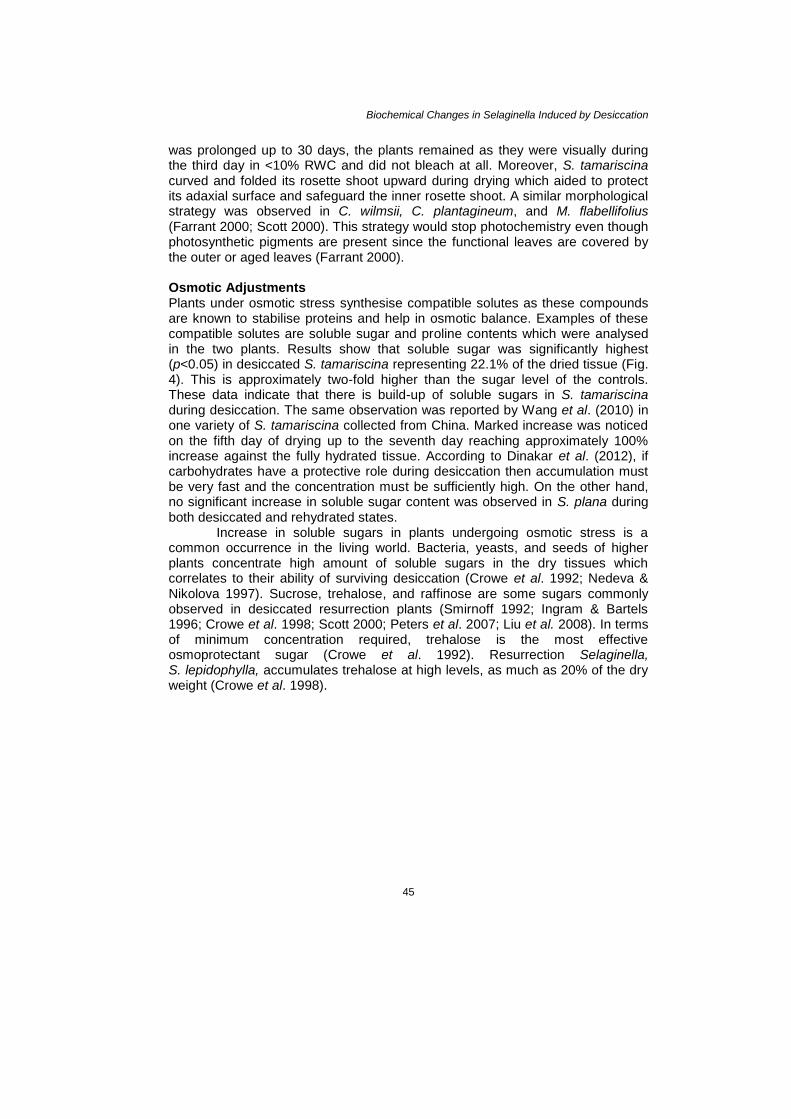
was prolonged up to 30 days, the plants remained as they were visually during the third day in <10% RWC and did not bleach at all. Moreover, S. tamariscina curved and folded its rosette shoot upward during drying which aided to protect its adaxial surface and safeguard the inner rosette shoot. A similar morphological strategy was observed in C. wilmsii, C. plantagineum, and M. flabellifolius (Farrant 2000; Scott 2000). This strategy would stop photochemistry even though photosynthetic pigments are present since the functional leaves are covered by the outer or aged leaves (Farrant 2000). Osmotic Adjustments Plants under osmotic stress synthesise compatible solutes as these compounds are known to stabilise proteins and help in osmotic balance. Examples of these compatible solutes are soluble sugar and proline contents which were analysed in the two plants. Results show that soluble sugar was significantly highest (p<0.05) in desiccated S. tamariscina representing 22.1% of the dried tissue (Fig. 4). This is approximately two-fold higher than the sugar level of the controls. These data indicate that there is build-up of soluble sugars in S. tamariscina during desiccation. The same observation was reported by Wang et al. (2010) in one variety of S. tamariscina collected from China. Marked increase was noticed on the fifth day of drying up to the seventh day reaching approximately 100% increase against the fully hydrated tissue. According to Dinakar et al. (2012), if carbohydrates have a protective role during desiccation then accumulation must be very fast and the concentration must be sufficiently high. On the other hand, no significant increase in soluble sugar content was observed in S. plana during both desiccated and rehydrated states.
Increase in soluble sugars in plants undergoing osmotic stress is a common occurrence in the living world. Bacteria, yeasts, and seeds of higher plants concentrate high amount of soluble sugars in the dry tissues which correlates to their ability of surviving desiccation (Crowe et al. 1992; Nedeva & Nikolova 1997). Sucrose, trehalose, and raffinose are some sugars commonly observed in desiccated resurrection plants (Smirnoff 1992; Ingram & Bartels 1996; Crowe et al. 1998; Scott 2000; Peters et al. 2007; Liu et al. 2008). In terms of minimum concentration required, trehalose is the most effective osmoprotectant sugar (Crowe et al. 1992). Resurrection Selaginella, S. lepidophylla, accumulates trehalose at high levels, as much as 20% of the dry weight (Crowe et al. 1998).
Angelo Rellama Agduma and Maribel Dionisio Sese
46
Figure 4: Total soluble sugar contents of S. tamariscina and S. plana following desiccation
and rehydration. Error bars represent standard deviation within the test group or treatment (p<0.05), obtained from three replicates.
Although trehalose is considered a major factor that determines the
anhydrobiotic ability of resurrection plants (Zentella et al. 1999), sucrose and other sugars, however, may also act as osmoprotectants (Crowe et al. 1992). In water-deprived Xerophyta viscosa, sucrose concentration in the leaves increased to nearly 5-fold at 5% RWC from the original sucrose content while raffinose increased to approximately 3-fold at same RWC (Peters et al. 2007). Others suggest that there is redirection of carbon flow from reserve substances such as starch or octulose to soluble saccharides (Ingram et al. 1997). In Craterostigma species, leaves of well-watered C. wilmsii and C. plantagineum have high amount of eight carbon sugar 2-octulose (Bianchi et al. 1991; Norwood et al. 2000). This eight-carbon sugar is converted to sucrose upon drying (Willige et al. 2009). This massive conversion of stored carbohydrates upon dehydration will concentrate sucrose in the dried tissue which consequently will comprise about 40% of the plant’s dry weight. In some cases, sucrose may make up as much as 50% of the dry weight (Crowe et al. 1998). In another desiccation-tolerant plant Ramonda sp., instead of 2-octulose, starch is converted to sucrose that serves the same function (Müller et al. 1997).
Crowe et al. (1998) stated two hypotheses concerning the roles of sugars in desiccated plants; they form supersaturated liquid known as biological glasses to stabilise internal structures and they prevent fusion of membranes and denaturation of proteins in the cell rendering maintenance of cell integrity during desiccation. The process of forming biological glasses is known as vitrification (Hoekstra 2005). This process is obligatory to survival during desiccation as this protects organelles from damage. This biological glass forms cavity inside the cell to prevent cellular collapse. It also restricts production of free radicals by slowing
Biochemical Changes in Selaginella Induced by Desiccation
47
molecular mobility in the cytoplasm to prevent chemical reactions to occur (Koster 1991; Ingram & Bartels 1996; Hoekstra 2005). Moreover, sugars protect membranes by altering the properties of the dry membranes to resemble those of fully hydrated ones. It is suggested that the hydroxyl groups of sugars substitute for water and provide the required hydrophilic interactions for membrane, the “water replacement hypothesis” (Crowe et al. 1992). Similarly, sugars stabilise proteins through the formation of hydrogen bonds between sugar hydroxyl groups and polar residues in proteins (Crowe et al. 1992). This mechanism of direct bonding of sugars with biomolecules is indeed imperative in the stabilisation of proteins, membranes, and whole cells under conditions of dehydration (Peters et al. 2007).
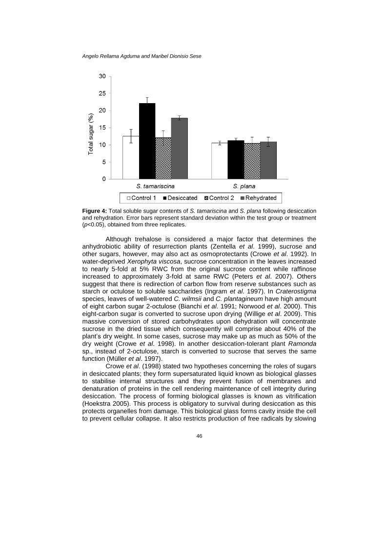
The present study showed that desiccated S. tamariscina had the highest proline content (p<0.05); approximately 15.0% increase in proline content was noted in desiccated plant compared to control plant (Fig. 5). In contrast, rehydrated S. tamariscina was shown to have lower proline content relative to the desiccated samples. These results validate the important role of proline during desiccation as shown in its increase during stress and its decrease when the plant was relieved from stress.
Figure 5: Proline contents of S. tamariscina and S. plana following desiccation and
rehydration. Error bars represent standard deviation within the test group or treatment (p<0.05), obtained from three replicates.
Proline is also known to accumulate in response to a wide range of
abiotic stresses in many plants (Hare & Cress 1997). Its presumed roles are in stabilising protein structures against denaturation, interacting with phospholipids to protect cell membranes, and in functioning as a hydroxyl radical scavenger in stressed plants (Santoro et al. 1992; Ingram & Bartels 1996; Claussen 2005). Pandey et al. (2010) and Wang et al. (2010) used proline as one of their parameters to assess desiccation tolerance in S. bryopteris and S. tamariscina, respectively. Proline in dried S. bryopteris frond was more or less 5-fold higher than in fully hydrated fronds (Pandey et al. 2010). On the other hand, Wang et al.
Angelo Rellama Agduma and Maribel Dionisio Sese
48
(2010) observed that relatively stable proline levels were noted from zero to third day of drying in S. tamariscina. It was only during the fifth day that sharp increase in proline concentration was observed which continued and was highest during the seventh day of drying. Moreover, proline concentrations during the 12th hour of rehydration and initial phase of drying were comparable indicating that high concentration of proline during fully hydrated and rehydrated phases is not a requirement.
Figure 5 also shows that the second control of S. tamariscina plants had higher proline concentration than the first control plants and was not significantly different compared to the desiccated plants. This is in contradiction with the initial statement that proline accumulates in response to desiccation. If the second control is taken into account and compared it with the desiccated data, statistics suggest that proline did not rise during desiccation. However, it should also be noted that the desiccated and the second control data were taken at different time intervals and perhaps at different prevailing environmental conditions. That is, the second control data were taken at a time corresponding to the time when rehydrated samples were analysed. Since proline is also implicated in other stressful environmental conditions like salinity stress (Amirjani 2010), drought stress (Mafakheri et al. 2010), high light intensity, and temperature (Claussen 2005), it might be that the increase in proline concentration in the second control of S. tamariscina plants were due to environmental stress which is not osmotic in nature. While desiccated and rehydrated S. plana was observed to have higher proline content than the fully hydrated ones (controls), this increase, however, was not significantly different from the control plants.
Despite evolutionary relatedness, it is apparent that S. tamariscina and S. plana behave differently toward desiccation stress. Most of the Selaginella species, including S. plana, are tropical growing in damp shady habitats (Kramer & Green 1990) but others such as S. lepidophylla and S. sartorii are adapted for seasonal drought or xerophytic conditions (Setyawan 2011) just like S. tamariscina. They grow on dry rocky cliffs or on soil that dries periodically. Besides physiological adaptations, desiccation-tolerant plants have also evolved the ability to overcome the drought-induced stress morphologically. To name a few, leaf curling, excessive cell volume reduction, and cell wall folding or shrinkage as the plant dries but they re-expand as they are moistened (Farrant et al. 2007). The aerial parts of S. tamariscina undergo gradual morphological rolling and wilting forming a ball in the dry state but as it is re-watered, the aerial parts fully open and recover in just 12 h (Wang et al. 2010). All these things together are manifestations of active strategy of S. tamariscina for protecting itself and avoiding damages brought by desiccation. CONCLUSION Desiccation-tolerance mechanisms have been found in S. tamariscina while desiccation-sensitive characteristics were found in S. plana. In S. tamariscina, evident protection mechanisms are engaged such as low electrolyte leakage and retention of some of the chlorophylls and carotenoid contents during the
Biochemical Changes in Selaginella Induced by Desiccation
49
desiccation and rehydration phases. The former indicates stability of the membranes while the latter means protection of the photosynthetic apparatus. Compatible solutes like sugars and proline also increased during desiccation inferring decreased rates of chemical reactions, reduced diffusion of molecules, and prevention of oxidative damage. Meanwhile, there appears to be no mechanism of subcellular protection operating in S. plana. As the drying process progressed, being intolerant of desiccation, drying up of tissues became irreversible. ACKNOWLEDGEMENT This work was supported by grants to the first author by the Commission on Higher Education Faculty Development Program Phase II (CHED FDP-II) and Department of Science and Technology-Accelerated Science and Technology Human Resource Development Program (DOST-ASTHRDP). The authors are particularly grateful to Dr. Calixto M. Protacio and Prof. Nerissa K. Torreta as well as the staff and faculty of Plant and Microbiology Divisions of Institute of Biological Sciences, University of the Philippines Los Baños for their help in the successful conduct of the study. REFERENCES Alpert P. (2006). Constraints of tolerance: Why are desiccation-tolerant organisms so
small or rare? Journal of Experimental Biology 209(9): 1575–1584. doi.org/ 10.1242/jeb.02179
____. (2005). The limits and frontiers of desiccation-tolerant life. Integrative and Comparative Biology 45(5): 685–695. doi.org/10.1093/icb/45.5.685
____. (2000). The discovery, scope, and puzzle of the desiccation tolerance in plants. Plant Ecology 151(1): 5–17. doi.org/10.1023/A:1026513800380
Amirjani M R. (2010). Effect of salinity stress on growth, mineral composition, proline content, antioxidant enzymes of soybean. American Journal of Plant Physiology 5(6): 350–360. doi.org/10.3923/ajpp.2010.350.360
Barrs H D and Weatherley P E. (1962). A re-examination of the relative turgidity technique for estimating water deficit in leaves. Australian Journal of Biological Sciences
15(3): 413–428. Bartels D. (2005). Desiccation tolerance studied in the resurrection plant Craterostigma
plantagineum. Integrative and Comparative Biology 45(5): 696–701. doi.org/10. 1093/icb/45.5.696
Bates L, Waldren R P and Teare I D. (1973). Rapid determination of free proline for water-stress studies. Plant and Soil 39(1): 205–207. doi.org/10.1007/BF00018060
Bianchi G, Gamba A, Murelli C, Salamini F and Bartels D. (1991). Novel carbohydrate metabolism in the resurrection plant Craterostigma plantagineum. The Plant Journal 1(3): 355–359. doi.org/10.1046/j.1365-313X.1991.t01-11-00999.x
Brighigna L, Bennici A, Tani C and Tani G. (2002). Structural and ultrastructural characterization of Selaginella lepidophylla, a desiccation-tolerant plant, during the rehydration process. Flora-Morphology, Distribution, Functional Ecology of Plants 197(2): 81–91. doi.org/10.1078/0367-2530-00018
Angelo Rellama Agduma and Maribel Dionisio Sese
50
Cagampang G B and Rodriguez F M. (1980). Methods of analysis for screening crops of appropriate qualities. IPB Bulletin 2. Philippines: Analytical Services Laboratory, Institute of Plant Breeding, University of the Philippines at Los Baños.
Claussen W. (2005). Proline as a measure of stress in tomato plants. Plant Science 168(1): 241–248. doi.org/10.1016/j.plantsci.2004.07.039
Cooper K and Farrant J M. (2002). Recovery of the resurrection plant Craterostigma wilmsii from desiccation: Protection versus repair. Journal of Experimental Botany
53(375): 1805–1813. doi.org/10.1093/jxb/erf028 Crowe J H, Hoekstra F A and Crowe L M. (1998). The role of vitrification in anhydrobiosis.
Annual Review of Physiology 60(1): 73–103. doi.org/10.1146/annurev.physiol. 60.1.73
____. (1992). Anhydrobiosis. Annual Review of Physiology 54(1): 579–599. doi.org/10.1146/annurev.ph.54.030192.003051
Dinakar C, Djilianov D and Bartels D. (2012). Photosynthesis in desiccation tolerant plants: Energy metabolism and antioxidative stress defense. Plant Science 182: 29–41.
doi.org/10.1016/j.plantsci.2011.01.018 Dubois M, Gilles K A, Hamilton J K, Rebers P A and Smith F. (1956). Colorimetric method
for determination of sugars and related substances. Analytical Chemistry 28(3): 350–356. doi.org/10.1021/ac60111a017
Farrant J M. (2000). A comparison of mechanisms of desiccation tolerance among three angiosperm resurrection plant species. Plant Ecology 151(1): 29–39. doi.org/10.1023/A:1026534305831
Farrant J M, Brandt W and Lindsey G G. (2007). An overview of mechanisms of desiccation tolerance in selected angiosperm resurrection plants. Plant Stress 1(1): 72–84.
Farrant J M, Cooper K, Kruger L A and Sherwin H W. (1999). The effect of drying rate on the survival of three desiccation-tolerant angiosperm species. Annals of Botany
84(3): 371–379. doi.org/10.1006/anbo.1999.0927 Farrant J M, Vander Willigen C, Loffell D A, Bartsch S and Whittaker A. (2003). An
investigation into the role of light during desiccation of three angiosperm resurrection plants. Plant, Cell & Environment 26(8): 1275–1286. doi.org/10.
1046/j.0016-8025.2003.01052.x Hare P D and Cress W A. (1997). Metabolic implications of stress-induced proline
accumulation in plants. Plant Growth Regulation 21(2): 79–102. doi.org/10.1023/ A:1005703923347
Hoekstra F A. (2005) Differential longevities in desiccated anhydrobiotic plant systems. Integrative and Comparative Biology 45(5): 725–733. doi.org/10.1093/icb/ 45.5.725
Ingram J and Bartels D. (1996). The molecular basis of dehydration tolerance in plants. Annual Review of Plant Biology 47(1): 377–403. doi.org/10.1146/annurev.arplant.
47.1.377 Ingram J, Chandler J W, Gallagher L, Salamini F and Bartels D. (1997). Analysis of cDNA
clones encoding sucrose-phosphate synthase in relation to sugar interconversions associated with dehydration in the resurrection plant Craterostigma plantagineum Hochst. Plant Physiology 115(1): 113–121. doi.org/ 10.1104/pp.115.1.113
Kenrick P and Crane P R. (1997). The origin and early evolution of plants on land. Nature 389(6646): 33–39. doi.org/10.1038/37918
Koster K L. (1991). Glass formation and desiccation tolerance in seeds. Plant Physiology 96(1): 302–304. doi.org/10.1104/pp.96.1.302
Biochemical Changes in Selaginella Induced by Desiccation
51
Kramer P S and Green P S. (1990). Pteridophytes and gymnosperms. In K Kubitzki (ed.). The families and genera of vascular plants. Berlin: Springer-Verlag. doi.org/10. 1007/978-3-662-02604-5
Layton B E, Boyd M B, Tripepi M S, Bitonti B M, Dollahon M N R and Balsamo R A. (2010). Dehydration-induced expression of a 31-kDa dehydrin in Polypodium polypodioides (Polypodiaceae) may enable large, reversible deformation of cell walls. American Journal of Botany 97(4): 535–544. doi.org/10.3732/ajb.0900285
Levitt J. (1980). Responses of plants to environmental stresses, vol 2. Water, radiation, salt and other stresses. New York: Academic Press.
Liu M S, Chien C T and Lin T P. (2008). Constitutive components and induced-gene expression are involved in the desiccation tolerance of Selaginella tamariscina. Plant and Cell Physiology 49(4): 653–663. doi.org/10.1093/pcp/pcn040
Mafakheri A, Siosemardeh A, Bahramnejad B, Struik P C and Sohrabi Y. (2010). Effect of drought stress on yield, proline and chlorophyll contents in three chickpea cultivars. Australian Journal of Crop Science 4(8): 580–585.
Moore J P, Lindsey G G, Farrant J M and Brandt W F. (2007). An overview of the biology of the desiccation-tolerant resurrection plant Myrothamnus flabellifolia. Annals of Botany 99(2): 211–217. doi.org/10.1093/aob/mcl 269
Müller J, Sprenger N, Bortlik K, Boller T and Wiemken A. (1997). Desiccation increases sucrose levels in Ramonda and Haberlea, two genera of resurrection plants in the Gesneriaceae. Physiologia Plantarum 100(1): 153–158. doi.org/10.1111/j. 1399-3054.1997.tb03466.x
Nedeva D and Nikolova A. (1997). Desiccation tolerance in developing seeds. Bulgarian Journal of Plant Physiology 23(3–4): 100–113.
Norwood M, Truesdale M R, Richter A and Scott P. (2000). Photosynthetic carbohydrate metabolism in the resurrection plant Craterostigma plantagineum. Journal of Experimental Botany 51(343): 159–165. doi.org/10.1093/jexbot/51.343.159
Oliver M J, Tuba Z and Mishler B D. (2000). The evolution of vegetative desiccation tolerance in land plants. Plant Ecology 151(1): 85–100. doi.org/10.1023/A:102655 0808557
Oliver M J, Velten J and Mishler B D. (2005). Desiccation tolerance in bryophytes: A reflection of the primitive strategy for plant survival in dehydrating habitats? Integrative and Comparative Biology 45(5): 788–799. doi.org/10.1093/icb/45.5. 788
Pandey V, Ranjan S, Deeba F, Pandey A K, Singh R, Shirke P A and Pathre U V. (2010). Desiccation-induced physiological and biochemical changes in resurrection plant, Selaginella bryopteris. Journal of Plant Physiology 167(16): 1351–1359. doi.org/ 10.1016/j.jplph.2010.05.001
Peters S, Mundree S G, Thomson J A, Farrant J M and Keller F. (2007). Protection mechanisms in the resurrection plant Xerophyta viscosa (Baker): Both sucrose
and raffinose family oligosaccharides (RFOs) accumulate in leaves in response to water deficit. Journal of Experimental Botany 58(8): 1947–1956. doi.org/10. 1093/jxb/erm056
Porra R J, Thompson W A and Kriedemann P E. (1989). Determination of accurate extinction coefficients and simultaneous equations for assaying chlorophylls a and b extracted with four different solvents: Verification of the concentration of chlorophyll standards by atomic absorption spectroscopy. Biochimica et Biophysica Acta (BBA)-Bioenergetics 975(3): 384–394. doi.org/10.1016/S0005-
2728(89)80347-0 Santoro M M, Liu Y, Khan S M, Hou L X and Bolen D W. (1992). Increased thermal
stability of proteins in the presence of naturally occurring osmolytes. Biochemistry 31(23): 5278–5283. doi.org/10.1021/bi00138a006
Angelo Rellama Agduma and Maribel Dionisio Sese
52
Scott P. (2000). Resurrection plants and the secrets of eternal leaf. Annals of Botany 85(2): 159–166. doi.org/10.1006/anbo.1999.1006
Setyawan A D. (2011). Recent status of Selaginella (Selaginellaceae) research in Nusantara. Biodiversitas 12(2): 112–124. doi.org/10.13057/biodiv/d120209
Sherwin H W and Farrant J M. (1998). Protection mechanisms against excess light in the resurrection plants Craterostigma wilmsii and Xerophyta viscosa. Plant Growth Regulation 24(3): 203–210. doi.org/10.1023/A:10 05801610891
____. (1996). Differences in rehydration of three desiccation-tolerant angiosperm species. Annals of Botany 78(6): 703–710. doi.org/10.1006/anbo.1996.0180
Smirnoff N. (1992). The carbohydrates of bryophytes in relation to desiccation tolerance. Journal of Bryology 17(2): 185–191. doi.org/10.1179/jbr.1992.17.2.185
Tuba Z, Proctor M C F and Csintalan Z. (1998). Ecophysiological responses of homoichlorophyllous and poikilochlorophyllous desiccation tolerant plants: A comparison and an ecological perspective. Plant Growth Regulation 24(3): 211–217. doi.org/10.1023/A:1005951908229
Wang X, Chen S, Zhang H, Shi L, Cao F, Guo L, Xie Y, Wang T, Yan X and Dai S. (2010). Desiccation tolerance mechanism in resurrection fern-ally Selaginella tamariscina revealed by physiological and proteomic analysis. Journal of Proteome Research 9(12): 6561–6577. doi.org/10.1021/pr100767k
Willige B C, Kutzer M, Tebartz F and Bartels D. (2009). Subcellular localization and enzymatic properties of differentially expressed transketolase genes isolated from the desiccation tolerant resurrection plant Craterostigma plantagineum. Planta 229(3): 659–666. doi.org/10.1007/s00425-008-0863-5
Zentella R, Mascorro-Gallardo J O, Van Dijck P, Folch-Mallol J, Bonini B, Van Vaeck C, Gaxiola R et al. (1999). A Selaginella lepidophylla trehalose-6-phosphate synthase complements growth and stress-tolerance defects in a yeast tps1 mutant. Plant Physiology 119(4): 1473–1482. doi.org/10.1104/pp.119.4.1473