cellular kinetics in growth anomalies of the scleractinian
TRANSCRIPT
DISEASES OF AQUATIC ORGANISMSDis Aquat Org
Vol. 102: 1–11, 2012doi: 10.3354/dao02530
Published December 3
INTRODUCTION
Growth anomaly (GA) is a common and easilyidentifiable disease that has been reported for hydro-zoans, gorgonians, and scleractinian corals (re -viewed by Sutherland et al. 2004). GAs in scleractin-ian corals have been observed on reefs throughoutthe Caribbean Sea and the Indo-Pacific Ocean (e.g.Sutherland et al. 2004, Aeby et al. 2011) and fromshallow waters (0.5 m; Aeby et al. 2011) to the deepsea (635 m; Squires 1965). GAs in scleractinian coralscommonly manifest as protuberant shapes with fewor no corallites/polyps and abnormal corallite/polypstructures (Yamashiro et al. 2000, Work et al. 2008,
Yasuda et al. 2012), and in most cases, the zooxan-thellae density is low compared to healthy regions ofhost colonies (Domart-Coulon et al. 2006, Work et al.2008, Yasuda et al. 2012).
Histological studies have described several re mar -kable structural anomalies of GA tissues, includingdegenerated polyps or complete lack of polyp struc-tures (Yamashiro et al. 2000, Work et al. 2008, Burns& Takabayashi 2011), no or reduced mesenterial fila-ments (Peters et al. 1986, Work & Rameyer 2005,Burns & Takabayashi 2011), and a hyperplastic basalbody wall (Work et al. 2008, Burns & Takabayashi2011, Williams et al. 2011). Furthermore, Domart-Coulon et al. (2006) demonstrated that the proteins
© Inter-Research 2012 · www.int-res.com*Corresponding author. Email: [email protected]
Cellular kinetics in growth anomalies of the scleractinian corals Porites australiensis
and Montipora informis
Naoko Yasuda1, Michio Hidaka2,*
1Marine and Environmental Science, Graduate School of Engineering and Science, and 2Department of Chemistry, Biology and Marine Science, Faculty of Science; University of the Ryukyus, Nishihara, Okinawa 903-0213, Japan
ABSTRACT: Growth anomalies (GAs) in corals are characterized by morphological abnormalitiesof the skeleton as well as polyps and coenosarcs. GAs commonly appear as protuberances withfewer polyps and are paler in color due to decreased zooxanthellae density. To test the hypothesisthat morphological anomalies in GAs may be caused by unregulated cellular kinetics, the relativeabundances of apoptotic cells and proliferating cells were compared between GAs and apparentlyhealthy regions in 2 corals, Porites australiensis and Montipora informis. Apoptotic cells and pro-liferating cells were detected using TUNEL assays and BrdU incorporation assays, respectively.The labeling indices for apoptotic nuclei and BrdU-labeled nuclei were measured in the epider-mis, oral gastrodermis, aboral gastrodermis, and calicodermis. The labeling index for apoptoticnuclei in the oral gastrodermis and the calicodermis was significantly lower in GAs than in healthyregions in both coral species. The index for BrdU-labeled cells in the calicodermis was signifi-cantly higher in GAs than in healthy regions in both coral species. When GA regions partiallydied, the GA tissues directly adjacent to the dead areas exhibited signs of necrosis, although someapoptotic cells were also present. Healthy oral gastrodermis adjacent to the border between thehealthy and GA regions exhibited higher frequencies of apoptotic cells. These results suggest thatapoptotic pathways were suppressed and cell proliferation was promoted in GA regions, althoughcells in GAs may die through both necrosis and apoptosis.
KEY WORDS: Apoptosis · Necrosis · Cell proliferation · Coral disease · Growth anomaly · Porites australiensis · Montipora informis
Resale or republication not permitted without written consent of the publisher
Dis Aquat Org 102: 1–11, 2012
MutY, Hsp90a1, GRP75, and metallo thionein, whichare hyperplasia-associated proteins, were upregu-lated in GAs in Porites compressa. Their results alsoindicate hyper-proliferation of cells in GA tissues.However, the mechanism and the process of cellularkinetics in GA regions are not yet understood.
The etiology of GA is still unclear, although rela-tionships between GA prevalence and several factorshave been suggested, including UVB radiation (Pe -ters et al. 1986, Coles & Seapy 1998), nutrients andorganic carbon (Kaczmarsky & Richardson 2010),water temperatures and photosynthetically activeradiation (Stimson 2010), water turbidity and depth(Williams et al. 2010), host density and human popu-lation size (Aeby et al. 2011), and aging (Irikawa etal. 2011). GA regions are frequently associated withmicroorganisms in the tissue as well as skeletal space(Coles & Seapy 1998, Work & Rameyer 2005, Work etal. 2008), and the microorganisms have been sug-gested to be involved in GAs (Work & Rameyer 2005,Domart-Coulon et al. 2006). However, microorgan-isms are not always detected in GAs (Yamashiro et al.2000, Burns & Takabayashi 2011), and the linkagesbetween their presence and GA development arecurrently unknown.
GA-affected corals show deteriorated physiologicalstates, such as low fecundity in Montipora spp. (Ya-mashiro et al. 2000, Burns & Takabayashi 2011), Acro-pora spp. (Irikawa et al. 2011), and Porites spp.(Hunter & Field 1997, Domart-Coulon et al. 2006, Ya-suda et al. 2012), low lipid storage in M. informis (Ya-mashiro et al. 2001), and low photosynthetic produc-tivity in A. cytherea and P. australiensis due toreduced numbers of zooxanthellae in the gastrodermis(Irikawa et al. 2011, Yasuda et al. 2012). However,previous field investigations demonstrated that GAsprogressively spread toward healthy tissue on thehost colonies (Cheney 1975, Bak 1983, Irikawa et al.2011, Yasuda et al. 2012). Healthy fragments graftedto GA regions or fused with GA fragments exhibitedreduced growth rates compared with non-graftedhealthy fragments (Stimson 2010, Yasuda et al. 2012).These findings suggest that GAs grow utilizing nutri-ent supplies from surrounding healthy regions (Ya-mashiro 2001 et al., Domart-Coulon et al. 2006, Ya-suda et al. 2012). However, the processes leading toGA progression, i.e. how GA tissues invade surround-ing healthy regions, have never been investigated.
The partial death of GAs on coral colonies has beenreported at gross and microscopic levels (Work et al.2008, Irikawa et al. 2011, Williams et al. 2011, Yasudaet al. 2012). The microscopic studies revealed that tis-sue necrosis of GAs are often, though not always,
associated with infiltrating microorganisms such asalgae and fungi (Work & Rameyer 2005, Work et al.2008, Williams et al. 2011). However, the cause of thenecrosis in GA is not yet fully understood.
In this study, we histologically assessed cellular ki-netics in apparently healthy and GA tissues in 2 scler-actinian corals, Porites australiensis and Montiporainformis, whose GAs have been well studied morpho-logically and physiologically (e.g. Yamashiro et al.2000, 2001, Yasuda et al. 2012). We compared the rel-ative abundances of apoptotic cells and proliferatingcells between healthy and GA tissues using the termi-nal deoxynucleotidyl transferase dUTP-biotin nick-end labeling (TUNEL) and 5-bromo-2-deoxyuridine(BrdU) incorporation assays, respectively. We alsomade histological observations of the interface be-tween healthy and GA regions, and GAs directly ad-jacent to dead areas, to clarify the processes of pro-gression of GAs and cause of necrosis in GAs.
MATERIALS AND METHODS
Sample collection
Pieces of 3 to 10 cm square (9 to 100 cm2) were col-lected from each of the following regions: apparentlyhealthy, GAs, interface between healthy and GAregions, and GAs directly adjacent to the dead areaof each of 4 Porites australiensis colonies (n = 4 foreach region). Two of the 4 colonies were manifestingmultiple GAs and an additional 2 to 3 pieces of inter-face region and GA adjacent to the dead area weresampled from different GA regions and used as sup-plementary materials for histology and the TUNELassay. These additional samples were used only forobservation and were not included in the statisticalanalyses. Pieces of similar size were collected fromapparently healthy, GA, and interface regions ofeach of 3 Montipora informis colonies (n = 3 for eachregion). The pieces were collected using a hammerand chisel at a depth of 2 to 3 m in the back reefmoats of Kayo (26° 33’ N, 128° 08’ E) and Odo(26° 05’ N, 127° 42’ E) on Okinawa Island, Japan. Col-lected samples of P. australiensis from Kayo and sam-ples of P. australiensis and M. informis from Odowere brought to the laboratory and fixed for histolog-ical observations and TUNEL assays within 3 h. Thesamples were cut into <1 cm2 pieces and fixed in10% neutral buffered formalin (pH 7.4). The remain-ing samples for BrdU incorporation assays weretransported to a flow-through water tank at SesokoStation on the same day.
2

Yasuda & Hidaka: Cellular kinetics of coral growth anomalies
Histological observations
Fixed specimens of both corals were trimmed into5 × 5 mm square pieces after decalcification in 10%EDTA in phosphate buffered saline (pH 7.8). Theywere dehydrated in a graded series of ethanol andembedded in Paraplast plus (Sigma P3683). Seriallongitudinal sections 5 µm thick were cut and stainedwith hematoxylin and eosin.
Detection of apoptotic cells
To detect free 3’-ends of DNA fragments, histo-logical sections from each paraffin-embeddedspecimen were processed for TUNEL assays usingan ApopTag® Plus Fluorescein In Situ ApoptosisDetection kit (Chemicon S7111). The sections werealso stained with 4’, 6-diamidino-2-phenylindole(DAPI) to visualize all nuclei in the histologicalsections. The sections were observed under a fluo-rescent microscope (Ni kon Optiphot-2) using ablue excitation filter (wave length: 495 nm) foranti-digoxigenin-fluorescein-labeled (TUNEL-posi-tive) nuclei or an ultraviolet filter (wave length:365 nm) for nuclei counterstained with DAPI. Pho-tomicrographs of the same areas were taken underblue and then UV excitation using a digital camera(Nikon Digital Sight DS-L1).
In order to quantitate the percentage of apoptoticcells in each region of 2 coral species, the labelingindex (LI) for TUNEL-positive cells was calculated bydividing the number of the fluorescein-labeled nucleiby the total number of nuclei stained with DAPI.Three histological sections were assayed for eachspecimen. Every third section was selected fromserial sections of the specimen so that the 3 sectionsassayed were distant from each other by at least15 µm. Three photographs were taken for each of theoral and aboral regions per histological section.Areas mostly occupied by the oral or aboral regionswere photographed without overlap. In total, 18photo graphs (each photographed area 5.1 × 104 µm2)were used for measurements of the number ofTUNEL-positive cells for each specimen, and aver-age LI was calculated for each of epidermis, oral gas-trodermis, aboral gastrodermis, and calicodermis.Dense apoptotic signals were occasionally observedin some regions. In such cases, the signals appearedas smears over a small area, and counting the num-ber of TUNEL-positive nuclei was difficult. There-fore, dense signal areas were not included in the LImeasurements.
Detection of proliferating cells
A thymidine analog, BrdU incorporated cells wereimmunohistologically detected as proliferating cellsin each specimen. Samples of apparently healthyregions and samples containing both apparentlyhealthy and GA regions of Porites australiensis andMontipora informis (n = 3 each for healthy and GAregions) were trimmed into approximately 1 × 1 cmsquare pieces and acclimatized for 1 wk in an indoortank with running seawater that was exposed to nat-ural sunlight. The samples were then placed individ-ually in closed chambers filled with 300 ml of 100 µMBrdU in filtered seawater, and incubated with aera-tion for 3 d. These chambers were placed in a ther-mostatic bath at 25°C under light (approximately400 µmol m−2 s−1) from a metal halide lamp (DuplaElectra) for 16 h (8:00−24:00 h) per day. The BrdUsolution was changed daily, and the chamber posi-tions were changed randomly every day. After incu-bation, the specimens were rinsed 3 times in filteredseawater with aeration each for 10 min. The speci-mens were fixed, decalcified, and processed for his-tology as described above.
Histological sections were processed for the incor-porated BrdU assay using a BrdU In Situ Detectionkit (BD PharmingenTM) and a Streptavidin/Biotinblocking kit (Vector Laboratories). Hematoxylin wasused to stain all nuclei in the histological sections.
In order to quantitate proliferating cells in eachregion of the 2 coral species, photomicrographs weretaken using a digital camera (Nikon Digital Sight DS-L1), and the LI for BrdU-labeled cells was calculatedby dividing the number of BrdU-labeled cells thatwere stained with diaminobenzidine (DAB) by thetotal number of nuclei stained with hematoxylin.Three histological sections were assayed for eachspecimen as described for the TUNEL assay. Threephotographs were taken for each of the oral and ab oral regions per histological section. In total, 18photo graphs (each photographed area 5.5 × 104 µm2)were used for measurements of each specimen, andthe average LI was calculated for each of epidermis,oral gastrodermis, aboral gastrodermis, and calico-dermis, as well as for zooxanthellae.
Statistical analysis
The mean LI (percent data) of TUNEL-positivecells and BrdU incorporated cells in each tissue werecompared between the apparently healthy and GAregions in both coral species. As the data did not
3
Dis Aquat Org 102: 1–11, 2012
meet parametric requirements (Shapiro-Wilk testand F-test), Welch’s t-test, which does not requireequal variance, or the non-parametric Mann-Whit-ney U-test were conducted. All statistical analyseswere performed using R Ver. 2.9.1.
RESULTS
Histological observations
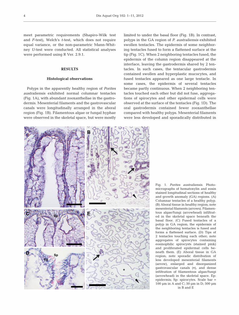
Polyps in the apparently healthy region of Poritesaustraliensis exhibited normal columnar tentacles(Fig. 1A), with abundant zooxanthellae in the gastro-dermis. Mesenterial filaments and the gastrovascularcanals were longitudinally arranged in the aboralregion (Fig. 1B). Filamentous algae or fungal hyphaewere observed in the skeletal space, but were mostly
limited to under the basal floor (Fig. 1B). In contrast,polyps in the GA region of P. australiensis exhibitedswollen tentacles. The epidermis of some neighbor-ing tentacles fused to form a flattened surface at thetip (Fig. 1C). When 2 neighboring tentacles fused, theepidermis of the column region disappeared at theinterface, leaving the gastrodermis shared by 2 ten-tacles. In such cases, the tentacular gastrodermiscontained swollen and hyperplastic mucocytes, andfused tentacles appeared as one large tentacle. Insome cases, the epidermis of several tentaclesbecame partly continuous. When 2 neighboring ten-tacles touched each other but did not fuse, aggrega-tions of spirocytes and other epidermal cells wereobserved at the surface of the tentacles (Fig. 1D). Theoral gastrodermis contained fewer zooxanthellaecompared with healthy polyps. Mesenterial filamentswere less developed and sporadically distributed in
4
A
C
D
B
EEp
EpSp
*
Fig. 1. Porites australiensis. Photo -micrographs of hematoxylin and eosinstained longitudinal sections of healthyand growth anomaly (GA) regions. (A)Columnar tentacles of a healthy polyp.(B) Aboral tissue in healthy region; notemesenterial filaments (arrows). Filamen-tous algae/fungi (arrowhead) infiltrat -ed in the skeletal space beneath thebasal floor. (C) Fused tentacles of apolyp in GA region; the epidermis ofthe neighboring tentacles is fused andforms a flattened surface. (D) Tips of2 tentacles touching each other; noteaggregates of spirocytes containingeosinophilic spirocysts (stained pink)and proliferated epidermal cells be -neath them. (E) Aboral tissue in GAregion; note sporadic distribution ofless developed mesenterial filaments(arrow), enlarged and disorganizedgastrovascular canals (*), and denseinfiltration of filamentous algae/fungi(arrowhead) in the skeletal space. Ep:epidermis; Sp: spirocytes. Scale bar =100 µm in A and C; 50 µm in D; 500 µm
in B and E

Yasuda & Hidaka: Cellular kinetics of coral growth anomalies
the aboral region of the GA (Fig. 1E). The aboralregion was mainly composed of disorganized gas-trovascular canals (Fig. 1E). The coenosarc in the GAregion swelled and protruded to almost the samelevel as polyps (data not shown). Dense filamentousalgae or fungi were observed throughout the skeletalspace in the GA region, and they sometimes infil-trated even into the oral regions of the GA. Necrotictissue which manifested as clusters of lysed cellswithout intact plasma membranes were observedadjacent to the accumulation of organic matrix com-ponents in both oral and aboral regions at the inter-face between GA and dead regions of P. australiensis(data not shown). The occurrence of necrotic tissuewas often associated with filamentous algae/fungi.
Detection of apoptotic cells
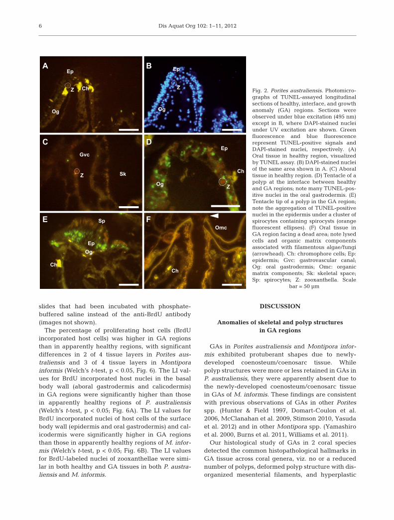
Apoptotic nuclei appeared as bright green dotsagainst the yellow autofluorescence of the host tissueunder blue excitation (wave length: 495 nm), whileall nuclei stained with DAPI exhibited blue fluores-cence under UV excitation (Fig. 2A,B). Symbiontsappeared bright to dull orange or were non-fluores-cent and showed no apoptotic signals in the presentassay (Fig. 2A, C, D). No TUNEL-positive signal wasdetected in the negative control slides that had beenincubated with deionized water instead of TdTenzyme (image not shown).
TUNEL-positive nuclei were few in both oral andaboral regions of apparently healthy tissues of Poritesaustraliensis (Fig. 2A, C). However, TUNEL-positivenuclei were occasionally observed in the oral gastro-dermis in the tentacles (Fig. 2D) or mesenterial fila-ments of apparently healthy polyps at the interfacebetween healthy and GA regions (5 of 7 interface re-gions observed). In contrast, they were rarely detectedin the aboral regions of polyps at the interface.
TUNEL-positive nuclei were also rare in the oraland aboral regions in the GA regions of Porites aus-traliensis. However, many labeled nuclei wereobserved in the cells exhibiting a fibrous structurenear the site where numerous spirocytes aggregatedin the tentacles of GA polyps (Fig. 2E; 4 of 4 speci-mens). GAs facing dead areas generally exhibited noapoptotic signals (Fig. 2F; 5 of 6 regions), although inone case, small numbers of apoptotic signals weredetected both in the surface and basal body walls.The apoptotic cells were associated with dense infil-tration of filamentous algae/fungi. Chromophorecells, which have been described in GAs of P. com-pressa (Domart-Coulon et al. 2006), were occasion-
ally observed as amoeboid or round cells with brightyellow autofluorescence in both apparently healthyand GA regions (Fig. 2A,E,F).
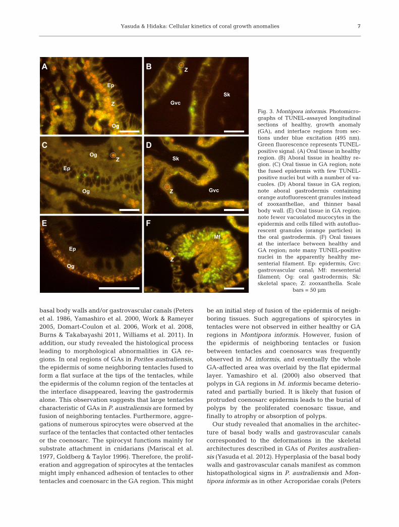
Few TUNEL-positive nuclei were observed in bothoral and aboral regions of apparently healthy regionsof Montipora informis (Fig. 3A, B). TUNEL-positivecells were also rare in both oral and aboral regions ofthe GAs (Fig. 3C−E). In some oral regions of GAs, thesurface of epidermis was fused leaving the epidermislaid between 2 layers of gastrodermis (Fig. 3C). Thistype of epidermis with oral gastrodermis on bothsides was observed in all GA samples. In other oralregions in the GAs, fewer vacuolated mucus secre-tory cells were observed in the epidermis, and thegastrodermis contained orange autofluorescentgranules but few zooxanthellae (Fig. 3E). In the abo-ral regions of GAs, thinner basal body walls contain-ing numerous orange granules in the aboral gastro-dermis were visible (Fig. 3D). Filamentous algae/fungi occasionally invaded the skeletal space of bothapparently healthy and GA regions. Aggregations oflabeled nuclei were occasionally seen in the mesen-terial filaments of apparently healthy polyps at theinterface between healthy and GA regions (Fig. 3F; 3of 3 specimens). They were rare in the aboral regionat the interface between apparently healthy and GAregions.
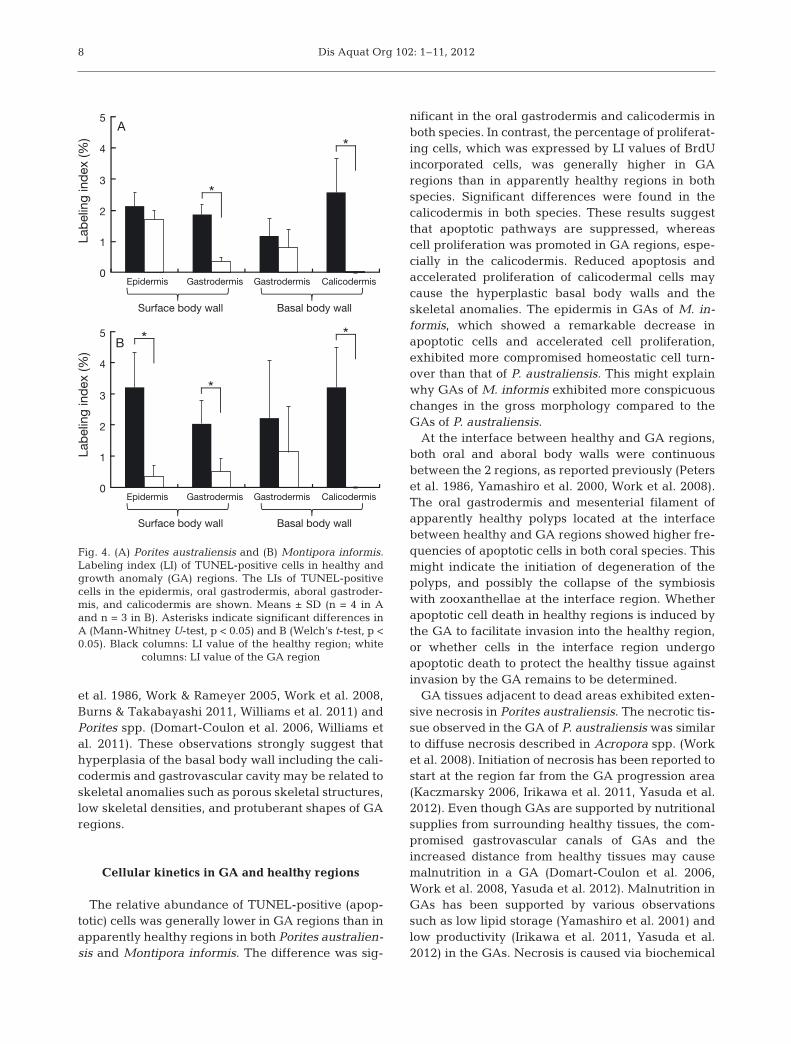
Significant differences in apoptotic cells werefound between healthy and GA regions in some tis-sues but not in all 4 tissues (p < 0.05, Fig. 4). In Po ri -tes australiensis, the LI values for TUNEL-positivecells in the oral gastrodermis and the calicodermis inapparently healthy regions were significantly higherthan those in the GAs (Mann-Whitney U-test, p <0.05; Fig. 4A). In Montipora informis, the LI values ofTUNEL-positive cells of the surface body wall (epi-dermis and gastrodermis) and the calicodermis inapparently healthy regions were significantly higherthan those in the GAs (Welch’s t-test, p < 0.05;Fig. 4B).
Detection of proliferating cells
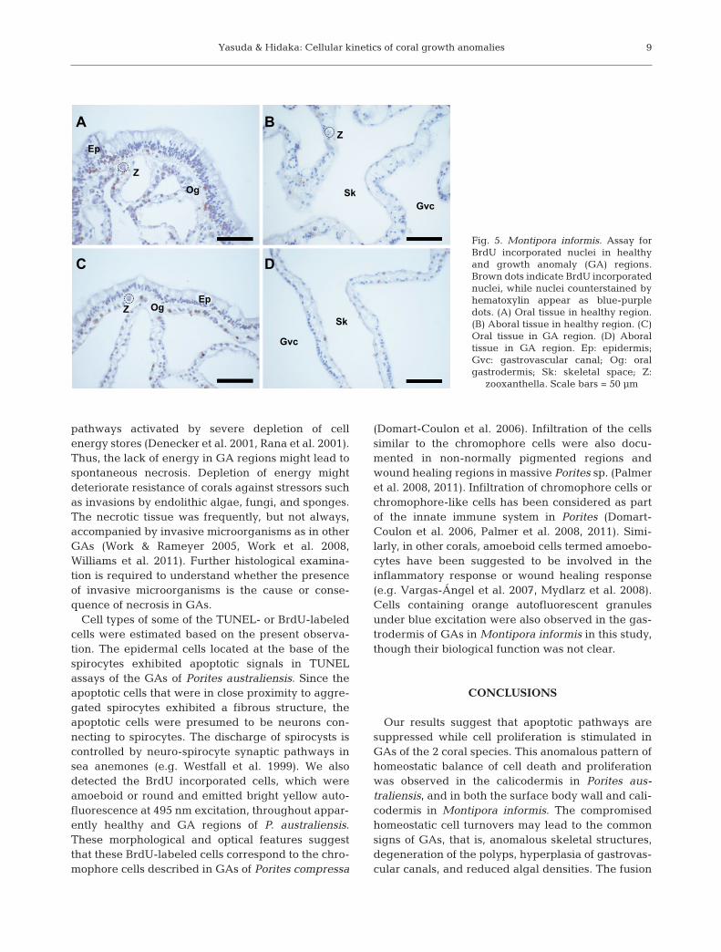
Host and symbiont nuclei with incorporated BrdUwere scattered in both healthy and GA tissues inPorites australiensis and Montipora informis (Fig. 5).BrdU incorporated chromophore cells were occasion-ally seen in both apparently healthy and GA regionsof P. australiensis. Clusters of BrdU incorporated cellswere not observed except in gonads in the appar-ently healthy tissue of M. informis. No BrdU incorpo-rated cells were detected in the negative control
5
Dis Aquat Org 102: 1–11, 2012
slides that had been incubated with phosphate-buffered saline instead of the anti-BrdU antibody(images not shown).
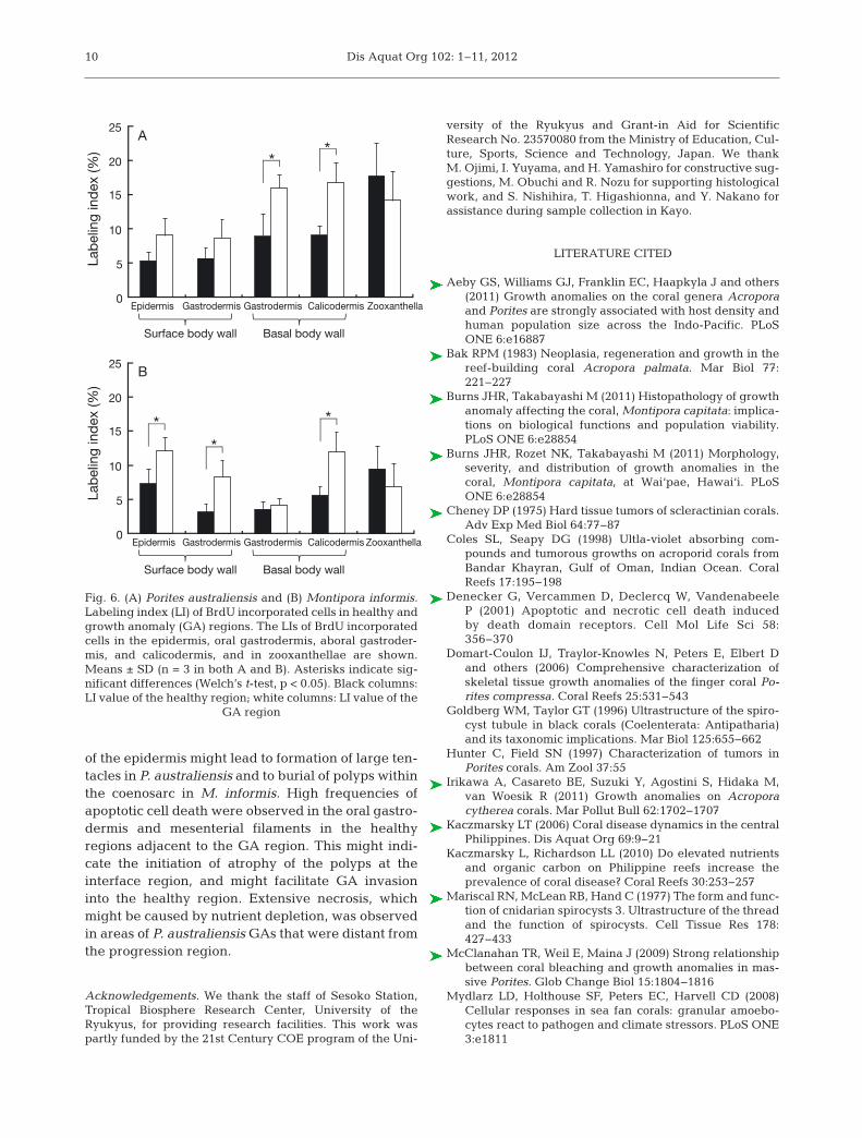
The percentage of proliferating host cells (BrdUincorporated host cells) was higher in GA regionsthan in apparently healthy regions, with significantdifferences in 2 of 4 tissue layers in Porites aus-traliensis and 3 of 4 tissue layers in Montiporainformis (Welch’s t-test, p < 0.05, Fig. 6). The LI val-ues for BrdU incorporated host nuclei in the basalbody wall (aboral gastrodermis and calicodermis)in GA regions were significantly higher than thosein apparently healthy regions of P. australiensis(Welch’s t-test, p < 0.05; Fig. 6A). The LI values forBrdU incorporated nuclei of host cells of the surfacebody wall (epidermis and oral gastrodermis) and cal-icodermis were significantly higher in GA regionsthan those in apparently healthy regions of M. in for -mis (Welch’s t-test, p < 0.05; Fig. 6B). The LI valuesfor BrdU-labeled nuclei of zooxanthellae were simi-lar in both healthy and GA tissues in both P. austra -liensis and M. informis.
DISCUSSION
Anomalies of skeletal and polyp structures in GA regions
GAs in Porites australiensis and Montipora in for -mis exhibited protuberant shapes due to newly-developed coenosteum/coenosarc tissue. Whilepolyp structures were more or less retained in GAs inP. australiensis, they were apparently absent due tothe newly-developed coenosteum/coenosarc tissuein GAs of M. informis. These findings are consistentwith previous observations of GAs in other Poritesspp. (Hunter & Field 1997, Domart-Coulon et al.2006, McClanahan et al. 2009, Stimson 2010, Yasudaet al. 2012) and in other Montipora spp. (Yamashiroet al. 2000, Burns et al. 2011, Williams et al. 2011).
Our histological study of GAs in 2 coral speciesdetected the common histopathological hallmarks inGA tissue across coral genera, viz. no or a reducednumber of polyps, deformed polyp structure with dis-organized mesenterial filaments, and hyperplastic
6
A B
C
F
D
E
ChZ
Ep
Og
Z
Ep
Og
Z
Gvc
Sk
Z
Ch
Ep
Og
Ch
Ep
Og
Sp
Ch
Omc
Fig. 2. Porites australiensis. Photomicro-graphs of TUNEL-assayed longitudinalsections of healthy, interface, and growthanomaly (GA) regions. Sections wereobserved under blue excitation (495 nm)except in B, where DAPI-stained nucleiunder UV excitation are shown. Greenfluorescence and blue fluorescence represent TUNEL-positive signals andDAPI-stained nuclei, respectively. (A)Oral tissue in healthy region, visualizedby TUNEL assay. (B) DAPI-stained nucleiof the same area shown in A. (C) Aboraltissue in healthy region. (D) Tentacle of apolyp at the interface between healthyand GA regions; note many TUNEL-pos-itive nuclei in the oral gastrodermis. (E)Tentacle tip of a polyp in the GA region;note the aggregation of TUNEL-positivenuclei in the epidermis under a cluster ofspirocytes containing spirocysts (orangefluorescent ellip ses). (F) Oral tissue inGA region facing a dead area; note lysedcells and organic matrix componentsassociated with filamentous algae/fungi(arrowhead). Ch: chromo phore cells; Ep:epidermis; Gvc: gastrovascular canal;Og: oral gastrodermis; Omc: organicmatrix components; Sk: skeletal space;Sp: spirocytes; Z: zooxanthella. Scale
bar = 50 µm
Yasuda & Hidaka: Cellular kinetics of coral growth anomalies
basal body walls and/or gastrovascular canals (Peterset al. 1986, Yamashiro et al. 2000, Work & Rameyer2005, Domart-Coulon et al. 2006, Work et al. 2008,Burns & Takabayashi 2011, Williams et al. 2011). Inaddition, our study revealed the histological processleading to morphological abnormalities in GA re -gions. In oral regions of GAs in Porites australiensis,the epidermis of some neighboring tentacles fused toform a flat surface at the tips of the tentacles, whilethe epidermis of the column region of the tentacles atthe interface disappeared, leaving the gastrodermisalone. This observation suggests that large tentaclescharacteristic of GAs in P. australiensis are formed byfusion of neighboring tentacles. Furthermore, aggre-gations of numerous spirocytes were observed at thesurface of the tentacles that contacted other tentaclesor the coenosarc. The spirocyst functions mainly forsubstrate attachment in cnidarians (Mariscal et al.1977, Goldberg & Taylor 1996). Therefore, the prolif-eration and aggregation of spirocytes at the tentaclesmight imply enhanced adhesion of tentacles to othertentacles and coenosarc in the GA region. This might
be an initial step of fusion of the epidermis of neigh-boring tissues. Such aggregations of spirocytes intentacles were not observed in either healthy or GAregions in Montipora informis. However, fusion ofthe epidermis of neighboring tentacles or fusionbetween tentacles and coenosarcs was frequentlyobserved in M. informis, and eventually the wholeGA-affected area was overlaid by the flat epidermallayer. Yamashiro et al. (2000) also observed thatpolyps in GA regions in M. informis became deterio-rated and partially buried. It is likely that fusion ofprotruded coenosarc epidermis leads to the burial ofpolyps by the proliferated coenosarc tissue, andfinally to atrophy or absorption of polyps.
Our study revealed that anomalies in the architec-ture of basal body walls and gastrovascular canalscorresponded to the deformations in the skeletalarchitectures described in GAs of Porites australien-sis (Yasuda et al. 2012). Hyperplasia of the basal bodywalls and gastrovascular canals manifest as commonhistopathological signs in P. australiensis and Mon-tipora informis as in other Acroporidae corals (Peters
7
A B
C D
E F
Z
Ep
Og
Z
GvcSk
Og
Og
ZEp
Z Gvc
Sk
Og
Ep
Z
Mf
Fig. 3. Montipora informis. Photomicro-graphs of TUNEL-assayed longitudinalsections of healthy, growth anomaly(GA), and interface regions from sec-tions under blue excitation (495 nm).Green fluorescence represents TUNEL-positive signal. (A) Oral tissue in healthyregion. (B) Aboral tissue in healthy re -gion. (C) Oral tissue in GA region; notethe fused epidermis with few TUNEL-positive nuclei but with a number of va -cuoles. (D) Aboral tissue in GA region;note aboral gastrodermis containingorange autofluorescent granules insteadof zooxanthellae, and thinner basalbody wall. (E) Oral tissue in GA region;note fewer vacuolated mucocytes in theepidermis and cells filled with autofluo-rescent granules (orange particles) inthe oral gastrodermis. (F) Oral tissuesat the interface between healthy andGA region; note many TUNEL- positivenuclei in the apparently healthy me -senterial filament. Ep: epidermis; Gvc:gastrovascular canal; Mf: mesenterialfilament; Og: oral gastrodermis; Sk:skeletal space; Z: zooxanthella. Scale
bars = 50 µm
Dis Aquat Org 102: 1–11, 2012
et al. 1986, Work & Rameyer 2005, Work et al. 2008,Burns & Takabayashi 2011, Williams et al. 2011) andPorites spp. (Domart-Coulon et al. 2006, Williams etal. 2011). These observations strongly suggest thathyperplasia of the basal body wall including the cali-codermis and gastrovascular cavity may be related toskeletal anomalies such as porous skeletal structures,low skeletal densities, and protuberant shapes of GAregions.
Cellular kinetics in GA and healthy regions
The relative abundance of TUNEL-positive (apop-totic) cells was generally lower in GA regions than inapparently healthy regions in both Porites australien-sis and Montipora informis. The difference was sig-
nificant in the oral gastrodermis and calicodermis inboth species. In contrast, the percentage of proliferat-ing cells, which was expressed by LI values of BrdUincorporated cells, was generally higher in GAregions than in apparently healthy regions in bothspecies. Significant differences were found in thecalicodermis in both species. These results suggestthat apoptotic pathways are suppressed, whereascell proliferation was promoted in GA regions, espe-cially in the calicodermis. Reduced apoptosis andaccelerated proliferation of calicodermal cells maycause the hyperplastic basal body walls and theskeletal anomalies. The epidermis in GAs of M. in -formis, which showed a remarkable decrease inapoptotic cells and accelerated cell proliferation,exhibited more compromised homeostatic cell turn-over than that of P. australiensis. This might explainwhy GAs of M. informis exhibited more conspicuouschanges in the gross morphology compared to theGAs of P. australiensis.
At the interface between healthy and GA regions,both oral and aboral body walls were continuousbetween the 2 regions, as reported previously (Peterset al. 1986, Yamashiro et al. 2000, Work et al. 2008).The oral gastrodermis and mesenterial filament ofapparently healthy polyps located at the interfacebetween healthy and GA regions showed higher fre-quencies of apoptotic cells in both coral species. Thismight indicate the initiation of degeneration of thepolyps, and possibly the collapse of the symbiosiswith zooxanthellae at the interface region. Whetherapoptotic cell death in healthy regions is induced bythe GA to facilitate invasion into the healthy region,or whether cells in the interface region undergoapoptotic death to protect the healthy tissue againstinvasion by the GA remains to be determined.
GA tissues adjacent to dead areas exhibited exten-sive necrosis in Porites australiensis. The necrotic tis-sue observed in the GA of P. australiensis was similarto diffuse necrosis described in Acropora spp. (Worket al. 2008). Initiation of necrosis has been reported tostart at the region far from the GA progression area(Kaczmarsky 2006, Irikawa et al. 2011, Yasuda et al.2012). Even though GAs are supported by nutritionalsupplies from surrounding healthy tissues, the com-promised gastrovascular canals of GAs and theincreased distance from healthy tissues may causemalnutrition in a GA (Domart-Coulon et al. 2006,Work et al. 2008, Yasuda et al. 2012). Malnutrition inGAs has been supported by various observationssuch as low lipid storage (Yamashiro et al. 2001) andlow productivity (Irikawa et al. 2011, Yasuda et al.2012) in the GAs. Necrosis is caused via biochemical
8
GastrodermisEpidermis Gastrodermis Calicodermis
Surface body wall Basal body wall
A
Lab
elin
g in
dex
(%)
*
*
0
1
2
3
4
5
B
Lab
elin
g in
dex
(%)
*
*
*
GastrodermisEpidermis Gastrodermis Calicodermis
Surface body wall Basal body wall
0
1
2
3
4
5
Fig. 4. (A) Porites australiensis and (B) Montipora informis.Labeling index (LI) of TUNEL-positive cells in healthy andgrowth anomaly (GA) regions. The LIs of TUNEL-positivecells in the epidermis, oral gastrodermis, aboral gastroder-mis, and calicodermis are shown. Means ± SD (n = 4 in Aand n = 3 in B). Asterisks indicate significant differences inA (Mann-Whitney U-test, p < 0.05) and B (Welch’s t-test, p <0.05). Black columns: LI value of the healthy region; white
columns: LI value of the GA region
Yasuda & Hidaka: Cellular kinetics of coral growth anomalies
pathways activated by severe depletion of cellenergy stores (Denecker et al. 2001, Rana et al. 2001).Thus, the lack of energy in GA regions might lead tospontaneous necrosis. Depletion of energy mightdeteriorate resistance of corals against stressors suchas invasions by endolithic algae, fungi, and sponges.The necrotic tissue was frequently, but not always,accompanied by invasive microorganisms as in otherGAs (Work & Rameyer 2005, Work et al. 2008,Williams et al. 2011). Further histological examina-tion is required to understand whether the presenceof invasive microorganisms is the cause or conse-quence of necrosis in GAs.
Cell types of some of the TUNEL- or BrdU-labeledcells were estimated based on the present observa-tion. The epidermal cells located at the base of thespirocytes exhibited apoptotic signals in TUNELassays of the GAs of Porites australiensis. Since theapoptotic cells that were in close proximity to aggre-gated spirocytes exhibited a fibrous structure, theapoptotic cells were presumed to be neurons con-necting to spirocytes. The discharge of spirocysts iscontrolled by neuro-spirocyte synaptic pathways insea anemones (e.g. Westfall et al. 1999). We alsodetected the BrdU incorporated cells, which wereamoeboid or round and emitted bright yellow auto-fluorescence at 495 nm excitation, throughout appar-ently healthy and GA regions of P. australiensis.These morphological and optical features suggestthat these BrdU-labeled cells correspond to the chro-mophore cells described in GAs of Porites compressa
(Domart-Coulon et al. 2006). Infiltration of the cellssimilar to the chromophore cells were also docu-mented in non-normally pigmented regions andwound healing regions in massive Porites sp. (Palmeret al. 2008, 2011). Infiltration of chromophore cells orchromophore-like cells has been considered as partof the innate immune system in Porites (Domart-Coulon et al. 2006, Palmer et al. 2008, 2011). Simi-larly, in other corals, amoeboid cells termed amoebo-cytes have been suggested to be involved in theinflammatory response or wound healing response(e.g. Vargas-Ángel et al. 2007, Mydlarz et al. 2008).Cells containing orange autofluorescent granulesunder blue excitation were also observed in the gas-trodermis of GAs in Montipora informis in this study,though their biological function was not clear.
CONCLUSIONS
Our results suggest that apoptotic pathways aresuppressed while cell proliferation is stimulated inGAs of the 2 coral species. This anomalous pattern ofhomeostatic balance of cell death and proliferationwas observed in the calicodermis in Porites aus-traliensis, and in both the surface body wall and cali-codermis in Montipora informis. The compromisedhomeostatic cell turnovers may lead to the commonsigns of GAs, that is, anomalous skeletal structures,degeneration of the polyps, hyperplasia of gastrovas-cular canals, and reduced algal densities. The fusion
9
A B
C D
Z
Ep
Og
ZEp
Og
Z
Gvc
Sk
GvcSk
Fig. 5. Montipora informis. Assay forBrdU incorporated nuclei in healthyand growth anomaly (GA) regions.Brown dots indicate BrdU incorporatednuclei, while nuclei counterstained byhematoxylin appear as blue-purpledots. (A) Oral tissue in healthy region.(B) Aboral tissue in healthy region. (C)Oral tissue in GA region. (D) Aboraltissue in GA region. Ep: epidermis;Gvc: gastrovascular canal; Og: oralgastrodermis; Sk: skeletal space; Z:
zooxanthella. Scale bars = 50 µm
Dis Aquat Org 102: 1–11, 201210
of the epidermis might lead to formation of large ten-tacles in P. australiensis and to burial of polyps withinthe coenosarc in M. informis. High frequencies ofapoptotic cell death were observed in the oral gastro-dermis and mesenterial filaments in the healthyregions adjacent to the GA region. This might indi-cate the initiation of atrophy of the polyps at theinterface region, and might facilitate GA invasioninto the healthy region. Extensive necrosis, whichmight be caused by nutrient depletion, was observedin areas of P. australiensis GAs that were distant fromthe progression region.
Acknowledgements. We thank the staff of Sesoko Station,Tropical Biosphere Research Center, University of theRyukyus, for providing research facilities. This work waspartly funded by the 21st Century COE program of the Uni-
versity of the Ryukyus and Grant-in Aid for ScientificResearch No. 23570080 from the Ministry of Education, Cul-ture, Sports, Science and Technology, Japan. We thankM. Ojimi, I. Yuyama, and H. Yamashiro for constructive sug-gestions, M. Obuchi and R. Nozu for supporting histologicalwork, and S. Nishihira, T. Higashionna, and Y. Nakano forassistance during sample collection in Kayo.
LITERATURE CITED
Aeby GS, Williams GJ, Franklin EC, Haapkyla J and others(2011) Growth anomalies on the coral genera Acroporaand Porites are strongly associated with host density andhuman population size across the Indo-Pacific. PLoSONE 6: e16887
Bak RPM (1983) Neoplasia, regeneration and growth in thereef-building coral Acropora palmata. Mar Biol 77: 221−227
Burns JHR, Takabayashi M (2011) Histopathology of growthanomaly affecting the coral, Montipora capitata: implica-tions on biological functions and population viability.PLoS ONE 6: e28854
Burns JHR, Rozet NK, Takabayashi M (2011) Morphology,severity, and distribution of growth anomalies in thecoral, Montipora capitata, at Wai‘pae, Hawai‘i. PLoSONE 6: e28854
Cheney DP (1975) Hard tissue tumors of scleractinian corals.Adv Exp Med Biol 64: 77−87
Coles SL, Seapy DG (1998) Ultla-violet absorbing com-pounds and tumorous growths on acroporid corals fromBandar Khayran, Gulf of Oman, Indian Ocean. CoralReefs 17: 195−198
Denecker G, Vercammen D, Declercq W, VandenabeeleP (2001) Apoptotic and necrotic cell death inducedby death domain receptors. Cell Mol Life Sci 58: 356−370
Domart-Coulon IJ, Traylor-Knowles N, Peters E, Elbert Dand others (2006) Comprehensive characterization ofskeletal tissue growth anomalies of the finger coral Po -rites compressa. Coral Reefs 25: 531−543
Goldberg WM, Taylor GT (1996) Ultrastructure of the spiro-cyst tubule in black corals (Coelenterata: Antipatharia)and its taxonomic implications. Mar Biol 125: 655−662
Hunter C, Field SN (1997) Characterization of tumors inPorites corals. Am Zool 37: 55
Irikawa A, Casareto BE, Suzuki Y, Agostini S, Hidaka M,van Woesik R (2011) Growth anomalies on Acroporacytherea corals. Mar Pollut Bull 62: 1702−1707
Kaczmarsky LT (2006) Coral disease dynamics in the centralPhilippines. Dis Aquat Org 69: 9−21
Kaczmarsky L, Richardson LL (2010) Do elevated nutrientsand organic carbon on Philippine reefs increase theprevalence of coral disease? Coral Reefs 30: 253−257
Mariscal RN, McLean RB, Hand C (1977) The form and func-tion of cnidarian spirocysts 3. Ultrastructure of the threadand the function of spirocysts. Cell Tissue Res 178: 427−433
McClanahan TR, Weil E, Maina J (2009) Strong relationshipbetween coral bleaching and growth anomalies in mas-sive Porites. Glob Change Biol 15: 1804−1816
Mydlarz LD, Holthouse SF, Peters EC, Harvell CD (2008)Cellular responses in sea fan corals: granular amoebo-cytes react to pathogen and climate stressors. PLoS ONE3: e1811
A
Lab
elin
g in
dex
(%)
Gastrodermis Gastrodermis Calicodermis
Surface body wall Basal body wall
Epidermis Zooxanthella0
5
10
15
20
25
**
B
Lab
elin
g in
dex
(%)
0
5
10
15
20
25
Gastrodermis Gastrodermis Calicodermis
Surface body wall Basal body wall
Epidermis Zooxanthella
**
*
Fig. 6. (A) Porites australiensis and (B) Montipora informis.Labeling index (LI) of BrdU incorporated cells in healthy andgrowth anomaly (GA) regions. The LIs of BrdU incorporatedcells in the epidermis, oral gastrodermis, aboral gastroder-mis, and calicodermis, and in zooxanthellae are shown.Means ± SD (n = 3 in both A and B). Asterisks indicate sig-nificant differences (Welch’s t-test, p < 0.05). Black columns:LI value of the healthy region; white columns: LI value of the
GA region
Yasuda & Hidaka: Cellular kinetics of coral growth anomalies 11
Palmer CV, Mydlarz LD, Willis BL (2008) Evidence of aninflammatory-like response in non-normally pigmentedtissues of two scleractinian corals. Proc R Soc Lond B BiolSci 275: 2687−2693
Palmer CV, Traylor-Knowles NG, Willis BL, Bythell JC(2011) Corals use similar immune cells and wound-heal-ing processes as those of higher organisms. PLoS ONE 6: e23992
Peters EC, Halas JC, McCarty HB (1986) Calicoblastic neo-plasms in Acropora palmata, with a review of reports onanomalies of growth and form in corals. J Natl CancerInst 76: 895−912
Rana A, Sathyanarayana P, Lieberthal W (2001) Role ofapoptosis of renal tubular cells in acute renal failure: therapeutic implications. Apoptosis 6: 83−102
Squires DF (1965) Neoplasia in a coral? Science 148: 503−505Stimson J (2010) Ecological characterization of coral growth
anomalies on Porites compressa in Hawai‘i. Coral Reefs30: 133−142
Sutherland KP, Porter JW, Torres C (2004) Disease andimmunity in Caribbean and Indo-Pacific zooxanthellatecorals. Mar Ecol Prog Ser 266: 273−302
Vargas-Ángel B, Peters EC, Kramarsky-Winter E, GilliamDS, Dodge RE (2007) Cellular reactions to sedimentationand temperature stress in the Caribbean coral Montas-traea cavernosa. J Invertebr Pathol 95: 140−145
Westfall JA, Landers DD, Mccallum JD (1999) Ultrastructureof neuro-spirocyte synapses in the sea anemone Aiptasiapallida (Cnidaria, Anthozoa, Zoantharia). J Morphol 241: 165−173
Williams GJ, Aeby GS, Cowie ROM, Davy SK (2010) Predic-tive modeling of coral disease distribution within a reefsystem. PLoS ONE 5: e9264
Williams GJ, Work TM, Aeby GS, Knapp IS, Davy SK (2011)Gross and microscopic morphology of lesions in Cnidariafrom Palmyra Atoll, Central Pacific. J Invertebr Pathol106: 165−173
Work TM, Rameyer RA (2005) Characterizing lesions incorals from American Samoa. Coral Reefs 24: 384−390
Work TM, Aeby GS, Coles SL (2008) Distribution and mor-phology of growth anomalies in Acropora from across theIndo-Pacific. Dis Aquat Org 78: 255−264
Yamashiro H, Yamamoto M, van Woesik R (2000) Tumor for-mation on the coral Montipora informis. Dis Aquat Org41: 211−217
Yamashiro H, Oku H, Onaga K, Iwasaki H, Takara K (2001)Coral tumors store reduced level of lipids. J Exp Mar BiolEcol 265: 171−179
Yasuda N, Nakano Y, Yamashiro H, Hidaka M (2012) Skele-tal structure and progression of growth anomalies inPorites australiensis in Okinawa, Japan. Dis Aquat Org97: 237−247
Editorial responsibility: Garriet Smith, Aiken, South Carolina, USA
Submitted: June 4, 2012; Accepted: August 30, 2012Proofs received from author(s): November 28, 2012