cellular pathology prof orla sheils causes of disease adaptive responses 2nd year pathology 2011
TRANSCRIPT
Cellular PathologyProf Orla Sheils
Causes of Disease Adaptive Responses
2nd year Pathology 2011
2nd year Pathology 2011
References, Reading and Websites
Pathologic Basis of Disease - Robbins.
Cell, Tissue and Disease. The Basis of Pathology- Woolf
Pathology Secrets – Damjanov. Chapters 1 and 7
http://www.pathguy.com
http://medlib.med.utah.edu/WebPath/webpath.html

2nd year Pathology 2011
http://www.medicine.tcd.ie/Histopathology/courses/studentarea.htm
Lecture available on line at:

2nd year Pathology 2011
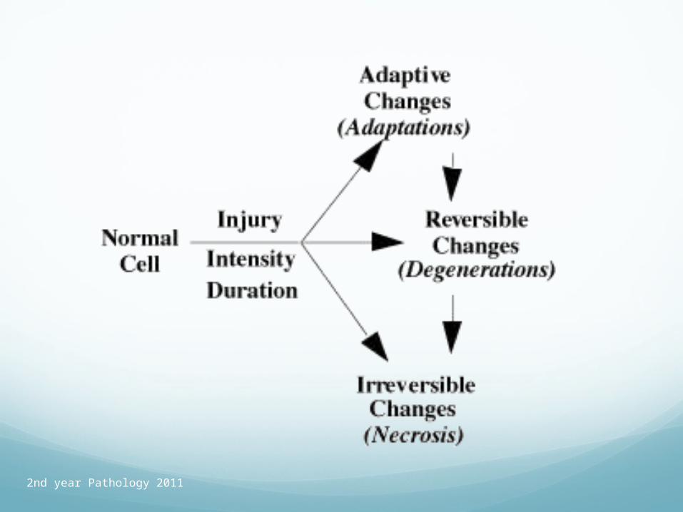
Causes of DiseaseDisease does not exist except as a reaction to
injury.
Concept of Homeostasis. “The Steady State” or equilibrium with the environment.
Cellular adaptation.
Physiologic.
Morphologic.
At the limits of cellular adaptation or in cases where adaptation is not possible then “cell injury” may occur.
2nd year Pathology 2011
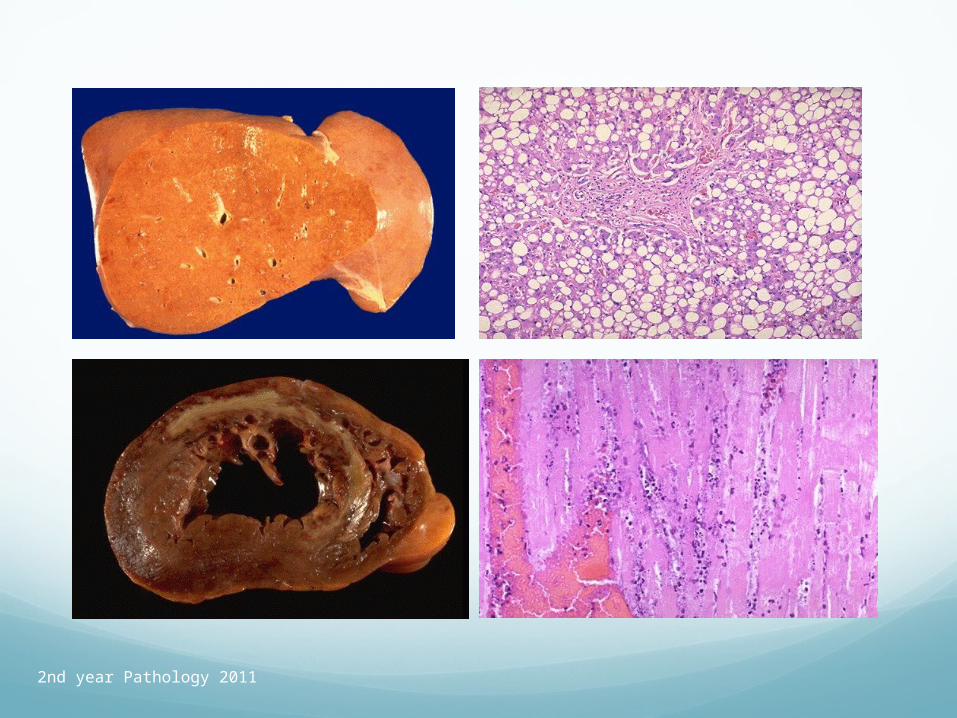
Cell injury
Reversible. Cell swelling/Hydropic change.
Fatty Change.
Irreversible.
Cell Death (Myocardial Infarction)
2nd year Pathology 2011
2nd year Pathology 2011

2nd year Pathology 2011
Types of Cell Injury
1) Oxygen Deprivation / Re-oxygenation • (Free radicals).
2) Physical Agents.
3) Chemical Agents / Drugs.
4) Infectious Agents.
5) Immunologic Reactions.
6) Genetic Derangements.
7) Nutritional Imbalances.
8) Aging (See next lecture)
2nd year Pathology 2011
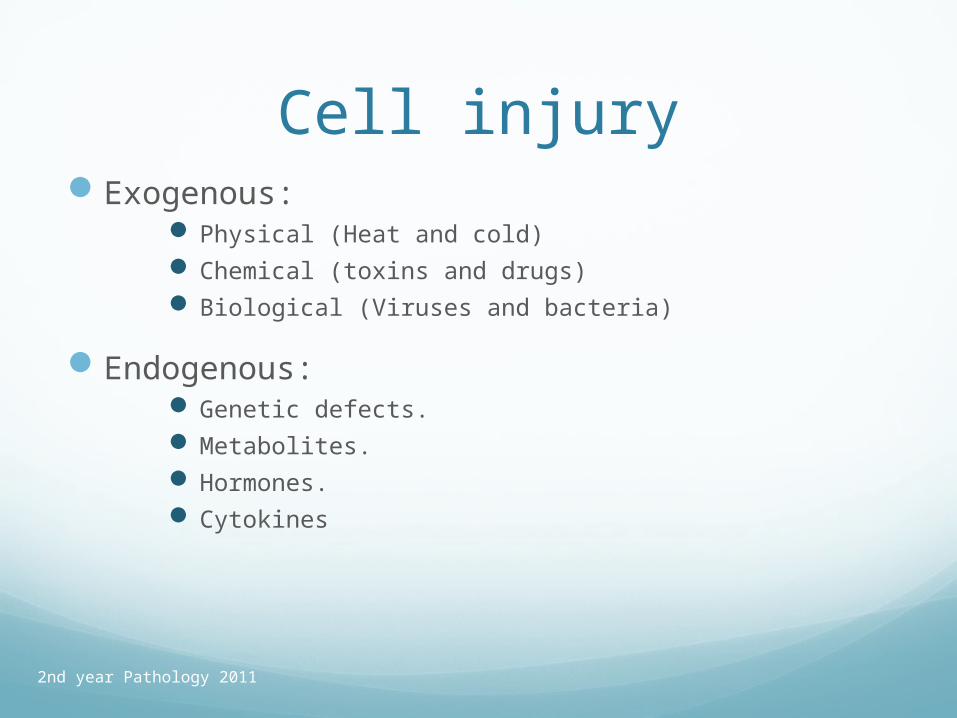
Cell injuryExogenous:
Physical (Heat and cold) Chemical (toxins and drugs) Biological (Viruses and bacteria)
Endogenous: Genetic defects. Metabolites. Hormones. Cytokines

2nd year Pathology 2011
1) Oxygen DeprivationTerms: Hypoxia/Anoxia.
Ischaemia.
Hypoxia is a reduction of the amount of oxygen delivered to cells. It is the most common cause of cell injury and death.
Ischaemia is a reduction in the perfusion of a body part or organ in relation to its needs.
2nd year Pathology 2011
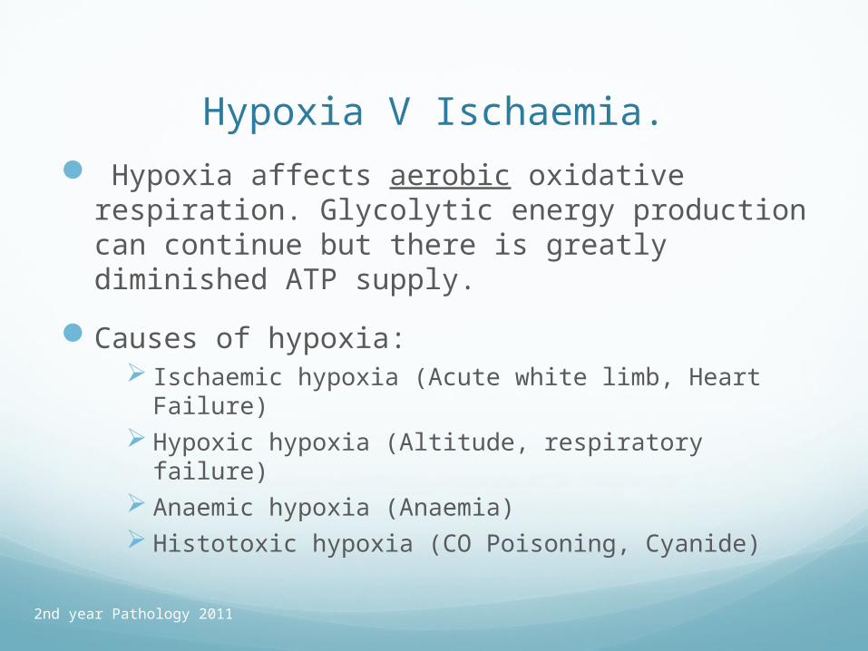
Hypoxia V Ischaemia.
Hypoxia affects aerobic oxidative respiration. Glycolytic energy production can continue but there is greatly diminished ATP supply.
Causes of hypoxia: Ischaemic hypoxia (Acute white limb, Heart Failure) Hypoxic hypoxia (Altitude, respiratory failure) Anaemic hypoxia (Anaemia) Histotoxic hypoxia (CO Poisoning, Cyanide)

2nd year Pathology 2011
CO poisoning- cherry pink skin discolouration
2nd year Pathology 2011
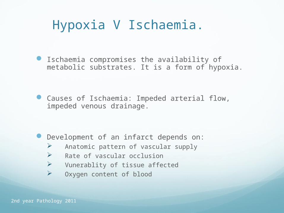
Hypoxia V Ischaemia.
Ischaemia compromises the availability of metabolic substrates. It is a form of hypoxia.
Causes of Ischaemia: Impeded arterial flow, impeded venous drainage.
Development of an infarct depends on: Anatomic pattern of vascular supply Rate of vascular occlusion Vunerablity of tissue affected Oxygen content of blood

2nd year Pathology 2011
Hypoxia Neurons: Frank necrosis after being deprived of
oxygen for 3-5 minutes at normal temperature clinically, brain damage follows much shorter intervals.
Heart muscle cells can last 30-60 minutes.
Liver cells and renal tubular cells can last for 1-2 hours without oxygen before they are irreversibly damaged ( but easy to replace.)
Skin fibroblasts can last for many hours.
2nd year Pathology 2011
2) Physical Agents.
Mechanical Trauma.
Extremes of Temperature.
Barotrauma.
Electric Shock.
Radiation.
2nd year Pathology 2011
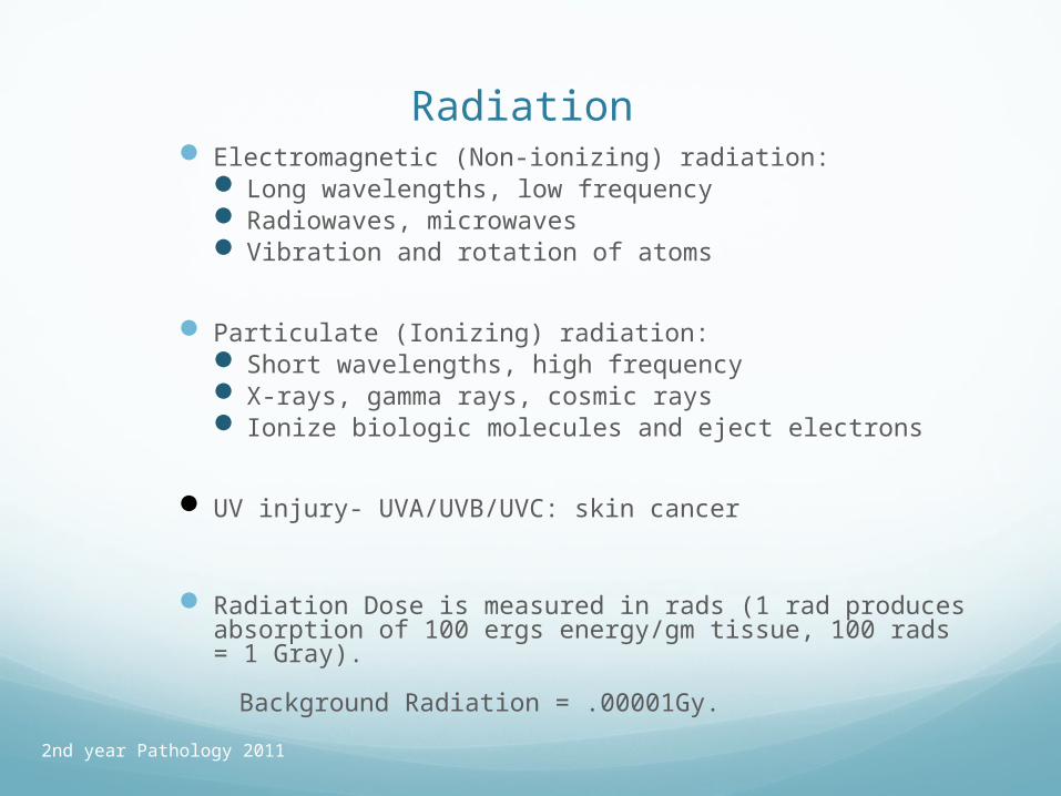
Radiation Electromagnetic (Non-ionizing) radiation:
Long wavelengths, low frequency Radiowaves, microwaves Vibration and rotation of atoms
Particulate (Ionizing) radiation: Short wavelengths, high frequency X-rays, gamma rays, cosmic rays Ionize biologic molecules and eject electrons
UV injury- UVA/UVB/UVC: skin cancer
Radiation Dose is measured in rads (1 rad produces absorption of 100 ergs energy/gm tissue, 100 rads = 1 Gray).
Background Radiation = .00001Gy.
2nd year Pathology 2011
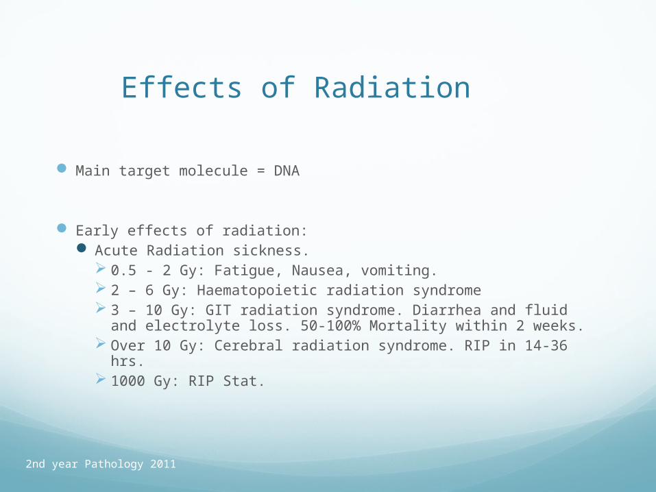
Effects of Radiation
Main target molecule = DNA
Early effects of radiation: Acute Radiation sickness.
0.5 - 2 Gy: Fatigue, Nausea, vomiting. 2 – 6 Gy: Haematopoietic radiation syndrome 3 – 10 Gy: GIT radiation syndrome. Diarrhea and fluid and
electrolyte loss. 50-100% Mortality within 2 weeks. Over 10 Gy: Cerebral radiation syndrome. RIP in 14-36 hrs. 1000 Gy: RIP Stat.
2nd year Pathology 2011
Late effects of radiation: Atrophy. Narrowing of blood vessels. Fibrosis. Inflammation Cataracts Carcinoma. Teratogenic.
2nd year Pathology 2011
3) Chemical Agents and Drugs
Hypertonic Solutions.
Oxygen.
Poisons: Arsenic, Cyanide, Mercury.
Environmental Pollutants.
Insecticides/ herbicides.
CO
Asbestos 1) Interstitial lung fibrosis. 2) Bronchogenic carcinoma. 3) Pleural Effusions. 4) Pleural plaques. 5) Mesotheliomas.
Recreational Drugs / C2H5OH
2nd year Pathology 2011
Chemical Injury
Biological molecules react like any other chemicals. Acids and alkalis hydrolyze membranes Poisons like mercuric ion tie up sulfhydryl groups and
destroy the cell. Formalin / formaldehyde crosslink amino groups on
proteins and nucleic acids. Histopathologists use this chemistry to “fix tissues”.
Current thinking is that most simple poisons that cause actual cell necrosis require activation to form free radicals. For example, carbon tetrachloride (old-fashioned cleaning fluid) is turned into CCl3
.- radical in the smooth endoplasmic reticulum of the liver.

2nd year Pathology 2011
Chemical Injury Other classic poisons affect the more vulnerable parts of
the cells.
Depending on the poison and dose, there may or may not be necrosis: Cell membranes: digitalis Oxidative phosphorylation: cyanide Ribosomes: toadstools Genes: chemotherapeutic agents Synapses: strychnine, ergot
2nd year Pathology 2011
4) Infectious Agents.Bacteria
Viruses
Fungi
Chlamydiae,Rickettsiae,Mycoplasma
Protozoa
Helminths
Ectoparasites
Bacteriophages, Plasmids
Prions.
2nd year Pathology 2011
How microrganisms cause disease
Entering cells
Releasing toxins
Damaging blood vessels
Inducing host responses with additional damage Suppuration Scarring Hypersensitivity reactions

2nd year Pathology 2011
Exotoxin Endotoxin
Secreted from living organism
Part of dead organism
Protein LPS
Elicits immune reaction No immune reaction (weak)
Heat labile Heat stable
2nd year Pathology 2011
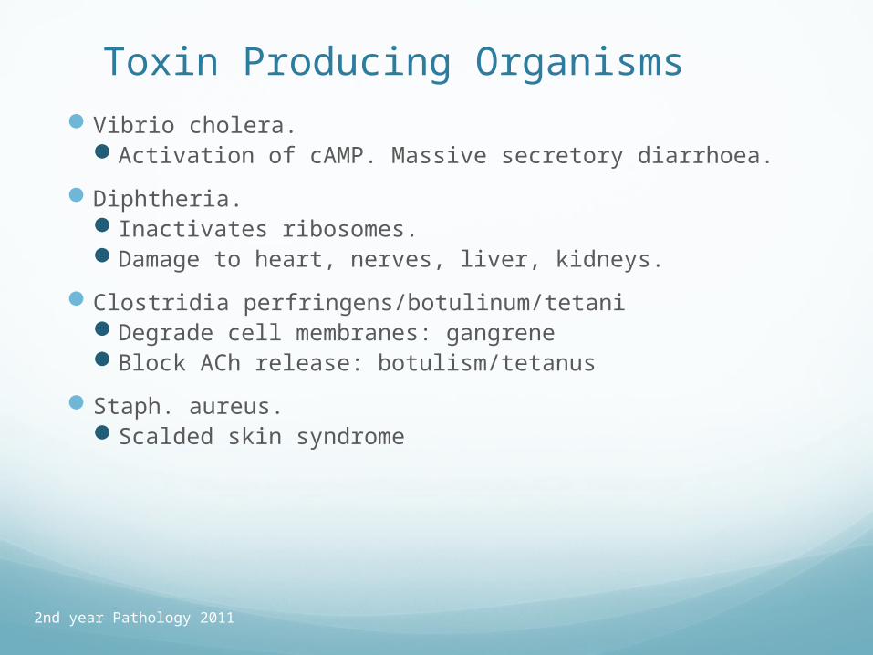
Toxin Producing OrganismsVibrio cholera.
Activation of cAMP. Massive secretory diarrhoea.
Diphtheria. Inactivates ribosomes. Damage to heart, nerves, liver, kidneys.
Clostridia perfringens/botulinum/tetaniDegrade cell membranes: gangreneBlock ACh release: botulism/tetanus
Staph. aureus.Scalded skin syndrome
2nd year Pathology 2011
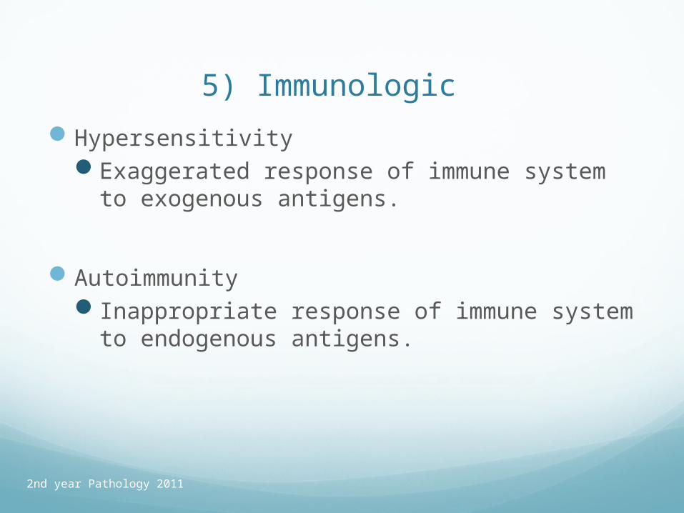
5) Immunologic Hypersensitivity
Exaggerated response of immune system to exogenous antigens.
AutoimmunityInappropriate response of immune system to
endogenous antigens.
2nd year Pathology 2011
6) Genetic Derangements
Chromosomal abnormalities.
Single gene disorders.

2nd year Pathology 2011
7) Nutritional Imbalances. Protein energy deficiency (PEM)
Marasmas. Muscle wasting, wrinkled skin, Hair loss. Kwashiorkor. Excess protein deficiency: Scaly skin, Swollen
abdomen (ascites), swollen ankles, Hypoalbuminemia.
Specific vitamin deficiencies Vit C: (ascorbic acid) Vit. D: Rickets, Osteomalacia. Vit A: Xeropthalmia, Bitot’s spots, keratomalacia, night blindness Niacin: Pellagra- Dermatitis, Diarrhoea, Dementia
Anorexia nervosa.
Dietary indiscretion – Cholesterol.
2nd year Pathology 2011
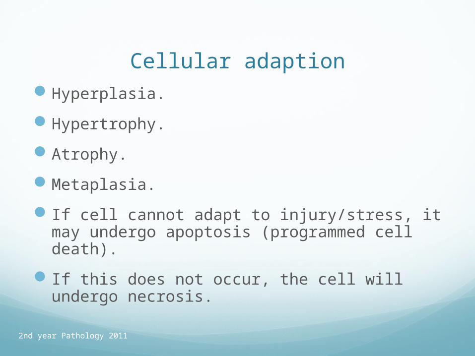
Cellular adaptionHyperplasia.
Hypertrophy.
Atrophy.
Metaplasia.
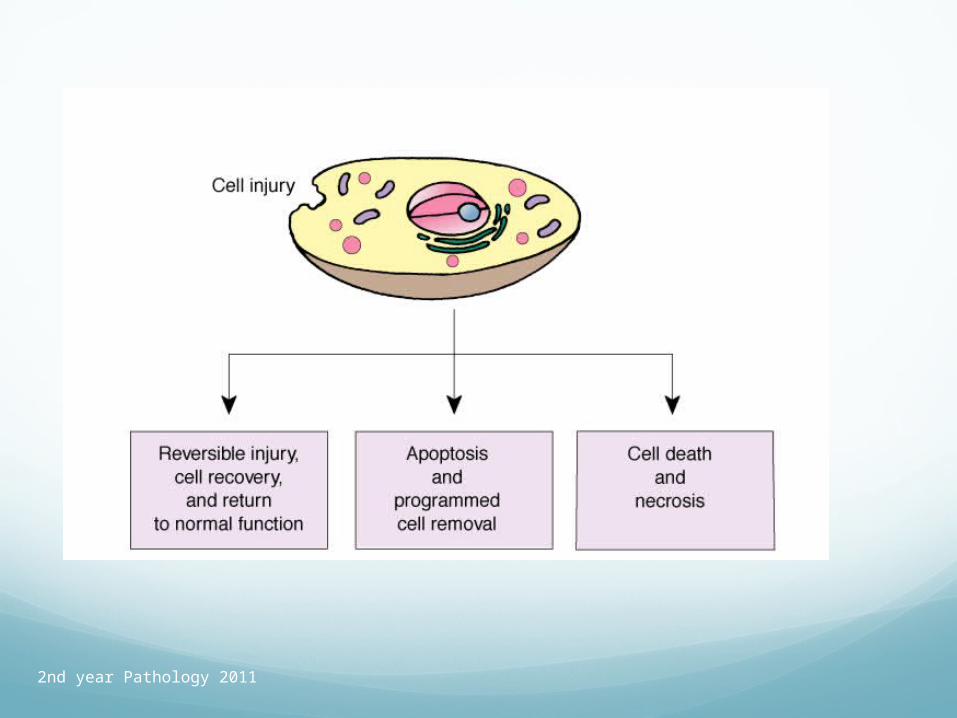
If cell cannot adapt to injury/stress, it may undergo apoptosis (programmed cell death).
If this does not occur, the cell will undergo necrosis.
2nd year Pathology 2011
Hyperplasia
An increase in the Number of cells in an organ or tissue.
Hyperplasia means cells growing more numerous.
Usually accompanied by hypertrophy.
Can only occur in cells capable of making new DNA (capable of division).

2nd year Pathology 2011
Physiologic Hyperplasia
A Demand - led physiological event
Hormonal: Endometrial proliferation after oestrogen stimulation.
Compensatory: Hyperplasia of liver after partial hepatectomy.
Breast and Thyroid at times of puberty and pregnancy
2nd year Pathology 2011
Pathologic Hyperplasia
Hyperoestrogenism and atypical endometrial hyperplasia.
Squamous hyperplasia induced by viruses. HPV (wart) virus.

2nd year Pathology 2011
Endometrial hyperplasia

2nd year Pathology 2011
Benign Prostatic Hyperplasia
MicroscopyMacroscopy

2nd year Pathology 2011
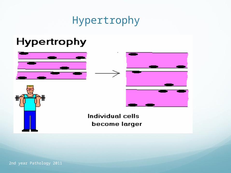
HypertrophyHYPERTROPHY: Increase in the sizes of
cells, and hence the size of the organ.
Often occurs in cells that have limited abilities to divide e.g. muscle
Physiological: Skeletal muscle hypertrophy due to exercise
Pathological: Hypertrophy of the overworked heart of an aerobic athlete, hypertension victim, or victim of aortic valve stenosis or other cardiac structural defect
2nd year Pathology 2011
Hypertrophy

2nd year Pathology 2011
Left Ventricular Hypertrophy
2nd year Pathology 2011
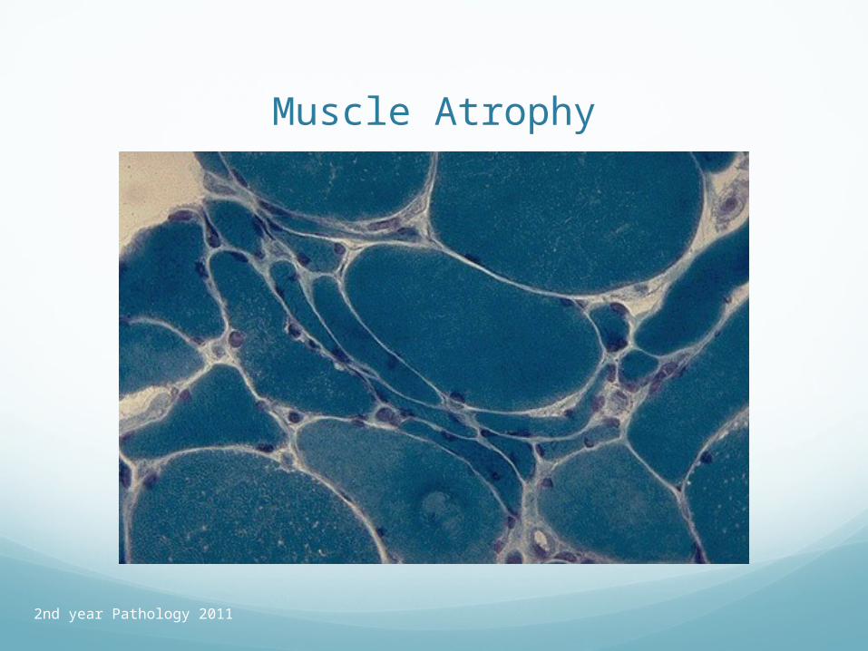
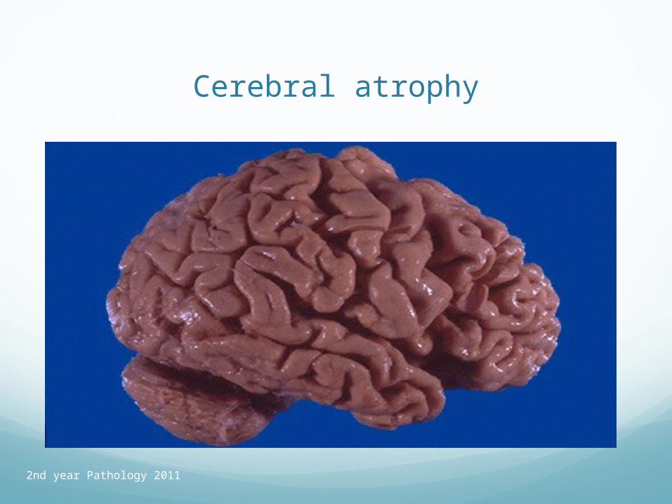
Atrophy
ATROPHY: "Shrinkage in the size of the cell by loss of cell substance" (Robbins), without the cell actually dying. When many cells each become smaller, the organ itself become smaller. Defined this way, atrophy is very reversible.

2nd year Pathology 2011
Causes of Atrophy
Disuse Atrophy - Workload.
Denervation atrophy.
blood supply.
Inadequate nutrition.
Loss of endocrine stimulation.
Senile atrophy.
Pressure/Involution.
2nd year Pathology 2011
Muscle Atrophy
2nd year Pathology 2011
Cerebral atrophy
2nd year Pathology 2011
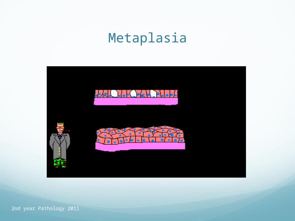
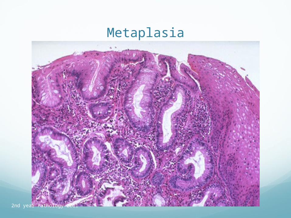
Metaplasia
METAPLASIA: (Adaptive) substitution of one type of adult or fully differentiated cell for another type of adult (or fully differentiated) cell. -Robbins.
"A reversible change in which one adult cell type (epithelial or mesenchymal) is replaced by another adult cell type." -Robbins.
"Conversion of a differentiated cell type into another" -- R&F.
2nd year Pathology 2011
Metaplasia
2nd year Pathology 2011
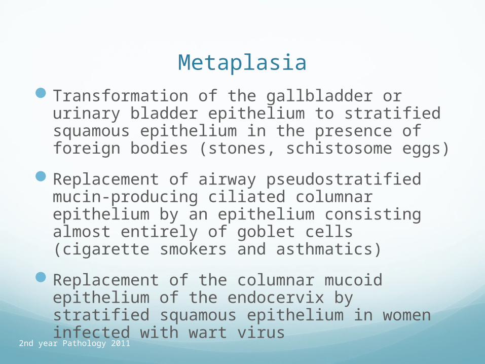
MetaplasiaTransformation of the gallbladder or urinary
bladder epithelium to stratified squamous epithelium in the presence of foreign bodies (stones, schistosome eggs)
Replacement of airway pseudostratified mucin-producing ciliated columnar epithelium by an epithelium consisting almost entirely of goblet cells (cigarette smokers and asthmatics)
Replacement of the columnar mucoid epithelium of the endocervix by stratified squamous epithelium in women infected with wart virus
2nd year Pathology 2011
Metaplasia
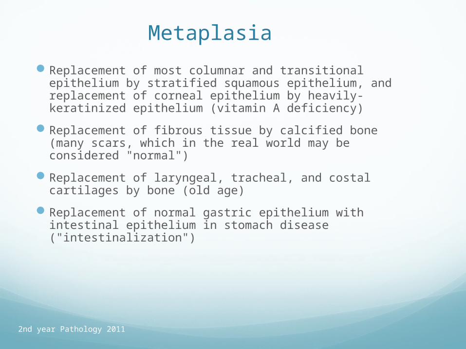
Replacement of most columnar and transitional epithelium by stratified squamous epithelium, and replacement of corneal epithelium by heavily-keratinized epithelium (vitamin A deficiency)
Replacement of fibrous tissue by calcified bone (many scars, which in the real world may be considered "normal")
Replacement of laryngeal, tracheal, and costal cartilages by bone (old age)
Replacement of normal gastric epithelium with intestinal epithelium in stomach disease ("intestinalization")
2nd year Pathology 2011
Metaplasia
2nd year Pathology 2011

2nd year Pathology 2011
AdaptationIf underlying stimulus is removed, cells can return to
normal.
Hyperplasia cell loss due to apoptosis normal number of cells
Hypertrophy lysosome ingestion of excess cell organelles normal cell size
Atrophy production of additional organelles normal cell size
Metaplasia differentiation back to original cell type
2nd year Pathology 2011
AdaptationIf stimulus persists, pathology results
Cell death
Loss of cell function
Malignant change
The latter is most likely to occur in the setting of metaplasia (of any type) which is often a significant risk factor for the development of carcinoma.
Often preceded by development of pre-invasive neoplastic change = dysplasia.

2nd year Pathology 2011
Summary
Causes of disease Types of injurious agents Hypoxia & Ischaemia Physical agents including Radiation Chemical injury Infectious agents Immune / Genetic / Nutritional
Mechanisms of cellular adaption Consequences