cellulose digestion and cellulase regulation distribution...
TRANSCRIPT
Vol. 56, No. 5APPLIED AND ENVIRONMENTAL MICROBIOLOGY, May 1990, p. 1221-12280099-2240/90/051221-08$02.00/0Copyright C 1990, American Society for Microbiology
Cellulose Digestion and Cellulase Regulation and Distribution inFibrobacter succinogenes subsp. succinogenes S85
LI HUANGt AND CECIL W. FORSBERG*Department of Microbiology, University of Guelph, Guelph, Ontario NIG 2WI, Canada
Received 25 August 1989/Accepted 14 February 1990
Fibrobacter succinogenes subsp. succinogenes S85 initiated growth on microcrystalline cellulose without a lagwhether inoculated from a glucose, ceUlobiose, or cellulose culture. During growth on celulose, there was noaccumulation of soluble carbohydrate. When the growth medium contained either glucose or ceUlobiose incombination with microcrystaline cellulose, there was a lag in cellulose digestion until all of the soluble sugarhad been utilized, suggesting an end product feedback mechanism that affects cellulose digestion. Cl-stimulatedcellobiosidase and periplasmic cellodextrinase were produced under all growth conditions tested, indicatingconstitutive synthesis. Both celobiosidases were cel associated until the stationary phase of growth, whereasproteins antigenically related to the Cl-stimulated ceUlobiosidase and a proportion of the endoglucanase werereleased into the extracellular culture fluid during growth, irrespective of the substrate. Immunoelectronmicroscopy of cells with a polyclonal antibody to Cl-stimulated ceUlobiosidase as the primary antibody and10-nm-diameter gold particles conj'ugated to goat anti-rabbit antibodies as the second antibody revealedprotrusions of the outer surface which were selectively labeled with gold, suggesting that Cl-stimulatedcelobiosidase was located on the protrusions. These data support the contention that the protrusions have arole in cellulose hydrolysis; however, this interpretation is complicated by reactivity of the antibodies with alarge number of other proteins that possess related antigenic epitopes.
Fibrobacter succinogenes subsp. succinogenes S85, for-merly Bacteroides succinogenes (27), is a major cellulolyticbacterium within the rumen (3, 33). One of the reasons for itspredominance is its ability to readily degrade various formsof crystalline cellulose (9, 10). However, the physiologicalmechanism by which the bacterium degrades cellulose isincompletely understood. Elucidation of the mechanism hasbeen tackled by purification and characterization of theenzymes involved in cellulose digestion. The enzymes puri-fied include two cellobiosidases, a Cl-stimulated cellobiosi-dase (14), a periplasmic cellodextrinase (13), and threeendoglucanases designated EG1, EG2, and EG3 (23, 25).Although we have detailed knowledge of the properties ofthese enzymes and their presence in Fibrobacter succino-genes subsp. succinogenes S85 (15), there is limited infor-mation on the regulation of cellulose digestion and of theenzymes involved in the process (9). Therefore, in a recentstudy by McGavin et al. (24), the regulation and distributionof the endoglucanases was examined. The major objective ofthe present study was to determine the effect of the carbonsource for growth on the rate of cellulose digestion and tocorrelate this with the production and distribution of cello-biosidase enzymes.
MATERIALS AND METHODSOrganism and growth conditions. F. succinogenes subsp.
succinogenes S85 was grown in a 500-ml round-bottom flaskwith gyratory shaking at 150 rpm at 37°C in the medium ofScott and Dehority (31) with the following modifications.Ammonium sulfate was the sole source of nitrogen. Glucose(0.5%, wt/vol), cellobiose (0.5%, wt/vol), Avicel microcrys-talline cellulose PH105 (0.3%, wt/vol; FMC Corp., Marcus
* Corresponding author.t Present address: Department of Biochemistry, School of Hy-
giene and Public Health, The Johns Hopkins University, Baltimore,MD 21205.
Hook, Pa.), and amorphous cellulose (acid-swollen cellu-lose; 0.2%, wt/vol; 30) were used individually or in combi-nation as carbon sources. Samples were removed for proc-essing at the times indicated.
Localization of cellulase components. The cellular distribu-tion of cellulase components of F. succinogenes subsp.succinogenes S85 grown under various conditions was ex-amined as described previously (12).Enzyme assays. All enzyme assays were conducted as
previously described (12).Analytical methods. Soluble sugars, i.e., glucose and cel-
lobiose, in culture fluids were measured by using the phenol-sulfuric acid method (4). Cellulose in samples was firstwashed repeatedly by sedimentation and suspension in wa-ter to remove soluble carbohydrates. Sedimented cellulosewas then solubilized in 67% (wt/vol) sulfuric acid as de-scribed by Updegraff (34) and quantified by using the phenol-sulfuric acid method for soluble carbohydrates (4). Glucosewas used as the standard. Protein was determined by themethod of Bradford (2), with bovine serum albumin as thestandard.
Electrophoresis. Sodium dodecyl sulfate-polyacrylamidegel electrophoresis was performed as described by Laemmli(18), with several modifications (13), by using a 10% acryl-amide separating gel.
Lectin binding. Fluorescence-labeled lectin GS-1 fromGriffonia simplicifolia (Vector Laboratories, Inc., Burlin-game, Calif.) was tested for binding to F. succinogenessubsp. succinogenes S85 by fluorescence microscopy (20).Antiserum preparation and immunoblotting. Antisera to
cellodextrinase and Cl-stimulated cellobiosidase were pre-pared as described elsewhere (15). Immunoblotting of thesamples prepared from various cultures was conducted asdescribed previously (15).Immunogold labeling of cells. For prefixation labeling, a
sample (0.5 ml) of an exponentially growing glucose orcellobiose culture (9 h) or a rapidly grown cellulose culture
1221
on July 27, 2018 by guesthttp://aem
.asm.org/
Dow
nloaded from

1222 HUANG AND FORSBERG
120
i--
E
D
E
.-80
a)
0
0
0
a)
A-0.8
E0.6
E
0-0.40
1- ~~~~~0-0.2 a-
0 10 20Time (h)
30
3.0
E
E
2.0 a) '
2 c
C) c0
-.
&) °CL
JoTime (h)
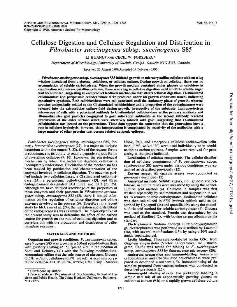
FIG. 1. Growth of F. succinogenes subsp. succinogenes S85 in batch cultures with glucose (A), cellobiose (B), Avicel microcrystallinecellulose (C), or amorphous cellulose (D) as the carbon source. Symbols: 0, total cellobiosidase activity; A, total protein concentration; 0,extracellular cellobiosidase activity; A, extracellular protein concentration; *, residual cellulose concentration.
(40 h) which contained a substantial amount of residualcellulose was harvested by centrifugation. With cellulose-grown cells, a low centrifugal force was maintained (2,000 x
g) to reduce sedimentation of unbound cells. The cell pelletwas suspended in 100 ,ul of 50-fold-diluted rabbit antibody toCl-stimulated cellobiosidase in 20 mM HEPES (N-2-hydrox-yethylpiperazine-N'-2-ethanesulfonic acid) buffer (pH 6.7)-0.8% (wt/vol) NaCl (HBS). After incubation at room tem-perature for 30 min, the sample was centrifuged and washedtwice in HBS. The washed cell pellet was suspended in 50 ,lIof 10-fold-diluted goat anti-rabbit immunoglobulin G (IgG)-gold conjugate (10-nm-diameter gold particles; Sigma Chem-ical Co., St. Louis, Mo.) in HBS supplemented with 0.2%(wt/vol) bovine serum albumin (HBS-BSA). Following incu-bation at room temperature for 30 min, the sample was
centrifuged and washed twice in HBS-BSA and once in 20mM HEPES buffer (pH 6.7). The cells were then fixed in0.5% (vol/vol) glutaraldehyde in 20 mM HEPES buffer (pH6.7). For postfixation labeling, a glutaraldehyde stock solu-tion (1%, wt/vol) in 20 mM HEPES buffer (pH 6.7) was
added to the culture to give a final concentration of 0.2%(vol/vol). After incubation at 4°C for 30 min, the sample was
centrifuged and washed twice in HBS. The cells were thensuspended in HBS containing 0.2 M NH4Cl and incubated at
room temperature for 30 min to inactivate residual reactivealdehyde groups. The samples was then centrifuged, washedtwice in HBS, and processed in the same way as forprefixation labeling. The control was a sample of cellstreated as described above, except that the first antibodywas replaced by preimmune serum or buffer.
Transmission electron microscopy. Transmission electronmicroscopy was performed in a way similar to that describedby Lam et al. (19). Equal volumes of immunogold-labeledcells and 1% (wt/vol) ammonium molybdate (pH 7) weremixed, and a carbon-Formvar-coated 200-mesh copper gridwas briefly floated on the surface. Excess solution wasremoved with filter paper. The samples were examined byusing a Philips 300 transmission electron microscope oper-ating at an accelerating voltage of 60 kV.
RESULTS
Growth and cellulase activity of cultures in medium con-taining a single carbon source. F. succinogenes subsp. suc-cinogenes S85 exhibited a generation time of 3.5 h whengrown in chemically defined medium with either glucose orcellobiose as the carbon source (Fig. IA and B). Growth oncrystalline cellulose was slower, with a generation time of 10
APPL. ENVIRON. MICROBIOL.
on July 27, 2018 by guesthttp://aem
.asm.org/
Dow
nloaded from

CELLOBIOSIDASES OF F. SUCCINOGENES 1223
TABLE 1. Specific activities of cellulolytic enzymes ofF. succinogenes subsp. succinogenes S85 grown
on various carbon sources'
Sp act (nmol- min-' mg of protein-')Carbon sources
Cellobiosidaseb Endoglu- Cello-canase biase
Glucose 219 (1.10 ± 0.03) 603 276Cellobiose 134 (1.08 ± 0.02) 296 368Amorphous cellulose 154 (ND) 439 210Avicel 90 (1.11 ± 0.02) 468 192
a Cells grown in synthetic medium (31) with glucose (0.5%, wt/vol),cellobiose (0.5%, wt/vol), or amorphous cellulose (approximately 0.2%,wt/vol) as the carbon source were harvested in the late exponential phase (10h), while cells grown with Avicel microcrystalline cellulose PH105 (0.3%,wt/vol) as the sole carbon source were harvested after complete digestion ofcellulose (50 h). The cells were sonicated for nine 20-s bursts with 1-minintervals on ice. The cellobiosidase activities were measured in the presenceof chloride.
b The values in parentheses represent the ratios of the cellobiosidaseactivity of the disrupted cell suspensions assayed in 0.2 M NaCl to thatassayed without NaCl. Each value is expressed as the mean and standarddeviation of six measurements. ND, Not determined.
h (Fig. 1C). An unexpected finding was that cells grew onamorphous cellulose (acid swollen) with a generation time of3.5 h, which was identical to that for cells grown on glucose(Fig. 1D).When cells were grown on glucose, cellobiose, or crystal-
line cellulose, the patterns of cellobiosidase synthesis weresimilar to those of cell biomass increase (Fig. 1A to C).However, the cellulose-grown culture exhibited low cellobi-osidase and cellobiase activities (Table 1). The glucose-grown culture exhibited the highest specific activities ofcellobiosidase and endoglucanase, whereas the cellobiose-grown culture expressed the highest cellobiase activity.
Cellulose digestion and cellobiosidase activity of culturesgrown in medium containing two carbon sources. Both glu-cose- and cellobiose-adapted inocula initiated growth andcellulose digestion with no obvious lag when transferred tomedium containing cellulose as the sole carbon source (Fig.2). Furthermore, during growth on cellulose, there was noincrease in soluble carbohydrate in the medium.
In a separate set of experiments, F. succinogenes wasgrown in medium containing either glucose plus cellulose orcellobiose plus cellulose. Each of these media was inocu-lated with cells grown through three serial subcultures oneither glucose or cellobiose. In cultures grown with eitherglucose or cellobiose in conjunction with cellulose, thesoluble carbohydrate was consumed in preference to the
0o A
~0
0 10 20 30Time (h)
E0.6 - 2 0a66
-04 aa40.4-I.
20 0-I020 0
2'2 t0 (h)0 0
0 J000 10 20 0 Qou
Time (h)
FIG. 2. Growth of F. succinogenes subsp. succinogenes S85 inbatch cultures with Avicel cellulose (0.2%) as the carbon source. Aglucose-grown culture (A) or a cellobiose-grown culture (B) was
used as the inoculum. Symbols: 0, total cellobiosidase activity; A,
protein content; U, cellulose concentration; E, soluble carbohy-drate.
3
EE
2C000
0
InSD
Jo
3
r6 EE -
4c
-
2 0
c-EVwE
2 r,0
aXI
0D1
Jo
FIG. 3. Growth of F. succinogenes subsp. succinogenes S85 inbatch cultures with glucose (0.3%) and Avicel cellulose (0.2%) as hecarbon sources. A glucose-grown culture (A) or a cellobiose-grownculture (B) was used as the inoculum. Symbols: 0, total cellobiosi-dase activity; A, protein content; *, cellulose concentration; O,soluble carbohydrate concentration.
cellulose (Fig. 3 and 4). Immediately after inoculation, somecellulose was solubilized but solubilization proceeded at adecreasing rate and came to a virtual stop. After the solublecarbohydrate had been consumed, hydrolysis of celluloseresumed at a high rate with no definite lag. Despite thepresence of the two distinct phases of cellulose digestion inthese cases, the cellobiosidase activity increased in parallelwith increasing cell biomass, thereby giving a constantcellular specific activity (Fig. 5).
Cellular distribution of cellulases in cultures grown onvarious carbon sources. Most cellobiosidase activity (>90%)remained associated with cells which were allowed to growto the late exponential phase on glucose, cellobiose, andamorphous cellulose or to consume all of the cellulose (Table2, data shown only for cellulose-grown cells). In contrast tocellobiosidase, considerably larger proportions of endoglu-canase activity were released from the cells. Further frac-tionation of cells grown on cellulose indicated that nearlyhalf of the cell-associated cellobiosidase activity could bereleased by osmotic shock, suggesting its periplasmic loca-tion (Table 2). Small amounts were present in the extracel-lular culture fluid, the sucrose wash, and the residual mem-brane fraction. A particularly interesting feature was that allfractions except the periplasmic one exhibited a higheractivity when assayed in 0.2 M NaCl, suggesting the pres-ence of Cl-stimulated cellobiosidase in all fractions exceptthe periplasm. This result was similar to that previously
80 A 0.8 3 80 B,0.8 3
-0D A0.20C600.6E20 0E E0.6!-:EC o o 0
<40 OA4 lu ,40 0.4 c0 0~~~ ~~0 0 0
0U 0Time 0h) Time 0h)
batchc0.3 an Aie cthe-02Z-b Z200A2g~~~~20~ ~ ~ ~0 0~~~~~a 0 a.
o 0) 00
0i10o 30O 0 0J 60 10 2u 30 .0 -0Time (h) Time (h)
FIG. 4. Growth of F. succinogenes subsp. succinogenes S85 inbatch cultures with cellobiose (0.3%) and Avicel cellulose (0.2%) asthe carbon sources. A glucose-grown culture (A) or a cellobiose-grown culture (B) was used as the inoculum. Symbols: 0, totalcellobiosidase activity; A, protein content; *, cellulose concentra-tion; O, soluble carbohydrate concentration.
E6
E-41CX
0
o 2.2
VOL. 56, 1990
on July 27, 2018 by guesthttp://aem
.asm.org/
Dow
nloaded from

1224 HUANG AND FORSBERG
CL
01> 10E
U 0.I0-
0 0.05
O 0 20 30 40Time (h)
FIG. 5. Specific cellobiosidase activities of F. succinogenessubsp. succinogenes S85 during growth in batch cultures undervarious conditions. The growth medium contained glucose (0.3%)plus Avicel cellulose (0.2%) (0 and 0), cellobiose (0.3%) plusAvicel cellulose (0.2%) (A and A), or Avicel cellulose (0.2%) only(D and O). The inoculum was a glucose-grown culture (closedsymbols) or a cellobiose-grown culture (open symbols).
observed for slowly growing chemostat cultures (Table 1 ofreference 12), except that there was little indication of celllysis products in batch culture.
Cellodextrinase and Cl-stimulated cellobiosidase in cellsgrown on various carbon sources. The production of the twocellulases known to be responsible for most of the cellobio-sidase activity, i.e., cellodextrinase and Cl-stimulated cello-biosidase, of F. succinogenes subsp. succinogenes S85grown on various carbon sources was individually analyzedby immunoblotting. Cellodextrinase was present at similarlevels in cells grown to the late exponential phase in mediumcontaining glucose, cellobiose, or Avicel cellulose (Fig. 6).No cellodextrinase was detected by immunoblotting in theculture fluid of glucose- and cellobiose-grown cells, and onlya small amount was found in the culture fluid of cellulose-grown cells. Immunoblotting of the same samples with anaffinity-purified polyclonal antibody to Cl-stimulated cellobi-osidase revealed numerous bands of cross-reactive antigenspresent both in the cells and in cell-free culture fluid of cellsgrown in each medium (Fig. 7A). A distinct immunoreactiveband migrated just ahead of the Cl-stimulated cellobiosidasestandard. The supernatant and cell-associated proteins ex-
hibited some differences in banding patterns. In a separate
1 2 3 4 5 6 7I_.mmw-~:
FIG. 6. Detection of periplasmic cellodextrinase in cultures
grown on different carbon sources. Lanes: 1, 14 p.g of extracellular
proteins from an Avicel-grown culture; 2, 14 jig of extracellular
proteins from a cellobiose-grown culture; 3, 14 pg of extracellular
proteins from a glucose-grown culture; 4, 0.4icg of periplasmiccellodextrinase; 5, 27 jig of cellular proteins from an Avicel-grownculture; 6, 27 p.g of cellular proteins from a cellobiose-grownculture; 7, 27 ,ug of cellular proteins from a glucose-grown culture.The first antibody was a polyclonal antibody to periplasmic cello-dextrinase, and the second antibody was a 3,000-fold-diluted Bio-Rad goat anti-rabbit IgG antibody conjugated to alkaline phos-phatase.
experiment, when monoclonal antibody H4A was used todetect the specific antigens, they were also found in both thecell extracts and culture fluid. A protein that comigrated withCl-stimulated cellobiosidase was found in each of the sam-
ples (Fig. 7B). Most of the proteins recognized by themonoclonal antibody were smaller than Cl-stimulated cello-biosidase. The banding pattern of the proteins recognized byeither the affinity-purified polyclonal antibody or the mono-
clonal antibody was not affected by the age of the F.succinogenes subsp. succinogenes S85 culture. The reason
for the differences in the labeling of proteins by polyclonaland monoclonal antibodies is unknown, although it is knownthat the intensity of labeling is different because of thevariation in access to antigenic sites on proteins and thequantitative difference in epitopes on each protein.Immunolabeling of cells. To determine the relationship
between proteins that reacted with the antibody to Cl-stimulated cellobiosidase with respect to their locations on
the cell surface of F. succinogenes subsp. succinogenes S85,glucose-grown cells were incubated with diluted antibodiesto the enzyme and then treated with a goat anti-rabbit IgG
TABLE 2. Location of the cellulase components in F. succinogenes subsp. succinogenes S85 grown in a batch culturea
% of sum of activities in each fractionbFraction Cellobiosidase Carboxymethyl- Cellobiase p-Nitrophenyl- Phosphoglucose Protein
cellulase C-D-cellobioside isomerase
Cell-free culture fluid 6 (2) [1.30] 45 (79) 5 (4) 10 (0.7) 11 (13) 9 (0.05)Buffer washes ND 3 (4) ND ND ND 2 (0.01)Sucrose wash 6 (2) [1.54] 17 (30) ND ND 4 (4) 9 (0.05)Osmotic shock fluid 44 (14) [1.02] 11 (19) ND 8 (0.4) 34 (42) 17 (0.09)Supernatant of sonicated sample 38 (12) [1.15] 13 (23) 8 (6) 82 (5.8) 31 (38) 34 (0.18)Membrane pellet of sonicated sample 6 (2) [1.11] 11 (19) 87 (70) ND 20 (25) 29 (0.15)
% Recoveryc 110 86 96 114 111 100
a Cells grown in a batch culture with 0.3% (wt/vol) cellulose as the carbon source were harvested after virtually complete digestion of cellulose (60 h).b The values in parentheses are units of enzyme activity (nanomoles per minute per milliliter) or protein concentrations (milligrams per milliliter) in the
fractions. The values in brackets represent the ratios of cellobiosidase activities of disrupted cell suspensions assayed in NaCl (0.2 M) to those assayed withoutNaCI. ND, Not detectable.
c Recovery was calculated on the basis of the total activity or protein content of a sonicated culture.
APPL. ENVIRON. MICROBIOL.
on July 27, 2018 by guesthttp://aem
.asm.org/
Dow
nloaded from

CELLOBIOSIDASES OF F. SUCCINOGENES 1225
A1 2 3 4 5 6 7
B4
FIG. 7. Detection of Cl-stimulated cellobiosidase in culturesgrown on different carbon sources. Lanes: 1, 14 ,ug of extracellularproteins from an Avicel-grown culture; 2, 14 ,ug of extracellularproteins from a cellobiose-grown culture; 3, 14 ,ug of extracellularproteins from a glucose-grown culture; 4, 0.2 p.g of Cl-stimulatedcellobiosidase; 5, 27 ,ug of cellular proteins from an Avicel-grownculture; 6, 27 pg of cellular proteins from a cellobiose-grownculture; 7, 27 ,ug of cellular proteins from a glucose-grown culture.The first antibody was a polyclonal antibody to Cl-stimulatedcellobiosidase (A) or undiluted monoclonal antibody H4A (B), andthe second antibody was 3,000-fold-diluted protein A conjugated toalkaline phosphatase (A) or a 5,000-fold-diluted goat anti-mouseF(ab')2 antibody conjugated to alkaline phosphatase (B).
antibody conjugated to gold particles. Upon centrifugation,samples incubated with antiserum produced pink cell pellets,while cells first reacted with preimmune serum had un-
stained pellets, indicating the specific interaction betweenthe antibodies to Cl-stimulated cellobiosidase and cell sur-face antigens. Similar results were achieved when affinity-purified antibodies were used. When glucose-grown cellswere labeled with gold before being fixed with 1% (wt/vol)glutaraldehyde, the cell surface structure was not well pre-served because of repeated washing of the cells. However,clusters of gold particles were found, indicating that theantigens were not evenly distributed on the cell surface (Fig.8). No nonspecific background staining by the gold markerwas discernible on cells treated with preimmune serum inplace of the antiserum. Glucose-grown cells prepared bypostfixation labeling retained a well-defined shape, and sur-face structures were apparent (Fig. 9A). Membranous pro-tuberant structures were observed, and a patchy distributionof the gold particles was observed, except that they bound tothe protuberances. When a sample of the sedimented cellu-lose with attached cells was processed by using the postfix-ation labeling method, patches of gold particles were alsofound to be associated with cell surface protuberances (Fig.9B).
FIG. 8. Immunolabeling of glucose-grown cells. The first anti-body was a polyclonal antibody to Cl-stimulated cellobiosidase, andthe second antibody was a goat anti-rabbit IgG antibody conjugatedto gold particles. The cells were fixed with 1% (wt/vol) glutaralde-hyde after gold labeling. Bar, 0.5 ,um.
Lectin binding. F. succinogenes was tested for lectinbinding by using fluorescence-labeled lectin GS-1 from G.simplicifolia, since binding has been correlated with thepresence of cellulosomes in some cellulolytic bacteria (20).However, no binding of the lectin to F. succinogenes subsp.succinogenes S85 cells was detected.
DISCUSSION
In this study, we examined the influence of growth condi-tions on digestion of cellulose and production and distribu-tion of cellobiosidases in F. succinogenes subsp. succino-genes S85. A fascinating observation on the growthcharacteristics was that cells grew as rapidly on amorphouscellulose as on glucose or cellobiose, whereas growth wasthreefold slower on crystalline cellulose. Therefore, it ap-pears that the actual solubilization process, involving disrup-tion of the crystalline structure of cellulose and not hydrol-ysis of the glucan strands of amorphous cellulose, limits cellgrowth on crystalline cellulose (5). In other words, as weproposed previously, endoglucanase action which catalyzesthe breakdown of amorphous cellulose is not a growth-limiting factor (6). This conclusion is further supported bythe observation that soluble carbohydrate did not accumu-late during growth of F. succinogenes subsp. succinogenesS85 on crystalline cellulose.When cells were grown in medium containing a combina-
tion of either glucose or cellobiose with crystalline cellulose,the growth rate decreased after utilization of the solublecarbohydrate, but no distinct lag in growth was observed andhydrolysis of cellulose followed immediately. Furthermore,no lag in initiation of growth on crystalline cellulose wasobserved when either glucose- or cellobiose-grown cellswere used as the inoculum. These observations suggest thatcells constitutively synthesize the enzymes necessary for
VOL. 56, 1990
on July 27, 2018 by guesthttp://aem
.asm.org/
Dow
nloaded from

1226 HUANG AND FORSBERG
FIG. 9. Immunolabeling of cells grown on glucose (A) or Avicel
cellulose (B). The first antibody was a polyclonal antibody to
Cl-stimulated cellobiosidase, and the second antibody was a goat
anti-rabbit IgG antibody conjugated to colloid gold. The cells were
fixed with 0.2% (wt/vol) glutaraldehyde for 30 min before labeling.
Bar, 0.5 p.m. The arrowheads indicate cell surface protrusionslabeled with immunogold.
digestion of crystalline cellulose. This is indeed true of
cellobiosidase synthesis. Cellobiosidase production by F.
succinogenes subsp. succinogenes 885 correlated closelywith the increase in cell protein under all conditions of
growth. Immunoblots of cellular and extracellular proteinsof cells grown on glucose, cellobiose, or cellulose demon-
strated that periplasmic cellodextrinase and Cl-stimulated
cellobiosidase were synthesized in similar proportions by
cells grown on each of the three carbon sources. In addition
to cellobiosidase activity, endoglucanase and cellobiase ac-
tivities were also found in cells grown on glucose, cellobiose,or cellulose medium.However, there was a lag in the digestion of cellulose
when either glucose or cellobiose was present in conjunctionwith cellulose. The lag was not immediate, as may have beenexpected, but this could be attributed to the fact that Avicelmicrocrystalline cellulose contains as much as 30% amor-phous cellulose (17). The physiological basis of the lag indigestion is not known, but it must be due to a type offeedback inhibition of enzyme catalysis or binding or inhibi-tion of cell adherence rather than repression of synthesis.The data also suggest that the inhibition is of an enzyme(s)that acts at an early stage in the disaggregation of crystallinecellulose. The type of enzyme affected may be one with theunique ability to partially degrade insoluble cellulose tolower-molecular-weight insoluble fragments, and thus it maybe similar to the endoglucanase isolated from Ruminococcusalbus (29). Inhibition by cellobiose is readily explained byinhibition of binding by the cellulase-binding domain, asdemonstrated by Ghangas and Wilson (7); however, inhibi-tion by glucose is difficult to account for but must affectcatalysis.The observation of the constitutive nature of cellulase
action in F. succinogenes subsp. succinogenes S85 made inthis study differs from data presented by Hiltner and Dehor-ity (11). They observed that inocula from either glucose- orcellobiose-grown cultures of F. succinogenes subsp. succi-nogenes S85 exhibited a lag before initiating cellulose diges-tion, and it was suggested that the lag was due to theinducible nature of cellulase action, removal of a metabolicinhibitor, or time required for attachment. Their observedlag in cellulose digestion can perhaps be accounted for by acarryover of either glucose or cellobiose which affectscellulose digestion, since the cellulase enzymes seem to beconstitutive and glucose-grown cells bind efficiently to crys-talline cellulose (8).
Cellobiose has been reported to cause repression of cellu-lase production in both Clostridium thermocellum (16) andBacteroides cellulosolvens (28). In addition, feedback inhi-bition of the cellulase system of B. cellulosolvens by cello-biose was noted but glucose had no deleterious effect oneither cellulase production or activity (28). However, thecellulase ofAcetovibrio cellulolyticus was not subject to endproduct inhibition by either glucose or cellobiose (21),whereas the cellulase of Streptomyces flavogriseus wasinhibited by cellobiose but not by glucose (22). In contrast tothe various responses described for other bacteria, bothglucose and cellobiose have been reported to cause inhibi-tion of cellulase activity of R. albus (32). These data indicatethat regulation of cellulose digestion by F. succinogenessubsp. succinogenes S85 resembles that in R. albus by beingfeedback inhibited by both glucose and cellobiose.The cellobiosidase activity was mostly cell associated,
while 45% of the endoglucanase was released from the cells;these data were confirmed by McGavin et al. (24). Thecellular location of cellobiosidase activity determined underthese conditions is consistent with the initial observations byHuang and Forsberg (12, 13) which showed that the cellobi-osidase not stimulated by chloride was mainly periplasmic,while Cl-stimulated cellobiosidase was present in all cellfractions except the periplasm and the extracellular culturefluid. By using immunoelectron microscopy with polyclonalantibodies specific for Cl-stimulated cellobiosidase, the en-zyme and/or other related antigens were found located inpatches on the cell surface. In cells fixed with glutaraldehydebefore labeling, the antibody was found associated with
APPL. ENVIRON. MICROBIOL.
vqlnglA 0
4w
4
A.1],
j.
.k4
on July 27, 2018 by guesthttp://aem
.asm.org/
Dow
nloaded from

CELLOBIOSIDASES OF F. SUCCINOGENES 1227
membranous protrusions or blebs in both glucose- andcellulose-grown cells. These structures are similar in appear-ance to the polycellulosomes initially identified in Clostrid-ium thermocellum (1) and apparently related structuresobserved in other cellulolytic bacteria, including B. cellulo-solvens, A. cellulolyticus (20), and more recently, R. flave-faciens and F. succinogenes (26). The distribution of cello-biosidase-common antigen(s) is different from that of EG2,another cell-associated enzyme from F. succinogenes subsp.succinogenes S85 (24). EG2 was rarely detected at the cellsurface by immunoelectron microscopy, and then onlywhere there was disruption of the integrity of the outermembrane. The probable presence of Cl-stimulated cellobi-osides on the cell surface is complicated by the fact that theenzyme shares epitopes with a large number of other cellularproteins of F. succinogenes subsp. succinogenes S85 (15).Because of this complication, it cannot be concluded un-equivocally that Cl-stimulated cellobiosidase is at the cellsurface. Obviously, it is of considerable interest to elucidatethe nature and functions of those proteins immunologicallyrelated to Cl-stimulated cellobiosidase.The experimental data available strongly support the cell
surface as the location for crystalline cellulose digestion;however, there are a number of missing pieces in this puzzle.Furthermore, the question remains as to what the preciseactivators are that bring the various cellulase enzymes intojuxtaposition for highly efficient crystalline cellulose hydrol-ysis.
ACKNOWLEDGMENTS
We are grateful to M. McGavin for a gift of monoclonal antibodyand Bozena Kristof for assistance with the production of polyclonalantibodies. We thank Billie Baughan for preparation of the manu-script on a word processor.
This research was supported by an operating grant from theNatural Sciences and Engineering Research Council of Canada toC.W.F.
LITERATURE CITED
1. Bayer, E. A., and R. Lamed. 1988. The cellulosome of Clostrid-ium thermocellum. Adv. Appl. Microbiol. 33:1-46.
2. Bradford, M. M. 1976. A rapid and sensitive method for thequantification of microgram quantities of protein utilizing theprinciple of protein-dye binding. Anal. Biochem. 72:248-254.
3. Cheng, K.-J., C. S. Stewart, D. Dinsdale, and J. W. Costerton.1984. Electron microscopy of bacteria involved in the digestionof plant cell walls. Anim. Feed Sci. Technol. 10:93-120.
4. Dubois, M., K. A. Gilles, J. K. Hamilton, P. A. Rebers, and F.Smith. 1956. Colorimetric method for determination of sugarsand related substances. Anal. Chem. 28:350-356.
5. Enari, T.-M., and M.-L. Niku-Paavola. 1987. Enzymatic hydrol-ysis of cellulose: is the current theory of the mechanisms ofhydrolysis valid? Crit. Rev. Microbiol. 5:67-87.
6. Forsberg, C. W., T. J. Beveridge, and A. Helistrom. 1981.Cellulase and xylanase release from Bacteroides succinogenesand its importance in the rumen environment. Appl. Environ.Microbiol. 42:886-896.
7. Ghangas, G. S., and D. B. Wilson. 1988. Cloning of the Ther-momonospora fusca endoglucanase E2 gene in Streptomyceslividans: affinity purification and functional domains of thecloned gene product. Appl. Environ. Microbiol. 54:2521-2526.
8. Gong, J., and C. W. Forsberg. 1989. Factors affecting adhesionof Fibrobacter succinogenes subsp. succinogenes S85 and ad-herence-defective mutants to cellulose. Appl. Environ. Micro-biol. 55:3039-3044.
9. Groleau, D., and C. W. Forsberg. 1981. The cellulolytic activityof the rumen bacterium Bacteroides succinogenes. Can. J.
Microbiol. 27:517-530.10. Halliwell, G., and M. P. Bryant. 1963. The cellulolytic activity of
pure strains of bacteria from the rumen of cattle. J. Gen.Microbiol. 32:441-448.
11. Hiltner, P., and B. A. Dehority. 1983. Effect of soluble carbo-hydrates on digestion of cellulose by pure cultures of rumenbacteria. Appl. Environ. Microbiol. 46:642-648.
12. Huang, L., and C. W. Forsberg. 1987. Isolation of a cellodext-rinase from Bacteroides succinogenes. Appl. Environ. Micro-biol. 53:1034-1041.
13. Huang, L., and C. W. Forsberg. 1988. Purification and compar-ison of the periplasmic and extracellular forms of the cellodex-trinase from Bacteroides succinogenes. Appl. Environ. Micro-biol. 54:1488-1493.
14. Huang, L., C. W. Forsberg, and D. Y. Thomas. 1988. Purifica-tion and characterization of a chloride-stimulated cellobiosidasefrom Bacteroides succinogenes S85. J. Bacteriol. 170:2923-2932.
15. Huang, L., M. McGavin, C. W. Forsberg, J. S. Lam, and K. J.Cheng. 1990. Antigenic nature of the chloride-stimulated cello-biosidase and other cellulases of Fibrobacter succinogenessubsp. succinogenes S85 and related fresh isolates. Appl. En-viron. Microbiol. 56:1229-1234.
16. Johnson, E. A., F. Bouchot, and A. L. Demain. 1985. Regulationof cellulase formation in Clostridium thermocellum. J. Gen.Microbiol. 131:2303-2308.
17. Johnson, E. A., M. Sakajoh, G. Halliwell, A. Madia, and A. L.Demain. 1982. Saccharification of complex cellulosic substratesby the cellulase system from Clostridium thermocellum. Appl.Environ. Microbiol. 43:1125-1132.
18. Laemmli, U. K. 1970. Cleavage of structural proteins during theassembly of the head of bacteriophage T4. Nature (London)227:680-685.
19. Lam, J. S., M. Y. C. Lam, L. A. MacDonald, and R. E. W.Hancock. 1987. Visualization of Pseudomonas aeruginosa 0antigens by using a protein A-dextran-colloidal gold conjugatewith both immunoglobin G and immunoglobin M monoclonalantibodies. J. Bacteriol. 169:3531-3538.
20. Lamed, R., J. Naimark, E. Morgenstern, and E. A. Bayer. 1987.Specialized cell surface structures in cellulolytic bacteria. J.Bacteriol. 169:3792-3800.
21. MacKenzie, C. R., and D. Bilous. 1982. Location and kineticproperties of the cellulase system of Acetovibrio cellulolyticus.Can. J. Microbiol. 28:1158-1164.
22. MacKenzie, C. R., D. Bilous, and K. G. Johnson. 1984. Strep-tomycesflavogriseus cellulase: evaluation under various hydrol-ysis conditions. Biotechnol. Bioeng. 26:590-594.
23. McGavin, M., and C. W. Forsberg. 1988. Isolation and charac-terization of endoglucanases 1 and 2 from Bacteroides succino-genes S85. J. Bacteriol. 170:2914-2922.
24. McGavin, M., J. Lam, and C. W. Forsberg. 1990. Regulationand distribution of Fibrobacter succinogenes subsp. succino-genes S85 endoglucanases. Appl. Environ. Microbiol. 56:1235-1244.
25. McGavin, M. J., C. W. Forsberg, B. Crosby, A. W. Bell, D.Dignard, and D. Y. Thomas. 1989. Structure of the cel-3 genefrom Fibrobacter succinogenes S85 and characteristics of theencoded gene product, endoglucanase 3. J. Bacteriol. 171:5587-5595.
26. Miron, J., M. T. Yokoyama, and R. Lamed. 1989. Bacterial cellsurface structures involved in lucerne cell wall degradation bypure cultures of cellulolytic rumen bacteria. Appl. Microbiol.Biotechnol. 32:218-222.
27. Montgomery, L., B. Flesher, and D. Stahl. 1988. Transfer ofBacteroides succinogenes (Hungate) to Fibrobacter gen. nov.as Fibrobacter succinogenes comb. nov. and description ofFibrobacter intestinalis sp. nov. Int. J. Syst. Bacteriol. 38:430-435.
28. Murray, W. D. 1987. Effects of cellobiose and glucose oncellulose hydrolysis by both growing and resting cells of Bac-teroides cellulosolvens. Biotechnol. Bioeng. 29:1151-1154.
29. Ohmiya, K., K. Maeda, and S. Shimizu. 1987. Purification andproperties of endo-(1-4)-p-D-glucanase from Ruminococcus al-
VOL. 56, 1990
on July 27, 2018 by guesthttp://aem
.asm.org/
Dow
nloaded from

1228 HUANG AND FORSBERG
bus. Carbohydr. Res. 166:145-155.30. Schelihorn, H. E., and C. W. Forsberg. 1984. Multiplicity of
extracellular ,-(1,4)-endoglucanases of Bacteroides succino-genes S85. Can. J. Microbiol. 30:930-937.
31. Scott, H. W., and B. A. Dehority. 1965. Vitamin requirements ofseveral cellulolytic rumen bacteria. J. Bacteriol. 89:1169-1175.
32. Smith, W. R., I. Yu, and R. E. Hungate. 1973. Factors affect-
APPL. ENVIRON. MICROBIOL.
ing cellulolysis by Ruminococcus albus. J. Bacteriol. 114:729-737.
33. Stewart, C. S., and H. J. Flint. 1989. Bacteroides (Fibrobacter)succinogenes, a cellulolytic anaerobic bacterium from the gas-trointestinal tract. Appl. Microbiol. Biotechnol. 30:433-439.
34. Updegraff, D. M. 1969. Semimicro determination of cellulose inbiological materials. Anal. Biochem. 32:420-424.
on July 27, 2018 by guesthttp://aem
.asm.org/
Dow
nloaded from