center for annual report biomedical fy2016 imaging...
TRANSCRIPT
1
Center for Biomedical Imaging
Annual Report FY2016
(issued 10 April 2017)
The Center for Biomedical Imaging provides the resources to enable basic and clinical scientists to collaborate to discover new insights into normal and disease processes and to apply this knowledge to clinically relevant research.

2
Introduction & Background
The Center for Biomedical Imaging (CBI) was established by the Board of Trustees in 2010 as a University Designated center. This decision has enabled MUSC to remain competitive with other academic institutions and to establish the infrastructure and environment to reach the next level in this crucial research area.
The CBI is a resource for basic and clinical scientists collaborating to discover new
information about normal and disease processes and how to apply this knowledge to clinically relevant research. Central to the mission objectives of the CBI are 1) service to the MUSC imaging research community, 2) training and mentorship of graduate students and future leaders in biomedical imaging, 3) recruitment of outstanding senior and young investigators, 4) discovery of new clinical applications of imaging and their practice in the clinical arena and 5) promotion of basic research in medical imaging and related fields. The CBI’s website can be found at: http://academicdepartments.musc.edu/cbi/
The CBI central offices are located on the 2nd floor of the Bioengineering Building (BEB)
at 68 President Street. In FY2016, the CBI managed six research dedicated advanced imaging devices including a 3T human MRI system, a 7T animal MRI system, an animal PET/CT imaging system, a bioluminescence & fluorescence imaging system and a small animal in vivo fluorescence imaging system. Space is divided into two locations: human imaging located at 30 Bee Street and small animal imaging located on the second floor of the BEB. The CBI is open to all investigators in SC and serves as a foundation for the development of numerous applications that benefit from the use of biomedical imaging.
Mission Statement:
The mission of the CBI is to provide the leadership and infrastructure in the imaging
sciences necessary for basic and clinical scientists to collaborate, discover new ways to study normal and disease processes, develop and apply this knowledge to clinically relevant research, and to translate these advances to the patient community while providing a quality graduate education environment.
Vision Statement:
The vision of the CBI is to be recognized as an integrated and multidisciplinary center for
biomedical imaging research with mutually supportive and valued interactions among basic science and clinical departments, to recruit outstanding faculty and educate the future leaders of the field.
In fiscal year 2016, the CBI provided imaging support and resources for a total of 76 grants, 56 of which were federal grants to MUSC. The CBI also supports MUSC faculty by offering development time to be used for collaborations and the collection of pilot data. In fiscal year 2016, the CBI underwrote approximately $288K of this development time for MUSC researchers.
3
Administration
General: In FY2016, the leadership of the CBI included:
Dr. Joseph A. Helpern, Director Dr. Truman R. Brown, Scientific Director Dr. Ann-Marie Broome, Director of Molecular Imaging and Dr. U. Joseph Schoepf, Director of Cardiovascular Imaging.
Ms. Haley Godfrey serves as the Administrative Assistant (0.2 FTE) and Mr. Kevin Hildreth as the Fiscal Manager (0.2 FTE). CBI Internal Advisory Committee:
The CBI’s Internal Advisory Committee (IAC) comprises the CBI Directors as well as both
early stage and senior researchers from across the University. Many of these individuals are experienced in participating in large research programs as well as in the management of shared facilities. The IAC advises the Director on the administrative operation of the CBI, coordinates resources, and ensures that the research conducted within the CBI is appropriately prioritized to reflect the overall goals of MUSC.
Current members of the IAC are: Dr. Kathleen Brady Dr. Ann-Marie Broome Dr. Truman Brown Dr. Phil Costello Dr. Chris Davies Dr. Mark Eckert Ms. Anita Harrison Dr. Joseph A. Helpern Dr. Peter Kalivas (Chair) Dr. Steven Kautz Dr. Amanda LaRue Dr. Vincent Pellegrini Dr. Thomas Uhde
In FY2016, CBI leadership continued to hold quarterly “town hall meetings” in which all
users were able to express their views and opinions.
Scheduling: Scheduling of time on imaging systems is performed through a web-based system called
Calpendo (https://musc.calpendo.com/), that allows researchers with approved IRB or IACUC protocols to examine and schedule CBI equipment and facilities.
4
Operations
Staff: The following are full- and part-time staff were employed by CBI in FY2016.
R. Deardorff, M.S. (0.50 FTE) Lab Manager M. Van Horn, Ph.D. (0.76 FTE) Image Analysis & IT J. Doose, M. Eng. (0.50 FTE) Biomedical Engineer K. Hildreth (0.20 FTE) Fiscal Manager H. Godfrey (0.20 FTE) Admin. Assistant J. Purl (1.00 FTE) MRI Technologist (Siemens 3T) D. Montgomery (0.10 FTE) Program Coordinator I (IRB compliance) A. Moore, M.S. (0.85 FTE) Research Specialist (Animal Imaging - other) X. Nie, MD. (1.00 FTE) Research Specialist (Animal Imaging – 7T MRI)
Preclinical (Small Animal) Imaging:
Maestro 2 In Vivo Imaging: The Maestro 2 in vivo imaging system (Caliper Life Sciences)
provides state-of-the-art fluorescence imaging of small animals, including the capability to generate anatomic organ maps and to anatomically target co-localization using DyCE, a Caliper-developed all-optical imaging platform.
Xenogen IVIS 200 Bioluminescence Preclinical Imaging System: The IVIS 200 can image up to 5 animals at a time and can provide limited 3D depth information.
Siemens Micro-CT/PET: The Siemens Micro-CT/PET is a dual-modality system to acquire both micro-CT and micro-PET images. Image data can be co-registered so that PET image data can be anatomically localized with the micro-CT imaging data.
Bruker 7T MRI: The BioSpec 70/30 MRI scanner is a multipurpose system for high-resolution MR spectroscopy and imaging operating at 7 Tesla (T). The 7T MRI is ideal for 2D and/or 3D high-resolution anatomical imaging as well as diffusion and diffusion tensor, flow, cardiac, dynamic contrast, functional MRI and chemical shift imaging.
Surgery Room: The Surgery Room is booked concurrently with the 7T MRI and is available for pre-imaging preparation. Human imaging Resources:
Siemens 3T TIM Trio MRI Scanner: The Siemens 3T MRI is equipped with integrated fMRI
paradigm presentation equipment and offers visual, auditory and olfactory stimulus delivery with tactile and verbal feedback. The scanner and fMRI set-up have been designed to integrate seamlessly with other research MR scanners in South Carolina to allow for multi-center studies. The scanner operates with a 100% mandate for research use and is covered by a master research agreement with Siemens Medical. This scanner is now 9 years old and will need to be replaced/upgraded in the near future. In May of 2016, a $1,044,116 High End Instrumentation Proposal (HEIP) with Dr. Helpern as the PI was submitted to NIH for to upgrade this system to a Siemens Prisma system.
Mock Scanner: The Mock Scanner is a full-size replica of the 3T MRI made from plywood
and other building materials to look and sound like the real MRI. The Mock Scanner is available to be used for ‘trial runs’ with patients who are wary of undergoing the full scanning procedure and can be also booked for use as a training or demonstration tool.
5
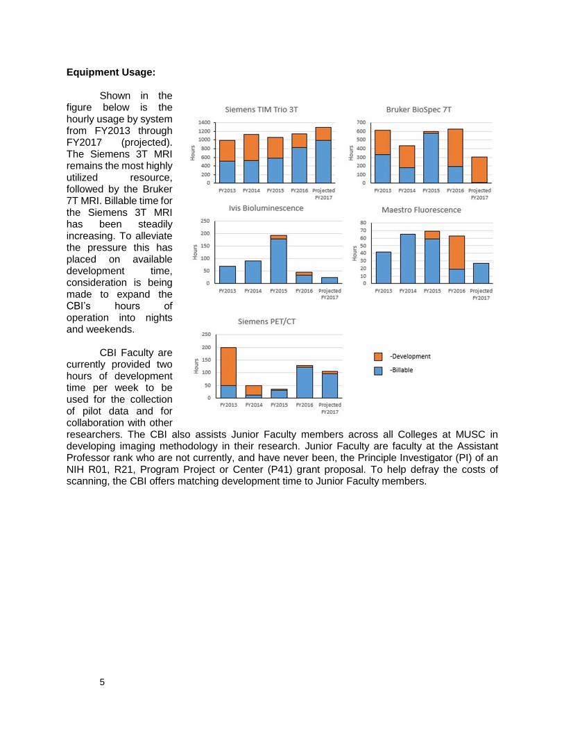
Equipment Usage:
Shown in the figure below is the hourly usage by system from FY2013 through FY2017 (projected). The Siemens 3T MRI remains the most highly utilized resource, followed by the Bruker 7T MRI. Billable time for the Siemens 3T MRI has been steadily increasing. To alleviate the pressure this has placed on available development time, consideration is being made to expand the CBI’s hours of operation into nights and weekends.
CBI Faculty are
currently provided two hours of development time per week to be used for the collection of pilot data and for collaboration with other researchers. The CBI also assists Junior Faculty members across all Colleges at MUSC in developing imaging methodology in their research. Junior Faculty are faculty at the Assistant Professor rank who are not currently, and have never been, the Principle Investigator (PI) of an NIH R01, R21, Program Project or Center (P41) grant proposal. To help defray the costs of scanning, the CBI offers matching development time to Junior Faculty members.
6
Faculty
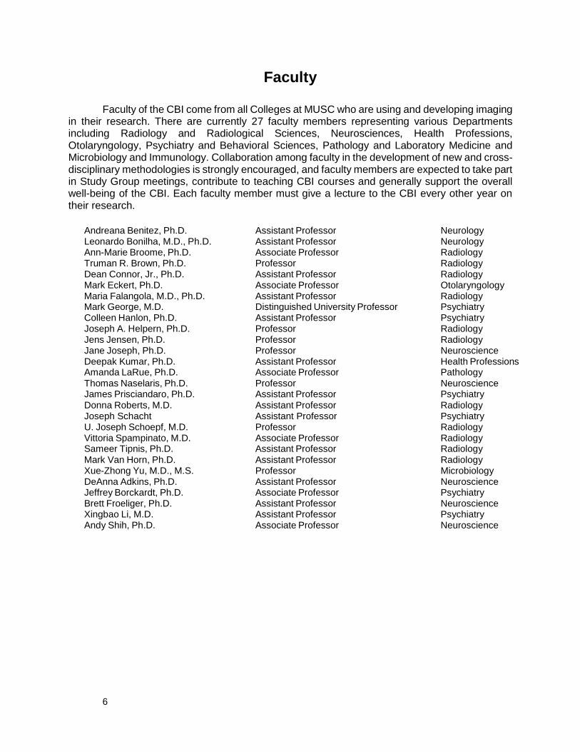
Faculty of the CBI come from all Colleges at MUSC who are using and developing imaging in their research. There are currently 27 faculty members representing various Departments including Radiology and Radiological Sciences, Neurosciences, Health Professions, Otolaryngology, Psychiatry and Behavioral Sciences, Pathology and Laboratory Medicine and Microbiology and Immunology. Collaboration among faculty in the development of new and cross-disciplinary methodologies is strongly encouraged, and faculty members are expected to take part in Study Group meetings, contribute to teaching CBI courses and generally support the overall well-being of the CBI. Each faculty member must give a lecture to the CBI every other year on their research.
Andreana Benitez, Ph.D. Assistant Professor Neurology Leonardo Bonilha, M.D., Ph.D. Assistant Professor Neurology Ann-Marie Broome, Ph.D. Associate Professor Radiology Truman R. Brown, Ph.D. Professor Radiology Dean Connor, Jr., Ph.D. Assistant Professor Radiology Mark Eckert, Ph.D. Associate Professor Otolaryngology Maria Falangola, M.D., Ph.D. Assistant Professor Radiology Mark George, M.D. Distinguished University Professor Psychiatry Colleen Hanlon, Ph.D. Assistant Professor Psychiatry Joseph A. Helpern, Ph.D. Professor Radiology Jens Jensen, Ph.D. Professor Radiology Jane Joseph, Ph.D. Professor Neuroscience Deepak Kumar, Ph.D. Assistant Professor Health Professions Amanda LaRue, Ph.D. Associate Professor Pathology Thomas Naselaris, Ph.D. Professor Neuroscience James Prisciandaro, Ph.D. Assistant Professor Psychiatry Donna Roberts, M.D.
Assistant Professor Radiology
Joseph Schacht Assistant Professor Psychiatry U. Joseph Schoepf, M.D. Professor Radiology
Vittoria Spampinato, M.D. Associate Professor Radiology Sameer Tipnis, Ph.D. Assistant Professor Radiology Mark Van Horn, Ph.D. Assistant Professor Radiology Xue-Zhong Yu, M.D., M.S. Professor Microbiology DeAnna Adkins, Ph.D. Assistant Professor Neuroscience Jeffrey Borckardt, Ph.D. Associate Professor Psychiatry Brett Froeliger, Ph.D. Assistant Professor Neuroscience Xingbao Li, M.D. Assistant Professor Psychiatry Andy Shih, Ph.D. Associate Professor Neuroscience
7
Education
Biomedical Imaging Ph.D. Program
In 2014, the CBI received full approval for the Ph.D. in Biomedical Imaging curriculum and currently has 4 students enrolled. The mission of the Biomedical Imaging Ph.D. program is to train students in a basic core of knowledge and skills that will prepare them to become leaders in the application of biomedical imaging technology to problems in basic and clinical research. The program provides a strong foundation in the fundamentals of image acquisition technologies and data analysis methods, while exposing students to the application of specific imaging modalities through a series of individual electives in their chosen area of interest.
The Biomedical Imaging PhD program is designed to provide students with the education
and training needed to pursue careers applying cutting edge developments in biomedical imaging to solving scientific and healthcare problems within academia or industry.
Through the CBI, MUSC offers a comprehensive and integrated graduate training program
combining biomedical sciences through the College of Graduate Studies core curriculum, with a strong emphasis on imaging science and its biomedical applications leading to a Ph.D. in Biomedical Imaging. The core curriculum is designed to provide a strong foundation in the fundamentals of imaging acquisition technologies, data analysis methods, and research design, all within the context of applying these techniques in clinical and basic research projects in academic and industrial medical and research settings. Through this program, students are able to gain hands-on experience with advanced imaging systems dedicated to both preclinical (bioluminescence, fluorescence, Micro-CT/PET, 7T MRI) and human (3T MRI) research. The students have opportunities to rotate as research assistants in laboratories of faculty who actively conduct research within many departments throughout the University, such as Neurosciences, Psychiatry, Radiology, Rehabilitation, Cardiology, Pediatrics, Surgery, and Oncology. The students are required to demonstrate scientific proficiency in the area of biomedical sciences, with an emphasis on biomedical imaging through the completion of a qualifying examination and an individual doctoral dissertation.
Upon the completion of this degree, graduates have the foundation on which they can
build careers as independent investigators or key collaborators who possess a unique combination of skills: a fund of technical knowledge of imaging sciences and its most critical innovations as well as a distinct perspective that is focused on applying these advances in biomedical imaging to a breadth of preclinical and human research areas, from basic physiological processes to phenotypically complex diseases.
Our program begins with two years of didactic coursework during which students master
the fundamental physical and biological principles behind biomedical imaging. Through laboratory rotations students identify a faculty mentor and a research topic for their thesis. The facilities at MUSC are quite extensive (bioluminescence, fluorescence, Micro-CT/PET, animal 7T MRI and human 3T MRI), and faculty research interests are broad (Neurosciences, Psychiatry, Radiology, Rehabilitation, Cardiology, Pediatrics, Surgery, and Oncology), allowing students to select a mentor and topic of research closely suited to their interests. More information on this program can be found at: http://academicdepartments.musc.edu/biomi/
8
Study Groups
Addiction - Leader: Joseph Schacht, Ph.D.
The Addiction Study Group is a forum for discussion of neuroimaging of addictive disorders. Topics include 1) discussion of the clinical presentation of disorders including alcohol, nicotine, cocaine, heroin, and prescription opioid abuse and dependence; 2) application of a broad spectrum of neuroimaging modalities, including functional and structural MRI, diffusion tensor and kurtosis imaging, and MRS, and methods, including network connectivity analysis; and 3) opportunities for collaboration among investigators for grant submissions. Meetings have varied formats, including journal club discussion, data presentation, grant ideas forums, and practice job or conference talks. Both clinical and basic science investigators currently involved with addiction neuroimaging projects or hoping to pursue such projects are encouraged to attend. User Group Meeting (Nuts & Bolts)
The CBI Nuts and Bolts User Group meets twice a month and provides a forum for in-
depth discussions by researchers about imaging, statistical methods, data analysis techniques and administrative issues. The first portion of the meeting is dedicated to discussion of CBI equipment and administrative issues, and the remainder of the meeting typically consists of a presentation of a discussion topic chosen by the group. Lectures
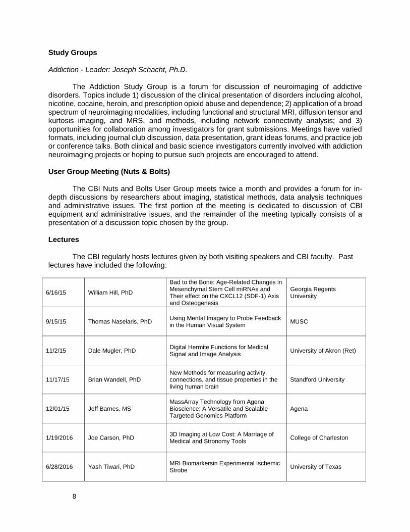
The CBI regularly hosts lectures given by both visiting speakers and CBI faculty. Past
lectures have included the following:
6/16/15 William Hill, PhD
Bad to the Bone: Age-Related Changes in Mesenchymal Stem Cell miRNAs and Their effect on the CXCL12 (SDF-1) Axis and Osteogenesis
Georgia Regents University
9/15/15 Thomas Naselaris, PhD Using Mental Imagery to Probe Feedback in the Human Visual System
MUSC
11/2/15 Dale Mugler, PhD Digital Hermite Functions for Medical Signal and Image Analysis
University of Akron (Ret)
11/17/15 Brian Wandell, PhD New Methods for measuring activity, connections, and tissue properties in the living human brain
Standford University
12/01/15 Jeff Barnes, MS MassArray Technology from Agena Bioscience: A Versatile and Scalable Targeted Genomics Platform
Agena
1/19/2016 Joe Carson, PhD 3D Imaging at Low Cost: A Marriage of Medical and Stronomy Tools
College of Charleston
6/28/2016 Yash Tiwari, PhD MRI Biomarkersin Experimental Ischemic Strobe
University of Texas

9
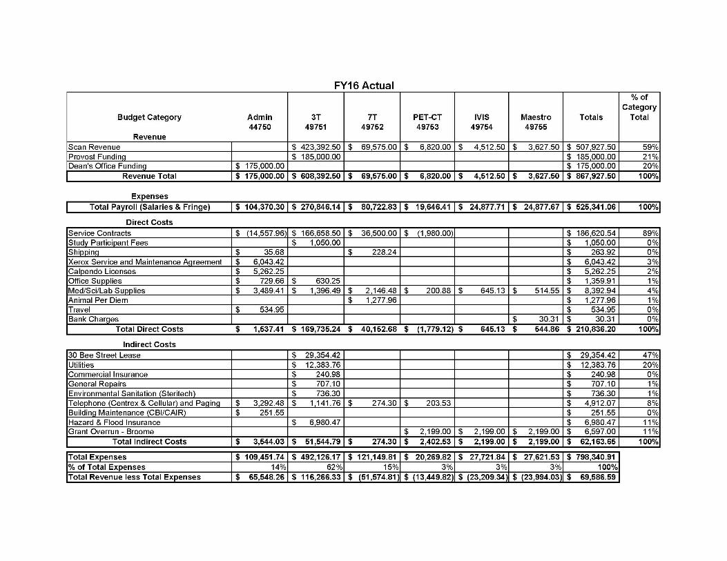
Appendix I: Budget
10

11
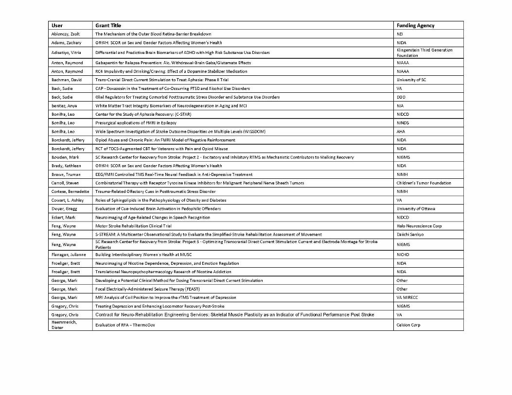
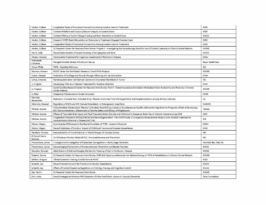
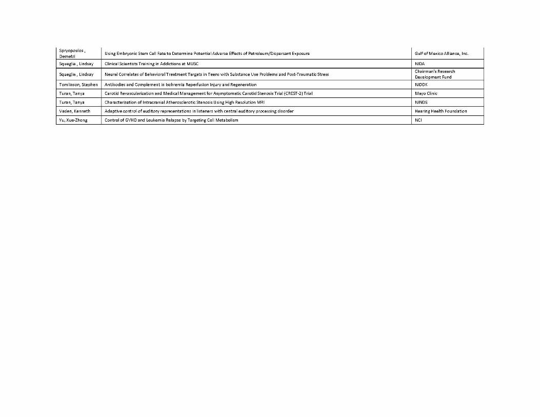
Appendix II: Grants Supported by CBI
12
13
14

15
Center for Biomedical Imaging Medical University of South Carolina 68 President Street Charleston, South Carolina 29425 Tel: (843) 876-2460 Fax: (843) 876-2469 www.musc.edu/cbi